Introduction
GnRH released in a pulsatile fashion from nerve terminals in the median eminence stimulates pituitary gonadotrophs to secrete LH and FSH episodically in both sexes. These gonadotrophins in turn stimulate aspects of gonadal function including steroidogenesis in both sexes and ovulation in the female. Gonadal steroids regulate the frequency and amplitude of GnRH release in both males and females. In females of spontaneously ovulating species including rats, hamsters and sheep, oestrogens exert both negative and positive feedback actions on the hypothalamus and pituitary gland to control GnRH and LH secretion (reviewed in Kalra and Kalra, 1983, 1989). In contrast, in females of induced ovulating species including voles, ferrets and rabbits (Gray et al., 1976; Carroll et al., 1985; Pau et al., 1986) there is only evidence for a negative feedback action of oestrogens on GnRH and LH secretion. Regimens of oestradiol with or without concurrent progesterone treatment that induced LH surges in rats were unsuccessful in augmenting LH secretion in rabbits (Sawyer and Markee, 1959), voles (Milligan, 1978) or ferrets (Baum et al., 1990). However, oestradiol acts in the female hypothalamus to induce aspects of proceptive and receptive sexual behaviour that are required to allow receipt of an intromission from a male conspecific, ultimately leading to a preovulatory LH surge (for example see Beyer and McDonald, 1973; Baum and Schretlen, 1978; Dluzen and Carter, 1979).
The ability to generate preovulatory LH surges is sexually dimorphic in non-primate mammalian species. Males show neither mating- nor steroid-induced LH surges (Neill, 1972; Karsch and Foster, 1975; Gray et al., 1976; Teresawa et al., 1979; Albers et al., 1984; Carroll et al., 1987). In males of all non-primate mammalian species studied to date, testosterone, probably acting after neural aromatization to oestradiol (Carroll and Baum, 1989; Spratt and Herbison, 1997), exerts only negative feedback actions on hypothalamic GnRH release and pituitary LH secretion.
The negative feedback action of gonadal steroids on LH secretion is sexually differentiated in rats; females are more sensitive than males to inhibition by oestrogen (Gay and Midgley, 1969; King and Rubin, 1995). In contrast, in ferrets, the negative feedback control of LH secretion by oestrogen is not sexually dimorphic (Carroll and Baum, 1989). The time course of the rise in pulsatile LH secretion is very similar in gonadectomized male and female ferrets after withdrawal of oestradiol. Several studies (reviewed in Kalra and Kalra, 1989) have shown that GnRH content and release, measured in the mediobasal hypothalamus using either in vitro or in vivo methods, is diminished after gonadectomy in rats of both sexes. Likewise, ovariectomy of oestrous female ferrets causes a decrease in the in vitro release and content of GnRH peptide in the mediobasal hypothalamus (Lambert et al., 1992). Ovariectomized ferrets have significantly fewer GnRH mRNA-containing cells in the arcuate region than intact controls in either oestrus or anoestrus (Bakker et al., 1999). This decrease in the number of cells probably reflects a reduction in cellular GnRH mRNA cells rather than
Effect of gonadal steroids on pituitary LH secretion and mediobasal
hypothalamic GnRH mRNA in ferrets
J. Bakker and M. J. Baum
Department of Biology, Boston University, 5 Cummington St, Boston, MA 02215, USA
In vitro release and content of GnRH in mediobasal hypothalamic slices are reduced by
ovariectomy of female ferrets but are not affected by castration of male ferrets in
breeding condition. The aim of the present study was to determine whether this sex
difference reflects a sexually dimorphic effect of gonadal steroids on mediobasal
hypothalamic GnRH mRNA content of male and female ferrets killed 4 weeks after
gonadectomy, either with or without steroid hormone replacement. This time interval
exceeds the 6–10 days needed for increments in plasma LH concentrations to stabilize
after gonadectomy of ferrets of both sexes. In situ hybridization using an
35S-labelled
oligoprobe complementary to the human GnRH coding region showed that the number
of mediobasal hypothalamic neurones and the cellular content of GnRH mRNA did not
differ significantly among groups of male and female ferrets that were either in breeding
condition or that had been gonadectomized and treated with sex steroids or oil vehicle.
These results indicate that gonadal hormones regulate mediobasal hypothalamic GnRH
biosynthesis and release in both sexes via post-transcriptional events that may include
GnRH mRNA translation or the conversion of pre-pro GnRH precursor into mature
GnRH.
© 2000 Journals of Reproduction and Fertility Ltd 0022–4251/2000
apoptosis of these neurones, so that cells expressing decreased mRNA content were no longer detected with the in situ hybridization method used. Castration of male ferrets does not affect the in vitro release and content of mediobasal hypothalamus GnRH (Lambert et al., 1992) nor does castration affect the number of GnRH mRNA-containing neurones in any region of the mediobasal hypothalamus, although it was reported to augment cellular GnRH mRNA content in the preoptic area (Tang et al., 1997). These findings indicate that gonadal steroids may exert different negative feedback actions on mediobasal hypothalamus GnRH release in male and female ferrets by differentially affecting GnRH gene transcription in the mediobasal hypothalamus. To date, the effects of gonadectomy on mediobasal hypothalamus GnRH mRNA of male and female ferrets have been assessed in separate studies (Tang et al., 1997; Bakker et al., 1999), and the effects of steroid replacement have been assessed only in males (Tang et al., 1997). Therefore, in the present study, the effects of long-term gonadectomy and steroid replacement on the number of mediobasal hypothalamic neurones and the cellular content of GnRH mRNA were systematically compared in ferrets of both sexes. Animals were studied at a time (4 weeks) after gonadectomy when plasma LH concentrations are known to have increased to a stable plateau in ferrets of both sexes (Carroll and Baum, 1989).
Materials and Methods
Animals and experimental design
Male and female European ferrets (Mustela furo) were purchased from Marshall Farms (North Rose, NY) at the age of 5–8 months. Animals were housed individually in modified rabbit cages under a long-day photoperiod (16 h light:8 h dark; lights on at 07:00 h). All ferrets were fed moistened Purina ferret chow (Ralston Purina Co., St Louis, MO) once a day. Water was available ad libitum. Adult oestrous female ferrets (n = 8) were anaesthetized using ketamine (Ketaset; Bristol Laboratories, Syracuse, NY; 35 mg kg–1) and xylazine (Rompun; Haver-Miles Laboratories,
Shawnie, KS; 4 mg kg–1) and ovo-hysterectomized via a
single midline incision. Females were injected s.c. once a day either with oestradiol benzoate dissolved in sesame oil (15µg kg–1) or with oil vehicle for 4 weeks before the animals
were killed. This dose of oestradiol benzoate fully restores feminine sexual behaviour in ovariectomized female ferrets (Baum et al., 1985). In addition, gonadally intact females (n = 4) in oestrus were killed at the same time. Male ferrets (n = 9) were castrated under anaesthesia and injected s.c. once a day with testosterone propionate (5 mg kg–1) for 2
weeks. This dose of testosterone propionate fully restores masculine sexual behaviours in castrated male ferrets (Lambert and Baum, 1991). After 2 weeks, five males continued to receive testosterone propionate and four males received injections of oil vehicle once a day for 4 weeks. After 4 weeks, the animals were killed. Gonadally intact males in breeding condition (n = 3) were killed at the same time. These experiments were conducted in accordance with the
guidelines of the NIH Guiding Principles for the Care and Use of Research Animals and were approved by the Boston University IACUC (protocol No. 97-017).
Blood and brain collection
Ferrets were anaesthetized quickly using CO2and were then decapitated. Brains were removed and immediately frozen in powdered dry ice before being stored at –80⬚C. Trunk blood was collected into heparinized tubes, centrifuged (at 313 g for 30 min) and plasma was collected and stored at –20⬚C before being transported elsewhere on dry ice for hormone assays.
Hormone assays
The hormone assays did not require extraction of the plasma. Plasma LH concentrations were quantified in duplicate by radioimmunoassay using the GDN 15 anti-ovine LH antiserum (Ryan et al., 1985). The minimum detection level of the assay was 0.45 ng ml–1. The LH assay
was generously performed by Dr Kathleen Ryan (Magee-Womens Research Institute, Pittsburgh, PA). Plasma oestradiol concentrations were measured in duplicate using a double antibody radioimmunoassay kit (Diagnostic Products Corp., Los Angeles, CA). The minimum detection level of the assay was 2 pg ml–1. All samples were included in
one assay and the intra-assay coefficient of variation (CV) was < 5%. Plasma testosterone concentrations were measured in duplicate using a total testosterone-coated tube assay (Diagnostic Products Corp., Los Angeles, CA). The antibody used to measure testosterone has < 5% crossreactivity with dihydrotestosterone. The inter-assay CV was 3.2% at 65 ng dl–1and 9.3% at 320 ng dl–1. The intra-assay
CV was < 5%. The oestradiol and testosterone assays were generously performed by Dr Geralyn Lambert Messerlian (Women and Infants Hospital, Providence, RI).
In situ hybridization for GnRH mRNA
Frozen brains were sectioned coronally at 14µm using a cryostat, and brain sections were mounted on to Vectabond-coated slides. Brain sections were collected beginning rostrally at the organum vasculosum of the lamina terminalis and extending caudally to the mammillary bodies. Slides were stored in boxes containing desiccant at –80⬚C until in situ hybridization was performed.
In situ hybridization was performed on every tenth brain section (140 µm intervals between adjacent sections) using a 48-base synthetic oligonucleotide probe complementary to the GnRH coding region (bases 102–149) of the human complementary DNA (Adelman et al., 1986). This oligoprobe has been used successfully in ferrets (Tang et al., 1997; Bakker et al., 1999). The GnRH oligoprobe was 3’-end labelled by incubation with [35S]deoxy-ATP (75 pmol; NEN Life
Sciences, Boston, MA) and terminal deoxynucleotidyl transferase (25 U; Boehringer Mannheim, Indianapolis, IN).
The in situ hybridization protocol was as described by Bakker et al. (1999).
Data analysis
Cell counts. Brain sections from 12 male and 12 female ferrets distributed over four in situ hybridization runs were analysed. Each run contained brain sections from every treatment group for each sex. All slides were coded to conceal the identity of the animal. All hybridized cells present in every section run for in situ hybridization were counted in several brain regions including the preoptic area, anterior hypothalamus, arcuate region and median eminence. Cells containing GnRH mRNA were identified by the presence of silver grains overlying a counterstained cell body (see Fig. 1).
Image analysis. Silver grains clustered over individual cells in two or three anatomically matched sections from the preoptic area, anterior hypothalamus, arcuate region and median eminence were analysed using an image analysis system (Image-pro, Media Cybernetics, Silver Springs, MD)
with a Hitachi digital colour camera attached to an Olympus BH-2 microscope. For each hybridized cell, a threshold intensity was set at the level of the underlying counterstained cell body so that only silver grains overlying this cell were above the threshold. Each cell body was circumscribed manually, and the total hybridization area per cell was estimated by computing the sum of areas occupied by silver grains. Only cells with five times the hybridization area (sum of areas occupied by grains) compared with the adjacent background were considered to be labelled. The average hybridization area (in µm2) per cell was calculated
for each brain region for each animal, and these values were used to determine the group mean and SEM.
Statistical analysis. The number of cells containing GnRH mRNA and the cellular content of GnRH mRNA were compared using two-way ANOVA (sex ⫻ treatment). Plasma hormone concentrations were analysed within each sex using a one-way ANOVA or a Student’s t test.
Results
Plasma hormone concentrations
Plasma LH concentrations were significantly higher in ovariectomized oil-treated ferrets than in intact oestrous females or in ovariectomized females treated with oestradiol benzoate (F(2/11) = 10.1, P < 0.01; Table 1). Plasma oestradiol concentrations were significantly higher in gonadally intact oestrous and ovariectomized oestradiol benzoate-treated females than in ovariectomized oil-treated females (F(2/11) = 5.7, P < 0.05; Table 1). Post-hoc analysis with Fisher’s LSD showed that plasma oestradiol concentrations did not differ significantly between oestrous females and ovariectomized oestradiol benzoate-treated females. Thus, daily treatment with 15µg oestradiol benzoate kg–1was sufficient to restore
plasma oestradiol concentrations and to suppress plasma LH concentrations in ovariectomized females to those found in gonadally intact oestrous females.
Plasma LH concentrations were significantly higher in castrated oil-treated male ferrets than in castrated male ferrets treated with testosterone propionate (t = 3.5; P < 0.05; Table 1). Plasma testosterone concentrations were significantly higher in castrated testosterone propionate-treated males than in castrated oil-propionate-treated males (t = 5.3, P < 0.01; Table 1). Treatment with 5 mg testosterone propionate kg–1 once a day led to supraphysiological
concentrations (average of 76.8⫾ 14.4 ng ml–1) of
testosterone in these animals compared with those found in gonadally intact breeding males (21.5⫾ 2.2 ng ml–1; Lambert
and Baum, 1991). The plasma testosterone and LH concentrations in the two intact breeder males for which data were available (Table 1) were variable and differed from those measured in gonadally intact breeding males in several studies (Sisk and Desjardins, 1986; Carroll et al., 1987; Carroll and Baum, 1989; Lambert and Baum, 1991). This profile of low plasma testosterone coupled with high LH indicates that the gonadally intact males used in the present study were
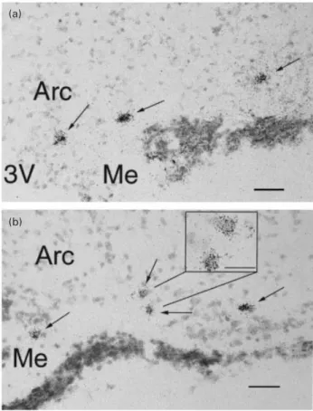
(a)
(b)
Fig. 1. Photomicrographs of cells expressing GnRH mRNA in the
arcuate region (Arc) and the median eminence (Me) of representative gonadally intact female (a) and male (b) ferrets in breeding condition. The high magnification inset in (b) shows two labelled cells with silver grains over a counterstained cell body. Arrows indicate labelled cells. Scale bars represent (a,b) 50 µm, inset 10 µm.
approaching the end of a breeding season even though their testes had not regressed in size (data not shown).
Distribution of neurones containing GnRH mRNA
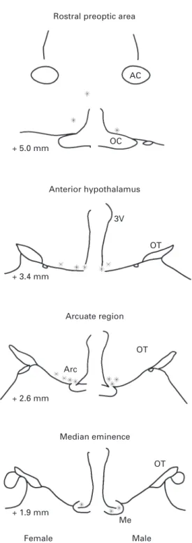
Examples of GnRH mRNA-positive neurones in the arcuate region of a gonadally intact oestrous female and of a gonadally intact breeding male are shown (Fig. 1). No sex differences were observed in the distribution of GnRH mRNA-containing neurones throughout the preoptic area and mediobasal hypothalamus. In contrast to their dense rostral distribution in rats, in ferrets, GnRH neurones are dispersed across the base of the hypothalamus and there is no evidence of any conspicuous grouping (Fig. 2). The observed distribution of cells hybridized for GnRH mRNA was similar to that reported in the female (Bakker et al., 1999) and male ferret (Tang et al., 1997) and to that found to be immunoreactive for GnRH peptide in ferrets of both sexes (King and Anthony, 1984; Tang and Sisk, 1992; Wersinger and Baum, 1996). The number of GnRH mRNA-positive cells detected in the various regions of the mediobasal hypothalamus was approximately the same as that reported by Bakker et al. (1999) and by Tang et al. (1997).
Effect of gonadal steroids on the content of GnRH mRNA in
the mediobasal hypothalamus
The number of cells containing GnRH mRNA as well as mean cellular GnRH mRNA content did not differ significantly among any of the experimental groups; this was the case in all brain regions studied (Table 2).
Discussion
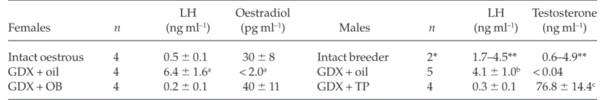
The main finding of the present study is that the content of GnRH mRNA in the mediobasal hypothalamus did not differ among male and female ferrets that were either in breeding condition or that had been gonadectomized for 4 weeks and treated once a day with sex steroids or oil vehicle. This result indicates that the content of GnRH mRNA in the mediobasal hypothalamus is not regulated by gonadal steroids in ferrets. The absence of an effect of gonadectomy on GnRH mRNA abundance in the mediobasal hypothalamus was in marked contrast to the effect of this procedure on plasma LH concentrations, which were increased in gonadectomized steroid-free ferrets. The results of the present study indicate that the long-term post-gonadectomy increase in LH Table 1. Plasma LH, oestradiol and testosterone concentrations in ferrets used in GnRH mRNA in situ
hybridization studies
LH Oestradiol LH Testosterone
Females n (ng ml–1) (pg ml–1) Males n (ng ml–1) (ng ml–1)
Intact oestrous 4 0.5 ⫾ 0.1 30 ⫾ 8 Intact breeder 2* 1.7–4.5** 0.6–4.9** GDX + oil 4 6.4 ⫾ 1.6a < 2.0a GDX + oil 5 4.1 ⫾ 1.0b < 0.04
GDX + OB 4 0.2 ⫾ 0.1 40 ⫾ 11 GDX + TP 4 0.3 ⫾ 0.1 76.8 ⫾ 14.4c Data are expressed as the mean ⫾SEM.
*Plasma sample of one breeding male was lost. **Range is given because n = 2.
GDX: gonadectomy; OB: oestradiol benzoate; TP: testosterone propionate. aSignificantly different (P < 0.05) from oestrous and GDX + OB values bSignificantly higher (P < 0.05) than GDX + TP values.
cSignificantly higher (P < 0.05) than GDX + oil values.
Table 2. Effect of gonadectomy and steroid replacement on number of cells containing GnRH mRNA and on the cellular content of GnRH mRNA in the mediobasal hypothalamus of male and female ferrets
Preoptic area Anterior hypothalamus Arcuate region Median eminence Number of Cellular Number of Cellular Number of Cellular Number of Cellular
n cells content cells content cells content cells content
Females Intact oestrous 4 8.3 ⫾ 6.6 83 ⫾ 11 15.3 ⫾ 4.9 44 ⫾ 10 34.8 ⫾ 6.6 58 ⫾ 7 4.3 ⫾ 1.5 63 ⫾ 17 GDX + oil 4 7.8 ⫾ 3.2 95 ⫾ 10 20.5 ⫾ 4.9 60 ⫾ 13 39.0 ⫾ 8.1 62 ⫾ 12 6.5 ⫾ 2.0 53 ⫾ 11 GDX + OB 4 6.0 ⫾ 3.3 97 ⫾ 1 20.8 ⫾ 4.9 62 ⫾ 7 28.3 ⫾ 6.3 62 ⫾ 7 5.8 ⫾ 2.2 47 ⫾ 11 Males Intact breeder 3 11.0 ⫾ 4.0 73 ⫾ 8 16.3 ⫾ 3.5 63 ⫾ 9 23.0 ⫾ 4.1 71 ⫾ 7 7.7 ⫾ 2.4 65 ⫾ 10 GDX + oil 4 4.5 ⫾ 1.0 71 ⫾ 1 30.5 ⫾ 4.9 71 ⫾ 13 29.3 ⫾ 6.6 70 ⫾ 13 12.3 ⫾ 2.1 78 ⫾ 21 GDX + TP 5 11.0 ⫾ 2.4 87 ⫾ 6 17.0 ⫾ 3.8 59 ⫾ 9 42.2 ⫾ 5.3 55 ⫾ 9 13.2 ⫾ 2.0 50 ⫾ 5
Data are expressed as mean number of GnRH mRNA-labelled cells per brain region and as the mean ⫾SEMhybridization area (µm2) per cell. GDX: gonadectomy; OB: oestradiol benzoate; TP: testosterone propionate.
secretion by the pituitary, which is equivalent in male and female ferrets (Carroll and Baum,1989), does not depend on increased GnRH gene transcription leading to an increase in
the content of GnRH mRNA in the mediobasal
hypothalamus. It is possible that immediate perturbations in GnRH mRNA in the mediobasal hypothalamus induced by steroid hormone withdrawal were not detected since ferrets were killed at only one interval (4 weeks) after gonadectomy. Indeed, Emanuele et al. (1996) reported small transient increases in hypothalamic GnRH mRNA content after castration in male rats: the content of GnRH mRNA increased nearly twofold above that in intact controls at 1 day after castration, whereas they were unchanged at 3, 5, 7, 14, 21 or 28 days after castration despite marked increases in plasma LH and FSH concentrations at all of these time points. This result points to a transient response of hypothalamic GnRH mRNA to the withdrawal of gonadal hormones. It seems unlikely that such a change reflects the primary mechanism whereby the pituitary glands of long-term gonadectomized steroid-free animals persistently secrete large amounts of LH.
The increased LH secretion that occurs in both sexes after gonadectomy may result from the withdrawal of sex steroid action in pituitary gonadotrophs as opposed to GnRH neurones in the mediobasal hypothalamus. The number of pituitary GnRH receptors and their mRNA content increase after gonadectomy in both male and female rats (Clayton and Catt, 1981; Frager et al., 1981; Kaiser et al., 1993), whereas GnRH secretion in the mediobasal hypothalamus, measured by in vitro and in vivo methods, decreases after gonadectomy in rats of both sexes (reviewed in Kalra and Kalra, 1989). Thus, the hypersecretion of LH that follows steroid withdrawal may result from an increase in the sensitivity of pituitary gonadotrophs to lower amplitude GnRH pulses from the median eminence in both rats and ferrets (Kalra and Kalra, 1989; Lambert et al., 1992). In contrast to rats and ferrets, the post-gonadectomy increase in LH is associated with an enhanced secretion of GnRH from nerve terminals of the mediobasal hypothalamus in rabbits (Pau et al., 1986) and sheep (Karsch et al., 1987; Caraty and Locatelli, 1988). These findings suggest that different neuroendocrine mechanisms may underlie the post-gonadectomy rise in LH secretion in these species.
The finding of the present study that castration of male ferrets had no effect on the number of cells containing GnRH mRNA or on the cellular content of GnRH mRNA in the mediobasal hypothalamus is in agreement with the study by Tang et al. (1997). However, in contrast to the results of Tang et al. (1997), in the present study, no increase was observed in the cellular content of GnRH mRNA in the preoptic area after castration. This discrepancy cannot be explained by differences in post-gonadectomy interval: in both studies (Tang et al., 1997; present study) males were killed at 4 weeks after castration. In the present study, there was no difference in the number of cells containing GnRH mRNA in the arcuate region among gonadally intact oestrous females and ovariectomized oil-treated and oestradiol benzoate-treated subjects. This result is in contrast to the study of Bakker et al. (1999) in which the number of cells containing GnRH mRNA was significantly lower in the arcuate region of ovariectomized females than in gonadally intact oestrous or anoestrous females. The post-gonadectomy interval differs slightly between the two studies: in the study of Bakker et al. (1999) brains were collected 3 weeks after ovo-hysterectomy
Rostral preoptic area
AC OC + 5.0 mm Anterior hypothalamus 3V OT + 3.4 mm Arcuate region OT Arc + 2.6 mm Median eminence OT Me + 1.9 mm Female Male
Fig. 2. Camera lucida drawings of coronal sections through the
rostral preoptic area (+ 5.0 mm anterior to the interaural line), anterior hypothalamus (+ 3.4 mm), arcuate region (+ 2.6 mm), and median eminence (+ 1.9 mm) showing the distribution of GnRH mRNA-containing neurones (*) in representative gonadally intact female and male ferrets in breeding condition. 3V: third ventricle; AC: anterior commissure; Arc: arcuate region; Me: median eminence; OC: optic chiasma; OT: optic tract.
as opposed to 4 weeks in the present study. However, it is unlikely that this difference in post-gonadectomy interval can explain the discrepancy in the content of GnRH mRNA in the arcuate region in the two studies. In gonadectomized ferrets of both sexes, plasma LH concentrations reached a plateau approximately 6–10 days after steroid withdrawal and remained stable for at least 20 days thereafter (Sisk and Desjardins, 1986; Carroll and Baum, 1989). Thus, it seems likely that similar mechanisms cause the hypersecretion of LH from the pituitary as late as 3–4 weeks after ovo-hysterectomy. It should also be noted that Bakker et al. (1999) reported that the content of GnRH mRNA in the mediobasal hypothalamus was equivalent in gonadally intact oestrous and anoestrous females even though plasma oestradiol concentrations were significantly lower in the intact anoestrous females than in oestrous subjects. Bakker et al. (1999) concluded that ovarian hormones other than oestradiol may normally facilitate the GnRH mRNA content in the mediobasal hypothalamus in anoestrous as well as oestrous female ferrets. This view is consistent with the findings of the present study that oestrogen replacement had no effect on GnRH mRNA content in the mediobasal hypothalamus in ovariectomized females.
The results of the present study are in general agreement with several studies in rats that did not detect any significant changes in hypothalamic GnRH mRNA content after steroidal manipulations (Kelly et al., 1989; Wiemann et al., 1990; Malik et al., 1991; Emanuele et al., 1996). Although there are many reports showing either increases or decreases in hypothalamic GnRH mRNA content after gonadectomy in rats (reviewed in Sagrillo et al., 1996; Gore and Roberts, 1997), these effects of gonadectomy were small and transient. Thus, the post-gonadectomy increase in LH in rats and ferrets does not appear to depend on marked increases in hypothalamic GnRH gene transcription. A number of studies in rats (Kelly et al., 1989; Emanuele et al., 1996; Gore and Roberts, 1997) indicate that GnRH biosynthesis and release are primarily regulated via post-transcriptional mechanisms such as GnRH mRNA translation or conversion of precursor into mature GnRH decapeptide. Similarly, the observed (Lambert et al., 1992) sex difference in the in vitro release of GnRH from mediobasal hypothalamic tissue may reflect sex differences in translation of GnRH mRNA or conversion of pre-pro GnRH precursor into mature GnRH in the mediobasal hypothalamus. In addition, there is independent evidence that in the mediobasal hypothalamus in ferrets, GnRH biosynthesis and release are regulated primarily by post-transcriptional mechanisms. The mating induced preovulatory LH surge in the oestrous female is associated with an increase in GnRH biosynthesis in the mediobasal hypothalamus (Lambert et al., 1992), but not with a change in
the content of GnRH mRNA in the mediobasal
hypothalamus (Bakker et al., 1999). Finally, the postcoital increase in LH output is associated with an increase in the number of protein-synthesizing Golgi complexes present in immunoreactive GnRH neurones in the mediobasal hypothalamus (Bibeau et al., 1991).
In conclusion, the post-gonadectomy increase in plasma LH concentration, which is equivalent in male and female ferrets, does not appear to depend on increased GnRH gene
transcription in the mediobasal hypothalamus leading to an increase in GnRH mRNA content. This result indicates that gonadal hormones regulate mediobasal hypothalamic GnRH biosynthesis and release in both sexes via post-transcriptional events such as GnRH mRNA translation or the conversion of pre-pro GnRH precursor into mature GnRH.
This work was supported by NIH grant HD21094 and by NIMH Senior Research Scientist Award MH 00392 (to M. J. Baum). The authors thank J. A. Cherry (Department of Psychology, Boston University) for generously providing laboratory facilities, and the staff of the Boston University Animal Facility for their care of the ferrets.
References
Adelman JP, Mason AJ, Hayflick JS and Seeburg PH (1986) Isolation of the gene and hypothalamic cDNA for the common precursor of gonadotropin-releasing hormone and prolactin release-inhibiting factor in human and rat
Proceedings National Academy of Sciences USA 83 179–183
Albers HE, Moline ML and Moore-Ede MC (1984) Sex differences in circadian control of LH secretion Journal of Endocrinology 100 101–105
Bakker J, Rubin BS and Baum MJ (1999) Changes in mediobasal hypothalamic gonadotropin-releasing hormone messenger ribonucleic acid levels induced by mating or ovariectomy in a reflex ovulator, the ferret
Endocrinology 140 595–602
Baum MJ and Schretlen PJ (1978) Oestrogenic induction of sexual behaviour in ovariectomized ferrets housed under short or long photoperiods Journal
of Endocrinology 78 295–296
Baum MJ, Stockman ER and Lundell LA (1985) Evidence of proceptive without receptive defeminization in male ferrets Behavioural Neuroscience 99 742–750
Baum MJ, Carroll RS, Cherry JA and Tobet SA (1990) Steroidal control of behavioural, neuroendocrine and brain sexual differentiation: studies in a carnivore, the ferret Journal of Neuroendocrinology 2 401–418
Beyer C and McDonald P (1973) Hormonal control of sexual behaviour in the female rabbit Advances in Reproductive Physiology 6 185–219
Bibeau CE, Tobet SA, Anthony ELP, Carroll RS, Baum MJ and King JC (1991) Vaginocervical stimulation of ferrets induces release of luteinizing hormone-releasing hormone Journal of Neuroendocrinology 3 29–36 Caraty A and Locatelli A (1988) Effect of time after castration on secretion of
GnRH and LH in the ram Journal of Reproduction and Fertility 82 263–269 Carroll RS and Baum MJ (1989) Evidence that oestrogen exerts an equivalent
negative feedback action on LH secretion in male and female ferrets Journal
of Reproduction and Fertility 86 235–245
Carroll RS, Erskine MS, Doherty PC, Lundell LA and Baum MJ (1985) Coital stimuli controlling luteinizing hormone secretion and ovulation in the female ferret Biology of Reproduction 32 925–933
Carroll RS, Erskine MS and Baum MJ (1987) Sex difference in the effect of mating on the pulsatile secretion of luteinizing hormone in a reflex ovulator, the ferret Endocrinology 121 1349–1359
Clayton RN and Catt KJ (1981) Regulation of pituitary gonadotropin-releasing hormone receptors by gonadal hormones Endocrinology 108 887–895
Dluzen DE and Carter CS (1979) Ovarian hormones regulating sexual and social behaviors in female prairie voles Microtus ochrogaster. Physiology and
Behavior 23 597–600
Emanuele NV, Jurgens J, La Paglia N, Williams DW and Kelley MR (1996) The effect of castration on steady state levels of luteinizing hormone-releasing hormone (LHRH) mRNA and proLHRH processing: time course study utilizing semi-quantitative reverse transcription/polymerase chain reaction Journal of Endocrinology 148 509–515
Frager MS, Pieper DR, Tonetta SA, Duncan JA and Marshall JC (1981) Pituitary gonadotropin-releasing hormone receptors: effects of castration, steroid replacement, and the role of gonadotropin-releasing hormone in modulating receptors in the rat Journal of Clinical Investigation 67 615–623 Gay VL and Midgley AR (1969) Response of the adult rat to orchidectomy
and ovariectomy as determined by LH radioimmunoassay Endocrinology 84 1359–1364
Gore AC and Roberts JL (1997) Regulation of gonadotropin-releasing hormone gene expression in vivo and in vitro. Frontiers in Neuroendocrinology 18 209–245
Gray GD, Davis HN, McKenney AM and Dewsbury DA (1976) Effect of mating on plasma levels of LH and progesterone in montane voles (Microtus
montanus) Journal of Reproduction and Fertility 47 89–91
Kaiser UB, Jakubowiak A, Steinberger A and Chin WW (1993) Regulation of rat pituitary gonadotropin-releasing hormone receptor mRNA levels in vivo and in vitro. Endocrinology 133 931–934
Kalra SP and Kalra PS (1983) Neural regulation of luteinizing hormone secretion in the rat Endocrinology Reviews 4 311–351
Kalra SP and Kalra PS (1989) Do testosterone and estradiol-17βenforce inhibition or stimulation of luteinizing hormone-releasing hormone secretion? Biology of Reproduction 41 559–570
Karsch FJ and Foster DL (1975) Sexual differentiation of the mechanism controlling the preovulatory discharge of luteinizing hormone in sheep
Endocrinology 97 373–379
Karsch FJ, Cummins JT, Thomas GB and Clarke IJ (1987) Steroid feedback inhibition of pulsatile secretion of gonadotropin-releasing hormone in the ewe Biology of Reproduction 36 1207–1218
Kelly MJ, Garrett J, Bosch MA, Roselli CE, Douglass J, Adelman JP and Ronnekleiv OK (1989) Effects of ovariectomy on GnRH mRNA, proGnRH and GnRH levels in the preoptic hypothalamus of the female rat
Neuroendocrinology 49 88–97
King JC and Anthony ELP (1984) LHRH neurons and their projections in humans and other mammals: species comparisons Peptides 5 195–207 King JC and Rubin BS (1995) Dynamic alterations in luteinizing
hormone-releasing hormone (LHRH) neuronal cell bodies and terminals of adult rat
Cellular and Molecular Neurobiology 15 89–106
Lambert GM and Baum MJ (1991) Reciprocal relationships between pulsatile androgen secretion and the expression of mating behavior in adult male ferrets Hormones and Behavior 25 382–393
Lambert GM, Rubin BS and Baum MJ (1992) Sexual dimorphism in the effects of mating on the in vitro release of LHRH from the ferret mediobasal hypothalamus Physiology and Behavior 52 809–813
Malik KF, Silverman A-J and Morrell JI (1991) Gonadotropin-releasing hormone mRNA in the rat: distribution and neuronal content over the estrous cycle and after castration of males Anatomical Record 231 457–466 Milligan SR (1978) The feedback of exogeneous steroids on LH release and
ovulation in the intact female vole (Microtus agrestis) Journal of Reproduction
and Fertility 54 309–311
Neill JD (1972) Sexual differences in the hypothalamic regulation of prolactin secretion Endocrinology 90 1154–1159
Pau KY, Orstead KM, Hess DL and Spies HG (1986) Feedback effects of ovarian steroids on the hypothalamic–hypophyseal axis in the rabbit
Biology of Reproduction 35 1009–1023
Ryan KD, Siegel SF and Robinson SL (1985) Influence of day length and endocrine status on luteinizing hormone secretion in intact and ovariectomized adult ferrets Biology of Reproduction 33 690–697
Sawyer CH and Markee JE (1959) Estrogen facilitation of release of pituitary ovulating hormone in the rabbit in response to vaginal stimulation
Endocrinology 65 614–621
Sagrillo CA, Grattan DR, McCarthy MM and Selmanoff M (1996) Hormonal and neurotransmitter regulation of GnRH gene expression and related reproductive behaviors Behavior Genetics 26 241–277
Sisk CL and Desjardins C (1986) Pulsatile release of luteinizing hormone and testosterone in male ferrets Endocrinology 119 1195–1203
Spratt DP and Herbison AE (1997) Regulation of preoptic area gonadotrophin-releasing hormone (GnRH) mRNA expression by gonadal steroids in the long-term gonadectomized male rat Molecular Brain Research 47 125–133
Tang YP and Sisk CL (1992) LHRH in the ferret: pubertal decrease in the number of immunopositive arcuate neurons Peptides 13 241–247
Tang YP, Kashon ML and Sisk CL (1997) Brain region-specific regulation of luteinizing hormone-releasing hormone messenger ribonucleic acid in the male ferret: interactions between pubertal maturation and testosterone
Endocrinology 138 4740–4747
Teresawa E, Rodriquez JS, Bridson WE and Wiegand SJ (1979) Factors influencing the positive feedback action of estrogen upon the luteinizing hormone surge in the ovariectomized guinea pig Endocrinology 104 680–686
Wersinger SR and Baum MJ (1996) The temporal pattern of mating-induced immediate-early gene product immunoreactivity in LHRH and non-LHRH neurons of the estrous ferret forebrain Journal of Neuroendocrinology 8 345–359
Wiemann JN, Clifton DK and Steiner RA (1990) Gonadotropin-releasing hormone messenger ribonucleic acid levels are unaltered with changes in the gonadal hormone milieu of the adult male rat Endocrinology 127 523–532