Pituicyte Stellation is Prevented by RhoA-or Cdc42-Dependent Actin
Polymerization
Lia Rosso1,2, Patricia M. Pierson1, Claire Golfier1, Brigitta Peteri-Brunbäck1, Christophe Deroanne3,4, Ellen Van Obberghen-Schilling3, Jean-Marc Mienville1
1 CNRS UMR 6548, Laboratoire de Physiologie Cellulaire et Moléculaire Faculté des Sciences, Université de Nice-Sophia Antipolis, 28 Avenue Valrose, Nice Cedex 2 06108, France
2 Centre Intégratif de Génomique, Université de Lausanne, Lausanne 1015, Switzerland
3 CNRS UMR 6543, Centre Antoine Lacassagne, Université de Nice-Sophia Antipolis, Nice, France
4 Laboratoire de Biologie des Tissus Conjonctifs, Tour de Pathologie B23/3, Sart-Tilman, Liège 1 4000, Belgium
Abstract
Our aim was to shed light on different steps leading from metabotropic receptor activation to changes in cell shape, such as those that characterize the morphological plasticity of neurohypophysial astrocytes (pituicytes). Using explant cultures of adult rat pituicytes, we have previously established that adenosine A1 receptor
activation induces stellation via inhibition of RhoA monomeric GTPase and subsequent disruption of actin stress fibers. Here, we rule out RhoA phosphorylation as a mechanism for that inhibition. Rather, our results are more consistent with involvement of a GTPase-activating protein (GAP). siRNA and pull-down experiments suggest that a step downstream of RhoA might involve Cdc42, another GTPase of the Rho family. However, RhoA activation, e.g., in the presence of serum, induces stress fibers, whereas direct Cdc42 activation appears to confine actin within a submembrane—i.e., cortical—network, which also prevents stellation. Therefore, we propose that RhoA may activate Cdc42 in parallel with an effector, such as p160Rho-kinase, that induces and maintains actin stress fibers in a dominant fashion. Racl is not involved in the stellation process per se but appears to induce a dendritogenic effect. Ultimately, it may be stated that pituicyte stellation is inducible upon mere actin depolymerization, and preventable upon actin organization, be it in the form of stress fibers or in a cortical configuration.
Keywords : Neurohypophysis ; Small GTPases ; Stress fibers ; Cortical actin ; Adenosine
Introduction
In recent years, considerable attention has been given to the morphofunctional plasticity of glial cells as an important means of maintaining homeostasis in the nervous system. The hypothalamus-neurohypophysis system represents an example where such a plasticity may have direct physiological significance. For instance, during relevant stimulations, such as dehydration or lactation, astrocytes of the posterior pituitary (pituicytes) retract from perivascular spaces, thereby increasing access of secreting terminals to the basal lamina, and facilitating release of vasopressin (VP) or oxytocin into the blood (reviewed in Theodosis and MacVicar 1996; Hatton 1999).
Our previous in vitro studies showed that the morphological plasticity of pituicytes can be controlled via specific membrane receptors of the G-protein-coupled receptor (GPCR) family, and downstream regulation of RhoA and Cdc42 GTPase activity (Rosso et al. 2002a, b). In culture conditions, pituicytes exhibit a flat, spread shape due to serum components (e.g., lysophosphatidic acid; LPA) that are powerful RhoA activators (Ramakers and
Moolenaar 1998). Spreading is maintained in serum concentrations as low as 0.025%, a condition that in turn allows induction of stellation by adenosine Al receptor activation and reversible inhibition of RhoA. Stellation can also be induced by C3 toxin, an irreversible RhoA inhibitor, and by Y-27632, an inhibitor of the RhoA effector pl60Rho-kinase. Stellation induced by adenosine, C3 toxin or Y-27632 can be reversed—i.e., spreading can be restored—by subnanomolar concentrations of the neurohormone VP acting at V1a receptors. These findings have physiological relevance because (1) adenosine is readily generated from breakdown of ATP, which is present in VP secretory vesicles (Gratzl et al. 1980), (2) exocytosis of these vesicles occurs locally from axon swellings, i.e. in the pituicyte environment (Morris et al. 1988), and (3) pituicyte shape in turn is likely to influence neurohormone output (Theodosis and MacVicar 1996; Hatton 1999).
The fact that VP reverses stellation induced by the irreversible RhoA inhibitor C3 argued against a RhoA-dependent effect of the hormone. Indeed, our pull-down assays showed no increase of activated RhoA in the presence of VP. Instead, consistent with our previous observation that VP induces the formation of short membrane protrusions resembling filopodia, a process known to be under the control of Cdc42 (Hall 1998), we have found that VP reverses stellation through Cdc42 activation. Thus, in pull-down assays VP increases the amount of activated Cdc42, and VP-mediated reversal of stellation is blocked by transfection of NWASP-GBD, the GTPase binding domain of a Wiscott-Aldrich syndrome-related protein that blocks endogenous Cdc42 (Rosso et al. 2002b). As RhoA inhibition alone is sufficient to induce stellation, our initial hypothesis was that in "resting" conditions (e.g., 0.025% serum) Cdc42 is inactive, or at least is not active enough to induce spreading. However, another possible interpretation is that RhoA and Cdc42 are functionally linked. For instance, it has been suggested that RhoA can act downstream of Cdc42 (Hall 1998). This scheme, nevertheless, is highly unlikely in pituicytes as Cdc42 activation by VP (1) reverses C3-induced stellation (whereas LPA fails to do so), and (2) does not change the level of activated RhoA as measured in pulldown assays (whereas LPA does). The converse possibility that RhoA may activate Cdc42 was not examined in our previous work. Here, we used small-interfering (si) RNA and mutant GTPase expression techniques in order to expand our understanding of the respective roles of RhoA and Cdc42 in pituicyte shape changes. Additional objectives were (i) to gain further insight into the mechanism of RhoA inhibition by adenosine; (ii) to investigate any potential role of Rac1, another monomeric GTPase involved in cell morphology; and ultimately (iii) to identify the cytoskeletal targets that transduce the observed plasticity of pituicytes.
Materials and Methods
Pituicyte Cultures and Morphological Analysis
Pituicyte explant cultures were prepared as described previously (Rosso et al. 2002a) from adult Wistar rats (150-200 g) anesthetized with pentobarbital (55 µg g-1) and decapitated in accordance with French/European ethical guidelines. We used 8-12-day-old cultures plated on collagen-coated Petri dishes. Due to the inhibitory effect of serum on stellation, cells were switched to a low-serum (0.025%) medium one hour prior to
experiments, a standard procedure followed by most authors (e.g., Ramsell and Cobbett 1997; Abe and Saito 1998). All drugs or their vehicle as control were added one hour (37°C) prior to phase-contrast image
acquisition. The latter was performed with a digital still camera and Adobe Photoshop software was used for cell counting. Digitized images were coded with respect to treatment in order to perform blind counting. Cells were considered as stellate when they displayed phase-bright somas with two or more processes at least 2 soma diameters long (Narumi et al. 1978). For each culture dish, relative stellation (number of stellate/total cells in field × 100) was assessed by counting 100-200 cells at 10× magnification over 5 arbitrarily chosen areas 0.9 × 0.7 mm wide, and taking the resulting average.
siRNA Transfection
RhoA and Cdc42 siRNAs (for sequences see Deroanne et al. 2005, the "first" siCdc42 sequence was used in the present study) were purchased from Eurogentec (Seraing, Belgium) and transfected into cells grown in regular (10%) serum medium using a Ca2+ phosphate protocol as described previously (Vouret-Craviari et al. 2004). Cells were analyzed 48 h after the second transfection. The efficiency of protein silencing was assessed by western blot using specific antibodies obtained from Santa Cruz Biotechnology (mouse anti-RhoA), Transduction Laboratories (mouse anti-Cdc42), or Sigma (mouse anti-α smooth muscle actin). Blots were analyzed with Scionlmage software to perform light intensity quantification over bands of interest. Relative light intensity was then normalized and is expressed as the ratio of GTPase signal intensity divided by corresponding actin signal intensity.
DNA Constructs
RhoA-S188A and S188D (see supplemental data in Rolli-Derkinderen et al. 2005, for primers used in mutagenesis), cloned in the pKH3-HA vector, were provided to us by G. Loirand (INSERM U533, Nantes, France). RhoA-G14V, cloned in the pSG5 vector, was given to us by P. Chardin (CNRS UMR 6097, Nice, France). The GFP fusion proteins of Cdc42-V12 and Rac1, cloned in the pEGFP-C1 vector (Roux et al. 1997; Gadea et al. 2002), were provided by P. Roux (CNRS FRE 2593, Montpellier, France).
Transfection, Retroviral Infection and Fluorescence Labeling
In order to monitor the morphological effects of RhoA mutants, we used a modified version of the GFP reporter method (Fincham et al. 1999); using the Ca2+ phosphate method, pituicytes were transfected with either GFP alone (pEGFP plasmid, Clontech) as control or cotransfected with GFP and RhoA mutants at a 1:10
concentration ratio. Regarding Cdc42 and Racl experiments, we have transduced pituicytes as follows: retroviral packaging HEK-293 Phoenix cells were transfected using a modified Ca2+ phosphate protocol in the presence of 25 mM chloroquine. The next day, the medium was replaced with fresh growth medium. After 48 h, conditioned medium was harvested, filtered through a 0.45-µm filter, and polybrene was added to a final concentration of 4 µg/ ml. For infection, pituicytes were trypsinized 4 h before the addition of the viral supernatant. After exposure to specific control and test conditions as indicated, the cells were fixed with 3% paraformaldehyde +2% sucrose at 37°C, washed 3 times in PBS, permeabilized for 4 min in PBS +0.2% Triton, and again rinsed 3 times in PBS and saturated 15 min in PBS +5% serum. Actin was labeled with rhodamine- or fluorescein-conjugated
phalloidin. Pituicytes transfected or transduced with the different vectors and/ or labeled with phalloidin were mounted on slides in the presence of Citifluor for observation on an epifluorescence microscope at appropriate wavelengths.
Cdc42 Activity Assay
Pull-down assays were performed using bacterially produced GST-CRIB (Cdc42/Rac interactive binding domain from PAK) as described previously (Rosso et al. 2002a, b) on five 60-mm culture dishes per experimental condition (see details in Results). Levels of activated and total Cdc42 were determined by Western blotting (monoclonal anti-Cdc42 # C70820, Transduction Laboratories) and used to quantify GTPase activation by densitometric analysis. The optical density of each band on the blot was analyzed with PCBAS software (Raytest, Straubenhardt, Germany) and, after background subtraction, the ratio of activated/total GTPase was calculated.
Drugs
Adenosine, phalloidin-TRITC or FITC, cytochalasin D, calyculin-A, and vasopressin were purchased from Sigma. Jasplakinolide was from Calbiochem.
Results
Effects of Monomeric GTPase Expression on Pituicyte Morphology
In order to gain insight into the role of RhoA and Cdc42 in pituicyte morphology, we used a loss of function approach to silence expression of each protein. Transfection of pituicytes with RhoA or Cdc42 siRNA duplexes resulted in a substantial decrease (~70 or 80% respectively), but not complete disappearance of protein
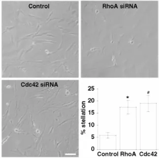
expression (Fig. 1). Transfected cells were analyzed for their morphological features, which revealed a moderate tendency for stellation in both RhoA- and Cdc42-depleted cells, as compared to control cells (Fig. 2). The key finding of these experiments is that Cdc42 silencing results in significant stellation indicating, contrary to our initial assumption, that the GTPase is somewhat active in basal conditions. This in turn suggests a functional link between RhoA and Cdc42, given the fact that RhoA inhibition per se induces stellation. As mentioned earlier, a control of Cdc42 over RhoA is highly unlikely; thus, we here tested the alternative possibility that Cdc42 acts downstream of RhoA. For that purpose, we performed a Cdc42 pull-down in conditions of RhoA inhibition by 10 µM adenosine versus its full activation by 10% serum (see Rosso et al. 2002a). For comparison, we included Cdc42 activity measurements during 5 or 10-min exposures to 10 nM VP, which we previously showed to be a reliable Cdc42 activator (Rosso et al. 2002b). The results indicate that Cdc42 activity in serum is comparable to that obtained with VP, and higher than that in the presence of adenosine (Fig. 3), i.e. in conditions of RhoA inhibition. Although we cannot exclude the possibility of a direct effect of serum on Cdc42, an interpretation consistent with our siRNA results is that Cdc42 activation is due to RhoA stimulation.
Fig. 1 Western blots of RhoA and Cdc42 protein expression in pituicytes transfected with siRNAs targeting either protein. Left panels show sample gels in which upper bands in left lanes correspond to a control signal obtained for each GTPase from cells transfected with siRNA targeting the other GTPase, while right lanes show the signal obtained from cells transfected with siRNA targeting the same GTPase. Lower bands correspond to actin detected in samples and used for normalizing the amounts of RhoA or Cdc42. Right panels summarize relative intensity of protein signal obtained from cells with or without transfection of siRNA targeting the corresponding protein, *p = 0.03 (n = 3 each); #p = 0.001 (n = 4 each) compared to control (without); Student's one-tail t-test
Fig. 2 Effects of RhoA or Cdc42 siRNA transfection on pituicyte morphology. Phase-contrast microphotographs show pituicyte cultures under each relevant condition as indicated. Scale bar = 40 µm. Lower right panel summarizes relative stellation quantified in each condition, *p = 0.02 (n = 11); #p = 0.01 (n = 22) compared to control (n = 10); Kruskal-Wallis test
Fig. 3 Pull-down of Cdc42 in various conditions as indicated. Upper panels show western blots of total and activated protein from pituicytes incubated in 0.025% serum with 10 µM adenosine (Ado) for 45 min, to which 10 nM VP was added for 5 or 10 min, or in 10% serum (no drug added). Lower panel shows the corresponding ratio of activated/total Cdc42
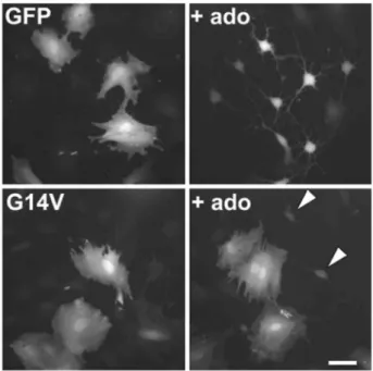
Fig. 4 Effects of constitutively active RhoA mutant (G14V) transfection on pituicyte morphology. Upper panels correspond to control conditions (GFP alone, no G14V). Based on a 1:10 concentration ratio cotransfection of GFP + mutant, bright cells in lower panels presumably reflect successful transfection of the mutant. Left: without adenosine; right: with 10 µM adenosine (ado). Notice two dim cells in lower right panel (arrowheads) that presumably fail to express G14V and therefore appear stellate. Scale bar: 50 µm
In order to confirm the major role of RhoA in pituicyte stellation and gain further insight into the mechanism involved, we transfected cells with GFP either alone or in combination with RhoA-G14V, a constitutively active mutant of the GTPase. GFP transfection did not induce any effect in pituicytes, which maintained their spread shape in control conditions, and became stellate in the presence of 10 µM adenosine (Fig. 4, upper panels). This rules out non-specific effects of the transfection procedure on pituicyte morphology. Likewise, cotransfection of
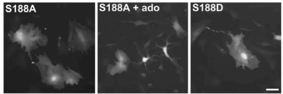
GFP + RhoA-G14V did not affect spread pituicytes (Fig. 4, lower left panel). On the other hand, RhoA-G14V expression clearly prevented adenosine from inducing pituicyte stellation (Fig. 4, lower right panel), consistent with our previous finding that adenosine induces stellation via RhoA inhibition. In order to determine whether the mechanism of inhibition involves serine phosphorylation of RhoA, as was shown in other cell types (Lang et al. 1996; Ellerbroek et al. 2003), we transfected pituicytes with two types of mutants: RhoA-S188A, a
phosphorylation-resistant mutant, or RhoA-S188D, a phosphomimetic mutant. The former had no effect on spread pituicytes nor did it prevent adenosine from inducing stellation, whereas the latter mutant by itself did not induce stellation (Fig. 5). This suggests that adenosine induces pituicyte stellation by inhibiting RhoA via a mechanism other than phosphorylation.
We then investigated in more detail the role of Cdc42 in pituicyte morphology. For that purpose, we transduced cells with Cdc42-V12, a constitutively active mutant of the GTPase, fused to GFP. The expression of GFP alone did not prevent adenosine from inducing stellation, which is characterized by a diffuse staining of actin in both transfected and non-transfected cells (Fig. 6, upper panels). Conversely, cells expressing Cdc42-V12 were resistant to adenosine-induced stellation, with actin rather organized as a submembrane—i.e., cortical—structure, despite the sparse presence of stress fibers (Fig. 6, lower panels). A more complete depiction of this phenomenon is presented in Fig. 7, in which control pituicytes display a dense alignment of stress fibers in resting conditions (i.e. with RhoA activated) versus a diffuse distribution of depolymerizing actin in the presence of adenosine (upper panels). Conversely, the actin of pituicytes expressing Cdc42-V12 retains a cortical localization following addition of adenosine(lower panels). As expected, adenosine-mediated stellation is prevented solely in those cells that express the Cdc42 mutant (lower right panel). Note that cortical actin also prevails a short time after exposure of stellate wild-type pituicytes to VP (inset), which has been shown to activate Cdc42 (Rosso et al. 2002b).
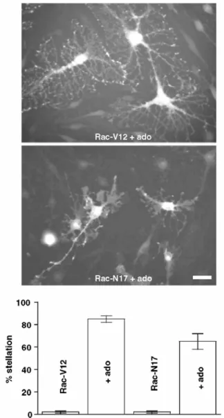
As Rac1 is known to be implicated in cell morphology (Hall 1998), we investigated its potential role in pituicyte stellation using constitutively active (V12) or dominant negative (N17) mutants of the GTPase. Neither V12- nor N17-Rac1 transduction had any effect on either "resting" pituicytes, which remained spread, or adenosine-stimulated cells, which became stellate in the presence of the purine (Fig. 8). On the other hand, a consistent finding was that adenosine-stimulated, stellate pituicytes expressing Rac1-V12 are larger and exhibit more numerous, long and branched processes than control cells (Fig. 8, top panel).
Fig. 5 Effects of phosphorylation-resistant (S188A) or phosphomimetic (S188D) RhoA mutant transfection (same method as in Fig. 4) on pituicyte morphology. Left image: S188A alone; middle image: S188A + 10 µM
adenosine; note that not all cells are stellate consistent with the fact that adenosine never induces 100% stellation; right image: S188D alone. Scale bar: 50 µm
Fig. 6 Effects of expression of a V12 constitutively active mutant of Cdc42 on adenosine-mediated pituicyte stellation. All pictures are in the presence of 10 µM adenosine. Actin is labeled with rhodamine-conjugated phalloidin (red). Upper row shows cells expressing GFP (green) alone (no V12); lower row shows cells expressing Cdc42-V12 protein fused to GFP. Notice two stellate, GFP-negative cells (arrowheads). Scale bar: 50 µm
Fig. 7 Effects of Cdc42 activation on pituicyte actin cytoskeleton. Upper two pictures and inset represent wild-type pituicytes in 0.025%-serum medium, which maintains basal RhoA activation and dense actin (red) stress fibers in control conditions, and allows stellation in the presence of 10 µM adenosine. Inset displays initiation of stellation reversal by 10 nM VP applied for 3 min. Lower pictures are overlays of actin (red) and GFP+Cdc42-V12 (green) fluorescence. Left image: two GFP+Cdc42-V12+ cells (center) have lost most of their stress fibers and instead display cortical actin. Right image: V12-negative cells (arrowheads) are stellate in the presence of 10 µM adenosine, while V12+ cells are not. Notice the even more pronounced cortical localization of actin (yellow overlay) in that case. Scale bar: 60 µm
Fig. 8 Effects of Rac1 constitutively active (V12) or dominant negative (N17) mutant expression on pituicyte morphology. Top panels show pituicytes expressing (GFP fluorescence) Rac1-V12 or Rac1-N17 in the presence of 10 µM adenosine. Scale bar: 40 µm. Bottom graph summarizes stellation observed in cells expressing Rac1-V12 or Rac1-N17 in the presence or absence of 10 µM adenosine (n = 3 dishes for each condition; only GFP+ cells were counted)
Pituicyte Stellation and the Actin Cytoskeleton
Given the fact that RhoA or Cdc42 activation appears to block stellation in association with specific states of actin polymerization, we hypothesized that actin depolymerization might be a sufficient factor for pituicyte stellation. Similar to a previous study using cytochalasin B (Miyata et al. 1999), we found that actin
depolymerization by cytochalasin D indeed was a powerful means of inducing pituicyte stellation, with results consistently near 100% efficiency (Fig. 9, lower left panel). Conversely, pituicyte incubation with increasing concentrations of jasplakinolide (JPK), a cell-permeant drug that stabilizes F-actin, provided proportionally increasing, i.e., dose-dependent, resistance to adenosine-mediated stellation up to almost complete blockade (Fig. 9). At high concentrations (1-5 µM), JPK induced shape changes somehow resembling stellation, though treated cultures consisted more of both net-like structures with shriveling, dark somas and rounded, phase-bright but processless cells. As these effects might be nonspecific, they were not further investigated.
Next, to test whether cortical actin organization was a sufficient condition to prevent stellation, we treated pituicytes with calyculin-A, a phosphatase inhibitor that dissolves stress fibers and induces cortical actin redistribution (Kreienbühl et al. 1992; Patterson et al. 1999). A striking result of that treatment was the rounding up of pituicytes along with a tendency to form aggregates, an effect consistently observed in various cell types (e.g. Patterson et al. 1999). It is unlikely that cell rounding was the result of a non-specific, toxic effect of
calyculin-A since (1) we used a concentration ~20 times lower than that used in most studies, and (2) we did not observe any tendency of treated cells to detach from substrate. Following addition of adenosine, pituicytes remained rounded but no processes were formed, i.e., stellation was inhibited by calyculin-A (Fig. 9, upper right panel). Finally, we verified that the actin cytoskeleton of JPK- or calyculin-A-treated cells was as predicted. Figure 10 shows that pituicytes incubated with 250 nM JPK or 5 nM calyculin-A display an actin cytoskeleton organized respectively as a stress-fiber or cortical network, both before and after addition of 10 µM adenosine.
Fig. 9 Effects of cytoskeleton-targeting drugs on pituicyte stellation. Phase-contrast pictures of pituicyte cultures in 0.025%-serum medium were taken in the various conditions indicated. Control image corresponds to no drug added (first zero on graph). Jasplakinolide (JPK) or calyculin-A were added 45 min before adenosine.
Treatment with 10 µM cytochalasin-D induced 93 ± 2% stellation (n = 5), while 5 nM calyculin-A resulted in virtually 0% stellation (n = 6). Scale bar: 70 µm. Bottom right: Concentration-dependent inhibition of 10 µM adenosine-induced pituicyte stellation by JPK (n = 3 or 4 dishes per condition). Adenosine was present as indicated by the bar above the graph
Fig. 10 Effects of jasplakinolide (JPK) or calyculin-A on pituicyte actin cytoskeleton. Note the same patterns of phalloidin labeling before or after addition of 10 µM adenosine. Scale bar = 50 µm
Discussion
Our siRNA experiments resulted in significant yet incomplete inhibition of RhoA and Cdc42. This is shown by the results of both our western blot and morphological analyses. In particular, if RhoA silencing was equivalent to its pharmacological inhibition, we would have expected a much higher level of stellation, which generally averages 70-80% in the presence of adenosine (e.g., Figs. 8 and 9). In addition to incomplete silencing, the fact that our knock-downs were performed in 10% serum suggests that remaining RhoA was in a fully activated form, making it difficult to disrupt stress fibers. Nevertheless, the fact that Cdc42 silencing results in significant stellation prompted us to test whether the GTPase might act downstream of RhoA, and this hypothesis is now strengthened by the observed increase in activity of Cdc42 in serum conditions that stimulate RhoA. In that perspective however, a difficulty arises as to the fact that each GTPase has widely different effects on the actin cytoskeleton, as discussed below. These conflicting notions might be resolved by hypothesizing that RhoA activates Cdc42 in parallel with an effector (such as p160Rho-kinase) that induces and maintains actin stress fibers in a dominant fashion. Thus, inhibiting RhoA would release pituicytes from cytoskeletal constraints that derive from both Cdc42 and the stress fiber-inducing effector, thereby triggering stellation. However, to
comprehensively account for our results, alternative and perhaps more subtle forms of crosstalk between the two GTPases cannot be excluded.
Relevant to adenosine-induced pituicyte stellation, protein kinase A phosphorylation of a Ser residue has been suggested as one mechanism for RhoA inhibition (Lang et al. 1996; Ellerbroek et al. 2003). This indeed might represent an intriguing link between metabotropic receptor stimulation and monomeric GTPase inhibition. Given our results with phosphorylation-resistant and phosphomimetic mutants, however, this pathway can be ruled out regarding adenosine-mediated RhoA inhibition and ensuing stellation. In fact, our results are not surprising considering the following points. Ser phosphorylation of RhoA is normally related to its inhibition by a guanine-nucleotide dissociation inhibitor (GDI; Lang et al. 1996; Ellerbroek et al. 2003). In this case, GTP-bound RhoA is not amenable to pull-down analysis (Ellerbroek et al. 2003), which is inconsistent with our previous pull-down data (Rosso et al. 2002a). RhoA inhibition by a GTPase-activating protein (GAP) in turn can be measured by pull-down (Ellerbroek et al. 2003). Furthermore, as RhoA-G14V, which is protected from GAP-mediated GTP hydrolysis, is an efficient stellation inhibitor in our hands, we believe that a GAP-related mechanism is more likely to mediate RhoA inhibition by adenosine. Not inconsistent with such a mechanism, Harrington et al. (2004) proposed that adenosine downregulates RhoA by decreasing its methylation state via inhibition of S-adenosylmethionine (SAM)-dependent methyltransferases. In order to rule out such non-receptor-mediated effects of adenosine, stellation experiments in the presence of nucleoside transport inhibitors will have to be performed.
Our previous studies suggested that actin stress fibers were responsible for pituicyte spreading and that their breakdown was the triggering event for stellation (Rosso et al. 2002a). This view is no longer consistent with the observation that Cdc42 activation prevents stellation and maintains spreading while inhibiting stress fibers (Figs. 6 and 7). We believe that the key observation to account for these effects is that Cdc42 does not simply inhibit stress fibers but also appears to reorganize polymerized actin in a cortical fashion, which is consistent with its reported induction of actin polymerization at the cell periphery (Gasman et al. 2004). In fact, a similar
phenomenon can be observed when pituicytes are exposed to VP (inset of Fig. 7), which, as we found, activates Cdc42 and reverses stellation (Rosso et al. 2002b). Consistent with these data, pituicyte stellation is completely blocked by calyculin-A, an agent that dissolves stress fibers and induces cortical actin redistribution (Kreienbühl et al. 1992; Patterson et al. 1999). Incidentally, it would be interesting to investigate, e.g. by pull-down analysis, whether calyculin-A directly activates Cdc42. In that respect, it is worth mentioning previous work by others (Feoktistov et al. 2000) showing that calyculin-A potentiates cAMP-mediated activation of Cdc42.
From the above considerations, we could conclude that depolymerized actin, which might represent a default state in the absence of monomeric GTPase activity, constitutes a necessary and sufficient factor to induce stellation. Indeed, cytochalasin D is a very—if not the most—efficient agent for triggering pituicyte stellation. Conversely, our experiments with JPK and calyculin-A indicate that actin polymerization constitutes a sufficient condition to prevent stellation. The molecular basis for such a phenomenon remains to be determined. Perhaps polymerized actin, whether organized as stress fibers or cortical ring, constitutes a physical or biochemical barrier for the microtubule extensions that characterize stellation (Rosso et al. 2002a). Contrary to the clear involvement of RhoA and Cdc42, our data suggest that in pituicytes Rac1 may not exert much effect upon actin as neither its constitutively active nor its dominant negative forms prevent or induce stellation. Intriguingly, in place of its known induction of lamellipodia and membrane ruffles (Hall 1998), Rac1 appears to have a distinct dendritogenic effect in pituicytes.
From a physiological viewpoint, our results with pituicytes may open a relevant avenue of investigation into the mechanisms that regulate the plasticity of other astrocytes of the nervous system in vivo.
Acknowledgements
We are grateful to Anne-Sophie Coldefy for her help with western blot analysis. We thank Pierre Roux for giving us the Cdc42 and Rac1 mutants, and Gervaise Loirand and Pierre Chardin for the gift of RhoA mutants.
References
Abe K, Saito H (1998) Adenosine stimulates stellation of cultured rat cortical astrocytes. Brain Res 804:63-71
Deroanne CF, Hamelryckx D, Ho TT, Lambert CA, Catroux P, Lapiere CM, Nusgens BV (2005) Cdc42 downregulates MMP-1 expression by inhibiting the ERK1/2 pathway. J Cell Sci 118:1173-1183
Ellerbroek SM, Wennerberg K, Burridge K (2003) Serine phosphorylation negatively regulates RhoA in vivo. J Biol Chem 278:19023-19031 Feoktistov I, Goldstein AE, Biaggioni I (2000) Cyclic AMP and protein kinase A stimulate Cdc42: role of A(2) adenosine receptors in human mast cells. Mol Pharmacol 58:903-910
Fincham VJ, Chudleigh A, Frame MC (1999) Regulation of pl90 Rho-GAP by v-Src is linked to cytoskeletal disruption during transformation. J Cell Sci 112:947-956
Gadea G, Lapasset L, Gauthier-Rouviere C, Roux P (2002) Regulation of Cdc42-mediated morphological effects: a novel function for p53. EMBO J 21:2373-2382
Gasman S, Chasserot-Golaz S, Malacombe M, Way M, Bader MF (2004) Regulated exocytosis in neuroendocrine cells: a role for subplasmalemmal Cdc42/N-WASP-induced actin filaments. Mol Biol Cell 15:520-531
Gratzl M, Torp-Pedersen C, Daertt D, Treiman M, Thorn NA (1980) Isolation and characterization of secretory vesicles from bovine neurohypophyses. Hoppe Seylers Z Physiol Chem 361:1615-1628
Hall A (1998) Rho GTPases and the actin cytoskeleton. Science 279:509-514
Harrington EO, Newton J, Morin N, Rounds S (2004) Barrier dysfunction and RhoA activation are blunted by homocysteine and adenosine in pulmonary endothelium. Am J Physiol Lung Cell Mol Physiol 287:L1091-1097
Hatton GI (1999) Astroglial modulation of neurotransmitter/peptide release from the neurohypophysis: present status. J Chem Neuroanat 16:203-222
Kreienbühl P, Keller H, Niggli V (1992) Protein phosphatase inhibitors okadaic acid and calyculin A alter cell shape and F-actin distribution and inhibit stimulus-dependent increases in cytoskeletal actin of human neutrophils. Blood 80:2911-2919
Lang P, Gesbert F, Delespine-Carmagnat M, Stancou R, Pouchelet M, Bertoglio J (1996) Protein kinase A phosphorylation of RhoA mediates the morphological and functional effects of cyclic AMP in cytotoxic lymphocytes. EMBO J 15:510-519
Miyata S, Furuya K, Nakai S, Bun H, Kiyohara T (1999) Morphological plasticity and rearrangement of cytoskeletons in pituicytes cultured from adult rat neurohypophysis. Neurosci Res 33:299-306
Morris JF, Pow DV, Shaw FD (1988) Release of neuropeptides from magnocellular neurons: does anatomical compartmentalization have a functional significance? In: Pickering BT, Wakerley JB, Summerlee AJS (eds), Neurosecretion: Cellular aspects of the production and release of neuropeptides, Plenum Press, New York, pp. 113-122
Narumi S, Kimelberg HK, Bourke RS (1978) Effects of norepinephrine on the morphology and some enzyme activities of primary monolayer cultures from rat brain. J Neurochem 31:1479-1490
Patterson RL, van Rossum DB, Gill DL (1999) Store-operated Ca2+ entry: evidence for a secretion-like coupling model. Cell 98:487-499
Ramakers GJA, Moolenaar WH (1998) Regulation of astrocyte morphology by RhoA and lysophosphatidic acid. Exp Cell Res 245:252-262 Ramsell KD, Cobbett P (1997) Serum uncouples elevation of cyclic adenosine monophosphate concentration from cyclic adenosine monophosphate dependent morphological changes exhibited by cultured pituicytes. Neurosci Lett 226:41-44
Rolli-Derkinderen M, Sauzeau V, Boyer L, Lemichez E, Baron C, Henrion D, Loirand G, Pacaud P (2005) Phosphorylation of serine 188 protects RhoA from ubiquitin/proteasome-mediated degradation in vascular smooth muscle cells. Circ Res 96:1152-1160
Rosso L, Peteri-Brunbäck B, Vouret-Craviari V, Deroanne C, Troadec J-D, Thirion S, Van Obberghen-Schilling E, Mienville J-M. (2002a) RhoA inhibition is a key step in pituicyte stellation induced by A1-type adenosine receptor activation. Glia 38:351-362
Rosso L, Peteri-Brunbäck B, Vouret-Craviari V, Deroanne C, Van Obberghen-Schilling E, Mienville J-M. (2002b) Vasopressin and oxytocin reverse adenosine-induced pituicyte stellation via calcium-dependent activation of Cdc42. Eur J Neurosci 16:2324-2332
Roux P, Gauthier-Rouvière C, Doucet-Brutin S, Fort P (1997) The small GTPases Cdc42Hs, Rac1 and RhoG delineate Raf-independent pathways that cooperate to transform NIH3T3 cells. Curr Biol 7:629-637
Theodosis DT, Macvicar B (1996) Neurone-glia interactions in the hypothalamus and pituitary. Trends Neurosci 19:363-367
Vouret-Craviari V, Boulter E, Grall D, Matthews C, Van Obberghen-Schilling E (2004) ILK is required for the assembly of matrix-forming adhesions and capillary morphogenesis in endothelial cells. J Cell Sci 117:4559-4569