The Role of DcR3 in
Systemic Lupus Erythematosus and
Islet β-Cell Viability and Function
Bing Han
Département de Médecine Faculté de Médecine Université de Montréal
Thèse présentée à la Faculté des études supérieures en vue de l’obtention du grade de
Philosophiae Doctor (Ph.D.) en Sciences Biomédicales
Université de Montréal Faculté des études supérieures
Cette thèse intitulée:
« The Role of DcR3 in Systemic Lupus Erythematosus and Islet β-Cell Viability and Function »
Présentée par: Bing Han
A été évaluée par un jury composé des personnes suivantes:
Dr. Manuela Santos ……… Président du jury Dr. Jiangping Wu ……… Directeur de recherché Dr. Hongyu Luo ……… Directeur de recherche Dr. Janos G. Filep ……… Membre du jury Dr. Alain Lamarre ……… Examinateur externe Dr. Daniel Lajeunesse ……… Représentant du doyen de la FES
Summary
Decoy receptor 3 (DcR3) is a member of the tumor necrosis factor (TNF) receptor family, and is widely expressed in human normal tissues and malignant tumors. It is a decoy receptor of three TNF family members, i.e., FasL, LIGHT and TL1A. The interaction of DcR3 and its ligands will not transmit signal into cells via DcR3 because DcR3 is a soluble protein without a transmembrane and intracellular segment. Thereby, DcR3 competitively inhibits signaling through three functional receptors, i.e., Fas, HVEM/LTβR and DR3.
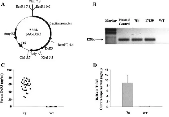
In previous studies, we found that DcR3 could modulate immune cell function, and protect islet viability. Herein, we generated DcR3 transgenic (Tg) mice driven by the human β-actin promoter to further investigate the function of DcR3.
Interestingly, the DcR3 Tg mice developed a lupus-like syndrome at 6 months of age. They presented a variety of autoantibodies including anti-nucleus and anti-dsDNA antibodies. They also manifested renal, dermal, hepatic and hematopoietic lesions. Compared to lpr and gld mouse lupus models, DcR3 Tg mice more closely resembled human SLE in terms of Th2-biased immune response and anti-Sm antibody production.
Furthermore, we found that DcR3-producing hematopoietic cell were sufficient to cause these pathological changes. Mechanistically, DcR3 may break T-cell homeostasis to interfere with peripheral tolerance, and then induce autoimmunity.
In humans, we detected high DcR3 levels in SLE patient sera. The high DcR3 levels were related to elevated IgE titer in some SLE patients, as was the case in the mouse model. Therefore, DcR3 may represent an important pathogenetic factor of human SLE.
Utilizing the DcR3 Tg mouse, we further elucidated the mechanism by which DcR3 protected islets from primary nonfunction (PNF). Blocking of LIGHT and TL1A signaling by DcR3 are involved in such protection. Moreover, by mRNA microarray we identified possible downstream molecules, which may mediate such protection. We confirmed that Adcyap1 and Bank1 played critical roles in mediating DcR3’s effect in islet protection.
Our studies resolved a puzzle about the relationship between the Fas/FasL apoptosis signaling pathway and the pathogenesis of human SLE. DcR3 can block Fas/FasL pathway even if there is no genetic mutation in Fas and FasL. DcR3 can simultaneously interfere with LIGHT and TL1A signaling to cause a more complex phenotype than the simple Fas or FasL mutation
in patients. DcR3 can also be employed as a potential diagnostic parameter for SLE. The discovery of the mechanism of DcR3 in protecting islets allows us to explore novel therapeutic targets to protect islet graft.
Key words: DcR3; transgenic; systemic lupus erythematosus; islet transplantation; primary nonfunction ( PNF).
Résumé
Le récepteur DcR3 (Decoy receptor 3) est un membre de la famille des récepteurs aux facteurs de nécrose tumorale (TNF). Il est fortement exprimé dans les tissus humains normaux ainsi que les tumeurs malignes. DcR3 est un récepteur pour trois ligands de la famille du TNF tels que FasL, LIGHT et TL1A. Étant une protéine soluble donc dépourvue de la portion transmembranaire et intracytoplasmique, le récepteur DcR3 est incapable d’effectuer une transduction de signal intracellulaire à la suite de son interaction avec ses ligands. De ce fait, DcR3 joue un rôle de compétiteur pour ces derniers, afin d’inhiber la signalisation via leurs récepteurs fonctionnels tels que Fas, HVEM/LTβR et DR3.
Lors de nos précédentes études, nous avons pu démontrer, que DcR3 pouvaist moduler la fonction des cellules immunitaires, et aussi protéger la viabilité des îlots de Langerhans. À la suite de ces résultats, nous avons généré des souris DcR3 transgéniques (Tg) en utilisant le promoteur du gène β-actine humaine afin d’étudier plus amplement la fonction de ce récepteur.
d'auto-anticorps comprenant des anticorps anti-noyaux et anti-ADN. Elles ont également manifesté des lésions rénales, cutanées, hépatiques et hématopoïétiques. Contrairement aux modèles de lupus murin lpr et gld, les souris DcR3 sont plus proche du SLE humain en terme de réponse immunitaire de type Th2 et de production d'anticorps d'anti-Sm. En péus, nous avons constaté que les cellules hématopoïétiques produisant DcR3 sont suffisantes pour causer ces pathologies. DcR3 peut agir en perturbant l’homéostasie des cellules T pour interférer avec la tolérance périphérique, et ainsi induire l'autoimmunité.
Chez l'humain, nous avons détecté dans le sérum de patients SLE des niveaux élevés de la protéine DcR3. Chez certains patients, comme chez la souris, ces niveaux sont liés directement aux titres élevés d’IgE. Par conséquent, DcR3 peut représenter un facteur pathogénique important du SLE humain.
L’étude des souris Tg DcR3, nous a permis aussi d’élucider le mécanisme de protection des îlots de Langerhans. Le blocage de la signalisation des ligands LIGHT et TL1A par DcR3 est impliqué dans une telle protection. D'ailleurs, nous avons identifié par ARN microarray quelques molécules en aval de cette interaction, qui peuvent jouer un rôle dans le mécanisme
d’action. Nous avons par la suite confirmé que Adcyap1 et Bank1 joue un rôle critique dans la protection des îlots de Langerhans médiée par DcR3. Notre étude a ainsi élucidé le lien qui existe entre la signalisation apoptotique médiée par Fas/FasL et la pathogénèse du SLE humain. Donc, malgré l’absence de mutations génétiques sur Fas et FasL dans le cas de cette pathologie, DcR3 est capable de beoquer cette signalisation et provoquer le SLE chez l’humain. Ainsi, DcR3 peut simultanément interférer avec la signalisation des ligands LIGHT et TL1A et causer un phénotype plus complexe que les phénotypes résultant de la mutation de Fas ou de FasL chez certains patients. DcR3 peut également être utilisé comme paramètre diagnostique potentiel pour le SLE. Les découvertes du mécanisme de protection des îlots de Langerhans par DcR3 ouvrent la porte vers de nouveaux horizons afin d'explorer de nouvelles cibles thérapeutiques pour protéger la greffe d'îlots.
Mots clés : DcR3; transgénique; systemic lupus erythematosus; transplantation d'îlots; PNF.
INDEX Summary………..……….…….….…..III Résumé……….………..………..…..……VI List of Figures……….…..……..…..…...…...XIII List of Abbreviations……….……….………...…..XVI Acknowlegements……….…….…………...…………..…..XX I. INTRODUCTION……….………….……….…..…..….1 1. Decoy receptor 3………..….……….…….…………...…...2
1-1 General information about DcR3…...………...……….…..2
1-2 Biological function of DcR3 and its molecular mechanism…...………4
1-2-1 Fas and FasL……….………...…………4
1-2-2 LIGHT and HVEM/LTβR …...………..….. 6
1-2-3 TL1A and DR3………...………..….. 7
1-3 Regulation of DcR3 expression………...………... ………8
1-4 Immunity and DcR3…………...……….………. ………10
1-5 Cancer and DcR3………...………..……. ………11
2. Systemic lupus erythematosus (SLE)………..……..……….. 14
2-1 Epidemiology of SLE ……...……….……….. 14
2-2 Pathoaetiology of SLE………...……….…….……….. 15
2-2-1 Hormones………...……..……….……….. 16
2-2-3 Environment factors….……….……….. 18
2-2-4 Apoptosis and SLE………..………...……….. 19
2-3 Mouse models of SLE….………..……….. 22
2-4 Clinical features of SLE……..………...………...….……….. 23
2-4-1 Symptoms……….……….……….. 23
2-4-2 Diagnosis……….………...……….. 23
2-4-3 Therapy………..…..…….……….. 25
2-4-4 Prognosis……….……….……….…………..27
3. Diabetes mellitus………...………...…….. 28
3-1 The Epidemiology of Diabetes Mellitus..….……….…….. 28
3-2 The pathoaetiology of Diabetes Mellitus……..…………..…….……….. 31
3-2-1 Genetic Factors……….……….. 33
3-2-2 Environmental Factors………..……..……….. 34
3-2-3 Accelerator Hypothesis………..…………... 36
3-3 The clinical features of DM…….……..………..…………... 37
3-3-1 Symptoms and complications…….………..…………... 37
3-3-2 Diagnosis…….………..…………... 39
3-3-3 Therapy…….…..………..…………... 40
3-3-3-1 Diabetes education and lifestyle intervention………..…………... 40
3-3-3-2 Insulin and other medications………..…………... 40
3-3-3-3 Organ and cell transplantation………..…………... 42
Hypothesis and Objective…….………..…………... 46 References for introduction……….………...…... 47 II. ARTICLES……….…………..…..66 Article 1: Overexpression of human decoy receptor 3 in mice results in a systemic lupus erythematosus-like syndrome……..………..…..…….…..67
Article 2: DcR3 as a diagnostic parameter and risk factor for systemic lupus erythematosus..………...….…...…...…....109
Article 3: DcR3 protects islet β-cells from apoptosis through modulating Adcyap1 and Bank1 expression………...……….………....145
III. DISCUSSION………..……...…..190 1. DcR3 and the pathogenesis of SLE…………...…….……….191 1-1 The DcR3 overexpression in self-reactive lymphocyte interferes with their
anergy………..………..…192
1-2 DcR3 impairs the clearance of apoptotic cells and increases autoantigen presentation………..…...…194
1-3 DcR3 increases the memory T cell pool and enhances the secondary response to autoantigens…………...…….…....…...….…….…195
1-4 DcR3 disturbs B-cell homeostasis and tolerance……….196 1-5 DcR3 favors Th2 development………..……….…...……….197 2. The mechanisms by which DcR3 protects pancreatic islets………….198
2-1 DcR3 Tg islet graft resisted PNF and presented long term normoglycemia in s y n g e n e i c d i a b e t e s m o u s e r e c e i v i n g a s u b o p t i m a l d o s e o f
2-2 DcR3 acts through the TL1A and LIGHT pathways……….….199 2-3 Using DNA microarray to identify molecules downstream of DcR3
action.……….…………..200
Conclusion and Future Perspectives...202 References for discussion……….……….……..…...…….………203
LIST OF FIGURES AND TABLES
Introduction
Fig 1. The homology of the amino-acid sequences between DcR3 and OPG (osteoprotegerin).…3
Fig 2. The apoptotic signal pathway of Fas / FasL (CD95/CD95L)………….………...……...5
Fig 3. Genomic structure of DcR3 and its adjacent genes….………..…………..…..10
Fig 4. The susceptibility loci of SLE……….……….…………..18
Fig 5. The geographic distribution of DM prevalence……….………30
Fig 6. The pathogenesis of T1DM………...……31
Fig 7. Pathophysiology of T2DM……….………...…32
Table 1. Characteristics of lupus-prone mouse strains…….………..….……..………...22
Table 2. Diagnostic criteria of diabetes mellitus and other categories of hyperglycemia….……39
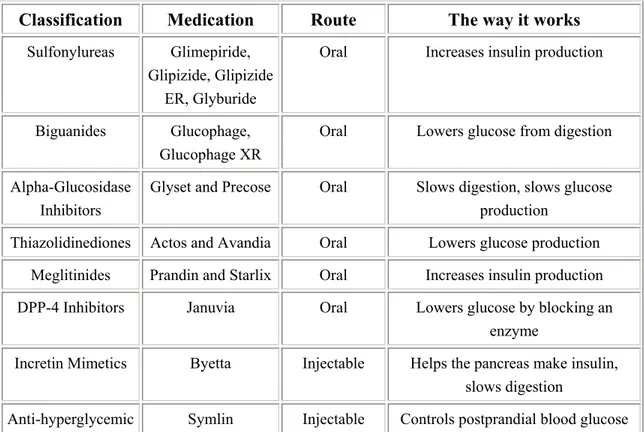
Table 3. Medications for Diabetes…...………...41
Article 1: Fig 1. Generation and characterization of decoy receptor 3 (DcR3)-transgenic mice...….99
Fig 2. Lymphadenopathy and survival rates in DcR3-Tg mice ………..……..…..100
Fig 3. Autoantibodies and B-1 cells in DcR3-Tg and WT mice………...…..…102
Fig 4. Tissue histopathology and urine and hematologic findings in aged DcR-Tg and WT mice……….…..104
Fig 6. Expanded CD44high,CD62Llow,CD25- CD4 population in peripheral blood mononuclear cells
(PBMCs) of DcR3-Tg mice….….…………..……….…………...…….….108
Article 2: Fig 1. Serum DcR3 levels and their relationship with SLEDAI in SLE patients...…….……...137
Fig 2. Relationship among medication, serum DcR3 levels and SLEDAI of SLE patients.……139
Fig 3. Elevated serum IgE levels in SLE patients……….………140
Fig 4. Th2-prone immune responses in young DcR3 Tg mice.………...….….. 141
Fig 5. Development of an SLE-like syndrome in WBI–BMTx mice….………...….142
Fig 6. Reduced survival rate in Tg BMTx recipients………....…144
Article 3: Fig 1. DcR3 secretion by Tg islets……….…………181
Fig 2. Tg islets present reduced apoptosis and maintain their function under cytokines assault...182
Fig 3. Expression of TL1A and LIGHT pathway components in islets according to real-time PCR……….………..…….…183
Fig 4. Recombinant mouse LIGHT and TL1A induced lpr/lpr islet cell apoptosis……...….…184
Fig 5. Reduced islet PNF in transplantation using DcR3 Tg islets………..185
Fig 6. Adcyap1 overexpression protected β-cells from cytokine-induced apoptosis…………..186
Fig 7. Inhibition of Bank1expression decreased cytokine-induced apoptosis in NIT-1 cells…...188
Table 2. Genes identified by DNA microarray and verified by real-time PCR from IFN-γ plus IL-1β-treated Tg versus WT islets….………..…179 Table 3. Genes identified by DNA microarray and verified by real-time PCR from TNF-α plus
LIST OF ABBREVIATIONS
ACR American College of Rheumatology
Adcyap1 adenylate cyclase activating polypeptide 1 AICD activation-induced cell death
ALPS lymphoproliferative syndrome ANA anti-nuclear antibody
AZA azathioprine
BMTx bone marrow transplantation CAPN10 calpain 10
CARD caspase-recruitment domain CCR-7 CC-chemokine receptor-7
cDNA complementary DNA
CFM cyclophosphamide CRD cysteine-rich domains
CTLA4 cytotoxic lymphocyte antigen 4 DAPK death-associated protein kinase DcR3 Decoy receptor 3
DD death domain
DED death-effector domain
DHEA dehydroepiandrosterone
DHEA-S dehydroepiandrosterone sulfate DISC death-inducing signalling complex DR death receptor
ds-DNA double stranded DNA
EBV Epstein-Barr virus
ELISA enzyme-linked immunosorbent assay
ERK1/2 extracellular signal-regulated kinase 1 and 2 FACS fluorescence-activated cell sorting
FADD Fas-associated death domain
FasL Fas ligand
FFA free fatty acid FLICE FADD-like ICE
GAD glutamic acid decarboxylase GnRH gonadotrophin releasing hormone
H&E hematoxylin and eosin
HCQ hydroxychloroquine HLA human leukocyte antigen
HPA hypothalamo–pituitary–adrenal
HVEM herpesvirus entry mediator protein
i.v. intravenously IEQ islet equivalents IFN-γ Interferon-γ
IGF insulin-like growth factor IGT impaired glucose tolerance IL-2 Interleukin-2
iNOS inducible nitric oxide synthase
IRS insulin receptor substrate
JNK c-Jun NH2-terminal kinase
LADA latent autoimmune diabetes of the adult
LIGHT lymphotoxin-like, exhibits inducible expression, and competes
with HSV glycoprotein D for HVEM
LTβR lymphotoxin receptor
MAPK mitogen-activated protein kinases
MICA-5 MHC class I chain-related genes allele 5
mTOR mammalian target of rapamycin
NF-κB Transcription factor nuclear factor-κB
NHL novel helicase-like gene
NSAIDs non-steroidal anti-inflammatory drugs
NZB New Zealand Black NZW New Zealand White OGTT oral glucose tolerance test
PBMCs peripheral blood mononuclear cells
PCR polymerase chain reaction
PDN prednisone
PI3K phosphatidylinositol 3-kinase PMA phorbol myristate acetate PNF primary non-function
RRE Rta-responsive element
SLE systemic lupus erythematosus
SLEDAI systemic lupus erythematosus disease activity index STAT signal transducer and activator of transcription STZ streptozocin
T1DM type 1 diabetes mellitus T2DM type 2 diabetes mellitus
TCR T cell receptor
Tg transgenic
TL1A TNF-like molecule 1A TNF tumor necrosis factor
TNFR tumor necrosis factor receptor
TRAIL TNF-related apoptosis-inducing ligand
TUNEL Terminal deoxynucleotidyl transferase-mediated dUTP nick
end-labelling
UV Ultraviolet
VEGI vascular endothelial growth inhibitor
WBI whole-body irradiation WT wild type
ACKNOWLEGEMENTS
I would like to express my sincere gratitude to my supervisor Dr. Jiangping Wu and Dr.
Hongyu Luo for their scientific guidance and encouragement throughout this study and
during the preparation of this thesis.
I would also like to thank all the colleagues in the lab for their splendid co-operation.
Thanks also extended to all my friends and those who always support me in different
ways.
Finally, I would like to thank all of my family members for their understanding,
patience, and their great support, which were critically important for me to complete
1. Decoy receptor 3
1-1. General information about DcR3
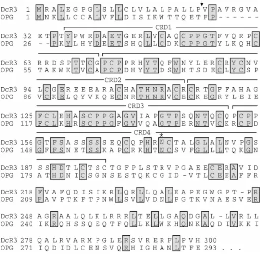
Decoy receptor 3 (DcR3) is a member of the tumor necrosis factor receptor (TNFR) family. Its full-length complementary DNA was first isolated from human fetal lung in 1998 by Pitti et al (1). The DcR3 gene is mapped to chromosome 20q13.3, a region associated with gene amplification and rearrangement in human cancer (2,3). The DcR3 cDNA encodes a 300-aa secreted protein that lacks transmembrane and intracellular domains in its sequence (1,4). It contains 4 conserved cysteine-rich domains (CRD) as other members of TNFR family, and one N-linked glycosylation site (Fig.1). There are two transcription variants of DcR3 at the 5' untranslated region but they encode the same protein (NM_032945.2 and NM_003823.2). DcR3 is expressed in humans, chicken (5), and Conger myriaster (6). The rodents do not have any homologue of DcR3 according to genome-wide computer search.
DcR3 is present in the normal human fetal lung, brain, and liver, also in the adult spleen, colon, lung, and activated T cells (1,7). More interestingly, it is highly expressed in many malignant tumors/cells. Overexpression of
(30/68) gastrointestinal tract adenocarcinomas (9), in 63%-73% (185/294- 163/223) colorectal tumors (10), in 60% (29/48) hepatocellular carcinoma (11), in 67% (10/15) pancreatic cancer (12), and in 83% (15/18) of high-grade gliomas (13). It is also found in lymphomas, renal cancers and ovarian cancers (14,15,16). According to our ELISA analysis, about 50–60% of various tumors overexpressed DcR3 (17).
Figure 1. The homology of the amino-acid sequences between DcR3 andOPG (osteoprotegerin, a typical member of TNFR family) (source: ref.1)
Arrow: the putative signal cleavage site; CRD: the cysteine-rich domains;
1-2. Biological function of DcR3 and its molecular mechanism
Although the DcR3 does not transmit signal into cells, it is able to bind three TNF family members, i.e., FasL (Fas ligand), LIGHT (lymphotoxin-like, exhibits inducible expression, and competes with HSV glycoprotein D for HVEM) and TL1A (TNF-like molecule 1A) (1,4,9,18). Such competition will disturb the interaction of these molecules with their functional receptors, i.e., FasL with Fas, LIGHT with HVEM (herpesvirus entry mediator protein) (19,20) and LTβR (lymphotoxin receptor) (21,22), and TL1A with DR3 (death domain-containing receptor 3) (18). As a consequence, DcR3 competitively suppresses signaling through these receptors, and interferes with their functions. A brief review of the functions of Fas, LIGHT and TL1A is given below.
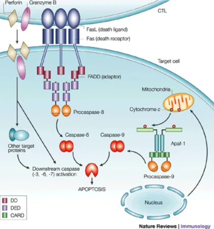
1-2-1. Fas and FasL: Fas antigen, also known as CD95 or APO-1, is a principal death receptor to trigger apoptosis signaling pathway (Fig.2). Various types of cells express Fas, including most immune cells (i.e., T cells, activated B cells, mononuclear phagocytes) and some non-immune cells in the liver, lung, heart (23) and islets of Langerhans (24). Fas contains a conserved “death domain” in its cytoplasmic region. FasL is a homotrimeric membrane protein which is predominantly expressed on activated T cells. When FasL binds to Fas, it clusters 3 or more Fas
molecules to form the death-induced signaling complex (DISC). The death domain of the Fas polymer recruits Fas-associated death domain (FADD). The death effector domain of FADD consequently binds to FADD-like ICE (FLICE), or more commonly known as pro-caspase 8. FLICE can be cleaved into p10 and p18 subunits through self-proteolysis, and form an active heterotetramer enzyme. Active caspase-8 is then released into the cytoplasm to cleave and activate downstream effector caspases, such as caspases 3, 6, or 7. This eventually leads to DNA fragmentation, membrane blebbing, and cell death.
The Fas/FasL apoptosis signaling pathway is involved in T-cell homeostasis, the establishment of immune privilege, cytotoxic T-cell activity (26) and tumor cell survival (27).
Besides triggering Fas positive cell to apoptosis, FasL may also act as a costimulator by retrograde signal transmission into FasL expressing cells by “reverse signaling” (28).
1-2-2. LIGHT and HVEM/LTβR: LIGHT is a type-II transmembrane protein (29), and is highly expressed on activated lymphocytes, CD8+ T cell lines, granulocytes, and monocytes (22). LIGHT, as a costimulator through the LIGHT/HVEM interaction, contributes to T-cell activation and modulates T-cell responses (30,31,32). The LIGHT/HVEM signaling has been involved in graft-versus-host diseases (33,34). On the other hand, the LIGHT/LTβR signaling plays various biological activities, including the induction of apoptosis (35,36,37), organogenesis of lymph nodes (38), restoration of secondary lymphoid structure and function (39,40), and production of cytokines (41). Moreover, LIGHT produced by activated lymphocytes can induce apoptosis of tumor cells expressing both LTβR and HVEM (22).
1-2-3. TL1A and DR3: TL1A (TNFSF15/VEGI) is the most recently identified member of the TNF superfamily. It has been shown to be highly expressed on the membrane of endothelial cells, dendritic cells and peripheral CD4+CCR9+ T cells (18,42). It is also a soluble protein (often called vascular endothelial growth inhibitor [VEGI]), which is present in body fluids or secreted by TNF-α−treated chondrocytes (43,44). DR3 is a high affinity receptor for TL1A (18) and contains a death domain similar to Fas. DR3 is not only a transmembrane receptor on lymphocytes, but also function as a "decoy" receptor when secreted without the transmembrane domain. The signaling through DR3 can either induce apoptosis or activate NF-κB (45). In T cells, TL1A activates NF-κB and cIAP-2 as a costimulator through ligation with DR3, and leads to elevated secretion of IFN- and GMCSF, but not apoptosis (46). This mechanism is involved in certain inflammatory diseases (inflammatory bowel disease, mucosal inflammation, and atherogenesis)
(47,48,49). Furthermore, the TL1A/DR3 signaling regulates osteoblast differentiation and apoptosis, and may contribute to arthritis and bone cancer (50).
In addition to FasL, LIGHT and TL1A, there is some indication that DcR3 might bind to other additional ligand(s) (51), but this has not been proven. Although mice do not express DcR3, studies by our team and Bossen et al. have showed that human that DcR3 can bind to mouse FasL, TL1A and LIGHT, and exert biological functions (4,52,53). DcR3 influences multiple physiological functions such as the induction of apoptosis (1,9,22,37,54), regulation of T-cell migration (55), regulation of angiogenesis (56), and modulation of macrophage and dendritic cell differentiation (57,58).
1-3. Regulation of DcR3 expression
The molecular mechanism regulating DcR3 expression in normal cells is not well elucidated. We know that DcR3 expression depends on the transcription factor NF-κB and the activation of mitogen-activated protein kinases (MAPK), such as extracellular signal-regulated kinase 1 and 2 (ERK1/2) and c-Jun NH2-terminal protein kinase (JNK) (59). A very recent study showed that serum DcR3 levels have cyclic changes according to the menstrual cycle and DcR3 expression in endometrial cells is modulated by sex hormones (60). This study also suggested that the sex hormone-related signaling pathways may participate in the regulation.
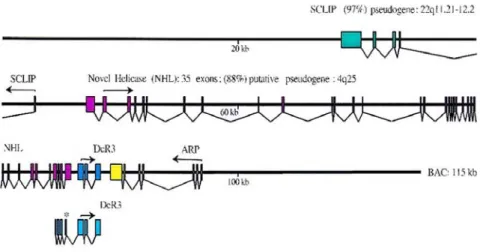
In tumors, gene amplification and overexpression of DcR3 are frequently observed (1, 9). However, they are not always concurrent (9,61). Protein overexpression may not depend on genomic change, and the gene amplification may not cause the protein overexpression either. These suggest that the gene amplification is neither sufficient nor necessary for the overexpression of DcR3, and there are other mechanisms to modulate its expression level. Further studies found that insulin-like growth factor-1 induced activation of the PI3K/Akt/NF- B signaling pathway is important to regulate endogenous DcR3 expression in human pancreatic carcinoma (62). Interestingly, DcR3 overexpression positively relates to Epstein-Barr virus (EBV) infection in lymphomas (63). Here, DcR3 expression is induced by Rta, a transcriptional activator encoded by EBV (64). Rta not only directly binds to the Rta-responsive element (RRE) sequence located in the DcR3 promoter region, but also enhances PI3-K activity. Besides, the coordinate transcriptional regulation of overlapped genes may be involved in the mechanism. The DcR3 gene is located in a gene-rich cluster and partially overlaps with exons 32 through 35 of the novel helicase-like gene (NHL) (Fig. 3), which relates to multiple inherited human neoplastic disorders (9). Such overlapping structures could cause coordinate transcriptional regulation ( 65 ) that may contribute to the
Figure 3. Genomic structure of DcR3 and adjacent genes. (source: ref. 9) Shaded boxes: Exons, each color represents one gene;
Arrows: The translation initiation methionine and the direction of protein translation;
*: One of DcR3 transcript variants overlaps the NHL helicase sequence with exons 32 through 35.
1-4. Immunity and DcR3
DcR3 plays a complex role in immune modulation. DcR3 primarily downregulates immune function. It could suppress T- and B-cell activation and T-cell proliferation (66), inhibit T-cell and macrophage chemotaxis (13,55,66), impair macrophage function (67), as well as induce apoptosis of dendritic cells (68). On the other hand, activated T cells secrete DcR3, which can prevent T cells from activation-induced cell death (AICD) (1)
costimulation (69,70,71). It also regulates the differentiation of dendritic cells and macrophages (57, 58), and the expression of adhesion molecules on endothelial cells (72). The complexity of the bioactivity of DcR3 hints that DcR3 may participate in multiple physiological procedures and precisely adjust different signaling pathways according to certain circumstances.
1-5. Cancer and DcR3
As described, DcR3 overexpression is found in diverse malignant tumors. Clinical studies also show relationship between DcR3 levels and malignancy, tumor stage and prognosis. Higher serum DcR3 levels are associated with poorer differentiation, later tumor stages and worse outcomes (8,15, 73 , 74 ). These observations and in vitro experiments (1,12,75) support the notion that tumors may benefit from the elevation of DcR3 levels to overcome immune surveillance. Indeed, DcR3 may provide several advantages to tumors:
1) DcR3 may inhibit NK cell and cytotoxic T lymphocyte activity through blocking of the Fas/FasL, LIGHT/ HVEM-LTβR death signaling pathway;
2) DcR3 may suppress the tumor-reactive lymphocyte activation and cytokine production by repressing costimulation of LIGHT and TL1A, or by modulation of dendritic cells;
3) DcR3 may restrain T lymphocyte chemotaxis by the activation of LIGHT reverse signaling;
4) DcR3 may induce angiogenesis of tumors by neutralization of TL1A; 5) DcR3 may disturb mononuclear phagocyte differentiation, adhesion and antigen presenting functions (57,72).
Several questions remain to be answered. Which cells do contribute to increased serum DcR3 levels in tumor patients? Tumor cells, tumor-reactive immune cells, or both? Is DcR3 elevation a cause or a consequence of oncogenesis? In other words, could occasional elevated DcR3 levels induce malignant changes, or existing malignant cells and/or tumor-reactive immune cells over secrete DcR3? We do not have definitive answers to these questions.
In our study, DcR3 transgenic mice do not produce any conspicuous tumors when expressing folds higher endogenous DcR3 than those observed from tumor patients (data not shown). This suggests that sole DcR3 overexpression may not be sufficient to induce but functions as an
caused by virus infection, endocrine disturbances, improper gene regulation, may assist the malignantly transformed cells to escape immune surveillance. In any case, DcR3 is becoming a useful early diagnostic and prognostic marker of various malignant tumors.
In summary, DcR3 is a multifunctional regulator. It is not absolutely needed for normal physiological functions since rodents do not have it at all. Its biological function in humans might be fine tuning of certain signaling pathways. It may involve multiple organ development, immune system maturation, peripheral tolerance maintenance and tissue repair. Dysfunction of DcR3 may result in a variety of disorders in humans, and its expression could be used to monitor conditions of these diseases.
2. Systemic lupus erythematosus (SLE)
Systemic lupus erythematosus (SLE) is an autoimmune disease characterized by the production of many different autoantibodies against cell components, especially nuclear components, with involvement of multiple systems, and variable symptoms. Excessive autoantibodies lead to formation of immune complexes, with deposition in different tissues causing inflammation and consequent tissue damage. The clinical manifestations of SLE vary greatly. The course of the disease is characterized by alternative relapse and remission.
2-1. Epidemiology of SLE
Although about 20 percent of SLE patients are diagnosed before age 16 (76), SLE mainly affects child-bearing age females, and the disease lasts for life (77). It is more frequent among Asians, Afro-Americans and Afro-Caribbeans than Caucasians and Blacks in Africa (78,79,80). The prevalence of SLE is highly varied according to regions. In the USA, it is from 40 to 150 cases per 100,000 (81,82); in Asia, 50 to 100 cases per 100,000 (83); about 20 to 70 per 100,000 in Europe (84,85,86). The
incidence has nearly tripled in the past 40 years, but this maybe due to improved detection of mild forms of the disease (87).
2-2. Pathoaetiology of SLE
Immune complex deposition, inflammation and vascular abnormalities are the basic pathological changes of SLE. Their central pathogenesis is the production of autoantibodies. Both T and B lymphocytes are necessary for the generation of autoantibodies. These antibodies aim several self molecules in the nucleus, cytoplasm, and cell surface, as well as some soluble molecules such as IgG and coagulation factors. Antinuclear antibodies are most common. The anti-double stranded DNA (ds-DNA) and anti-Sm antibodies are unique in SLE, and are thought to be the main reasons of tissue damage. When soluble self-antigens are exposed to autoantibodies, they form immune complexes, which are deposited in tissues, especially the joints, glomeruli and vascular wall. The deposited immune complexes will activate the complement system, which will then recruit and activate inflammatory cells, such as neutrophils and macrophages. These inflammatory cells will secrete reactive intermediates to mediate local inflammation. Subsequently, this leads to tissue damage and clinical symptoms.
In addition to T and B lymphocyte function which is important for autoantibody generation, other risk factors such as hormones, environmental elements and genetic susceptibility are also critical in SLE pathogenesis.
2-2-1. Hormones
Sex hormones are crucial in the pathogenesis of SLE. SLE is marked with female predominance. The female to male ratio of SLE patient rises from puberty (3: 1) and peak during child-bearing years (10-15: 1), it then decreases after menopause (8: 1) (88). High-estrogen levels, in the cases of early menarche, estrogen treatment, or Klinefelter’s syndrome that are characterized by hypergonadotrophic hypogonadism, could significantly increase the risk of SLE (89,90). Moreover, abnormalities of sex hormone metabolism including redundancy of 16 hydroxyestrone (91) and reduction of androgens (testosterone, dihydrotestosterone, dehydroepiandrosterone (DHEA) and dehydroepiandrosterone sulfate (DHEA-S) ( 92 , 93 )) are observed in both male and female SLE patients.
Besides sex hormones, gonadotrophin releasing hormone (GnRH) (94) and hormones in the hypothalamo–pituitary–adrenal (HPA) axis (such as
cortisol, prolactin and thyrotropin releasing hormone) ( 95 , 96 ) also influence the invasion and/or activity of SLE.
2-2-2. Genetic factors
SLE shows a strong familial aggregation (10–12%) among first degree relatives (97). Moreover, the concordance of SLE in identical twins is significantly higher than that in dizygotic twins (25–50% vs. 5%) (98). These observations suggest that the genetic factors play an important role in the pathogenesis of SLE. Although few SLE cases (<5%) are caused by single gene mutations (99), SLE is primarily a polygenic inheritance disease, which needs at least four susceptible genes to cause disease manifestation ( 100 ). More than 100 potential risk genes have been identified in the last few decades. Some of them were confirmed to have strong association with SLE. Those include: the genes of human leukocyte antigen (HLA) class II [HLA-DRB1*0301/*0302 (DR3), DRB1*1501/*1503 (DR2), DRB1*08 (DR8) (101,102,103)], genes of some classical complement activation pathway components [C1q, C1r/s (104), C2 (105,106), C4 (107,108,109)], the FCGR genes [FcgR IIa (110), IIb (111) and IIIa (112)], and some genes related to immune regulation, e.g., TNF-α (113,114), PDCD1 (115,116) and CTLA-4 (117,118).
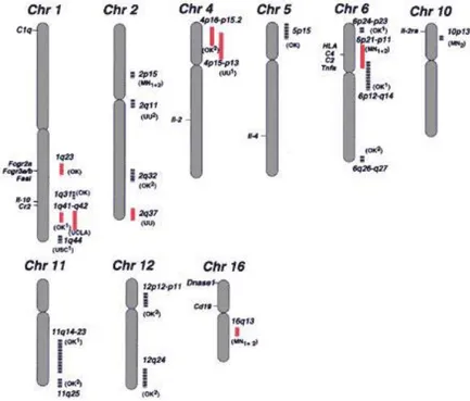
In addition to these candidate genes, 13 susceptibility loci show significant linkage to SLE, according to a genome-wide linkage analysis (Fig. 4) (119). Eight of them (1q23, 1q25-31, 1q41-42, 2q35-37, 4p16-15.2, 6p11-21, 12 q24, and 16q12) were confirmed by a number of studies (120,121,122,123, 124,125,126,127,128,129,130).
Fig. 4 The susceptibility loci of SLE (source: ref.131)
2-2-3. Environmental factors
initiation of SLE. Ultraviolet (UV) light, especially UVB, can bring about keratinocytes apoptosis, and then expose some self-antigens to the immune system triggering autoimmunity (132). Some chemicals, such as aromatic amines, hydrazines and their derivatives, are present in many drugs, agricultural and industrial products (even tobacco). They can potentially ignite a lupus-like syndrome ( 133 ). Infections in early childhood seem to affect the SLE risk in later life (134,135). Furthermore, the deficiency of vitamin D also has been found to be associated with SLE (136).
2-2-4. Apoptosis and SLE
Apoptosis, also called programmed cell death, is a process when cells undergo an ordered destruction and clearance, without releasing inflammatory intracellular contents into the extracellular environment. Apoptosis can be initiated by ligation of cell surface death receptors with their ligand(s), or by deficiency of survival stimuli. The former is called activation-induced cell death, and the latter is called passive cell death. In activation-induced cell death, ligated death receptor will activate caspase 8 (and caspase 10 in humans). While lack of survival stimuli will increase permeability of mitochondria and lead to release of cytochrome C.
Cytochrome C then couple with apoptosis activating factor-1 to induce activation of caspase 9. The activation of caspase 8 or caspase 9 is followed by the activation of an enzymatic cascade, nuclear condensation and fragmentation, and plasma membrane blebbing (137). Among all the cell death receptor/ligand signaling pathways, Fas/FasL-mediated apoptosis pathway is the best characterized (Fig 5). This pathway is crucial for the development of immune tolerance (138,139).
During the immune system maturation, self-tolerance is developed through apoptosis of auto-reactive lymphocytes in central lymphoid organs. Immature T/B cells with high affinity receptor of self-antigen will be induced to apoptosis. This is called central tolerance. But not all of the self-antigen can be presented in central lymphoid organs. Some auto-reactive lymphocytes can escape from negative selection. In peripheral, apoptosis of activated lymphocytes following an immune response maintains homeostasis of peripheral lymphocytes numbers, and contributes to peripheral tolerance to self-antigens (140,141). Disturbance in these apoptotic processes might break the balance present in the immune system and may predispose to autoimmunity. Some lupus-prone murine models support this assumption. The lpr/lpr and gld/gld mice, which have mutation in the Fas and FasL respectively, spontaneously develop
lymphadenopathy and lupus-like syndrome characterized by the presence of autoantibodies to nuclear antigens ( 142 ). Another model is the exogenous soluble Fas (sFas)-induced autoimmune symptoms in CD1 mouse (143). These models suggest that the disorder or blocking of Fas/FasL pathway will result in defect of peripheral autoreactive lymphocyte elimination that occurs through Fas-mediated apoptosis (144). However, in humans, Fas or FasL gene defects only cause the lymphoproliferative syndrome (ALPS) (140,145) with rare autoimmune manifestations (146). Most SLE patients do not have abnormality in their Fas/FasL gene structure and expression (147,148,149). Only sFas levels relate to SLE activity (150,151). Otherwise, apoptosis of peripheral lymphocytes in SLE patients is not compromised or even enhanced (152,153). This suggests that the relationship between apoptosis and self-tolerance in humans is much more complex than in mouse.
Apoptotic cells may also expose intracellular and cryptic epitopes to the immune system ( 154). The degradation and modification of cellular constituents during apoptosis could induce immunogenicity (155,156). If apoptotic residues can not be cleared in due time, e.g., due to deficiencies in complement factors or CD14 ( 157 , 158 ), autoimmunity may be stimulated.
2-3. Mouse models of SLE
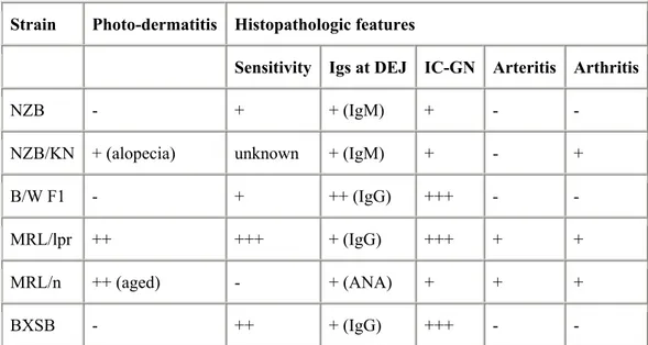
Although no single animal model perfectly recapitulates human SLE, some animal models can help us understand the etiology and pathogenesis of this disease. There are many inbred strains of spontaneous SLE-prone mice, including MRL/lpr, MRL/gld, NZB (New Zealand Black), F1 hybrids of NZB×NZW (New Zealand White) (B/W F1), and BXSB mice. Each of these have their own genetic background and autoimmune characteristics (table 1). More gene manipulated models are being generated, such as transgenic or gene knockout mice, which may help clarify the significance of target molecules in SLE.
Strain Photo-dermatitis Histopathologic features
Sensitivity Igs at DEJ IC-GN Arteritis Arthritis
NZB - + + (IgM) + - -
NZB/KN + (alopecia) unknown + (IgM) + - +
B/W F1 - + ++ (IgG) +++ - -
MRL/lpr ++ +++ + (IgG) +++ + +
MRL/n ++ (aged) - + (ANA) + + +
BXSB - ++ + (IgG) +++ - -
Table 1. Characteristics of lupus-prone mouse strains (source: ref.159) Igs at DEJ: immunoglobulins deposits at the dermoepidermal junction;
2-4. Clinical features of SLE 2-4-1. Symptoms
The clinical course and outcome of SLE are extremely variable. Glomerulonephritis, arthritis, systemic small arteries vasculitis, rashes, haemolytic anemia and thrombocytopenia, which are caused by immune complexes deposition and/or autoantibody-induced ADCC, are the most common clinical symptoms of SLE.
2-4-2. Diagnosis
The American College of Rheumatology (ACR) classification criteria, a generally-accepted diagnostic standard, were devised in 1982 and revised in 1997 (160). Any combination of 4 or more of the following 11 criteria, well-documented at any time during a patient's history, makes it likely that the patient has SLE (specificity and sensitivity are 95% and 75%, respectively).
1. Malar rash: fixed erythema, flat or raised, over the malar eminences; 2. Discoid rash: erythematous circular raised patches with adherent keratotic scaling and follicular plugging; atrophic scarring may occur; 3. Photosensitivity: exposure to ultraviolet light causes rash;
4. Oral ulcers: includes oral and nasopharyngeal ulcers, observed by a physician;
5. Arthritis: nonerosive arthritis of two or more peripheral joints, with tenderness, swelling, or effusion;
6. Serositis: pleuritis or pericarditis documented by ECG or rub or evidence of effusion;
7. Renal disorder: proteinuria >0.5 g/d or 3+, or cellular casts; 8. Neurologic disorder: seizures or psychosis without other causes;
9. Hematologic disorder: hemolytic anemia or leukopenia (<4000/L) or lymphopenia (<1500/L) or thrombocytopenia (<100,000/L) in the absence of offending drugs;
10. Immunologic disorder: anti-dsDNA, anti-Sm, and/or anti-phospholipid; 11. Antinuclear antibodies: an abnormal titer of ANA by immunofluorescence or an equivalent assay at any point in time in the absence of drugs known to induce ANAs.
2-4-3. Therapy
In the absence of specific therapy aiming at the pathogenesis, successful therapy of SLE depends on treating both underlying inflammation and symptoms. Currently immunosuppression is the major SLE therapy available. Four main classes of drugs, i.e., corticosteroids, non-steroidal anti-inflammatory drugs (NSAIDs), antimalarials and cytotoxic agents, are used.
High doses of corticosteroids remain the first line of treatment for many manifestations of SLE, especially in severe disease with renal, CNS and hematological involement. But it causes many hazardous side effects, such as infection, hyperlipidaemia, hypertension, osteoporosis, diabetes, and insomnia (161). The combined use of NSAIDs, antimalarials and cytotoxic agents can reduce the steroids’ dosage and side effects to a relative low level.
Antimalarials are commonly employed to treat patients with fatigue, arthralgia/arthritis and rash,but without major organ damage. Antimalarials can interfere with immune cellular functions, affect immune responses, and modulate cytokine levels in SLE patients. Antimalarials also improve skin lesions against the damaging effects of ultraviolet light. In these ways, antimalarials have the potential to keep SLE in remission.
Active lupus with major organ involvement often requires prompt, aggressive therapy with cytotoxicagents. Cytotoxicagents were primarily known to interrupt nucleic acid and protein synthesis in malignant cell. Then its immune inhibition was discovered. The use of cytotoxic agents in SLE benefits controlling active disease, reducing the rate of disease flares, and reducing steroid requirements.
Currently, there are many new treatments under investigation (162). Some biological agents (e.g., antibodies) have been tested in clinical trials. They either modulate or inhibit T-cell activation, T- and B-cell interactions, anti-dsDNA antibody production, immune complexes deposition, complement activation/deposition, and cytokine activity. A hormonal modulator, DHEA, has also shown promising therapeutic effect in SLE patients. In addition, a small number of very severe refractory lupus patients (7 cases) received high-dose chemotherapy and autologous stem cell transplantation to gain remission (163).
Other than pharmacological treatment, management of daily life can help SLE patients to control the disease. These therapies include avoiding sunlight over-exposure, low saturated fat and a high fish oil diet, stress avoidance, and smoking cessation.
2-4-4. Prognosis
In many countries, the survival rate of SLE exceeded 80-90% in 5 years post-diagnosis, but decreases to 60-80% in next 10 years (164,165, 166,167). The major causes of mortality are organ failure (especially renal failure), thrombocytopenia, cardiovascular diseases, infections, high SLE disease activity index (SLEDAI) (165,168). Proper and timely treatment in experienced hospitals is very important to improve the long-term survival rate (169).
SLE is a common chronic disease that seriously imperils the survival and life quality of patients. There is no cure due to the ambiguous etiology. Investigation of SLE pathogenesis will help us to develop specific treatments.
3. Diabetes Mellitus
Diabetes mellitus is a syndrome (includes excessive urine production, increased fluid and food intake, body weight loss, changes in energy metabolism, even blurred vision and renal failure) as results of abnormal high blood glucose levels. This metabolism disorder has two most common forms: type 1 diabetes mellitus (T1DM) is caused by insufficient insulin production that mainly follows after autoimmune damage to pancreatic islets; type 2 diabetes mellitus (T2DM) is caused by failed response to insulin in effective organ (such as liver and muscle), but the level of insulin are normal or even elevated before the late stage of the disease. Besides, some pregnant women may suffer gestational diabetes mellitus due to carbohydrate intolerance.
3-1. The Epidemiology of Diabetes Mellitus
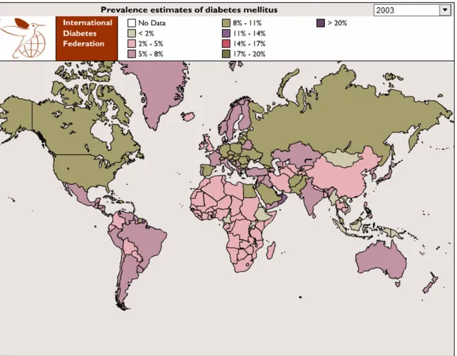
Nowadays, more than 170 million people live with diabetes worldwide (170), and that number will double in the next 20 years (171) (Fig. 5). T1DM is one of the most common chronic childhood disease whose incidence is about 20 to 30 per 100 000 children per year in the United
Kingdom, Finland, Norway and Sweden, but is much lower in Asian, Indian, Middle Eastern and African populations (172,173). The incidence of T1DM is increasing rapidly worldwide, and is estimated to exceed 30-50 per 100,000 a year by 2010 (173). Studies showed 2.5-3% annual global increase rate of incidence of T1DM with a larger increase in some central and eastern European countries, some Asian countries and Australia (174,175). The largest rate of increase was seen in 0-4 year old children (173).
T2DM usually occurs after the age of 30, but it can exist without any symptoms for many years. The first diagnosis is sometimes made through incidental abnormal blood or urine glucose tests, or through associated complications (176). It constitutes more than 90% of overall diabetes cases in the world (177) with large geographical prevalence variation, even within the same or similar ethnic groups (178,179). The greatest increase of T2DM is in the developing countries of Asia, Africa, and South America, which are evolving with rapid cultural and social changes, ageing populations, urbanization, and unhealthy lifestyle and behavioral patterns (180). Another worrisome T2DM increase is in children and adolescents. The proportion of T2DM in new-onset diabetes during children and adolescents has increased more than 15 folds (<3% vs. 45%) in last 15
years (181). The increase of T2DM incidence closely parallels the increase of obesity. This unprecedented twin epidemic is termed as "diabesity" (171).
Figure 5. The geographic distribution of DM prevalence (source: http://www.eatlas.idf.org/atlas.html?id=0)
3-2. The pathoaetiology of Diabetes Mellitus
T1DM had been defined as “insulin-dependent diabetes mellitus” due to the insufficient insulin in the body and the necessary treatment by artificial insulin even in its earliest stage. It is also called "juvenile diabetes" sometimes, because its onset is mainly in childhood although a few cases occur in adulthood, which is also called latent autoimmune diabetes of adults (182). T-cell-mediated autoimmune attack to the beta-cells of Langerhans islets is believed to be the major cause of the disease (Fig 6).
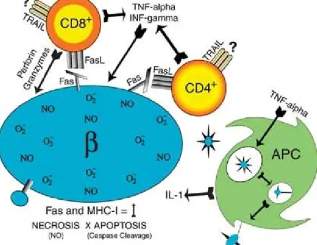
Fig. 6 The pathogenesis of T1DM. (source: ref. 183)
β-cell antigen is presented on the cell surface by MHC class I molecule. CD8+ T cells recognize this antigen and damage β-cell through the secretion of INF- or TNF/TRAIL or the perforin/granzyme system. The dendritic cells in islets take up cell components from dead β-cells, and present them to CD4+ T cells in lymph nodes. Active CD4+ T cells move into the islets to mediate killing through the Fas/FasL pathway.
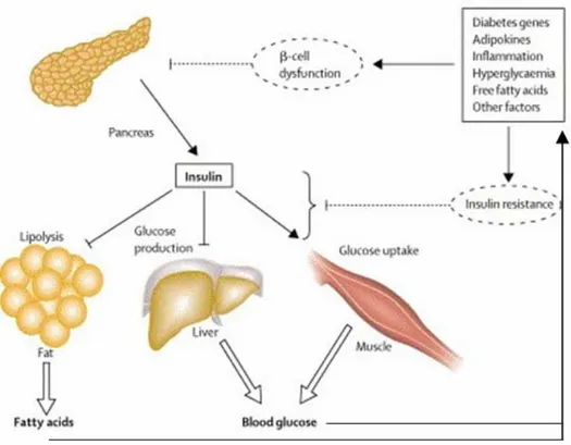
T2DM was called “non-insulin-dependent diabetes” in contrast to T1DM. A deficient reaction to insulin in liver and muscle initiates this disease. In these tissues, glucose processing is compromised. Thereafter, unprocessed glucose accumulates in circulation and reduces islet beta cells’ response to glucose (Fig7). T2DM patients are not ketosis-prone and do not depend on exogenous insulin. However, they may need insulin treatment to control hyperglycemia if the diet and /or oral hypoglycemic agents do not work well.
Figure 7. Pathophysiology of T2DM (source: ref. 170)
A variety of factors contribute to insulin resistance. Compromise of insulin action in major target tissues leads to increased circulating free fatty acids and hyperglycaemia, which will impair β-cell viability and worsen insulin resistance.
Like systemic lupus erythematosus, a combination of hereditary and environmental factors causes these metabolism dysfunctions, either in T1DM or T2DM. Clinical trials show that the concordance rates of monozygotic twins are around 50% in both T1DM and T2DM (184,185,186). These results suggest that the genetic factor is not the only protagonist in the etiology of DM. In other words, the trigger by environmental factors is also necessary to diabetes onset. T2DM seems to be familial (187,188). In addition to genetic factors, a similar lifestyle of members in the same family may also play a role.
3-2-1. Genetic Factors
Most diabetes, both T1DM and T2DM, are polygenic. There are only 1-5% cases (mainly neonatal diabetes mellitus and maturity-onset diabetes of the young) that are due to monogenic mutations (189). Although there are many diabetes risk genes, which have been identified by different studies, only a few of these findings are confirmed, i.e., HLA-DQ8 and DQ2, MICA-5 (MHC class I chain-related genes allele 5), KIR (killer immunoglobulin- like receptors) (190), CTLA4 (cytotoxic lymphocyte antigen 4), LYP (Lymphoid tyrosine phosphatase)/PTPN22 ( 191 ) to T1DM; TCF7L2 (Transcription Factor 7-Like 2) ( 192 ), CAPN10
(calpain 10) (193), PPARG (peroxisome proliferator-activated receptor gamma), KCNJ11 (194,195) to T2DM. As obesity contributes to T2DM, genes involved in obesity should also be considered risk factors for T2DM. More than 22 genes, which encode members of the leptin–melanocortin pathway, proinflammatory cytokines and uncoupling proteins (UCPs), are related to obesity risks (196).
3-2-2. Environmental Factors
Various environmental factors can influence diabetes morbidity.
a. Viral infection:
T1DM pathogenesis somewhat relates virus infection. Many viruses can trigger cell damaging processes in their host, such as Coxsackie B virus, mumps, echovirus, cytomegalovirus, Epstein-Barr virus (EBV), some retrovirus, rotavirus, parvovirus B19 and rubella virus (197). These viruses can induce autoimmunity through 1) direct infection of islets (198), 2) molecular mimicry, i.e., viral protein sharing similar peptide sequences with cellular autoantigens (199), or 3) activation of innate immunity (200).
b. Environmental contaminants and chemicals:
The increase of contaminants in the environment or occupational contact of certain chemicals could be potential risk factors of DM. For T1DM, higher intake of nitrates, nitrites, and N-nitroso compounds, as well as higher serum levels of polychlorinated biphenyls seem to link to higher incidence (201,202,203,204,205). Arsenic and 2,3,7,8-tetrachlorodibenzo-p-dioxin exposure is directly associated with T2DM risks (206,207,208,209).
c. Lifestyle and social factors:
Human behavior largely influences the incidence of DM (210). Decreased breastfeeding and early exposure to dietary cow-milk/cereals will raise the future risk of T1DM in the infants (211,212), while early supplement of vitamin D can decrease T1DM risks (213). The “Westernized food”, or energy-dense food, contributes to obesity, thereby increases the risk of T2DM (214). Physical activity is another effective behavior factor. Regular physical activity can reduce the risk of T2DM by 15-60% (195). Socio-economic and psychosocial factors could also indirectly affect the prevalence of DM through the change of lifestyle, nutrition, and natural environment (197,215,216).
d. Perinatal factors and postnatal growth:
Perinatal factors associated with increased T1DM risk to infant may include older maternal age at birth (217), excessive maternal weight gain, amniocentesis, preeclampsia (218), cesarean section delivery, complicated delivery, and maternal-fetal blood group incompatibility (219,220). Size at birth and early postnatal growth rates, which may reflects the fetal nutritional conditions, are significantly associated with the risk of diabetes, both T1DM and T2DM (221,222,223).
3-2-3. Accelerator hypothesis
Recent evidences indicate that incidence of both T1DM and T2DM increase in parallel with obesity (224,225). Meanwhile, the important role of insulin resistance in the early stages of T1DM development has been widely accepted (226,227). These findings led to a new theory called “accelerator hypothesis”. This hypothesis proposes that “T1DM and T2DM are the same disorder of insulin resistance set against different genetic backgrounds” (228). Excess bodyweight is the central point to the development of diabetes, both T1DM and T2DM. Weight gain increases insulin resistance and consequently results in hyperglycemia. Long-term
Based on these pathological changes, individual gene polymorphism and constitution, determine the tempo of beta cell loss and the age of onset, i.e., the manifestation of T1DM or T2DM. This hypothesis deems that T1DM and T2DM are subsets of the same disease. Thus, control of weight gain could prevent DM by slowing its progress.
3-3. The clinical features of DM 3-3-1. Symptoms and complications
Hyperglycemia is the principal cause of diabetes symptoms and complications. The classical diabetes triad symptoms are polyuria, polydipsia and polyphagia, i.e., frequent urination, increased thirst and fluid intake, increased appetite. Symptoms may develop quite rapidly (weeks or months) in T1DM, particularly in children. T1DM may also cause rapid weight loss and implacable fatigue. However, symptoms usually develop much slowly and mildly, sometimes may be even absent, in T2DM.
Ketoacidosis (DKA), an extreme state of metabolic dysregulation, may be the most common acute complication of DM. It is characterized by the acetone smell in the patient's breath, Kussmaul breathing, polyuria, nausea,
can cause coma and even death (229). It is mainly present in patients with T1DM, but also observed in patients with T2DM under certain conditions. Another serious acute complication is hyperosmolar nonketotic state, which is more common in T2DM (230). It is mainly the result of dehydration due to loss of body water.
Prolonged high blood glucose levels along with abnormal lipid levels lead to blood vessels lesion (angiopathy), which subsequently induces chronic multiple organ complications, including blindness, renal failure, lower limb gangrene and cardiovascular diseases.
Nosogenetic effects of hyperglycemia mainly attribute to the formation of advanced glycation end-products (AGEs). Hyperglycemia in diabetes increases the formation and accumulation of AGEs. AGEs can interact with cell-surface receptor of AGE (RAGE), leading to cell activation and increasing expression of extracellular matrix proteins, vascular adhesion molecules, cytokines, growth factors, and the generation of reactive oxygen intermediates. Moreover, certain AGEs precursor can covalently crosslink proteins and change their structure and function. These pathologic changes result in almost all the diabetes complications, both micro- and macroangiopathies.
3-3-2. Diagnosis
The diagnostic criteria are based on the WHO recommendations of 1999. They incorporate both fasting criterion and 2-h-after-glucose-load (oral glucose tolerance test (OGTT)) criterion into a practicable diagnostic classification (table 2) (231). About 7% of people with impaired fasting glucose and impaired glucose tolerance will eventually progress to overt diabetes every year without proper treatment (232,233). If a pregnant woman has any two of the followings, she will be diagnosed as gestational diabetes: 1) fasting plasma glucose more than 5.3 mmol/dl, 2) 1-hour glucose level of OGTT more than 10 mmol/dl, 3) 2-hour glucose level of OGTT more than 8.6 mmol/dl (234).
There are some other accessorial clinical tests to value the status of DM. For examples, urine ketones test can indicate the severity of T1DM; the glycosylated hemoglobin test will give information about how well the blood glucose is controlled.
Table 2. Diagnostic criteria of diabetes mellitus and other categories of hyperglycemia (source: ref. 231)
3-3-3. Therapy
Diabetes mellitus is a chronic disease currently without a cure. The goal of therapy is to maintain blood glucose level as close to normal as possible to prevent either acute or chronic complications.
3-3-3-1. Diabetes education and lifestyle intervention
Self-monitoring the blood glucose of diabetes patient is very important to keep both short-term and long-term blood glucose levels within acceptable bounds. Proper diet, regular physical exercise, moderate body weight and refrain from smoke and alcohol will help the patient to control the blood sugar and greatly reduce the risk of progression (232, 233). It is very necessary to impart related knowledge to the diabetes patients and help them modify their lifestyle.
3-3-3-2. Insulin and other medications
T1DM patients, as well as T2DM patients who do not respond to oral medications, need insulin therapy to survive. There are several different insulin types and administration methods available currently.
Besides insulin, many choices of drugs can be used to modulate glucose levels, especially for T2DM. The mechanism and active site of these medications are shown in Table 3
Additionally, since cardiovascular disorders, such as hypertension, coronary artery disease and cerebrovascular disease, occur very often in T2DM (235), the management of cardiovascular risk factors is necessary and beneficial (236).
Classification Medication Route The way it works
Sulfonylureas Glimepiride, Glipizide, Glipizide
ER, Glyburide
Oral Increases insulin production
Biguanides Glucophage, Glucophage XR
Oral Lowers glucose from digestion Alpha-Glucosidase
Inhibitors
Glyset and Precose Oral Slows digestion, slows glucose production
Thiazolidinediones Actos and Avandia Oral Lowers glucose production Meglitinides Prandin and Starlix Oral Increases insulin production DPP-4 Inhibitors Januvia Oral Lowers glucose by blocking an
enzyme
Incretin Mimetics Byetta Injectable Helps the pancreas make insulin, slows digestion
Anti-hyperglycemic Symlin Injectable Controls postprandial blood glucose
3-3-3-3. Organ and cell transplantation
The DM, in particular T1DM, has no cure. A potential cure for it is transplantation, either at the organ level or cell level.
a. Pancreas transplantation
Pancreas transplantation is an effective treatment for diabetes mellitus, especially for the end-stage renal complication. It was first successfully performed in combination with kidney transplant in 1966 (238). The improvements in surgery and immunosuppressors have significantly increased the graft survival rate; the 5-year graft survival rate is around 50%-70% ( 239 ). Pancreas transplant can achieve long-term normoglycemia, reduce mortality, and ameliorate diabetic complications (240). However, pancreas transplantation is still a high-risk operation with some severe postoperative complications. Moreover, the availability of organ donors and side effects of immunosuppressants are also limiting factors for popularization of this treatment.
b. Islet transplantation
Islet transplantation was applied to treat insulin-dependent diabetes since the 1970’s, but it had hardly achieved desirable long-term effectiveness
until the advent of a steroid-free immunosuppressive protocol in 2000 (241). The adoption of the Edmonton protocol, which uses steroid-free immunosuppression, has elevated the success of islet transplantation greatly. Most recipients could attain an insulin-independence immediately after transplantation (242,243). However, a large islet mass (> 10,000 islet equivalents (IEQ)/kg recipient body weight) derived from multiple donors (2-4) is necessary to achieve insulin independence (244) because of the loss of a large number of transplanted islets in the first 10–14 days after transplantation despite efficient immunosuppressive regimens (245,246). Experimental models also show that early damage results in 60% loss of the transplanted syngeneic islet mass in the peri-transplant period (247,248). This early stage non-rejective graft failure or the primary non-function (PNF) is becoming a bottleneck for the development of islet transplantation since the improvement of immunosuppressive agents. PNF reduces effective grafted islet mass, and then increases the metabolic load to surviving islets. This overloading reduces islet survival time and diminishes the long-term insulin independence, which is frequently (~90%) lost in 5 years after islet transplantation (249,250).
The reasons for PNF include oxidative stress in the islets during pretransplant manipulations (251,252), loss of trophic factors for the
isolated islets (253), host innate immunity and inflammatory responses ( 254 ), and failure of revascularization of islet graft ( 255 , 256 ). Proinflammatory cytokines (e.g., TNF-α, IL-1, and IFN-γ)-triggered multiple signaling pathways (e.g., c-Jun NH2-terminal kinase (JNK1) (257), Fas/FasL (258), nuclear factor- B, and transcription factor signal transducer and activator of transcription (STAT) (259)) contribute to the occurrence of PNF. Prevention of islet graft PNF through improvement of islet isolation and culture technology, application of inflammation inhibitors, and genetic modification to repress islet apoptosis would likely make a significant impact on the efficacy of islet transplantation.
c. Stem cell transplantation
Recent research showed that embryonic, fetal and adult stem/progenitor cells including putative multipotent pancreatic stem/progenitor cells have a potential for self-renewal of islet β-cells (260,261). The transplantation of insulin-producing β-cell derived from either wild-type or genetically modified stem/progenitor cells, or the expansion and differentiation of putative multipotent pancreatic stem/progenitor cells in vivo, may be promising alternative therapies for T1DM or T2DM in humans.
3-3-3-4. Gene therapy
Gene therapy of DM has been under investigation intensely in recent years. Studies include genetic modification of graft islets, genetically engineered ectopic insulin production, preventing or curing autoimmunity (262). All these therapies require an effective gene delivery system, which is the bottleneck of the gene therapy. The disadvantages (such as oncogenicity, efficacy and immunogenicity) of viral gene delivery systems limit its clinical usefulness. With future advances in gene delivery technologies, gene therapy may be a potential cure to DM.
Hypothesis and Objective
The etiology of SLE remains unclear. Disorder of lymphocyte apoptosis can lead to lupus-like syndromes in mouse models. But in humans, the relationship between SLE and lymphocyte apoptosis is a puzzle. DcR3, a native block of apoptosis, may play a role in pathogenesis of human SLE via interruption of lymphocyte apoptosis.
Our group pioneered the research on the biological functions of DcR3. We discovered its roles in the modulation of T-lymphocyte functions (7,55) and reported its expression in varied malignant tumors (17). Moreover, we showed protective effect of DcR3 on islet transplantation (53).
As part of my Ph.D. Program, the important- role of DcR3 in the pathogenesis of SLE both in a mouse model and in patients has been investigated. The mechanisms of DcR3’s effect on islet protection have been further studied. The details are provided in the following 3 chapters.