O R I G I N A L A R T I C L E
Selective disruption of
Tcf7l2 in the pancreatic β cell
impairs secretory function and lowers
β cell mass
Ryan K. Mitchell
1
, Angeles Mondragon
1
, Lingling Chen
2
, James A. Mcginty
2
,
Paul M. French
2
, Jorge Ferrer
3
, Bernard Thorens
4
, David J. Hodson
1
,
Guy A. Rutter
1,
*, and Gabriela Da Silva Xavier
1,
*
1Section of Cell Biology, Division of Diabetes, Endocrinology and Metabolism, Department of Medicine,
2Photonics Group, Department of Physics and
3Section of Genetics and Medicine, Division of Diabetes,
Endocrinology and Metabolism, Department of Medicine, Imperial College London, London, UK, and
4Center for
Integrative Genomics, Physiology Department, University of Lausanne, Genopode Building, CH-1015 Lausanne,
Switzerland
*To whom correspondence should be addressed at: Section of Cell Biology, 3rd Floor ICTEM Building, Hammersmith Hospital Campus, Imperial College, London W12 0NN, UK. Tel: +44 2075943358; Email: [email protected] (G.D.S.X.); Tel: +44 2075943340; Email: [email protected] (G.A.R.)
Abstract
Type 2 diabetes (T2D) is characterized byβ cell dysfunction and loss. Single nucleotide polymorphisms in the T-cell factor 7-like 2 (TCF7L2) gene, associated with T2D by genome-wide association studies, lead to impairedβ cell function. While deletion of the homologous murine Tcf7l2 gene throughout the developing pancreas leads to impaired glucose tolerance, deletion in theβ cell in adult mice reportedly has more modest effects. To inactivate Tcf7l2 highly selectively inβ cells from the earliest expression of the Ins1 gene (∼E11.5) we have therefore used a Cre recombinase introduced at the Ins1 locus. Tcfl2fl/fl::Ins1Cre mice display
impaired oral and intraperitoneal glucose tolerance by 8 and 16 weeks, respectively, and defective responses to the GLP-1 analogue liraglutide at 8 weeks. Tcfl2fl/fl::Ins1Cre islets displayed defective glucose- and GLP-1-stimulated insulin secretion and
the expression of both the Ins2 (∼20%) and Glp1r (∼40%) genes were significantly reduced. Glucose- and GLP-1-induced intracellular free Ca2+increases, and connectivity between individualβ cells, were both lowered by Tcf7l2 deletion in islets from
mice maintained on a high (60%) fat diet. Finally, analysis by optical projection tomography revealed∼30% decrease in β cell mass in pancreata from Tcfl2fl/fl::Ins1Cre mice. These data demonstrate that Tcf7l2 plays a cell autonomous role in the control ofβ
cell function and mass, serving as an important regulator of gene expression and islet cell coordination. The possible relevance of thesefindings for the action of TCF7L2 polymorphisms associated with Type 2 diabetes in man is discussed.
Introduction
A considerable body of evidence suggests there is a strong heredi-tary component to type 2 diabetes (T2D) (1–3). Indeed, genome-wide association studies (GWAS) have now identified over 90 loci that are associated with T2D risk (reviewed in4). Most of the identified single nucleotide polymorphisms (SNP) associated
with T2D appear to affectβ cell mass or function (5). However, most of these are in intronic or intergenic regions, making it dif-ficult to identify the causal gene(s) and thus the impact of the identified SNP(s) at the molecular and cellular level (6).
Recently, much effort has been devoted to elucidating how the T2D-associated SNP rs7903146, which lies in intron 3 of the T-cell factor 7 like-2 (TCF7L2) gene, may lead to increased risk of
Received: September 16, 2014. Revised and Accepted: October 26, 2014 © The Author 2014. Published by Oxford University Press.
This is an Open Access article distributed under the terms of the Creative Commons Attribution License (http://creativecommons.org/licenses/by/4.0/), which permits unrestricted reuse, distribution, and reproduction in any medium, provided the original work is properly cited.
doi: 10.1093/hmg/ddu553
Advance Access Publication Date: 29 October 2014 Original Article
diabetes. First described in Icelandic, Danish and US cohorts (7) SNP rs7903146 is presently the most strongly associated of the GWAS-identified variants with T2D (7–10) and is also associated with latent autoimmune diabetes in adults (11). Available clinical evidence points to an action of the risk alleles to impair insulin secretion in man with little, or a slightly protective effect, on insulin action (7–19).
TCF7L2 is a member of the TCF family of transcription factors involved in the control of cell growth and signalling downstream of wingless-type MMTV integration site family (Wnt) receptors (20) and was previously best known for its association with pros-tate and colon cancer development (21,22). Activation of the Wnt pathway leads to release of catenin from an inhibitory complex and its translocation to the nucleus, where it binds TCF7L2 and other related TCF factors (23). The function of this transcriptional complex is context dependent, i.e. it may act as either a transcrip-tional activator or repressor (5,23). In the pancreas, TCF7L2 and the Wnt signalling pathway are essential for proliferation of the pan-creatic epithelium (24) and enhanced Wnt signalling has been shown to lead to islet proliferation (25). While loss ofβ-catenin signalling leads to pancreatic hypoplasia (26), stabilization of β-catenin results in the formation of large pancreatic tumours (27). Studies from our own laboratory and others’ have shown that silencing of Tcf7l2 gene expression in clonal cell lines (28) and pri-mary islets (28,29) leads to increased apoptosis and impaired β cell function. Moreover, LoxP-mediated deletion specifically in the pancreas using a PDX1.Cre (30), led to glucose intolerance and impairedβ cell mass on a high fat diet. On the other hand, a recent report (31) indicated that whereas deletion in the liver led to lowered hepatic glucose output, consistent with earlier findings of perinatal mortality in global Tcf7l2 null mice (32), de-letion in theβ cell in adult mice using a tamoxifen-inducible rat insulin promoter 2-driven (RIP2.Cre-ERT2) deleter strain exerted no apparent effect on glucose homeostasis. The authors there-fore concluded that changes in Tcf7l2 expression in theβ cell in man are unlikely to contribute to diabetes risk. However, the lat-ter studies were limited to the examination of relatively young (<12 weeks old) mice maintained on a normal chow diet. More-over, deletion in adults precluded examination of the effects on β cell proliferation during early post-natal growth. Finally, it was unclear in these studies whether Tcf7l2 expression was af-fected in the hypothalamus of the resulting KO mice, as might be expected using the Pdx1.Cre line (33).
Gene expression analysis following Tcf7l2 deletion or silen-cing revealed changes in the expression of a number of genes in mouse pancreatic islets, including that encoding the GLP-1 re-ceptor (Glp1r; (15,28,30,34). Correspondingly, it has previously been reported that TCF7L2 may mediate GLP-1-inducedβ cell pro-liferation (35). Since GLP-1 has also been implicated inβ cell sur-vival, the increased incidence of apoptosis in TCF7L2-silenced islets (29,36), and in individuals carrying the risk variants of TCF7L2 (15), are both consistent with lowered GLP-1 signalling (15,36). Supporting this view is the decrease inβ cell mass in high fat-fed, pancreas-specific Tcf7l2 null mice (30) and in mice over-expressing a dominant negative form of Tcf7l2 inβ cells (37). Thus, the diminished insulinotropic effect of GLP-1 in islets lacking Tcf7l2 activity seems likely to be due, at least in part, to a lack of cognate receptors on the cell surface (28,30,37). Dimin-ished brain GLP-1 signalling in mice over-expressing a dominant negative form of Tcf7l2 was also reported to lead to impaired glu-cose tolerance and insulin sensitivity when mice were adminis-tered a high fat diet (38).
While the above evidence suggests that loss of Tcf7l2 from the β cell is likely to impair insulin production, and hence increase
T2D risk, adult Tcf7l2 knockout mice show reduced hepatic glucose production during fasting and improved glucose homeo-stasis when maintained on a high fat diet (31); loss of Tcf7l2 sig-nalling in the liver is associated with lowered expression of genes involved in glucose metabolism in this tissue (31,39). Such data together indicate that lowering Tcf7l2 activity, at least in the liver, may be beneficial in metabolic diseases. Moreover, it has also been reported that transgenic mice over-expressing TCF7L2 systemically have impaired glucose homeostasis, among other physiological anomalies (40). The latter data are consistent with those from Gaulton et al. (41), indicating that chromatin at the TCF7L2 gene is in an islet-specific ‘open’ conformation, and that inβ cell lines the enhancer activity of the at-risk T-allele is ele-vated compared with the C-allele. Moreover, Savic et al. (42) have identified tissue-specific enhancer activity within the asso-ciation interval of rs7903146 which may lead to the generation of different splice variants of TCF7L2, potentially leading to differ-ent functional effects in differdiffer-ent tissues. Thus, the appardiffer-ent discordance between the metabolic phenotype of the various mouse models could be partly due to the involvement of TCF7L2 in glucose homeostasis in more than one tissue, poten-tially in opposing directions. Further complicating the potential actions of genomic risk loci, TCF7L2 is subject to tissue-specific alternative splicing (43–46).
Given the existing controversy in the literature over the rela-tive importance of Tcf7l2 in theβ cell versus the liver and other tissues (30,31), and the contributions of extrapancreatic tissues to the action of risk variants on diabetes risk (47), the present study was designed to achieve highly selective deletion of Tcf7l2 in theβ cell, from the earliest possible stages in the estab-lishment of a definitive β cell status, i.e. expression of the insulin gene. To this end we have used a highlyβ cell selective Cre recom-binase, based on expression of the enzyme from the Ins1 locus. In contrast to the use of Pdx1- (30) or Pdx1ER-based Cres (31) this strategy allows recombination highly selectively and early in the development of individualβ cells, without the complications of deletion in other tissues including the brain (33,48).
We show (a) thatβ-cell deficient Tcf7l2 mice show impaired in-sulin release and glucose homeostasis, particularly in response to oral glucose challenge; (b) when examined in intact islets, an unsuspected action is observed on both intracellular calcium sig-nalling and cell–cell communication, likely to contribute to the deranged insulin secretion in vivo. Thesefindings provide further evidence for theβ-cell autonomous role of Tcf7l2, and imply that this factor maybe particularly important during expansion of these cells in the post-natal period or during adaptation to meta-bolic stresses including high fat diet
Results
Abnormal glucose tolerance and insulin secretion inβ cell selective Tcf7l2 null mice
In the present study, we used Ins1.Cre mice (48) (B. Thorens and J. Ferrer, manuscript in preparation) to effect highly selective and early (∼E11.5) (49,50) deletion of Tcf7l2 inβ cells, based on Cre ex-pression driven by the Ins1 gene (33,51). The resultant Tcf7l2 null (Tcf7l2fl/fl::Ins1.Cre+) mice were born at the expected Mendelian ratio and gained weight similarly to wild-type mice (Tcf7l2fl/fl:: Ins1.Cre−) when maintained on regular chow (Supplementary Material, Fig. S1).
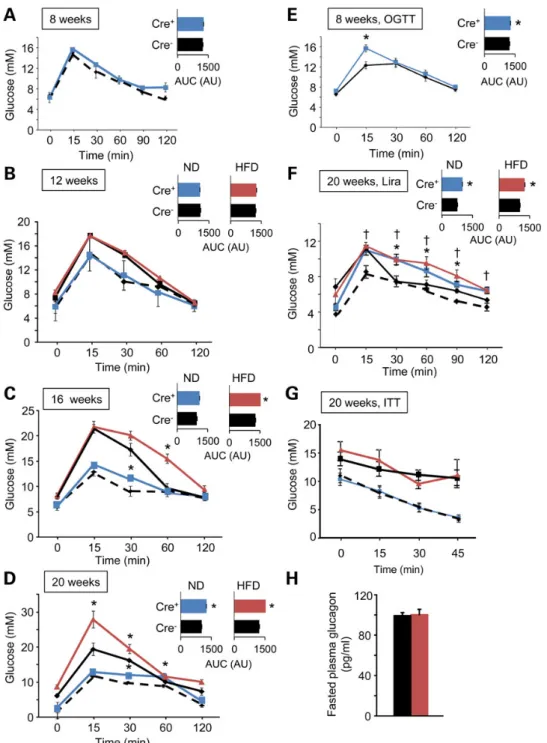
As shown in Figure1A–D, Tcf7l2fl/fl::Ins1.Cre+mice developed
impaired intraperitoneal glucose tolerance in an age-dependent manner, displaying significant impairments from 16 weeks
(increase in AUC of 13.6 ± 2.8%, n = 6 mice per genotype, P < 0.05). Impaired oral glucose tolerance (increase in AUC of 10.6 ± 1.3%, n = 6, P < 0.05) was apparent in younger animals (from 8 weeks; Fig.1E). Consistent with a more marked effect via altered incretin responses, the glucose excursion prompted by elevated glucose (1 g/kg) plus the GLP-1 analogue liraglutide (200 µg/kg) (52) was particularly strongly affected by Tcf7l2 deletion (Fig.1F). These changes were not associated with any alteration in sensitivity to intraperitoneal insulin (Fig.1G).
To determine whether the imposition of a metabolic stress may affect the glucose intolerance observed in Tcf7l2fl/fl::Ins1.Cre
+-mice, animals were maintained for the indicated times on a high fat (∼60% total calories) diet. Null animals under these conditions gained weight similarly to wildtype littermates, albeit with a small increase versus controls apparent from 20 weeks (Supple-mentary Material, Fig. S1). While glucose intolerance was not ap-parent at 12 weeks (Fig.1B), the dysglycemia observed at 16 and 20 weeks was further exaggerated compared with that apparent
Figure 1. Glucose and insulin tolerance in Tcf7l2fl/fl::Ins1.Cre+mice. Intraperitoneal glucose tolerance was assessed at 8 (A), 12 (B), 16 (C) and 20 (D) weeks of age in Tcf7l2fl/fl::
Ins1.Cre+(blue) and Tcf7l2fl/fl::Ins1.Cre−(black dotted) mice maintained on a normal chow diet (ND), and Tcf7l2fl/fl::Ins1.Cre+(red) and Tcf7l2fl/fl::Ins1.Cre−(black) maintained on
a high fat (HFD; 60%) diet. Glucose tolerance and area under the curve (inset) are shown. Oral glucose tolerance was assessed at 8 weeks in mice maintained on a normal chow diet (E). Intraperitoneal glucose (1 g/kg) tolerance and response to liraglutide (200 µg/kg) treatment (F) and insulin (0.75 units/kg) tolerance (G) was assessed at 20 weeks in mice maintained on a normal chow diet. (H) Fasting plasma glucagon concentration was measured on mice fed on a HFD (see Materials and Methods) n = 7–10 mice; *P≤ 0.05.
in animals maintained on regular chow (Fig.1C and D). Insulin tolerance did not differ between genotypes (Fig.1G), and fasting glucagon levels were identical (Fig.1H). Nonetheless, in vivo insu-lin release prompted by IP injection of 1 g/kg glucose was mark-edly impaired in the null mice (Fig.2A).
Confirming recombination in the β cell compartment, qRT-PCR revealed∼75% decrease in the expression of Tcf7l2 in the islet (Fig.2B). This was associated with decreased expression of both Ins2 and Glp1r (Fig.2B).
Consistent with the impaired glucose tolerance observed in vivo, islets of Langerhans isolated from Tcf7l2fl/fl::Ins1.Cre+ mice at
20 weeks displayed impaired glucose (P < 0.05) and GLP-1-(P < 0.05) stimulated insulin secretion versus islets from control Cre- mice (Fig.2C). These effects were even more marked in islets extracted from high fat-diet treated animals (Fig.2D), where the effects of GLP-1 to potentiate those of 17 m glucose were completely abolished.
Effects of Tcf7l2 elimination on glucose and GLP-1-induced intracellular free Ca2+dynamics
andβ cell connectivity
In healthyβ cells, high glucose induces elevations in intracellular ATP/ADP ratio, the closure of ATP-sensitive K+channels and the
opening of voltage-gated Ca2+channels to increase intracellular
(cytosolic) free calcium ([Ca2+]
cyt) (53,54). To further explore the
mechanisms underlying the altered secretory responses in Tcf7l2
nullβ cells we therefore determined whether glucose-induced [Ca2+]
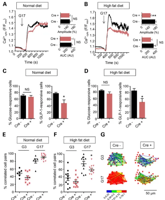
cytdynamics may be affected. Examined in whole islets
from normal diet-fed mice by functional multicellular imaging (55–57), we observed largely unaltered responses to glucose of individualβ cells after Tcf7l2 elimination (Fig.3A and C). In con-trast, those in response to GLP-1 were significantly impaired, with the proportion of GLP-1-responsive cells markedly reduced (Fig.3C). On the other hand, after maintenance on a high fat diet, both the amplitude of the [Ca2+]
cytresponse to high glucose
(Fig. 3B), and the proportion of cells responding to GLP-1 (Fig.3D), were reduced in Tcf7l2fl/fl::Ins1.Cre+mice versus controls.
Changes inβ cell ‘connectivity’, i.e. the degree to which β cells across the islet syncytium mount a coordinated (synchronized) re-sponse to stimulation (56), are associated with impaired insulin secretion (55). We have also recently demonstrated that humanβ cells depleted for another GWAS gene for T2D, adenylate cyclase V (ADCY5) (57), show impaired cellular connectivity. Whereas Tcf7l2 elimination exerted no effect on the number of connected cell pairs in islets from normal chow-fed mice (Fig.3E), inter-cellular connectivity was significant reduced by loss of this transcription factor from islets maintained on a high fat diet (Fig.3F and G).
Effects of Tcf7l2 elimination onβ cell expansion in response to high fat diet
To determine whether alterations inβ (or α) cell mass also might contribute to impaired insulin production in vivo after Tcf7l2
Figure 2. Tcf7l2 deletion impairs glucose and GLP1-stimulated insulin secretion from isolated islets. (A) Plasma insulin following intraperitoneal injection of glucose from 20-week-old mice that had been maintained on a high fat diet was measured as described in Materials and Methods. (B) Real-time quantitative PCR analysis was performed on islets from 20-weeks-old mice that had been maintained on a normal chow diet. n = 7–10 mice; *P ≤ 0.05. (C and D) Insulin secretion as assessed in isolated islets from 20-week old Tcf7l2fl/fl::Ins1.Cre+(red) and littermate Tcf7l2fl/fl::Ins1.Cre−(black) mice on normal chow (C) or high fat diet (D). n = 5 mice per genotype;
*P≤ 0.05; **P ≤ 0.01.
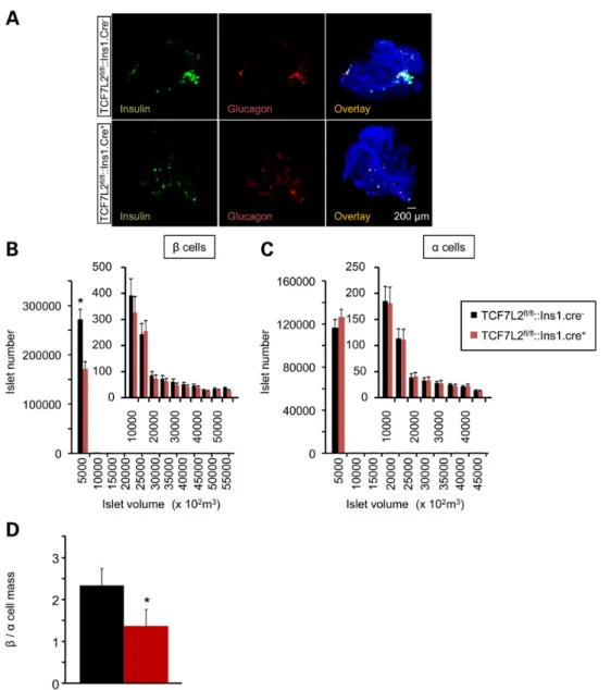
elimination, pancreata from 20-week-old Tcf7l2fl/fl::Ins1.Cre+mice
that had been maintained on a HFD for 12 weeks were stained for insulin and glucagon, respectively, and analyzed by optical projection tomography (OPT). Cre+islets displayed a markedly
(31.7%, P < 0.05; n = 4 mice per genotype) decreasedβ cell mass, but normal α-cell mass, compared with littermate controls (Fig.4), resulting in an overall decrease inβ to α cell ratio. The number of smaller insulin-stained islets was particularly sharply decreased in TCF7L2fl/fl::Ins1.Cre+mouse islets (Fig.4B).
Discussion
The chief goal of the present studies was to reassess the role in the β cell of the Wnt signalling dependent transcription factor TCF7L2. Using a Cre that is highly selective for the matureβ cell (48)
(B. Thorens and J. Ferrer unpublished), ourfindings show firstly that this factor is required for (a) the normal function of these cells, and (b) their expansion under a situation of metabolic stress. Moreover, we provide novel insights into the role of Tcf7l2 in the control of intracellular Ca2+dynamics andβ cell–β cell
communi-cation. The latter result was unexpected given previousfindings that [Ca2+]
cytchanges were apparently unaffected by
manipula-tion of Tcf7l2 expression (28). However, the earlier studies were performed using isolatedβ cells (or small clusters of cells) rather than intact islets as examined here, where complex inter-cellular cross-talk is likely to overlay cell-intrinsic responses to stimula-tion. Nonetheless, the present results are consistent with very recentfindings (58) showing decreased expression of the volt-age-gated calcium channel subunits including Cacna1d in rat insulinoma-derived INS1β cells silenced for Tcf7l2.
Figure 3. Impact of Tcf7l2 deletion on intra- and inter-cellular Ca2+dynamics. (A and C) Responses to glucose (17 m; G17) or GLP-1 (100 n) of β cells within intact islets of
Tcf7l2fl/fl::Ins1.Cre+(red) and Tcf7l2fl/fl::Ins1.Cre−(black) mice maintained on a normal chow diet. (B and D) as for (A and C) but for islets from animals maintained for 12 weeks
on a 60% fat diet. Correlation analysis (55,56) for islets from mice maintained on (E) normal diet and (F) high fat diet, and (G) quantitation of changes in typical islets from Tcf7l2fl/fl::Ins1.Cre+and Tcf7l2fl/fl::Ins1.Cre−mouse islets as indicated.
How might TCF7L2 controlβ cell connectivity? While the dis-ruption of connectivity in earlier studies, e.g. after exposure to li-potoxic conditions (55), could be ascribed to impairments in the expression of the gap junction protein connexin 36 (Gjd2), we were unable to detect changes in Gjd2 mRNA in Tcf7l2 null islets in the present study (Fig.2B), consistent with an earlier study in-vestigating the role of Wnt signalling in the expression of other gap junction proteins (59). Changes in level or post-translational modifications (60) of this protein nonetheless remain a possibil-ity as, of course do changes in the expression of other genes.
There is gathering evidence to suggest that the effect of TCF7L2 is mediated by resistance to incretin treatment at the level of theβ cell (15,16,61–63). The present studies provide fur-ther support for this view showing both marked alterations in the expression of the Glp1r gene in Tcf7l2 null islets (Fig.2B), and impaired glucose-stimulated insulin secretion (Fig.2C and D).
Whether reduced numbers of these receptors onβ cells also contributes to the apparent impairment inβ cell expansion in the face of a high fat diet is unknown, but would seem worthy of future investigation.
A particularly significant finding of the present study was the appearance in Tcf7l2fl/fl::Ins1.Cre+mice of glucose intolerance from 16 weeks onwards demonstrating that the appearance of a β cell phenotype is dependent on age or other stresses. Import-antly, in recent studies (31), the effects of Tcf7l2 inactivation in the adult mouseβ cell (using an alternative model in which exon 10, encoding the DNA-binding HMG box, was deleted) were not examined beyond 12 weeks of age, such that any effects later in life would have been missed. Of note, in the model described here, placing mice on a high fat diet increased the extent of the intolerance but did not bring forward its appearance substantially (Fig.1).
Figure 4. Impact of Tcf7l2 deletion onβ and α cell mass. (A) Representative 3D images from OPT (see Material and Methods) of Tcf7l2fl/fl::Ins1.Cre+and Tcf7l2fl/fl::Ins1.Cre−
pancreata labelled with anti-insulin and anti-glucagon primary antibodies, and revealed using AlexaFluor 568 and 680, respectively. Right hand-most panels are overlaid with the autofluorescence signal revealing the pancreatic ductal system. Quantification of β (B), α (C) and (D) β/α cell mass from Tcf7l2fl/fl::Ins1.Cre+(red) and
Tcf7l2fl/fl::Ins1.Cre−(black) knockout mice pancreata from 20-week-old mice that had been maintained on high fat diet. Quantification was conducted using Volocity software (Invitrogen), N = 4 mice per genotype.
Relevance of the presentfindings for understanding of the action of TCF7L2 T2D risk variants in man
We have previously speculated (6) that variants at rs7903146 and neighbouring SNPs may have differential effects on TCF7L2 expression the liver and onβ cells as a result of differing splicing patterns in the two tissues. In particular, alternative splicing at the boundaries of exon 13, 13b and 14 leads to the loss of a ‘CRARF’ motif that appears to be selectively expressed in the β cell and susceptible to modulation by the risk T allele (34). Thus, a decrease in TCF7L2 activity at the protein level may be observed in islets from T-allele carriers despite increases in the overall levels of TCF7L2 transcripts. Thus, the expression of mul-tiple Tcf7l2 alleles globally in mice (40), or forced inactivation of Tcf7l2 in the liver (31), with its consequent dramatic effect to impair hepatic glucose output, may be of limited relevance to the action of the human SNP. In contrast, the impact of lowered Tcf7l2 levels in theβ cell, at least during the stresses imposed by aging or high fat feeding, may be more pertinent. Thus, as shown by the present studies, these are likely to act at the same time on the secretory activity of singleβ cells (28), the co-ordination of these cells within the islet (likely to further impair insulin output) (55), andfinally on β cell mass. Nonetheless, further studies exploring in detail the relationship between Tcf7l2 action in different tissues would seem to be warranted.
On afinal note, ‘mice are not men’ and that, given recent find-ings in relation to the consequences of Slc30a8 deletion (64,65), a certain degree of circumspection is required in transposing find-ings from one to the other. There is currently no compelling evi-dence, other than immediate genomic proximity, tying TCF7L2 to the T2D-associated variants, and it remains formally possible, though rather unlikely, that the GWAS signal is mediated via another gene. Our new study reinforces the evidence (though does not prove) that TCF7L2 is the causal gene at this locus.
Material and Methods
MaterialsAll general chemicals and tissue culture reagents were purchased from Sigma (Dorset, UK) or Invitrogen (Paisley, UK), unless otherwise stated in the text.
Animals
All in vivo procedures were approved by the UK Home Office according to the Animals (Scientific Procedures) Act 1986 and were performed at the Central Biomedical Service, Imperial College, London, UK Rodents were culled by cervical dislocation. Mice were housed at two tofive animals per cage in a pathogen-free facility with a 12-h light–dark cycle. Animals were fed ad libitum with a standard mouse chow diet (Research Diet, New Brunswick, NJ) unless otherwise stated. For high fat diet treatment, mice were placed on a high fat diet at 8 weeks of age for 8 weeks (60% [wt/wt] fat content; Research Diet, New Bruns-wick, NJ, USA) prior to analysis. Mice were weighed weekly from eight weeks.
Generation and characterization ofβ cell-specific Tcf7l2 knockout mouse
Generation of mice carrying conditional knockout alleles of Tcf7l2 (Tcf7l2fl/fl) was as described in (30). All mouse strains were main-tained on a C57BL/6 background. Tcf7l2fl/flmice were crossed with mice expressing Cre recombinase under the control of the Insulin
1 promoter (Ins1.Cre mice; (48), J. Ferrer, B. Thorens, unpublished) to generate Tcf7l2 conditional knockout mice where exon 1 of the Tcf7l2 gene was removed selectively by Cre-mediated excision in pancreatic β cells (Tcf7l2fl/fl::ins1.Cre+). Mice were born at
the expected Mendelian ratios with no obvious abnormalities. Genotyping was performed by PCR using DNA from ear biopsies. Ablation of Tcf7l2 gene expression from pancreatic islets was assessed by real-time quantitative PCR (qPCR) on islet RNA, as described below.
Oral and intraperitoneal glucose tolerance test, insulin tolerance test
Glucose tolerance was assessed by oral and intraperitoneal administration of glucose (1 g/kg). Responsiveness to incretin treatment was assessed by co-injection of liraglutide [200 µg/kg (66); Bachem, Bubendorf, Switzerland] with glucose during an intraperitoneal glucose tolerance test. Mice were fasted for 16 h, with water available ad libitum. Glucose tolerance tests were con-ducted at 09:00 on each experimental day. Intraperitoneal insulin tolerance (075 Units per kg) test was performed as described in (67).
Measurement of plasma hormones
Plasma glucagon from mice fasted for 16 h were measured using radioimmunoassay (Millipore, Watford, U.K.). Blood (200 µl) was removed by cardiac puncture from mice killed by cervical disloca-tion. Plasma was collected using high speed (2000 g, 5 min at 4°C) centrifugation in heparin-coated Microvette®tubes containing EDTA (Sarstedt, Leicester, U.K.) with added DPP IV inhibitor (100 µM; Millipore, Watford, UK). For measurement of plasma in-sulin following intraperitoneal injection of glucose, blood (100 µl) was removed from the tail vein at various times points and plas-ma collected as described above. Plasplas-ma glucagon and insulin was measured by radioimmunoassay (Millipore).
Quantitative real-time PCR analysis
Primers were designed using Primer Express 3.0 (ABI, CA, USA). Specificity for all primers was verified using BLAST (http://www. ncbi.nlm.nih.gov/blast/). RNA was extracted using Trizol (Invitro-gen, UK). cDNA conversion was performed using High Capacity cDNA conversion kit (ABI, UK) after DNAse treatment (Ambion, TX, USA). Real-time PCR was performed on an ABI-Prism Fast 7500 device using powerSYBR reagent (ABI, UK). The primer sequences used were: Tcf7l2 (forward) TTCCCCCTTGACCTCC TAGTC, Tcf7l2 (reverse) GCACACGGTCAGTCCATGTT, Ins2 (for-ward) AGCCCTAAGTGATCCGCTACAA, Ins2 (reverse) CATGTTG AAACAATAACCTGGAAGA, Glp1r (forward) CCACGGTGTCCCTCT CAGA, Glp1r (reverse) ACTGCCGCCGGTATTCTCT, Gjd2 (forward) CCCAGTCTCTGTTTTATCACCTATTCT, Gjd2 (reverse) CGGCGTTC TCGCTGCTT.
Insulin secretion assay
Secreted insulin from groups of six pancreatic islets of Langer-hans was measured by radioimmunoassay (Linco, MA, USA) as previously described (68,69).
Calcium imaging and correlation analysis
Calcium imaging and correlation analysis were performed as described in (55).
Optical projection tomography and calculations of relativeα and β cell mass
Whole pancreatic OPT, to 19 µm resolution, was performed as previously described (67,70). Dual labelling for insulin and gluca-gon was performed using insulin antibody (DAKO) and anti-glucagon antibody (Sigma) revealed using Alexa Fluor 568 and 680 (Invitrogen), respectively.
Statistical analysis
Values presented are the mean ± SEM for the number of observa-tions indicated. Statistical significance and differences between means were assessed by a two-tailed Student’s t test or one-way ANOVA with Bonferroni correction for multiple analyses. Linear correlations were calculated using regression analyses. Analysis was performed using Excel™, R, GraphPad Prism (GraphPad Software) and IgorPro.
Supplementary Material
Supplementary Material is available at HMG online.
Acknowledgements
We thank Mr Daniel Davidsson (Section of Cell Biology, Imperial College) and Dr Steven Rothery (Facility for Imaging by Light Microscopy, Imperial College) for their help with image analysis. Conflict of Interest statement. None declared.
Funding
G.D.S.X. thanks the European Foundation for the Study of Diabetes (EFSD) and Diabetes UK (13/0004672; Alec and Beryl Warren Award) for Project grants. G.A.R. thanks the MRC (UK) for Programme grant MR/J0003042/1, the BBSRC (UK) for a Project grant (BB/J015873/1), the Royal Society for a Wolfson Research Merit Award and the Wellcome Trust for a Senior Investigator Award (WT098424AIA). D.J.H. is a Diabetes UK R.D. Lawrence Fellow. The work leading to this publication has received support from the Innovative Medi-cines Initiative Joint Undertaking under grant agreement no. 155005 (IMIDIA), resources of which are composed of afinancial contribution from the European Union′s Seventh Framework Pro-gramme (FP7/2007-2013) and EFPIA companies’ in kind contribu-tion (G.A.R.), and the Wellcome Trust (WT ISSF grant awarded to G.A.R. and P.M.F.). Funding to pay the Open Access publication charges for this article was provided by the Wellcome Trust.
References
1. Pierce, M., Keen, H. and Bradley, C. (1995) Risk of diabetes in offspring of parents with non-insulin-dependent diabetes. Diabet. Med., 12, 6–13.
2. Newman, B., Selby, J.V., King, M.C., Slemenda, C., Fabsitz, R. and Friedman, G.D. (1987) Concordance for type 2 (non-insulin-dependent) diabetes mellitus in male twins. Diabeto-logia, 30, 763–768.
3. Barroso, I. (2005) Genetics of Type 2 diabetes. Diabet. Med., 22, 517–535.
4. Basile, K.J., Johnson, M.E., Xia, Q. and Grant, S.F. (2014) Genetic susceptibility to type 2 diabetes and obesity: follow-up of find-ings from genome-wide association studies. Int. J. Endocrinol., 2014, 769671.
5. Perry, J.R. and Frayling, T.M. (2008) New gene variants alter type 2 diabetes risk predominantly through reduced beta-cell function. Curr. Opin. Clin. Nutr. Metab. Care, 11, 371–377. 6. Rutter, G.A. (2014) Understanding GWAS genes for Type 2
dia-betes. Diabet Med. 2014. doi: 10.1111/dme.12579. [Epub ahead of print].
7. Grant, S.F., Thorleifsson, G., Reynisdottir, I., Benediktsson, R., Manolescu, A., Sainz, J., Helgason, A., Stefansson, H., Emilsson, V. and Helgadottir, A., et al. (2006) Variant of transcription factor 7-like 2 (TCF7L2) gene confers risk of type 2 diabetes. Nat. Genet., 38, 320–323.
8. Scott, L.J., Mohlke, K.L., Bonnycastle, L.L., Willer, C.J., Li, Y., Duren, W.L., Erdos, M.R., Stringham, H.M., Chines, P.S. and Jackson, A.U., et al. (2007) A genome-wide association study of type 2 diabetes in Finns detects multiple susceptibility variants. Science, 316, 1341–1345.
9. Zeggini, E., Weedon, M.N., Lindgren, C.M., Frayling, T.M., Elliott, K.S., Lango, H., Timpson, N.J., Perry, J.R., Rayner, N. W. and Freathy, R.M., et al. (2007) Replication of genome-wide association signals in UK samples reveals risk loci for type 2 diabetes. Science, 316, 1336–1341.
10. Sladek, R., Rocheleau, G., Rung, J., Dina, C., Shen, L., Serre, D., Boutin, P., Vincent, D., Belisle, A. and Hadjadj, S., et al. (2007) A genome-wide association study identifies novel risk loci for type 2 diabetes. Nature, 445, 881–885.
11. Andersen, M.K., Sterner, M., Forsen, T., Karajamaki, A., Rolandsson, O., Forsblom, C., Groop, P.H., Lahti, K., Nilsson, P.M., Groop, L. and Tuomi, T. (2014) Type 2 diabetes suscepti-bility gene variants predispose to adult-onset autoimmune diabetes. Diabetologia 57:1859–1868.
12. Saxena, R., Voight, B.F., Lyssenko, V., Burtt, N.P., de Bakker, P.I., Chen, H., Roix, J.J., Kathiresan, S., Hirschhorn, J.N. and Daly, M.J., et al. (2007) Genome-wide association analysis identifies loci for type 2 diabetes and triglyceride levels. Science, 316, 1331–1336. 13. Saxena, R., Elbers, C.C., Guo, Y., Peter, I., Gaunt, T.R., Mega, J.L.,
Lanktree, M.B., Tare, A., Castillo, B.A. and Li, Y.R., et al. (2012) Large-scale gene-centric meta-analysis across 39 studies identifies type 2 diabetes loci. Am. J. Hum. Genet., 90, 410–425. 14. Saxena, R., Gianniny, L., Burtt, N.P., Lyssenko, V., Giuducci, C., Sjogren, M., Florez, J.C., Almgren, P., Isomaa, B. and Orho-Melander, M., et al. (2006) Common single nucleotide polymorphisms in TCF7L2 are reproducibly associated with type 2 diabetes and reduce the insulin response to glucose in nondiabetic individuals. Diabetes, 55, 2890–2895.
15. Villareal, D.T., Robertson, H., Bell, G.I., Patterson, B.W., Tran, H., Wice, B. and Polonsky, K.S. (2010) TCF7L2 variant rs7903146 affects the risk of type 2 diabetes by modulating incretin action. Diabetes, 59, 479–485.
16. Pilgaard, K., Jensen, C.B., Schou, J.H., Lyssenko, V., Wegner, L., Brons, C., Vilsboll, T., Hansen, T., Madsbad, S. and Holst, J.J., et al. (2009) The T allele of rs7903146 TCF7L2 is associated with impaired insulinotropic action of incretin hormones, reduced 24 h profiles of plasma insulin and glucagon, and increased hepatic glucose production in young healthy men. Diabetologia, 52, 1298–1307.
17. Le, B.O., Kerr-Conte, J., Gargani, S., Delalleau, N., Huyvaert, M., Gmyr, V., Froguel, P., Neve, B. and Pattou, F. (2012) TCF7L2 rs7903146 impairs islet function and morphology in non-diabetic individuals. Diabetologia, 55, 2677–2681.
18. Deng, S., Vatamaniuk, M., Huang, X., Doliba, N., Lian, M.M., Frank, A., Velidedeoglu, E., Desai, N.M., Koeberlein, B. and Wolf, B., et al. (2004) Structural and functional abnormalities in the islets isolated from type 2 diabetic subjects. Diabetes, 53, 624–632.
19. Yoon, K.H., Ko, S.H., Cho, J.H., Lee, J.M., Ahn, Y.B., Song, K.H., Yoo, S.J., Kang, M.I., Cha, B.Y. and Lee, K.W., et al. (2003) Selective beta-cell loss and alpha-cell expansion in patients with type 2 diabetes mellitus in Korea. J. Clin. Endocrinol. Metab, 88, 2300–2308.
20. Jin, T. and Liu, L. (2008) The Wnt signaling pathway effector TCF7L2 and type 2 diabetes mellitus. Mol. Endocrinol., 22, 2383–2392.
21. Roose, J. and Clevers, H. (1999) TCF transcription factors: molecular switches in carcinogenesis. Biochim. Biophys. Acta, 1424, M23–M37.
22. Saadeddin, A., Babaei-Jadidi, R., Spencer-Dene, B. and Nateri, A.S. (2009) The links between transcription, beta-catenin/JNK signaling, and carcinogenesis. Mol. Cancer Res., 7, 1189–1196. 23. Reya, T. and Clevers, H. (2005) Wnt signalling in stem cells
and cancer. Nature, 434, 843–850.
24. Papadopoulou, S. and Edlund, H. (2005) Attenuated Wnt signaling perturbs pancreatic growth but not pancreatic function. Diabetes, 54, 2844–2851.
25. Rulifson, I.C., Karnik, S.K., Heiser, P.W., ten, B.D., Chen, H., Gu, X., Taketo, M.M., Nusse, R., Hebrok, M. and Kim, S.K. (2007) Wnt signaling regulates pancreatic beta cell proliferation. Proc. Natl. Acad. Sci. U. S. A., 104, 6247–6252.
26. Wells, J.M., Esni, F., Boivin, G.P., Aronow, B.J., Stuart, W., Combs, C., Sklenka, A., Leach, S.D. and Lowy, A.M. (2007) Wnt/beta-catenin signaling is required for development of the exocrine pancreas. BMC Dev. Biol., 7, 4.
27. Heiser, P.W., Lau, J., Taketo, M.M., Herrera, P.L. and Hebrok, M. (2006) Stabilization of beta-catenin impacts pancreas growth. Development, 133, 2023–2032.
28. da Silva Xavier, G., Loder, M.K., McDonald, A., Tarasov, A.I., Carzaniga, R., Kronenberger, K., Barg, S. and Rutter, G.A. (2009) TCF7L2 regulates late events in insulin secretion from pancreatic islet beta-cells. Diabetes, 58, 894–905. 29. Shu, L., Sauter, N.S., Schulthess, F.T., Matveyenko, A.V.,
Oberholzer, J. and Maedler, K. (2008) Transcription factor 7-like 2 regulates beta-cell survival and function in human pancreatic islets. Diabetes, 57, 645–653.
30. da Silva Xavier, G., Mondragon, A., Sun, G., Chen, L., McGinty, J.A., French, P.M. and Rutter, G.A. (2012) Abnormal glucose tolerance and insulin secretion in pancreas-specific Tcf7l2-null mice. Diabetologia, 55, 2667–2676.
31. Boj, S.F., van Es, J.H., Huch, M., Li, V.S., Jose, A., Hatzis, P., Mokry, M., Haegebarth, A., van den Born, M. and Chambon, P., et al. (2012) Diabetes risk gene and Wnt effector Tcf7l2/ TCF4 controls hepatic response to perinatal and adult meta-bolic demand. Cell, 151, 1595–1607.
32. Korinek, V., Barker, N., Moerer, P., van, D.E., Huls, G., Peters, P. J. and Clevers, H. (1998) Depletion of epithelial stem-cell compartments in the small intestine of mice lacking Tcf-4. Nat. Genet., 19, 379–383.
33. Wicksteed, B., Brissova, M., Yan, W., Opland, D.M., Plank, J.L., Reinert, R.B., Dickson, L.M., Tamarina, N.A., Philipson, L.H. and Shostak, A., et al. (2010) Conditional gene targeting in mouse pancreatic ss-Cells: analysis of ectopic Cre transgene expression in the brain. Diabetes, 59, 3090–3098.
34. Prokunina-Olsson, L., Welch, C., Hansson, O., Adhikari, N., Scott, L.J., Usher, N., Tong, M., Sprau, A., Swift, A. and Bonny-castle, L.L., et al. (2009) Tissue-specific alternative splicing of TCF7L2. Hum. Mol. Genet., 18, 3795–3804.
35. Liu, Z. and Habener, J.F. (2008) Glucagon-like peptide-1 activa-tion of TCF7L2-dependent Wnt signaling enhances pancreat-ic beta cell proliferation. J. Biol. Chem., 283, 8723–8735.
36. Shu, L., Matveyenko, A.V., Kerr-Conte, J., Cho, J.H., McIntosh, C.H. and Maedler, K. (2009) Decreased TCF7L2 protein levels in type 2 diabetes mellitus correlate with downregulation of GIP- and GLP-1 receptors and impaired beta-cell function. Hum. Mol. Genet., 18, 2388–2399.
37. Takamoto, I., Kubota, N., Nakaya, K., Kumagai, K., Hashimoto, S., Kubota, T., Inoue, M., Kajiwara, E., Katsuyama, H. and Obata, A., et al. (2014) TCF7L2 in mouse pancreatic beta cells plays a crucial role in glucose homeostasis by regulating beta cell mass. Diabetologia, 57, 542–553.
38. Shao, W., Wang, D., Chiang, Y.T., Ip, W., Zhu, L., Xu, F., Colum-bus, J., Belsham, D.D., Irwin, D.M. and Zhang, H., et al. (2013) The Wnt signaling pathway effector TCF7L2 controls gut and brain proglucagon gene expression and glucose homeo-stasis. Diabetes, 62, 789–800.
39. Norton, L., Fourcaudot, M., Abdul-Ghani, M.A., Winnier, D., Mehta, F.F., Jenkinson, C.P. and Defronzo, R.A. (2011) Chroma-tin occupancy of transcription factor 7-like 2 (TCF7L2) and its role in hepatic glucose metabolism. Diabetologia, 54, 3132–3142.
40. Savic, D., Ye, H., Aneas, I., Park, S.Y., Bell, G.I. and Nobrega, M. A. (2011) Alterations in TCF7L2 expression define its role as a key regulator of glucose metabolism. Genome Res., 21, 1417– 1425.
41. Gaulton, K.J., Nammo, T., Pasquali, L., Simon, J.M., Giresi, P.G., Fogarty, M.P., Panhuis, T.M., Mieczkowski, P., Secchi, A. and Bosco, D., et al. (2010) A map of open chromatin in human pancreatic islets. Nat. Genet., 42, 255–259.
42. Savic, D., Bell, G.I. and Nobrega, M.A. (2012) An in vivo cis-regulatory screen at the type 2 diabetes associated TCF7L2 locus identifies multiple tissue-specific enhancers. PLoS One, 7, e36501.
43. Duval, A., Rolland, S., Tubacher, E., Bui, H., Thomas, G. and Hamelin, R. (2000) The human T-cell transcription factor-4 gene: structure, extensive characterization of alternative splicings, and mutational analysis in colorectal cancer cell lines. Cancer Res., 60, 3872–3879.
44. Le Bacquer, O., Shu, L., Marchand, M., Neve, B., Paroni, F., Kerr, C.J., Pattou, F., Froguel, P. and Maedler, K. (2011) TCF7L2 splice variants have distinct effects on beta-cell turnover and func-tion. Hum. Mol. Genet., 20, 1906–1915.
45. Mondal, A.K., Das, S.K., Baldini, G., Chu, W.S., Sharma, N.K., Hackney, O.G., Zhao, J., Grant, S.F. and Elbein, S.C. (2010) Genotype and tissue-specific effects on alternative splicing of the transcription factor 7-like 2 gene in humans. J. Clin. Endocrinol. Metab., 95, 1450–1457.
46. Weise, A., Bruser, K., Elfert, S., Wallmen, B., Wittel, Y., Wohrle, S. and Hecht, A. (2010) Alternative splicing of Tcf7l2 tran-scripts generates protein variants with differential pro-moter-binding and transcriptional activation properties at Wnt/beta-catenin targets. Nucleic Acids Res., 38, 1964–1981. 47. McCarthy, M.I., Rorsman, P. and Gloyn, A.L. (2013) TCF7L2 and
diabetes: a tale of two tissues, and of two species. Cell Metab., 17, 157–159.
48. Kone, M., Pullen, T.J., Sun, G., Ibberson, M., Martinez-Sanchez, A., Sayers, S., Nguyen-Tu, M.S., Kantor, C., Swisa, A. and Dor, Y., et al. (2014) LKB1 and AMPK differentially regulate pancre-atic beta-cell identity. FASEB J.28:4972–4985.
49. Yoshinari, M. and Daikoku, S. (1982) Ontogenetic appearance of immunoreactive endocrine cells in rat pancreatic islets. Anat. Embryol. (Berl), 165, 63–70.
50. Teitelman, G. and Lee, J.K. (1987) Cell lineage analysis of pan-creatic islet development: glucagon and insulin cells arise
from catecholaminergic precursors present in the pancreatic duct. Dev. Biol., 121, 454–466.
51. Tamarina, N.A., Roe, M.W. and Philipson, L. (2014) Character-ization of mice expressing Ins1 gene promoter driven CreERT recombinase for conditional gene deletion in pancreatic beta-cells. Islets, 6. [Epub ahead of print].
52. Mondragon, A., Davidsson, D., Kyriakidou, S., Bertling, A., Gomes-Faria, R., Cohen, P., Rothery, S., Chabosseau, P., Rutter, G. and da Silva Xavier, G. (2014) Divergent effects of liraglutide, exendin-4, and sitagliptin on beta-cell mass and indicators of pancreatitis in a mouse model of hypergly-caemia. PLoS One, 9, e104873.
53. Rutter, G.A. (2001) Nutrient-secretion coupling in the pancreatic islet beta-cell: recent advances. Mol. Aspects Med., 22, 247–284.
54. Rutter, G.A. (2004) Visualising insulin secretion. The Min-kowski Lecture 2004. Diabetologia, 47, 1861–1872.
55. Hodson, D.J., Mitchell, R.K., Bellomo, E.A., Sun, G., Vinet, L., Meda, P., Li, D., Li, W.H., Bugliani, M. and Marchetti, P., et al. (2013) Lipotoxicity disrupts incretin-regulated human beta cell connectivity. J. Clin. Invest, 123, 4182–4194.
56. Hodson, D.J., Tarasov, A.I., Gimeno, B.S., Mitchell, R.K., John-ston, N.R., Haghollahi, S., Cane, M.C., Bugliani, M., Marchetti, P. and Bosco, D., et al. (2014) Incretin-modulated beta cell en-ergetics in intact islets of Langerhans. Mol. Endocrinol., 28, 860–871.
57. Hodson, D.J., Mitchell, R.K., Marselli, L., Pullen, T.J., Gimeno, B. S., Semplici, F., Everett, K.L., Cooper, D.M., Bugliani, M. and Marchetti, P., et al. (2014) ADCY5 Couples Glucose to Insulin Secretion in Human Islets. Diabetes, 63, 3009–3021.
58. Zhou, Y., Park, S.Y., Su, J., Bailey, K., Ottosson-Laakso, E., Shcherbina, L., Oskolkov, N., Zhang, E., Thevenin, T. and Fa-dista, J., et al. (2014) TCF7L2 is a master regulator of insulin production and processing. Hum. Mol. Genet. [Epub ahead of print].
59. van der Heyden, M.A., Rook, M.B., Hermans, M.M., Rijksen, G., Boonstra, J., Defize, L.H. and Destree, O.H. (1998) Identification of connexin43 as a functional target for Wnt signalling. J. Cell Sci., 111 (Pt 12), 1741–1749.
60. Oyamada, M., Takebe, K. and Oyamada, Y. (2013) Regulation of connexin expression by transcription factors and epigen-etic mechanisms. Biochim. Biophys. Acta, 1828, 118–133. 61. Schafer, S.A., Tschritter, O., Machicao, F., Thamer, C., Stefan,
N., Gallwitz, B., Holst, J.J., Dekker, J.M., Hart’t, L.M. and Nijpels,
G., et al. (2007) Impaired glucagon-like peptide-1-induced in-sulin secretion in carriers of transcription factor 7-like 2 (TCF7L2) gene polymorphisms. Diabetologia, 50, 2443–2450. 62. Lyssenko, V., Lupi, R., Marchetti, P., Del, G.S., Orho-Melander,
M., Almgren, P., Sjogren, M., Ling, C., Eriksson, K.F. and Lethagen, A.L., et al. (2007) Mechanisms by which common variants in the TCF7L2 gene increase risk of type 2 diabetes. J. Clin. Invest, 117, 2155–2163.
63. Smushkin, G., Sathananthan, M., Sathananthan, A., Dalla, M. C., Micheletto, F., Zinsmeister, A.R., Cobelli, C. and Vella, A. (2012) Diabetes-associated common genetic variation and its association with GLP-1 concentrations and response to exogenous GLP-1. Diabetes, 61, 1082–1089.
64. Flannick, J., Thorleifsson, G., Beer, N.L., Jacobs, S.B., Grarup, N., Burtt, N.P., Mahajan, A., Fuchsberger, C., Atzmon, G. and Benediktsson, R., et al. (2014) Loss-of-function mutations in SLC30A8 protect against type 2 diabetes. Nat Genet., 46, 357–363.
65. Rutter, G.A. and Chimienti, F. (2014) SLC30A8 mutations in type 2 diabetes. Diabetologia. [Epub ahead of print].
66. Noyan-Ashraf, M.H., Momen, M.A., Ban, K., Sadi, A.M., Zhou, Y.Q., Riazi, A.M., Baggio, L.L., Henkelman, R.M., Husain, M. and Drucker, D.J. (2009) GLP-1R agonist liraglutide activates cytoprotective pathways and improves outcomes after experimental myocardial infarction in mice. Diabetes, 58, 975–983.
67. Sun, G., Tarasov, A.I., McGinty, J., McDonald, A., da Silva Xavier, G., Gorman, T., Marley, A., French, P.M., Parker, H. and Gribble, F., et al. (2010) Ablation of AMP-activated protein kinase alpha1 and alpha2 from mouse pancreatic beta cells and RIP2.Cre neurons suppresses insulin release in vivo. Diabetologia, 53, 924–936.
68. Ainscow, E.K., Zhao, C. and Rutter, G.A. (2000) Acute overex-pression of lactate dehydrogenase-A perturbs beta-cell mito-chondrial metabolism and insulin secretion. Diabetes, 49, 1149–1155.
69. da Silva Xavier, G., Leclerc, I., Varadi, A., Tsuboi, T., Moule, S.K. and Rutter, G.A. (2003) Role for AMP-activated protein kinase in glucose-stimulated insulin secretion and preproinsulin gene expression. Biochem. J., 371, 761–774.
70. Alanentalo, T., Asayesh, A., Morrison, H., Loren, C.E., Holmberg, D., Sharpe, J. and Ahlgren, U. (2007) Tomographic molecular imaging and 3D quantification within adult mouse organs. Nat. Methods, 4, 31–33.