DIFFERENCES IN ATRIAL FIBRILLATION PROPERTIES
UNDER VAGAL NERVE STIMULATION VERSUS ATRIAL
TACHYCARDIA REMODELING
par
Grigorios Katsouras MD
Programme en Sciences Biomédicales Faculté de Médecine
Thèse présentée à la Faculté des études supérieures en vue de l’obtention du grade de Maitrise
en Sciences Biomédicales
Août 2009
Faculté des études supérieures
Cette thèse intitulée :
DIFFERENCES IN ATRIAL FIBRILLATION PROPERTIES
UNDER VAGAL NERVE STIMULATION VERSUS ATRIAL
TACHYCARDIA REMODELING
présentée par : Grigorios Katsouras MD
a été évaluée par un jury composé des personnes suivantes :
Bruce Allen PhD, président-rapporteur Stanley Nattel MD, directeur de recherche
Résumé
Fond : Le substrat de fibrillation auriculaire (FA) vagale et celui secondaire à remodelage par tachycardie auriculaire (RTA) partagent beaucoup des caractéristiques : période réfractaire efficace (PRE) réduite, hétérogénéité accrue de PRE et quelques mécanismes moléculaires communs. Cette étude a comparé les 2 substrats à une abréviation comparable de PRE.
Méthodes : Chez chacun de 6 chiens de groupe de stimulation vagal (SV), les paramètres de stimulation cervicale bilatérale de nerves vagaux ont été ajustés pour produire la même PRE moyenne (calculé à 8 sites des oreillettes gauche et droite) avec 6 chiens de groupe de RTA assorti à sexe et poids. Des paramètres électrophysiologiques, la durée moyenne de la fibrillation auriculaire (DAF) et les fréquences dominantes (FD) locales ont étés calculés. Résultats : En dépit des PREs assorties (SV: 80±12msec contre RTA: 79±12msec) la DAF était plus longue (*), l’hétérogénéité de conduction était plus élevée (*), la FD était plus rapide (*) et la variabilité de FD plus grande (*) chez les chiens SV. Les zones de maximum FD qui reflètent les zones d’origine de FA étaient à côté de ganglions autonomes chez les chiens SV.
Conclusions : Pour un PRE atriale comparable, la FA secondaire à SV est plus rapide et plus persistante que la FA avec un substrat de RTA. Ces résultats sont consistants avec des modèles de travail suggérant que l'hyperpolarisation SV-induite contribue de façon important à la stabilisation et à l'accélération des rotors qui maintiennent la FA. La similitude de la distribution de FD du groupe vagal avec la distribution des lésions
d’ablation après cartographie des électrogrammes atriales fragmentés suggère des nouvelles techniques d’ablation. La distribution des FD entre le SV et le RTA fournit de nouvelles idées au sujet de possible rémodelage neuroreceptorial et indique des différences importantes entre ces substrats de FA superficiellement semblables.
Mots-clés : fibrillation auriculaire, période réfractaire effective, stimulation vagal, ganglion cardiaque, remodelage par tachycardie auriculaire, fréquence dominante, récepteurs
Abstract
Background: Vagal nerve stimulation (VS) and atrial tachycardia remodeled (ATR) atrial fibrillation (AF) substrates share many features: reduced effective refractory period (ERP), increased ERP heterogeneity and some common molecular mechanisms. This study compared VS and ATR substrates at comparable ERP abbreviation.
Methods: In each of 6 VS dogs, bilateral cervical VS parameters were adjusted to produce the same mean ERP as a sex and weight matched ATR dog. Electrophysiological parameters, mean duration of AF (DAF) and local dominant frequencies (DF) were determined (before (CTL) and after VS in VS dogs).
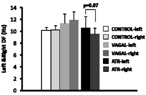
Results: Despite matched ERPs (VG: 80±12msec vs ATR: 79±12msec) DAF was greater (*), conduction heterogeneity was greater (*), DF was faster (*) and DF variability greater (*) in VS dogs. AF drivers reflected by maximum DF zones were adjacent to autonomic ganglia in VS dogs; there was a tendency (p<0.07) to faster driver zones in the left atrium comparing to the right in ATR dogs.
Conclusions: For a comparable atrial ERP, VS AF is faster and more persistent than AF with an ATR substrate. These results are consistent with modeling work suggesting that VS-induced hyperpolarization is an important contributor to AF-maintaining rotor stabilization and acceleration. Similarities in DF distribution in VS dogs with distribution of ablation lesions performed after Complex Fractionated Atrial Electrograms mapping suggests new curative ablation methods. DF distribution differences between VS and ATR
provides new ideas about possible neuroreceptorial remodeling and indicates important differences between these superficially similar AF substrates.
Keywords: Atrial fibrillation, atrial tachycardia remodeling, vagal stimulation, dominant frequencies, effective refractory period, cardiac ganglia, muscarinic receptors, conduction velocity
Table of contents
List of figures ... ix List of abbreviations ... x Remerciements ... xiv 1. Introduction ... 1 1.1 Atrial Fibrillation ... 1 1.1.1 Definition ... 1 1.1.2 Classification ... 21.1.3 Epidemiology and prognosis ... 4
1.2 Mechanisms of Atrial Fibrillation ... 6
1.2.1 Theories ... 6
1.2.2 Spiral waves and rotors ... 9
1.2.3 Dominant Frequencies and AF ... 11
1.2.4 Complex fractionated atrial electrograms (CFAEs) and DF ... 13
1.2.5 Autonomic nervous system and AF ... 14
1.2.6 Atrial Remodeling ... 19
1.3 Animal Models ... 25
1.3.1 Atrial tachycardia remodeling (ATR) animal model ... 26
1.3.2 Vagal stimulation (VS) animal model ... 28
2. Hypothesis ... 30
3. Differences in atrial fibrillation properties under vagal nerve stimulation versus atrial tachycardia remodeling ... 33
3.1 Abstract ... 34
3.2 Introduction ... 37
3.2 Methods ... 37
3.2.1 Animal models and experimental groups ... 37
3.2.2 Experimental protocol ... 38
3.2.4 Atrial frequency-spectrum analysis... 41
3.2.5 Statistical analysis ... 42
3.3 Results ... 43
3.3.1 Electrophysiological variables and properties of atrial fibrillation ... 43
3.3.2 Regional changes in electrophysiological properties ... 44
3.3.3 Local conduction abnormalities ... 44
3.3.4 Dominant frequency analysis ... 45
3.4 Discussion ... 46
3.4.1 Atrial electrophysiological consequences of vagal-nerve stimulation and ATR 46 3.4.2 Novel findings and potential underlying mechanisms ... 48
3.5 Potential limitations ... 50
3.6 Acknowledgment ... 51
3.7 References ... 52
3.8 Figure Legends ... 57
4. Discussion ... 67
4.1 Electrophysiological properties of VS and ATR substrate ... 67
4.2 Leading circle model, spiral wave theory and our study ... 70
4.3 Role of autonomic ganglia, spiral waves and rotors ... 72
4.4 Clinical implications ... 75
4.5 Potential limitations ... 76
4.6 IKACh , IKACh,c , muscarinic receptors, DF and future research ... 77
5. Conclusion ... 82 Bibliography ... I
List of figures
Fig. 1 Patterns of AF. page 3
Fig. 2 Example of electrogram and power spectra page 11 Fig. 3 Sympathetic and parasympathetic nervous system page 14 Fig. 4 Anatomic distribution of cardiac autonomic ganglia page 16 Fig. 5 Mean AERP and mean duration of AF under each condition page 59 Fig. 6 Mean CV and mean WL under each condition page 60 Fig. 7 AERP heterogeneity and CV heterogeneity under each condition page 61 Fig. 8 Effects of VS and ATR on electrophysiological indexes in different
areas of left and right atrium page 62
Fig. 9 Local conduction differences between groups under LAA
and RAA pacing page 63
Fig. 10 Example of electrogram recordings and power spectra under
each condition page 64
Fig. 11 Spatial distribution of mean DFs under each condition page 65 Fig. 12 Overall results of DF analysis. Comparison between groups page 66 Fig. 13 Similarities between anatomical distribution of epicardial
autonomic ganglia, DF distribution under VS and ablation
lesions following CFAEs. page 75
List of abbreviations
AERP: atrial effective refractory period AF: atrial fibrillation
AFCL: atrial fibrillation cycle length APD: action potential duration ASR: atrial structural remodeling ATR: atrial tachycardia remodeling AV: atrioventricular
BCL: basic cycle length CL: cycle length CTL: control
CV: conduction velocity
DAF: durée de fibrillation auriculaire DF: dominant frequency
ECG: electrocardiogram ERP: effective refractory period FA: fibrillation auriculaire FD: fréquences dominantes HF: heart failure
IKACh: inward rectifier potassium current legated to acetylcholine IKACh, c: IKACh constitutively active
IK1: inward rectifier potassium current IVC: inferior vena cava
LA: left atrial
LAA: left-atrial appendage
LBB: left-atrial side of Bachmann’s Bundle LIW: left-atrial inferior wall
LPW: left-atrial posterior wall mAChKir: muscarinic K+ channel Pa: pulmonary artery
PRE: période réfractaire efficace PV: pulmonary veins
RA: right atrial
RAA: right-atrial appendage
RBB: right-atrial side of Bachmann’s Bundle RIW: right-atrial inferior wall
RPV: right pulmonary vein RPW: right-atrial posterior wall
RTA: remodelage par tachycardie auriculaire RV: right ventricular
SV: stimulation vagal SVC: superior vena cava
VS: vagal stimulation
À mes parents Evangelos et Litsa pour leur soutien continue
Remerciements
Je voudrais remercier la fondation Bodossakis pour leur aide économique pendant mes années académiques, le journaliste Nick Gage pour le soutien et les conseils, Madames Louise Fortier, Sylvie Levesque et Marie-Claude Guertin pour leur conseils en Statistique, Madames Nathalie L’Heureux et Chantal St-Cyr pour leur aide aux techniques de laboratoire, la fondation de l’Institut de Cardiologie, les médecins de l’équipe d’électrophysiologie et particulièrement Dr Mario Talajic qui m’a aidé à compléter mon fellowship clinique et expérimentale. Sans la contribution et l’aide substantiel des coauteurs de l’article, cette thèse ne pourrait pas terminer. Enfin un grande merci à mon directeur de recherche Dr Stanley Nattel qui m’a permis de faire partie de son laboratoire et qui m’a aidé pendant toutes les phases du projet.
1. Introduction
1.1 Atrial Fibrillation
1.1.1 Definition
Atrial fibrillation (AF) is a supraventricular tachycardia characterized by erratic and uncoordinated electrical activation of the atria causing deterioration of atrial mechanical function and irregular heart rhythm1, 2. On the electrocardiogram, AF is characterized by the substitution of the normally present P waves with fibrillatory (f) waves that conduct variably to the ventricles, creating the characteristic “irregularly irregular” heart rhythm of AF.
The f waves are small, irregular baseline undulations of variable amplitude and morphology at a rate of 350-600 beats/min3. Atrial dimensions and underlying heart disease are the main factors influencing the amplitude of f waves. Occasionaly, electrical activity is not recognizable on the ECG, but only by atrial endocardial leads recording electrical activity of even higher frequency. It is believed that f waves don’t represent total atrial activity but depict only the larger vectors generated by the variable electrical atrial activation at any given moment3. However, if each atrial impulse were conducted to the ventricle the extremely rapid ventricular rate would lead to ineffective cardiac contraction and sudden death4. The filtering properties of the atrioventricular (AV) node, which has a
limited impulse-carrying capacity, prevent such an event. Thus, the ventricular response to AF depends on the interaction between atrial rate and filtering AV node function, the level of vagal and sympathetic tone, the presence or absence of accessory pathways, and the action of drugs1, 4.
Although AF can occur in patients without any evident heart problems (lone AF), it is generally associated with structural heart diseases such as congestive heart failure, valvular disease, ischemic heart disease, pericardial diseases, congenital defects and hypertensive or other cardiomyopathies. Atrial dilation and increased atrial pressure are the most plausible causes of AF promotion in such patients. However, the precise mechanistic links are incompletely defined4. Non-cardiac causes include electrolyte disturbances, ethanol intoxication, thyroid dysfunction, pulmonary disease, septic disease or febrile illnesses5.
1.1.2 Classification
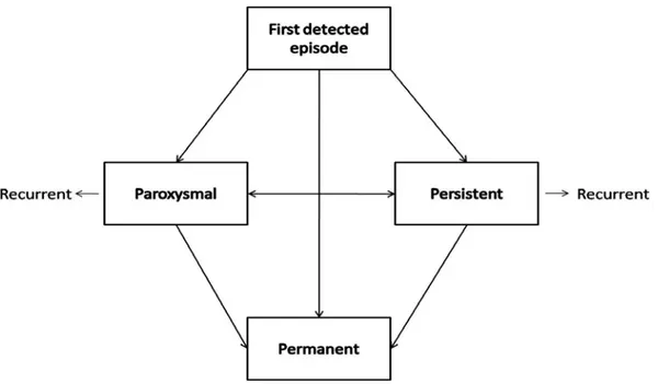
Various classifications have been proposed. According to ACC/AHA/ESC guidelines “the clinician should distinguish a first-detected episode of AF (fig. 1), recognizing that there may be uncertainty about the duration of the episode and about previous undetected episodes”1. If the patient had 2 or more episodes AF is recurrent. If an episode lasts longer than 7 days then AF is characterized as persistent. If AF terminates spontaneously or even after pharmacological or electrical cardioversion in less than 7 days
time then it should be classified as paroxysmal. AF lasting longer than a year becomes permanent.
Fig. 1 Patterns of AF. Paroxysmal AF for episodes lasting less than 7 days, persistent AF for episodes lasting between 7 days and one year and permanent AF for episodes greater than a year (adapted from Fuster et al1)
These categories are not mutually exclusive. A patient can present several times with paroxysmal and occasionally persistent AF and vice versa. It is practical to categorize the patient according to the most frequent presentation.
The term “lone AF” applies to arrhythmic episodes in young individuals (generally <60 years old) without evidence of cardiopulmonary disease1, 6. It represents one of the
most intriguing forms of AF, comporting interesting pathophysiological implications. It generally has a favorable prognosis.
1.1.3 Epidemiology and prognosis
Prevalence of a disease is defined as the proportion of a population affected by the disease at a point in time. The estimated prevalence of AF is 0.4% to 1% in the general population1, 7. It increases to 9% in subjects 80 years or older. AF prevalence is higher in men when compared to women at all ages. Nevertheless, due to the higher mean age, women comprise a larger proportion of the older groups8, 9.
AF prevalence is expected to increase proportionally with the increase of mean age of population. In USA the percentage of subjects older than 65 years is estimated to increase substantially by the year 2050 to more than 20% of the general population9. Considering that 75% of patients with AF are 65 years or older and the median age of U.S patients with AF is 75 years7-9, we would expect a considerable rise of AF prevalence in USA and proportionally in other countries with similar lifestyle.
The incidence of a disease is the rate at which new cases present in a population during a specified time period. In prospective studies the incidence of AF increases from less than 0.1% per year in patients under 40 years to more than 1.5% in women and 2% in men older than 80 years old1, 10, 11. Furthermore, in the Framingham study the age-adjusted incidence increased in a span of less than 30 years, suggesting a greater impact of AF in the
near future1, 11. The lifetime risk for development of AF was 1 in 4 for individuals 40 years of age and older9, 12. Thus, the burden of AF in the population, being already high, increases from year to year, comporting increases in costs, physical damage and mortality.
AF is associated with an increased risk of stroke, heart failure (HF) and all cause mortality1. In the Framingham Heart Study, it was associated with 1.5 to 1.9 fold excess mortality after adjustment for preexisting cardiovascular conditions. Main causes of mortality are thromboembolic events and/or HF.
The ventricular rate in AF is determined by the interaction between the high atrial rate and the filtering function of AV node4. High ventricular rates can per se cause severe congestive HF after several weeks or months13, whereas high heart rate in patients with HF can be deleterious and increase mortality. HF promotes AF and vice versa. Patients with either condition that develop the alternate have a poor prognosis1. Moreover, loss of effective atrial contraction leads to blood stasis in the atria and formation of clots that tend to propagate to the brain and other vital organs. The rate of ischemic stroke among patients with non valvular AF averages 5% per year with a relative risk 6 times higher of that of people without AF1. About 36% of ischemic strokes in individuals aged 80 to 89 years old are attributed to AF9.
1.2 Mechanisms of Atrial Fibrillation
1.2.1 Theories
AF is the most common arrhythmia in clinical practice. Therefore, researchers focused early on this arrhythmia and theories regarding its mechanisms have been put forward almost 100 years ago4, 5. The principal mechanisms described were 1) the automatic focus activity 2) the single circuit reentry and 3) the multiple-circuit reentry.
1.2.1.1 Rapid ectopic activity and single circuit reentry theories
Focal activity and single circuit reentry theories include the existence of a source producing high frequency wavefronts that interact with the spatially variable refractory and conductive properties of atrial tissue. A single rapid firing focus would be expected to produce a regular tachycardia. However, if the atrial rate is too rapid the atria cannot respond in a 1:1 fashion and a chaotic atrial rhythm will result. Haissaguere et al showed that AF is frequently initiated by rapid focal ectopic activity coming from the pulmonary veins (PV)14. While PV seem the most frequent source of ectopic activity, rapid foci have been found also in superior vena cava (SVC), ligament of Marshall, left posterior free wall, crista terminalis and coronary sinus1.
Seemingly, special conditions are necessary in order for a single-circuit reentry to stabilize and maintain AF. A microreentrant circuit, with exiting impulses firing at extremely high rates, encounters functional conduction barriers in the atria that obstacle a
1:1 conduction. Fibrillatory conduction can be due to spatially variable refractory periods or to the structural properties of atrial tissue5, 15. Schuessler et al. demonstrated in an isolated right atrial (RA) preparation that, with increasing concentrations of acetylcholine (ACh), a single, relatively stable, high frequency circuit resulted in fibrillatory conduction16. Studies from Mandapati et al. reinforced the concept that localized left atrial (LA) sources, which may be single microreentrant circuits, would be the basis for AF in isolated animal hearts17. Furthermore, Berenfeld et al. demonstrated in the same animal model that there exists a frequency in the sheep RA below which electrical activity is organized and above which it is disorganized like AF15. Consequently, although a dominant frequency (DF) of fibrillation in LA would be conducted in the RA with decreasing values, such frequency would be high enough to disorganize electrical propagation in RA. Similar DF differences between LA and RA have been observed either clinically or experimentally in other studies18, 19.
1.2.1.2 Multiple-circuit reentry
Over the past 50 years the hypothesis of multiple circuit reentry has been the dominant conceptual model of AF4. According to Moe and coworkers20, fractionation of wavefronts propagating through the atria results in multiple reentrant self- perpetuating wavelets. The number of wavelets at any time depends on the atrial mass, the refractory period and the conduction velocity (CV) in different parts of the atria. Large atrial mass, short refractory period and delayed conduction are conditions that increase the number of
wavelets favoring sustained AF. However, a sufficient number of these wave fronts must always find excitable tissue in order for the arrhythmia to persist.
Based on these concepts and in subsequent experimental work Allessie and colleagues21, 22 developed the “leading circle” model of functional reentry. The wavelength (WL) is the distance travelled by the electrical impulse during the time of effective refractory period (ERP) and equals the product of the ERP with the CV. If the dimensions of the tissue involved are shorter than the WL, the presuppositions of reentry are missing, as the electrical impulse encounters unexcitable tissue and consequently extinguishes. Normal size atrium can permit only a small number of leading circles, which tend to extinguish. On the contrary, a dilated atrium can host multiple leading circles promoting sustained AF. Based on this notion, the Maze procedure was designed to divide the atrium into regions too small to support reentry23. The great efficacy of this procedure strengthened the multiple circuit reentry concept5. Additionally, interventions that decrease WL such as vagal stimulation (VS) (which reduces atrial effective refractory period [AERP]) permit more leading circles to coexist and promote AF, while interventions that increase WL, such as antiarrhythmic drugs that increase AERP, suppress AF by reducing the number of possible circuits24. Accordingly, most of antiarrhythmic drugs in use prevent the recurrence of AF by prolonging the WL22, 25, 26.
However, AERP is not uniformly distributed in the atria and it has been demonstrated that higher heterogeneity of AERP promotes AF27. Shorter regional AERP and consequently shorter WL would need a circuit of small size for reentry to occur. As the
firing heart rate of reentrant tachycardia increases with decreasing circuit size, the smallest circuit size (or smallest WL) should control the overall atrial electrical activity24. Such notion would be compatible with single circuit reentry with a DF that is conducted with decreasing frequency to the remaining atrium. Thus, single circuit reentry or multiple circuit reentry could be the two faces of the same coin; that is, two expressions of the same mechanism that unfortunately we still try to understand.
1.2.2 Spiral waves and rotors
Experimental work in the last decades has sustained the theory of spiral wave activity as responsible for sustained reentrant activity in excitable systems such as the heart24, 28, 29. While in the leading circle model the electrical impulse excitation follows a circle with an unexcitable core (continuously excited by centripetal waves), according to the spiral wave theory the electrical impulse pursues a spiral trajectory continuously changing direction. There is a source sink relationship where the source represents the excited and depolarized tissue and the sink denotes the excitable tissue30, 31. If the source is too small it will extinguish itself in the vast sink; if it is too big it won’t find excitable tissue to continue its activity. The maintenance of the spiral wave requires an angle of curvature which is determined by tissue excitability and source-sink relationships. Low excitability (for example an increase in ERP or reduction in INa) or propagation strength (reduced CV) limit the curvature and enlarge the spirals with subsequent termination of AF24. Thus, inhibition of Na+ current should enlarge the spirals and terminate AF as Na-channel
blockers do. On the contrary, according to the leading circle they would have the opposite effect: the CV reduction would decrease the WL and would promote AF, which is not what has been observed clinically32, 33 and experimentally34, 35.
Furthermore, experimental work demonstrated the existence of spiral waves during AF with optical36 and high density conventional mapping24, 37. A mathematical model of atrial activity with representations of atrial ionic and conduction properties in a 2-dimensional grid indicated also a spiral wave activity during AF38. Moreover, recent studies of cholinergic AF in the isolated sheep heart demonstrated that high frequency sources in the PV region dominate and drive the fibrillatory activity throughout both atria39, 40. This and other experimental studies,17, 36, 40 has led to the hypothesis that “patients with AF have a focal or reentrant mechanism as the initiating cause of arrhythmia, while perpetuation of AF may depend on the uninterrupted periodic activity of a small number of discrete generators (rotors), most often localized in the LA and established by the interaction of propagating waves with anatomical heterogeneities in the atria”4, 39. The results of one of the first studies using rapid atrial pacing in dogs to promote AF support such a hypothesis41. Ablation of the area with the shortest cycle length (CL) during AF had a profound antiarrhythmic effect in that model. Clinical studies42-44 demonstrated that ablation in PV region and posterior wall, that is areas showing the fastest activity during AF as measured by CL in coronary sinus electrograms, prolonged at first the CL and subsequently terminated the arrhythmia. The areas of shortest CL would correspond to the generators (rotors) responsible for the maintenance of AF. Consequently, based on these
works researchers focused recently their interest on the study of atrial electrical activity and its spectral properties during AF.
1.2.3 Dominant Frequencies and AF
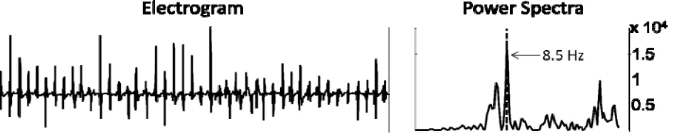
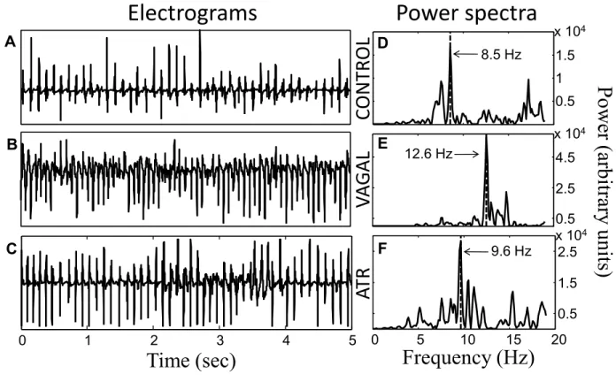
The theorem of Fourier tells that any time series, such as a cardiac electrogram, can be represented exactly as the superposition of sinusoidal waves with different frequencies and amplitudes45. Consequently, an atrial electrogram can be decomposed in its spectral components creating a power spectrum where the sinusoidal wave of major amplitude shows a dominant peak (fig. 2). The frequency of that sinusoidal wave constitutes the dominant frequency (DF) and conceptually represents the inverse of the most frequent and dominant CL in the registration of an atrial electrogram (DF= 1 sec/CL sec). For example atrial flutter, which is an atrial arrhythmia with regular CL, has generally a DF of 5Hz that would be equal to 1 sec divided by the classical CL of 0.2 sec (300 beats per minute).
Fig. 2 Example of atrial electrogram recording and its corresponding power spectra. The dominant frequency of 8.5 Hz corresponds to the frequency with the highest power.
Studies in isolated sheep hearts that have analyzed AF in frequency domains have provided evidence that AF has a high degree of spatiotemporal periodicity and high frequency rotors are responsible for maintaining AF17, 36, 40. Such rotors emanate generally from the PV region suggesting that these structures have an important role in maintaining AF45. Sanders et al46 creating DF maps from recordings of 120 points in both atria, correlated the DF distribution with ablation points and they found that ablation at PVs harboring high DF-sites resulted in an increase in CL of AF in either paroxysmal or permanent AF. However, arrhythmia termination during ablation occurred only in 88% of paroxysmal AF patients. In 87% arrhythmia termination was associated with ablation in high DF-sites.
Moreover, there is substantial evidence from animal and human studies that during AF dominant frequencies are higher in LA than in the RA18, 19, 39, 47. According to studies in isolated sheep heart, most of the high frequency sources exist in the LA and at a critical frequency, a gradient develops between LA and RA. In a recent study, Atienza et al48 demonstrated that elimination of LA to RA frequency gradients during ablation predicts long-term maintenance of sinus rhythm in AF patients. All these data clearly indicate that high-DF sites play a role in the maintenance of AF in a significant number of patients.
1.2.4 Complex fractionated atrial electrograms (CFAEs) and DF
There is evidence that high DF areas likely represent either rotors or triggered activity that is driving AF17. In the clinical setting new electogram based measures of fractionation and rate have recently been proposed to guide AF ablation49. Several studies describe AF termination after ablation of sites with CFAEs49-51. CFAES were originally defined by Nademanee49 as either 1) electrograms with two or more deflections or continuous electrical activity over a 10 second period or 2) electrograms with a mean CL< 120 msec over a 10 second recording period.
Kalifa et al52 studying AF in isolated sheep hearts showed that the most fractionated activity was found in the periphery of high frequency sources, while a similar correlation between DF areas and fractionated electrograms was demonstrated by Everett et al53 in various animal models. Recently, Zlochiver et al54 presented a very interesting work, providing insights into the mechanisms underlying CFAEs. They showed that complex electrogram signals can be divided into strictly periodic components (presumably due to underlying rotors) and residual components deriving from meandering of spiral wave sources55. Registrations farther from rotors have weaker signals, that is more residual components coming from spiral wave meandering. Interestingly, a recent study correlated the sites of fragmented electrograms with those of ganglionated plexi of the autonomic nervous system56.
1.2.5 Autonomic nervous system and AF
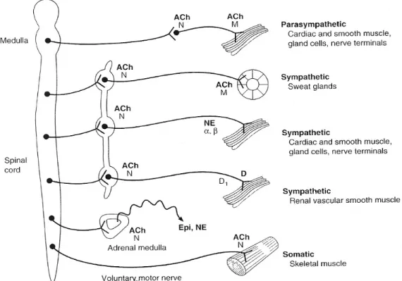
Autonomic nervous system plays a very important role in regulating heart rhythm. It is divided in two major portions: the craniosacral parasympathetic division and the thoracolumbar sympathetic division. Both divisions originate in nuclei within the central nervous system with preganglionic fibers that exit from the brain stem or spinal cord and terminate in ganglia57.
Fig. 3 Distribution of parasympathetic and sympathetic fibers from central nervous system to ganglia and subsequently to peripheral organs. Sympathetic preganglionic fibers terminate in ganglia of the paravertebral chain, while parasympathetic preganglionic fibers terminate in peripheral ganglia in straight relationship with the organs and tissues innervated (adapted from Katzung57).
Sympathetic cardiac preganglionic fibers leave the central nervous system through thoracic nerves and terminate in ganglia located in the paravertebral chains (superior cervical ganglion, middle cervical ganglion, vertebral ganglion, cervicothoracic (stellate) ganglion)58-60. Parasympathetic cardiac preganglionic fibers leave the central nervous system through the vagal nerves and terminate on ganglion cells distributed diffusely in a complex system of ganglia located in fat pads or near the superficial vessels of the heart (fig. 3). In the same complex system of ganglia and fat pads terminate also the postganglionic sympathetic fibers constituting a plexus of cardiac nerves with multiple bifurcations and synapses among them. According to Chiou et al61 there are three fat pads around the heart that constitute the primary stations of the preganglionic parasympathetic fibers. Most efferent vagal fibers travel through a fat pad located between the SVC and aortic root and then project to the right pulmonary vein (RPV) fat pad (adjacent to the right pulmonary vein-atrial junction), or the inferior vena cava (IVC) fat pad (at the junction of inferior vena cava and left atrium). Notably the RPV fat pad would innervate the sinus node, while the IVC pad would innervate the AV node. However, a few vagal fibers bypass the SVC fat pad and go directly to either of the other two pads. The vagal postganglionic neurons to the sinus node are located in the RPV fat pad (adjacent to the RPV-atrial junction), while the vagal postganglionic neurons innervating the AV node are located in the IVC fat pad (at the junction of IVC and LA). However, fibers coming from the fat pads terminate in a complex system of intrinsic cardiac neurons located in small ganglia
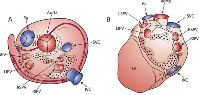
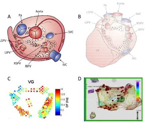
scattered mainly on the posterior surfaces of the atria and superior aspect of the ventricles, including AV groove and circumscribing most of the main heart vessels59, 62, 63 (fig. 4).
Fig. 4 Schematic representation of anatomical distribution of ganglia of the autonomous intrinsic cardiac nervous system on the epicardial surface. Most of the ganglia are located in the posterior atrial surface and superior atrial and ventricular surface circumscribing main heart vessels. A: Superior view. B: Posterior view IVC= Inferior vena cava, LIPV= Left Inferior Pulmonary Vein, LSPV= Left Superior Pulmonary Vein, LV= Left Ventricle, Pa= Pulmonary Artery, RIPV= Right Inferior Pulmonary Vein, RSPV= Right Superior Pulmonary Vein, SVC= Superior Vena Cava (adapted from Armour et al62, 64 and Yuan et al64).
Although epicardial fat pads and ganglionated plexuses contain mainly vagal ganglia, some sympathetic nerve fibers and even sympathetic neurons exist inside this complex system65. Whereas, electrical stimulation of the fat pads elicits a predominantly vagal response, parasympathetic blockade by atropine reveals the sympathetic excitation. Consequently, RF ablation or surgical removal of fat pads damages both vagal and sympathetic innervation65.
Vagus nerves exert a negative dromotropic and chronotropic effect on the sinus and AV nodes while sympathetic stimulation has a positive effect. Right VS primarily slows heart rate while right stellate ganglion stimulation increases heart rate. Conversely, left vagal or left stellate stimulation exerts its negative or positive dromotropic effects mainly on AV conduction66. However, both sympathetic and parasympathetic systems act contemporaneously and the final effect depends on the interaction between them. Thus, VS produces a greater reduction in heart rate in the presence of tonic sympathetic stimulation (accentuated antagonism), while changes in AV conduction during concomitant sympathetic and VS are the algebraic sum of the changes occurring independently if we stimulate only each one of the branches of autonomic nervous system66. Furthermore, cardiac responses to brief vagal bursts begin after a short latency and dissipate quickly, while cardiac responses to sympathetic stimulation commence and dissipate rather slowly. So, VS influences beat to beat regulation, obtaining rapid responses to vagal influences, while sympathetic stimulation produces a slow temporal response. Such phenomena are manifested also in the measurement of heart rate variability in long electrocardiographic
recordings. In the power spectra of such recordings (obtained with Fast Fourier Trasformation as we saw in the DF section) the high frequency (HF) component corresponds to rapid modifications of heart frequency and would express the vagal influence on heart rate. On the contrary, the low frequency (LF) component would rather correspond to slow temporal heart rate modifications and would express the sympathetic influence on heart rate.
Parasympathetic stimulation has long been recognized to play an important role in AF67. Coumel suggested that either limb of the autonomic nervous system, particularly the parasympathetic, can generate AF68. He distinguished a clinical form of vagal AF occurring generally in men without structural heart disease after meals or during rest (that is in situations of vagal preponderance). In contrast, adrenergic AF occurres generally in patients with heart disease, hyperthyroidism or pheocromocytoma, with arrhythmic episodes happening in daytime, during stress or exercise.
Vagal AF promotion is related to a spatially heterogeneous reduction in AERP69. VS substantially reduces AERP and even if slightly increases CV70, shortens atrial WL promoting reentry and AF according to the leading circle model65, 71. The heterogeneous spatial distribution of ganglia containing vagal neurons and the corresponding inhomogeneous distribution of ACh in the atria could be responsible for ERP heterogeneity and consequent AF promotion. Sympathetic stimulation provokes a more homogeneous reduction in AERP still promoting AF, however not as much as VS71.
Due to the proarrhythmic potential of VS, it has been supposed that denervation of the atria would be beneficial in preventing AF. Chiou et al61 demonstrated that radiofrequency ablation of the three fat pads prevented the inducibility of sustained AF during bilateral VS. Autonomic denervation during AF ablation seems to play an important role for its efficacy72, while ablation of autonomic ganglia at the base of the PVs suppresses vagal responses in an experimental model of AF73. As recently suggested56, CFAEs may result from activation of the intrinsic cardiac autonomic nervous system and the clinical efficacy of ablation of CFAEs49 may result by a partial atrial denervation65.
1.2.6 Atrial Remodeling
In 1995, Allessie’s group published one of the most important works of the last decades74, demonstrating that AF changes the electrophysiological properties of the atria in a way to promote its persistence or even its repeated and early initiation after several terminations. “AF begets AF” representing a common clinical scenario in humans, where AF presents initially as paroxysms that become more and more persistent with time, progressing subsequently to chronic AF. The changes in electrophysiological, structural, contractile, neuroreceptorial or endothelial properties of the atrium created by the persistence of AF have been named as atrial remodeling75. Two major categories of atrial remodeling have been described: atrial tachycardia remodeling (ATR) occurring after rapid atrial tachyarrhythmias, and atrial structural remodeling (ASR) which is associated with HF
and other fibrosis promoting conditions76. ATR would decrease AERP and ASR would affect CV, both promoting AF according to the leading circle model.
1.2.6.1 Atrial tachycardia remodeling (ATR)
The alterations in ionic currents and properties of cellular excitability are termed electrical remodeling75. The most characteristic changes are ERP reduction and loss of ERP rate-dependence5, 74 (ERP doesn’t become shorter proportionally to the increase in heart rate). Moreover, spatially heterogeneous ERP changes and anisotropic conduction are important in establishing favorable conditions for reentry and subsequently persistence of AF4, 27, 75.
ATR abbreviates atrial refractoriness by decreasing action potential duration (APD), primarily by ICaL downregulation and increased inward rectifier K+ currents76. ICaL is a calcium current passing through the L-type (long-lasting) calcium channels occurring in the later phases of calcium channel opening77. AF increases atrial rate ~10 fold, which increases atrial myocyte Ca2+ loading78. Therefore, atrial myocytes put forward different mechanisms in order to protect themselves from Ca2+. Yue et al79, studying an animal model of atrial tachycardia pacing to mimic AF, observed a progressive downregulation of the transient outward K+ current Ito and the L-type Ca2+ current ICaL, leading to APD shortening and impaired APD rate adaptation with consequent AF promotion. Subsequent work of the same group demonstrated transcriptional messenger RNA (mRNA) downregulation of the α-subunit of the L-type Ca2+ channel, as well as of the Kv4.3-Ito
α-subunit as the causative mechanism80. Furthermore, Bosch et al obtained similar findings in human AF81.
Intracellular Ca2+ loading by rapid atrial pacing leads to mitochondrial swelling and may jeopardize cell viability75. Declines in Ca2+ gradients between the cell compartments decreases atrial myocyte contractility and underlies the atrial contractile dysfunction observed in clinical AF75, 78. Such alterations in intracellular Ca2+ handling and cellular contractility have been named contractile remodeling75, 82.
The voltage gated transient outward K+ current Ito is consistently decreased in ATR83. Its role in promoting AF is not well established. Ito opposes the inward Na+ current in the first phase of action potential. Therefore, a decrease of Ito may facilitate wave propagation by increasing action potential amplitude76. Decreases of Ito are associated with reductions in both mRNA and protein expression of its pore forming Kv4.3 subunit80.
Moreover, substantial work in the last decade put in evidence the important role of the inward rectifier channel superfamily Kir in atrial cell repolarization and genesis of arrhythmia. Potassium channels of this family set the resting membrane potential of cardiac myocytes77. Kir passes an outward current when the membrane potential is above the potassium equilibrium potential and thus contributes on the late repolarization phase of the action potential, thereby regaining the resting membrane potential. Conversely, when cell membrane is hyperpolarized, Kir passes an inward current in order to maintain the high internal K+ activity and keep the resting membrane potential. The main background conductance that controls atrial resting membrane potential is IK1, a current passing through
Kir superfamily channels. AF increases expression levels of mRNA of Kir2.1 (channel subunit of Kir), consistent with larger IK176, 84. Moreover, an increase in IK1 has been observed in either human81, 85 or animal AF studies86, contributing to a faster repolarization, thus reducing further the APD75.
Kir superfamily channels include also ligand-operated channels, of which the most noted is the muscarinic operated channel. The muscarinic K+ channel (mAChKir) is a heterotetramer of Kir3.1 and Kir3.4 (also known as GIRK1 and GIRK4)87. Stimulation of cardiac M2 muscarinic receptors by cholinergic agonists activates IKACh (acetylcholine-regulated potassium current) which strongly promotes AF83 by stabilizing atrial reentrant rotors38. This is consistent with the clinical forms of AF beginning under vagotonic conditions68.
Although, other subtypes of muscarinic receptors have also been associated with different K+ currents, such currents seem to possess delayed-rectifier properties, distinguishing them from classical IKAch88, 89. A reduction of IKACh subunits (Kir3.1 and 3.4) mRNA and proteins in AF patients has been reported83, 90, 91, corresponding to a decreased current response to M2-receptor stimulation84, 85. Furthermore, Yeh et al92 report a decrease of both cholinergic receptor-coupled K+ currents and corresponding expression of channel proteins after 1 week rapid atrial pacing. This IKACh decrease would lead to an increase in APD and ERP with lower theoretical propensity to AF.
Nevertheless, even if functional IKACh is reduced, an increase in a constitutively active form of this current has been described as an important contributor in maintaining
AF93, 94. It has been shown that the inward rectifier potassium current IK1 is higher in patients with chronic AF and is associated with smaller muscarinic receptor-mediated IKACh84. Additionally, patients with chronic AF exhibit an agonist independent constitutive IKACh (IKACh,c) activity that contributes to the enhanced basal inward rectifier current93. In animal models, a similar constitutively active IKACh,c current is upregulated and independent of the presence of the agonist (ACh)94, while its inhibition in vitro by tertiapin increases APD and suppresses atrial tachyarrhythmias95. Whereas ATR94, 95 or chronic AF84, 93 decreases agonist stimulated IKACh, an agonist independent (“constitutive”) IKACh,c activity is enhanced93-96. Increased IKACh,c was supposed to be secondary to an increase in mAChKir channels that for unknown reasons were becoming independent of ACh binding. However, mRNA and protein expression of Kir3 subunits are unchanged in experimental ATR94, 96 whereas they are decreased in chronic AF76, 93. Consequently, the increased mAChKir channels hypothesis was rejected. Rather, recent studies proved that AF induced enhancement of IKACh,c is due to increased single channel open probability caused by slowed channel closure96, secondary to altered channel phosphorylation97, 98.
1.2.6.1.1 Neuroreceptorial remodeling
Long term rapid atrial pacing creates the above described electrical remodeling. One of the characteristics of such remodeling is an increased heterogeneity in atrial refractoriness. Analogous to studies in ventricular arrhythmias, it has been proposed that a potential mechanism of such spatial dispersion of refractoriness could be the heterogeneity of autonomic innervation99. Thus, heterogeneous atrial denervation with application of
phenol facilitated sustained AF in one study. Consequently, based on the idea that “AF begets AF” there have been studies evaluating the possibility of an autonomic remodeling in animal models of rapid atrial pacing. Jayachandran et al100 demonstrated that rapid rates of AF produce a heterogeneous increase in atrial sympathetic innervations, while Chang et al101 showed that there is a significant nerve sprouting and sympathetic hyperinnervation in a canine model of sustained AF. However, it remains unclear if such neural remodeling promotes sustained AF. Recently, Yeh et al92 demonstrated that atrial tachycardia induces downregulation of muscarinic receptors, adding further evidence in favor of a neuroreceptorial remodeling occurring after prolonged atrial tachycardia.
1.2.6.2 Atrial structural remodeling (ASR)
Clinical AF has been associated with atrial fibrosis.102 Changes that include increases in extracellular connective tissue composition, cellular size, glycogen accumulation, myolysis, mitochondrial changes, or chromatin redistribution have been termed structural remodeling103. Fibrosis is a hallmark of arrhythmogenic structural remodeling104-106. Fibrosis occurs generally as a reparative process substituting degenerating myocardium with connective tissue, which causes interstitial expansion.
Atrial fibrosis occurs in a variety of clinical settings such as senescence, cardiac dysfunction, mitral valvular disease, and possibly myocardial ischemia104. In a dog-model of congestive HF caused by ventricular tachypacing for 2-5 weeks, promotion of interstitial fibrosis induced sustained AF105. Complete recovery from tachycardia-induced congestive HF after 4-5 weeks of pacing cessation has been demonstrated107, 108. However, structural
remodeling with increased interstitial fibrosis remains unmodified, creating a substrate that can support prolonged AF109. The likelihood of AF increases with increasing extent of fibrosis110. Furthermore, Hanna et al showed that ventricular tachypacing-induced congestive HF was associated with substantially more fibrosis in the LA than in the left ventricle111. Although, AERP remains unchanged or increased after ventricular tachypacing, prominent conduction abnormalities in association with increased fibrosis create a favorable substrate to reentry according to leading circle model, thus promoting AF105. Consequently, therapeutic approaches targeting the prevention of the fibrous atrial remodeling could have a very important role in fighting the disease.
Several profibrotic factors have been described. TGFβ1 is an important profibrotic factor112. Targeted cardiac TGFβ1 overexpression produces prominent atrial fibrosis113, while TGFβ1 overexpressing mice show an enhanced susceptibility to AF114. Angiotensin II is another well-established profibrotic molecule. Transgenic mice with cardiac-restricted angiotensin converting enzyme overexpression show marked atrial dilation with focal fibrosis and AF76, 115. Recent evidence attributes significant roles also to Platelet Derived Growth Factor (PDGF) and Connective Tissue Growth Factor (CTGF)76.
1.3 Animal Models
Numerous clinical and experimental studies have been published in an effort to understand the complex pathophysiology of AF24, 116. Despite the undoubted progress in explaining causal factors and pathophysiological mechanisms leading to AF, many
unanswered questions remain4. Therefore, many animal models have been developed, assisting continuously in the progress of our knowledge and in the acquisition of new important information41, 74, 117, 118. Among them the “atrial tachycardia remodeling” (ATR) animal model and the “vagal stimulation” (VS) animal model are frequently used in order to evaluate new potential therapeutic methods.
1.3.1 Atrial tachycardia remodeling (ATR) animal model
Lone AF presents clinically as a paroxysmal form with auto limited episodes. However, by the recurrence and perseverance of new episodes, AF becomes initially persistent, requiring pharmacological or electrical cardioversions, and subsequently chronic or permanent. The longer AF persists the more difficult it becomes to maintain sinus rhythm119. Progression from paroxysmal AF to persistent and permanent AF is associated with structural and biochemical changes. As previously mentioned, the alterations in ionic currents and properties of cellular excitability are termed electrical remodeling75, 120. The changes in myocyte number, chamber size, extracellular connective tissue composition, fibroblast proliferation, glycogen accumulation, mitochondrial composition and chromatin distribution have been termed as structural remodeling75, 103, while the contractile dysfunction secondary to altered Ca+2 handling represents the contractile remodeling75, 82, 121.
Most of the above knowledge regarding atrial remodeling came from animal models. In 1995 Wijffels et al74 showed that AF causes electrophysiological changes of the
atrial myocardium which promote the maintenance of AF (AF begets AF). Pacemakers sustaining AF by right appendage pacing were implanted in goats. After a week of pacing-induced AF there was a notable shortening of the AERP associated with a remarkable increase in AF duration and inducibility.
Moreover, other experimental work demonstrated that the primary stimulus to remodeling in AF is atrial tachycardia41, 117. Atrial tachycardia reduces AERP in a spatially heterogeneous fashion, promoting AF by reducing WL and increasing its heterogeneity27. Such conditions favor multiple circuit reentry inducing and stabilizing AF4.
Following atrial tachycardia, significant changes in ionic currents and channels occur. There is a progressive reduction of the transient outward potassium current Ito and the L-type Ca+2 current ICa,L, leading to a net shortening of APD and impairment of the APD rate adaptation75, 79. Furthermore, an increase in IK1 has been observed, contributing to a faster repolarization (this current would act in the late phase of repolarization), thus reducing further the APD75, 81, 85, 86.
IKACh is the acetylcholine-regulated potassium current. As previously mentioned, it is an inwardly rectifying current generally influencing late repolarization and resting membrane voltage. Stimulation of cardiac M2 muscarinic receptors by cholinergic agonists activates IKACh which strongly promotes AF83. A reduction of IKACh subunits mRNA or proteins in AF patients has been reported83, 90. This would lead to an increase in APD and ERP with lower theoretical propensity to AF. Nevertheless, even if functional IKACh is
reduced, an increase in a constitutively active form of this current has been described as an important contributor in maintaining AF93, 94.
The ATR model is one of the animal models used in the present study. In each animal of the ATR group, a pacing lead was inserted into the right ventricular (RV) apex and connected to a subcutaneously-implanted VVI-pacemaker. Radiofrequency ablation of the AV node was performed and the RV-pacemaker was programmed at 80 bpm. A pacing lead in the RA appendage (RAA) was connected to a subcutaneously-implanted pacemaker to pace the atria at 400 bpm for 1 week. After 1 week, an electrophysiologic study was done, retrieving all the necessary information for the study.
Recapitulating, ATR animal model gave useful information and clarified many aspects of the complex AF pathophysiology. The similarity of the animal model with real human clinical conditions was ascertained by the similarities in electrophysiological and biochemical variations that occur after atrial tachycardia in the animal model or AF in humans. However, many aspects of the pathophysiology of AF have yet to be clarified, especially concerning the role of the autonomic nervous system in initiating or maintaining the arrhythmia.
1.3.2 Vagal stimulation (VS) animal model
The parasympathetic nervous system has long been recognized as an important factor in the onset and maintenance of AF67. A form of vagal AF, occurring after meals or during rest (thus in situations of vagal preponderance), has also been reported68. VS
provokes a spatially heterogeneous ERP abbreviation inducing and maintaining AF71. On the contrary, a similar ERP reduction provoked by sympathetic stimulation doesn’t promote AF as much, due to a more homogeneous distribution of ERP in the atria71.
The VS model is the second animal model that we used in our study. AF is easily induced and maintained for the whole time that vagus nerves are stimulated in this animal model. Both cervical vagal trunks are isolated, doubly ligated and divided. Bipolar hook electrodes are inserted into each nerve parallel to vagal fibers for several centimeters and subsequently bilateral vagal-nerve stimulation is delivered by a stimulator71. VS provokes ACh liberation at the terminal nerve endings. Stimulation of muscarinic receptors by acetylcholine activates IKACh resulting in shorter APD and therefore decreased AERP. On the contrary, a small increase in CV is observed. However, the final result is a net decrease of the WL, creating optimal conditions for microreentry and AF.
A direct relationship of the intensity of VS with the capability of inducing and maintaining AF has been reported69. This property promoted the application of VS in many animal models in order to obtain easily an AF episode and performed various experiments. Clinical AF often begins under vagotonic conditions. Nevertheless, it is unknown how comparable this artificial vagal AF could be with the clinical forms of AF.
2. Hypothesis
Both VS and ATR AF are not associated with structural heart disease and consequently they would represent two forms of idiopathic paroxysmal AF. VS and ATR animal models have common electrophysiological and molecular mechanisms in promoting AF. With the VS model we study the influence of the parasympathetic system in creating temporary electrophysiological alterations promoting AF, while with the ATR model we study the effect of atrial tachycardia in inducing more persistent electrophysiological alterations, leading to the perseverance of AF (AF begets AF74).
Vagal AF promotion is related to a spatially heterogeneous reduction in AERP and reduced AERP rate adaptation71. Similarly to the effects of VS, atrial tachycardia remodeling reduces the AERP, the AERP rate adaptation and the WL while increasing the AERP heterogeneity27, 41, 74, 105, 117. Nevertheless, the time course of AF promotion due to atrial tachycardia remodeling is slower than that of AERP changes, indicating the involvement of additional mechanisms24.
Interestingly, the VS and ATR substrate share some common molecular mechanisms. Stimulation of cardiac M2 muscarinic cholinergic receptors with cholinergic agonists in the VS model elicits a large inward rectifier K+ current, IKAch, which strongly promotes AF38, 83. Similarly, constitutively activated inward rectifier K+ current IKAch,c is also upregulated in cardiomyocytes from AF patients93 and contributes to ATR-induced APD abbreviation and atrial-tachyarrhythmia promotion in canine atrium83, 95. However,
ATR results in reduction in L-type Ca+2 current ICa,L, which contributes significantly to APD reduction and, accordingly, AERP. Thus, the main difference between VS and ATR model would be the reduction in Ca+2 current ICa,L.
Recently, Pandit et al122 demonstrated in a computer model that in two experimental situations with similar APD reduction, an increase in inward rectifier currents accelerates and stabilize rotors more than a reduction in Ca+2 current ICa,L. Even if the two situations produced the same APD reduction, they had a different response in rotor acceleration and stabilization and probably in AF promotion, if such conditions were applied clinically.
Based on the above observations, we hypothesized that similar situations could be reproduced by the VS and ATR animal models. Thus, we wanted to compare the two experimentally induced paradigms of AF in conditions of similar AERP shortening, in an effort to clarify different pathophysiological aspects of AF. Should a similar AERP shortening produce the same electrophysiological properties and similar AF promotion? Are there any electrophysiological differences between the models that can be paralleled to different clinical forms of AF or that can justify a different AF promotion?
In order to clarify such aspects, we followed a 1:1 matching of VS dogs with ATR dogs to ensure similar sex and weight distributions, and similar effects on mean AERP. We adjusted VS parameters in each VS dog according to the mean AERP recorded at 8 RA and left-atrial (LA) sites in the preceding ATR dog (basic cycle length, BCL, 300 ms). We obtained the same mean AERP in both models and we obtained epicardial maps of activation that permitted a detailed study of classical electrophysiological variables in
various anatomical areas. Moreover, DF analysis and its correlation with the electroanatomical properties of the two models gave interesting pathophysiological inputs.
Overall, the purpose of this work was to study the similarities and differences in the electrophysiological mechanisms underlying AF promotion in VS and ATR animal models, search for possible mechanisms that could explain such results, and finally try to correlate these models with clinical AF.
3. Differences in atrial fibrillation properties under vagal
nerve stimulation versus atrial tachycardia
remodeling
Grigorios Katsouras, MD,1 Masao Sakabe, MD, PhD,1 Philippe Comtois, PhD,2 Ange Maguy, PhD,1 Brett Burstein, PhD,1
Peter G. Guerra, MD,1 Mario Talajic, MD,1 Stanley Nattel, MD1
1Department of Medicine, 2Department of Physiology and Research Center, Montreal Heart Institute and Université de Montréal, 5000 Belanger Street, Montreal, Quebec, Canada, H1T 1C8
There are no conflicts of interest.
Short title: Vagal versus atrial tachycardia AF substrate
Word count: 4,994 (excluding short title, word count and abbreviation list)
Financial support: Supported by the Canadian Institutes of Health Research (MOP 44365) and the Fondation Leducq ENAFRA network grant (07/CVD-03).
Address for correspondence: Stanley Nattel, 5000 Belanger Street East, Montreal, Quebec H1T 1C8, Canada. Tel.: 514-376-3330; Fax: 514-376-1355. E-mail:
3.1 Abstract
BACKGROUND There are many similarities between atrial effects of
atrial-tachycardia remodeling (ATR) and vagal-nerve stimulation (VS): both promote atrial fibrillation (AF), reduce atrial effective refractory period (AERP) and AERP rate-accommodation, enhance AERP-heterogeneity and increase inward-rectifier K+-current.
OBJECTIVE To compare the consequences of ATR and VS at similar levels of AERP
abbreviation in dogs.
METHODS ATR-dogs (n=6) were subjected to 7-day atrial-tachypacing at 400 bpm,
with radiofrequency-induced atrioventricular block and ventricular-demand pacing (80 bpm) to control ventricular response. VS was applied in 6 matched dogs with stimulation-parameters selected to produce similar mean AERP-values to ATR-dogs.
RESULTS ATR and VS produced similarly-short AERPs (79±12 and 80±12 ms
respectively), AERP rate-adaptation loss and AERP heterogeneity increases. Although both ATR and VS increased AF duration, VS was significantly more effective in AF-promotion, with mean AF duration of 992±134 seconds, versus 440±240 seconds (P<0.05) under ATR. The greater AF-promoting effect of VS was associated with greater mean dominant-frequency values during AF (11.7±1.8 versus 10.0±1.3 Hz ATR,
P<0.05). VS greatly enhanced the spatial dominant-frequency variability, increasing
the coefficient of variation to 15.2±1.9 Hz, versus 8.9±1.5 Hz for ATR (P<0.05), primarily by increasing per-dog maximum dominant-frequency (15.4±0.6 Hz, versus 12.5±0.6 for ATR, P<0.01).
CONCLUSION For matched AERP-values, VS promotes AF more strongly than
ATR. Despite similar AERP-changes, VS produces considerably greater increases in dominant-frequencies, particularly maximum values, consistent with previous
suggestions that inward-rectifier K+-current enhancement is particularly effective at accelerating and stabilizing spiral-wave rotors that maintain AF.
KEYWORDS: Atrial remodeling; Arrhythmia mechanisms; Reentry; Autonomic
Abbreviation List
AERP: atrial effective refractory period AF: atrial fibrillation
ATR: atrial-tachycardia remodeling BCL: basic cycle length
CTL: control
CV: conduction velocity DF: dominant frequency
ERP: effective refractory period LA: left atrial
LAA: left-atrial appendage
LBB: left-atrial side of Bachmann’s Bundle LIW: left-atrial inferior wall
LPW: left-atrial posterior wall RA: right atrial
RAA: right-atrial appendage
RBB: right-atrial side of Bachmann’s Bundle RIW: right-atrial inferior wall
RPW: right-atrial posterior wall RV: right ventricular
VS: vagal stimulation WL: wavelength
3.2 Introduction
Numerous studies have addressed the complex pathophysiology of atrial fibrillation (AF).1 Animal models have been used for over 100 years and have provided extensive relevant information.2 The atrial-tachycardia remodeling (ATR) paradigm, by which rapid atrial activation promotes AF, has provided important insights into AF mechanisms.1-3 Vagotonic AF has also been widely used, particularly to study pharmacological control.2
There are many similarities in the atrial properties associated with vagal versus ATR-induced AF-promotion. Both involve reduced atrial effective refractory period (AERP) and AERP rate-adaptation,3,4 and both manifest increased spatial AERP-heterogeneity.5,6 Vagal effects on AERP and AF are due to enhancement of the acetylcholine-regulated K+-current IKACh,7 and a significant portion of the ATR-effect is due to enhanced constitutively-active IKACh (IKACh,c).8 These similarities in electrophysiological effects and ionic-current changes suggest common mechanisms of AF promotion. We therefore designed the present study to compare directly ATR-related and vagal AF at matched levels of AERP-shortening.
3.2 Methods
3.2.1 Animal models and experimental groups
Animal-handling procedures were approved by the local Animal Research Ethics Committee. Twelve mongrel dogs were studied in two groups: 1) 6 vagal-stimulation (VS) dogs; 2) 6 dogs subjected to 1-week ATR as previously described.8
ATR dogs were initially anesthetized with sodium pentobarbital (30 mg/kg IV, additional 4 mg/kg doses as needed). A pacing lead was inserted into the right ventricular (RV) apex and connected to a subcutaneously-implanted VVI-pacemaker. Radiofrequency ablation of the atrioventricular node was performed and the RV-pacemaker was programmed at 80 bpm. A pacing lead in the right atrial (RA) appendage (RAA) was connected to a subcutaneously-implanted pacemaker to pace the atria at 400 bpm for 1 week.
3.2.2 Experimental protocol
On study days, dogs were anesthetized with morphine (2 mg/kg sc) and α-chloralose (120 mg/kg iv, followed by 29.3 mg/kg/h), and ventilated mechanically. Body temperature was maintained at 37°C. In the ATR group the RA-pacemaker was deactivated. A median thoracotomy was performed and a pericardial cradle created. Four bipolar Teflon-coated stainless steel electrodes were inserted into the RAA and left atrial (LA) appendages (LAA) for recording and stimulation. Five silicon plaques containing 240 bipolar electrodes were sewn over the atrial epicardial surface.9 A programmable stimulator was used to deliver 2-ms twice-threshold current pulses. Signals were filtered (10-900 Hz) and digitized (12-bit resolution, 2-kHz sampling). In vagal dogs, a bipolar RV electrode and VVI-pacemaker were used to demand-pace the ventricles at 80 bpm.
3.2.2.1 Experimental design
VS dogs were matched 1:1 with ATR dogs to ensure similar sex and weight distributions, and similar effects on mean AERP. We first performed an open-chest electrophysiological study in each ATR dog, then matched the subsequent VS dog for sex and weight. We adjusted vagal-stimulation parameters in each VS dog according to the mean AERP recorded at 8 RA and left-atrial (LA) sites in the preceding ATR dog (basic cycle length, BCL, 300 ms).
3.2.2.2 Vagal stimulation
Both cervical vagal trunks were isolated, doubly ligated and divided. Bipolar hook electrodes (stainless steel, insulated with Teflon except for the distal 10-15 mm) were inserted into each nerve parallel to vagal fibers for several centimeters.5 Bilateral vagal-nerve stimulation (0.2-ms pulse-width) was delivered by a Grass SD-9F stimulator (Quincy, MA). Vagal stimulation voltage and frequency were selected to produce the target AERP (average values 3.9±0.7 V and 12.2±0.6 Hz). After vagal-stimulation parameters were established based on AERP, vagal vagal-stimulation was applied for 30 seconds and the sinus-node slowing response to vagal stimulation was assessed over the last 15 seconds (average baseline sinus cycle-length 360±8.5 ms, cycle-length during vagal stimulation 792±105 ms). This response was verified periodically during each experiment and vagal-stimulation parameters readjusted if necessary to produce a constant bradycardic response.
3.2.2.3 Electrophysiological study
AERP was measured at multiple BCLs in the RAA and LAA and at a BCL of 300 ms at 6 additional RA and LA sites: posterior wall (RPW, LPW), inferior wall
(RIW, LIW) and the Bachmann bundle (RBB, LBB). AERP was determined with 10 basic stimuli (S1) followed by a premature extrastimulus (S2) with 5-ms decrements. The longest S1-S2 failing to capture defined the AERP. The mean of 3 AERP values at each BCL was used for analysis. If AERP values differed by >10 ms, 2 additional AERP measurements were obtained and the mean of all determinations was taken to represent AERP. AF was induced with atrial burst-pacing at 10 Hz and 4 ×threshold-current. To estimate mean AF duration in each dog, AF was induced 10 times. AF≥20 min was considered sustained and was cardioverted with a synchronized direct-current shock. A 20-min rest period was then allowed before experimentation was resumed. After 2 cardioversions, no further AF inductions were performed.
3.2.3 Conduction and AERP analysis
Epicardial mapping was conducted with the Cardiomap system (Research Center, Sacre-Coeur Hospital and Biomedical Engineering Institute, École Polytechnique and Université de Montréal, Montreal, Quebec). Activation data were analyzed off-line with computer-determined peak amplitude criteria for activation, and were reviewed manually. Electrogram timing was compared with QRS complexes to exclude ventricular electrograms.
Conduction velocity (CV) was determined by analyzing activation at four electrode sites in the direction of rapid propagation in the RAA, RPW, RBB, LBB and LAA.5,9,10 Distance from the proximal site was plotted against activation time and CV was determined by linear regression (r>0.99 required for analysis). Because of variable conduction in the left inferior-posterior wall and right inferior wall, CV could not
always be measured accurately in LPW, LIW and RIW, which were therefore not included in the analysis. Local wavelengths (WLs) were calculated as the product of local CV and local ERP. CV and AERP heterogeneity were quantified by calculating coefficients of variation of 5 regional CV values (COVCV= SDCV/meanCV × 100%) and 8 regional AERP values (COVAERP= SDAERP/meanAERP × 100%).
To evaluate spatial conduction-inhomogeneities, phase maps were constructed, based on previously-described methods.10 In brief, activation-time differences between each electrode and its neighboring sites were normalized to interelectrode distances and maximum values at each site were taken to indicate local phase delays. A phase-delay histogram was then constructed and values corresponding to the 5th, 50th and 95th percentiles designated P5, P50 and P95 respectively. P50 represents the median phase-delay and is an overall conduction index. The P5-95 expresses the range of phase delays and the variation coefficient (P5-95/P50) is a conduction-heterogeneity index independent of CV.
3.2.4 Atrial frequency-spectrum analysis
Spectral characteristics of activation at each site during AF were studied as previously described.9,11,12 Periodogram estimation was performed on 4-second samples to analyze frequency content in the 0.5 to 80 Hz band. The mean of each signal was first subtracted to remove the DC-part of the signal. A split-cosine, bell-tapering window was applied, with 5% of data points being tapered. An estimate of the signal spectrum was obtained by using a smoothed periodogram with a weighting parameter M.12 M was determined semiautomatically with an unbiased risk estimator method to
choose the tuning parameter M for the smoothed periodogram. The dominant frequency (DF) at each recording-site was defined as the frequency of the highest peak of the smoothed periodogram.9 The variability in DFs was assessed in each dog based on the coefficient of variation (COVDF = SDDF/meanDF×100%).
3.2.5 Statistical analysis
All results are expressed as mean±SEM. Statistical comparisons between ATR and vagal-stimulation means were performed by unpaired t-tests, except in cases of repeated-measures data (such as AERP at multiple BCLs and AERP, CV and WL in multiple atrial regions), in which 2-factor ANOVA (ATR versus vagal-stimulation as one factor and the repeated-measure index such as BCL or region) was applied. All data satisfied statistical criteria for normal distribution (Kolmogrov-Smirnov test) except for AF durations, which were normally-distributed after log-transformation and were so analyzed. To test for differences between coefficients of variation, we performed an F-test of the variance ratio according to Lewontin.13 A two-tailed P<0.05 was considered statistically-significant. The control (baseline) values before vagal stimulation are shown as reference values for vagal and ATR conditions, but are not compared statistically.