O
pen
A
rchive
T
oulouse
A
rchive
O
uverte
(OATAO)
OATAO is an open access repository that collects the work of some Toulouse
researchers and makes it freely available over the web where possible.
This is
an author'sversion published in:
https://oatao.univ-toulouse.fr/23123Official URL :
https://doi.org/10.1302/1863-2548-11-160281
To cite this version :
Any correspondence concerning this service should be sent to the repository administrator:
tech-oatao@listes-diff.inp-toulouse.fr
Accadbled, Franck and May, Olivier and Thévenin-Lemoine, Camille and Sales de Gauzy,
Jérôme Slipped capital femoral epiphysis management and the arthroscope. (2017) Journal
of Children's Orthopaedics, 11 (2). 128-130. ISSN 1863-2521
Slipped capital femoral epiphysis management and
the arthroscope
F. Accadbled
1O. May
2C. Thévenin-Lemoine
3J. Sales de Gauzy
3 AbstractBackground In situ pinning of slipped capital femoral
epiph-ysis (SCFE) results in various degrees of deformity of the fem-oral head-neck junction. Repetitive trauma from cam-type femoroacetabular impingement (FAI) can lead to labral tears and injury to the articular cartilage causing loss of function. Arthroscopic osteoplasty is an alternative to open procedure and to Southwick/Imhäuser-type osteotomies in symptomatic selected cases.
Surgical technique The amount of bone to be resected has to
be carefully planned pre-operatively. Only gentle traction is applied on a well-padded perineal support. A spherical burr is used to gradually resect the prominence. Intra-operative fluoroscopy is very useful when checking adequate reshap-ing of the head-neck junction is obtained.
Results Arthroscopy often reveals acetabular cartilage lesions,
labrum hyperhemia and fraying which rarely require repair. Arthroscopic osteoplasty provides satisfactory pain relief and, to a lesser extent, restores hip internal rotation.
Conclusion Arthroscopic osteoplasty is more technically and
time-demanding in post SCFE than idiopathic FAI. It requires strong arthroscopic skills and experience in hip arthroscopy. It stands as a reasonable alternative to open procedure or flexion osteotomies in symptomatic FAI post mild to moder-ate SCFE. It provides pain relief and to a lesser extent restores internal rotation of the hip.
1 Department of Orthopaedics, Children’s Hospital, CHU de Toulouse, France
2 Clinique Médipôle Garonne, 45 rue de Gironis, 31036 Toulouse, France
3 Department of Orthopaedics, Children’s Hospital, CHU de Toulouse, France
Correspondence should be sent to: Professor F. Accadbled. Service de Chirurgie Orthopédique et Traumatologique, Hôpital des Enfants 330, avenue de Grande Bretagne, 31059 TOULOUSE cedex 9, France.
E-mail: faccadbled@gmail.com
Keywords: slipped capital femoral epiphysis; SCFE; femoroacetabular impingement,
osteochondroplasty; arthroscopy
Introduction
In situ pinning of slipped capital femoral epiphysis
(SCFE) results in various degrees of deformity of the femoral head-neck junction. Despite some remodelling over growth, functional loss may result from the residual deformity that impinges with the acetabular rim in hip flexion and internal rotation. Repetitive trauma from cam-type fem-oroacetabular impingement (FAI) can also lead to labral tears and injury to the articular cartilage.1 Arthroscopic osteoplasty after in situ
pinning is an alternative to open osteoplasty and to Southwick/Imhäuser-type osteotomies in symptomatic selected cases. Indication for this pro-cedure is based on symptoms and clinical and imaging signs of FAI. Symptoms are represented by loss of func-tion and sudden pain triggered in a given position of the hip (for example, getting on a bicycle). Internal rotation is typically restricted. Impingement test consists of gradual passive adduction and internal rotation on a 90° flexed hip suddenly exacerbating pain. Plain radiographs display post-slip morphology with excessive alpha angle and pis-tol grip deformity. MRI may demonstrate bone bruise and labral tears. Arthroscopic treatment aims to improve the clearance during movement of the hip and to eliminate the abutment of the prominent femoral head-neck junc-tion and the acetabular rim, therefore relieving pain and to a lesser extent restoring internal rotation.
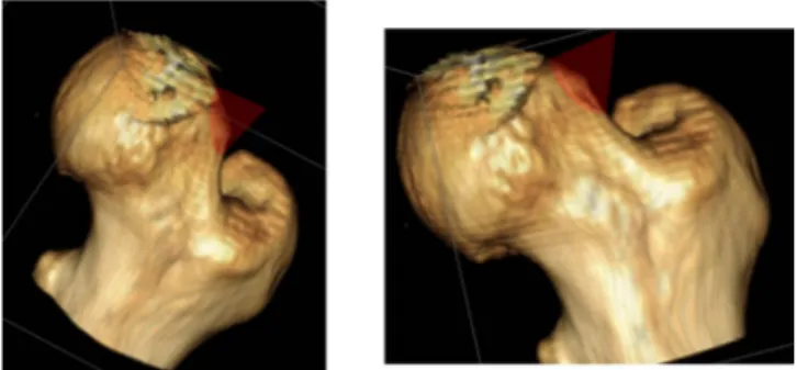
Surgical technique
Careful planning is necessary and is best based on pre-operative three-dimensional (3D) CT scan. The adequate amount of bone to be resected to restore the normal alpha angle is calculated (Fig. 1). The patient is positioned supine on a traction table. A 70° arthroscope and special trocars are helpful although the procedure can be performed using a standard 30° arthroscope.2
An anterolateral por-tal is established under fluoroscopic guidance with the
The age range at the time of the procedure was 24 to 39 years. At two-year minimum follow-up, the pain was relieved and at least neutral internal rotation of the hip was obtained. No complications were reported. Chen at al reported on 34 cases in 2014.5 The initial slip was mild in
21 hips, moderate in 16 and severe in three. The age range at the time of the procedure was 10 to 19 years. Labral damage was seen in all 34 hips and the labral lesions were descriptive only. Acetabular cartilage damage adjacent to the damaged labrum was seen in 26 hips. The damage appeared to be abrasive with roughening of the surface, without the softening or full-thickness flaps that are typi-cally seen in idiopathic cam impingement. Full-thickness cartilage loss noted in two hips was left untreated. At 12-month minimum follow-up, complete pain relief and correction of obligatory external rotation deformity (Dreh-mann sign) were obtained in 88% of cases. The mean internal rotation had improved from -20° to +10°. The mean alpha angle had improved from 88° to 54°. They reported two complications: one labral injury and one acetabular lesion during initial portal placement. Wylie et al reported on the results of nine patients aged 13.5 to 27 years in 2015 (six mild and three moderate slips).6 All
cases demonstrated acetabular cartilage and labral dam-age at the time of arthroscopy. Osteoplasty was combined with an acetabuloplasty in five cases and a labral repair in four cases. At 12-month minimum follow-up, the mean Harris hip score had improved from 63 to 91 and alpha angle from 75° to 46°. Four complications were reported: one capsular laxity requiring surgical revision, one peri-neal numbness, one lateral femoral cutaneous nerve numbness and one case of heterotopic calcifications. Our personal experience from the last ten years confirmed frequently associated acetabular cartilage lesions (mostly ICRS grade I and II) and labrum hyperhemia and fraying, none of which required specific treatment. Arthroscopic osteoplasty provided pain relief and restored some inter-nal rotation of the hip without complication.
Discussion
‘Acute’ arthroscopic osteochondroplasty at the time of in
situ fixation was first suggested by Leunig et al in 2010.7
The rationale of such management is the prevention of early acetabular cartilage and labral damage and poten-tially later osteoarthritis of the hip. Three patients aged 11 to 15 years, presenting with a mild and stable SCFE, were included. At six- to 23-month follow-up, they were all asymptomatic and had resumed sporting activities at the previous level. Hip internal rotation was still slightly reduced compared with the contralateral side. Tscholl et al confirmed in 2016 that the normal alpha angle could be
Fig. 1 3D CT scan view of the femoral head and neck. The cone
represents the correct amount of bone to be resected.
Fig. 2 Intra-operative fluoroscopic lateral view of the femoral
head and neck, before (left) and after (right) arthroscopic osteoplasty.
hip in 40° flexion into the peripheral compartment first. The anterior portal is placed under arthroscopic visuali-sation with a spinal needle and the guidewire technique, followed by an anterior capsulectomy, then gentle joint distraction. A 5.5 mm spherical burr is used to resect the prominence gradually (Video 1). Complete exposure of the bump may require hip mobilisation. Any retained metalware from previous surgeries is removed, if needed under arthroscopic visualisation. The amount of resection should not exceed 30% of the neck’s volume to limit the risk of fracture.3 Intra-operative fluoroscopy is very useful
to check adequate reshaping of the head-neck junction (Fig. 2). Portals are closed and injected with Ropivacaine. Patients are discharged home on the same day and are advised to use crutches and bear weight as tolerated for one month. Sports are usually resumed gradually after three months.
Results
Ilizaliturri et al first reported on the results of arthroscopic management of FAI post SCFE in eight patients in 2007.4
restored by systematic arthroscopic osteochondroplasty performed at a mean of three weeks (3 to 14) after in
situ fixation of 14 mild to moderate slips.8 One case of
post-operative hip arthrofibrosis r equired a rthroscopic revision. We do not perform arthroscopic osteoplasty as a prophylactic procedure in our practice. As acknowl-edged by the above-quoted authors, mid- and long-term outcomes have to be assessed and compared with controls.
Arthroscopic osteoplasty is more technically and time-demanding in post SCFE than idiopathic FAI. Indeed, the prominence is larger due to the epiphyseal slip, patients are often overweight and the presence of met-alwork makes the procedure more difficult. Arthroscopic skills and strong experience in hip arthroscopy are rec-ommended. Only gentle traction should be applied on a well-padded perineal support to prevent complications such as pudendal nerve palsy. It seems reasonable to limit the operative time two hours and 30 minutes. No more than 30% of the neck’s volume should be trimmed to pre-vent subsequent fracture.3 We believe FAI in severe slips
are best managed with Southwick/Imhaüser-type osteot-omies which provide satisfactory results.9
Arthroscopic osteoplasty stands as a reasonable alterna-tive to open procedure or flexion osteotomies in symptom-atic FAI post mild to moderate SCFE. It provides pain relief and, to a lesser extent, restores internal rotation of the hip. The benefit of a preventive procedure has yet to be demon-strated in a mid- to long-term series with a control group.
SUPPLEMENTARY VIDEO
A video showing arthroscopic osteoplasty of the left femur using a 5.5 mm ball burr in a 14-year-old female patient is available with the online version of this article at www.online.boneandjoint.org.uk/jco
COMPLIANCE WITH ETHICAL STANDARDS FUNDING STATEMENT
No benefits in any form have been received or will be received from a commercial party related directly or indirectly to the subject of this article.
OA LICENCE TEXT
This article is distributed under the terms of the Creative Commons Attribution-Non Commercial 4.0 International (CC BY-NC 4.0) licence (https://creativecommons.org/ licenses/by-nc/4.0/) which permits non-commercial use, reproduction and distribu-tion of the work without further permission provided the original work is attributed.
ETHICAL STATEMENT
Ethical approval: All procedures performed in studies involving human participants were in accordance with the ethical standards of the institutional and/or national re-search committee and with the 1964 Helsinki Declaration and its later amendments or comparable ethical standards.
No funding was received for this study.
REFERENCES
1. Leunig M, Casillas MM, Hamlet M, et al. Slipped capital femoral epiphysis: early mechanical damage to the acetabular cartilage by a prominent femoral metaphysis. Acta Orthop Scand 2000;71:370-375.
2. Accadbled F. Arthroscopic surgery in children. Orthop Traumatol Surg Res 2010;96:447-455.
3. Mardones RM, Gonzalez C, Chen Q, et al. Surgical treatment of femoroacetabular impingement: evaluation of the effect of the size of the resection. J Bone
Joint Surg [Am] 2005;87-A:273-279.
4. Ilizaliturri VM Jr, Nossa-Barrera JM, Acosta-Rodriguez E,
Camacho-Galindo J. Arthroscopic treatment of femoroacetabular impingement
secondary to paediatric hip disorders. J Bone Joint Surg [Br] 2007;89-B:1025-1030. 5. Chen A, Youderian A, Watkins S, Gourineni P. Arthroscopic femoral neck osteoplasty in slipped capital femoral epiphysis. Arthroscopy 2014;30: 1229-1234.
6. Wylie JD, Beckmann JT, Maak TG, Aoki SK. Arthroscopic treatment of mild to moderate deformity after slipped capital femoral epiphysis: intra-operative findings and functional outcomes. Arthroscopy 2015;31:247-253.
7. Leunig M, Horowitz K, Manner H, Ganz R. In situ pinning with arthroscopic osteoplasty for mild SCFE: A preliminary technical report. Clin Orthop Relat Res 2010;468:3160-3167.
8. Tscholl PM, Zingg PO, Dora C, et al. Arthroscopic osteochondroplasty in patients with mild slipped capital femoral epiphysis after in situ fixation. J Child Orthop 2016;10:25-30.
9. Hosalkar HS, Pandya NK, Bomar JD, Wenger DR. Hip impingement in slipped capital femoral epiphysis: a changing perspective. J Child Orthop 2012;6:161-172.