O
pen
A
rchive
T
OULOUSE
A
rchive
O
uverte (
OATAO
)
OATAO is an open access repository that collects the work of Toulouse researchers and
makes it freely available over the web where possible.
This is an author-deposited version published in :
http://oatao.univ-toulouse.fr/
Eprints ID : 17011
The contribution was presented at IUS 2016 :
http://sites.ieee.org/ius-2016/
To cite this version :
Zhao, Ningning and Wei, Qi and Basarab, Adrian and
Kouamé, Denis and Tourneret, Jean-Yves Blind Deconvolution of Medical
Ultrasound Images Using a Parametric Model for the Point Spread Function.
(2016) In: IEEE International Ultrasonics Symposium (IUS 2016), 18 September
2016 - 21 September 2016 (Tours, France).
Any correspondence concerning this service should be sent to the repository
administrator:
[email protected]
BLIND DECONVOLUTION OF MEDICAL ULTRASOUND IMAGES USING A PARAMETRIC
MODEL FOR THE POINT SPREAD FUNCTION
Ningning Zhao
1,2, Qi Wei
3, Adrian Basarab
2, Denis Kouam´e
2, Jean-Yves Tourneret
11
University of Toulouse, IRIT/INP-ENSEEIHT, 31071 Toulouse Cedex 7, France
2
University of Toulouse, IRIT, CNRS UMR 5505, Universit´e Paul Sabatier, Toulouse, France
3
University of Cambridge, Department of engineering, UK
{nzhao, jean-yves.tourneret}@enseeiht.fr, {qw245}@cam.ac.uk, {adrian.basarab, denis.kouame}@irit.fr
ABSTRACT
This paper addresses the problem of blind deconvolution of medical ultrasound (US) images. Specifically, a parametric model for the point spread function (PSF) established experi-mentally is used, i.e., the US PSF can be modeled by a Gaus-sian function modulated by a sinusoidal function. Given this parametric model, the estimation of the PSF in a blind de-convolution problem can be reduced to the estimation of its parameters. Moreover, due to the ill-posedness of blind
de-convolution problem, anℓp-norm (0 < p ! 2) regularization
term (including the widely consideredℓ1-norm,ℓ2-norm
reg-ularization terms) for the ultrasound tissue reflectivity func-tion (TRF) is employed, based on the assumpfunc-tion of gener-alized Gaussian distributed US images. An alternating opti-mization approach is proposed for the estimations of the US PSF and TRF. The behavior of the proposed algorithm is il-lustrated using simulated and in vivo US data.
Index Terms— Ultrasound imaging, blind deconvolu-tion, block circulant matrix, optimizadeconvolu-tion, variable metric forward backward splitting, proximal alternating linearized minimization.
1. INTRODUCTION
Medical ultrasound (US) imaging is widely used for clinical diagnosis such as cardiovascular medicine, urology and ob-stetrics. Compared to other medical imaging modalities, e.g., X-ray computed tomography (CT) and magnetic resonance imaging (MRI), US imaging has many advantages, including its harmless, cost-effective, portable and noninvasive proper-ties. However, US images suffer from a relatively low con-trast, reduced spatial resolution at a given frequency and low signal-to-noise ratio (SNR). Even though advances in ultra-sonic device-based solutions have improved the resolution of
Part of this work has been supported by the Chinese Scholarship Council and the thematic trimester on image processing of the CIMI Labex, Toulouse, France, under grant 11-LABX-0040-CIMI within the program ANR-11-IDEX-0002-02.
US images during the last decades, e.g., [1,2], post-processing techniques enhancing US image resolution are still appealing. In this paper, we explore a blind deconvolution method aiming at improving the quality of US images. The linear model used for US image blind deconvolution can be defined using the following matrix-vector formulation
y= Hx + n (1)
where y and x are vectors of RN ×1 obtained after
lexico-graphical order of the ultrasound radio-frequency (RF) im-age/observation and tissue reflectiviy function (TRF)/image to be estimated respectively, n is an additive white Gaussian
noise (AWGN) and H ∈ RN ×N is the system impulse
re-sponse/point spread function (PSF) assumed to be a circulant matrix [3, 4]. In US imaging systems, the PSF is usually un-known. Existing methods to address this problem include ei-ther the estimation of the PSF in a pre-processing step [3,5] or the estimation of the PSF and the TRF simultaneously [6, 7]. In this paper, we follow the second strategy to estimate the US TRF and PSF jointly. In particular, a parametric model for the PSF of the form of a modulated 2D Gaussian function is proposed. This parametric model allows us to reduce the estimation of the PSF during the blind deconvolution process to the estimation of a few parameters of the PSF model. In addition, a generalized Gaussian distribution is proposed for
the US TRF [3]. It includes the widely usedℓ1-norm andℓ2
-norm regularizers in US image deconvolution literature, see e.g. [8, 9].
This paper is organized as follows. Section 2 introduces the proposed parametric model for the PSF and the formu-lated problem for US image blind deconvolution. The pro-posed alternating method is presented in Section 3. Section 4 displays the simulation results and conclusions are reported in Section 5.
2. PROBLEM STATEMENT 2.1. PSF parametric model
We propose the following parametric model for the PSF of an US imaging system
hp(i, j) ≡ e(i, j) cos[ω0ta(i) + φ] (2)
with e(i, j) = t3 a(i) exp[−αt 2 a(i)] exp[−βt 2 l(j)] (3)
where the parametric model of PSF “hp” and its envelope “e”
belong to Rq×r, the integersi ∈ {1, · · · , q}, j ∈ {1, · · · , r}
denote the location of the PSF pixels,ω0= 2πf0is the
angu-lar central frequency of the transducer,φ is the phase of the
system PSF, the variablesα, β determine the envelope shape
of the PSF, the vectors taand tlare the temporal axes along
the axial and lateral directions, which related to the PSF band widths. Thus, the vectors ta∈ Rq×1and tl∈ R
1×rdetermine
the size of the PSF. Note that a similar model was considered in [6], where a Gaussian function modulated by a sinusoidal function has been shown to fit well the US PSFs.
With the a priori knowledge of the temporal axes or the size of the PSF1, there are three parametersφ, α, β to be esti-mated to completely determine the US PSF. The assumptions on the unknown PSF parameters considered herein are de-tailed hereafter.
• α and β: We denote θ = {α, β}. In this paper, we mainly focus on the estimation of the envelope shape parameters θ. Moreover, since the estimation of the two envelope shape parameters is ill-posed, we propose to constrain them as follows
ρ(α) = ıCα (4)
̺(β) = ıCβ (5)
whereρ(α) and ̺(β) are two indicator functions on sets
Cα= {α ∈ [αmin, αmax]} and Cβ= {β ∈ [βmin, βmax]}.
The definition of an indicator function is given by ıC(x) =
(
0 x ∈ C
+ ∞ x /∈ C (6)
• φ: In this paper, we estimate the phase term previously using the cepstrum-based method that exploits the min-imum phase assumption of US systems [10]. However, we emphasis that it is possible to pass by the
estima-tion ofφ by dealing with complex demodulated signals
following [6].
1The values of q and r or the size of the PSF are commonly assumed to
be known in advance in the problem of US image deconvolution. Moreover, since the size of the PSF is usually much smaller compared with the image size (i.e., q ≪ m, r ≪ n), zero padding of the PSF is necessary for the convolution computation. Without loss of generality, all the PSFs mentioned in this paper have been zero padded for the convolution computation.
2.2. Problem formulation
Taking into account the parametric model for the PSF, we for-mulate the US image blind deconvolution problem as follow-ing
minx,θ Ψ(x, h) + τ ϕ(x) + ρ(α) + ̺(β)
subject to h= hp. (7)
whereΨ(x, h) is the data fidelity term, ϕ(x) is the
regular-ization term for the TRF andτ is the corresponding
regular-ization parameter which weights the importance between the data fidelity term and the regularization term. Under the as-sumption of additive white Gaussian noise (AWGN), we have
Ψ(x, h) = 1
2*y − Hx*
2
. (8)
Given a generalized Gaussian distribution as the prior infor-mation of US TRF [11, 12], we have
ϕ(x) = *x*pp. (9)
3. PROPOSED METHOD
In order to solve the problem (7), we propose an alternat-ing minimization approach followalternat-ing the block-coordinate descent (BCD) framework [13]. Algorithm 1 outlines the proposed approach.
Algorithm 1: Alternating optimization algorithm Input: Observation y, Initial estimation h0,τ ,
Parameters of PSF modelα0,β0.
Repeat
// Update x with a known PSF
1 xˆ∈ argminxΨ(x, h) + τ ϕ(x);
// Update h by estimating α, β with a known TRF
2 α, ˆˆ β ∈ argminα,βΨ(x, h) + ρ(α) + ̺(β);
3 hˆ= hp(ˆα, ˆβ);
Output:ˆx, ˆh
We note that the steps♯ 1 and ♯ 2 can be solved using a
proximal algorithm. More related details about this algorithm can be found in [13, 14].
4. SIMULATION RESULTS
In order to study the performance of the proposed algorithm, experiments have been conducted on simulated and in vivo ultrasound images. Moreover, a comparison with a non-blind deconvolution algorithm, where the PSF is estimated in a pre-processing step using the cepstrum-based algorithm [10, 15]
has been conducted. For simulated US images, the perfor-mance of the algorithms is evaluated through the normalized root mean square error (NRMSE). The ground truth for the ultrasound TRF and PSF are not available for real US images. Therefore, the visually inspection has been used to evaluate the performance of TRF estimation for real images.
4.1. Simulated US images
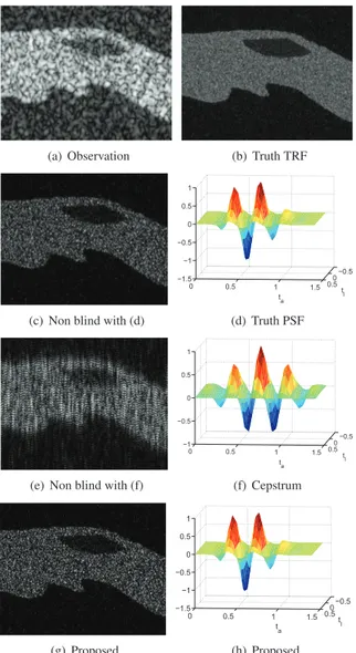
Simulated ultrasound TRF of size275 × 75 has been
gener-ated according to generalized Gaussian distribution, as shown in Fig. 1(b). The pixels in the different regions are distributed
according to GGDs with different parameters: p = 1.2 for
the bright region,p = 1.8 for the darker region surrounded
by the bright zone andp = 0.6 for the background. More
de-tails about this way of TRF generation can be found in [3]. The observed image shown in Fig. 1(a) has been blurred by a simulated PSF (displayed in Fig. 1(d)) that was gen-erated following the model (2) and contaminated by an ad-ditive white Gaussian noise with blurred signal-to-noise ratio
(BSNR) equal to30 dB. The parameters of the model (2) are
fixed at α = 4.8, β = 10 and φ = 3.2. Figs. 1(f), 1(h)
display the estimated PSFs using the cepstrum-based method and the proposed method. Figs. 1(c), 1(e) and 1(g) show the restored ultrasound TRFs using the true PSF, the estimated PSFs obtained with the cepstrum-based method [10] and the proposed method respectively. The prior used for the TRF is
anℓp-norm regularization (p = 1) for all experiments related
to simulated images. The TRFs estimated using the true PSF and the proposed method are visually very similar. The PSF obtained with the proposed method is also closer to the true PSF than the one obtained by the cepstrum-based method. The quantitative results displayed in Table 1 confirm the vi-sual impression in terms of NRMSE. Thus, for the simulated US images, the proposed blind deconvolution algorithm pro-vides better performance than the non-blind deconvolution al-gorithm using a PSF estimated with cepstrum-based method both visually and quantitatively.
Table 1. Blind deconvolution performance of simulated US images.
NRMSE
Method Prior x h
Non blind with (d) ℓ1 0.97 0
Non blind with (f) ℓ1 1.44 1.70
Proposed ℓ1 1.09 0.04
4.2. Experimental images
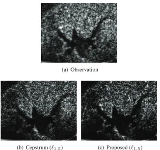
The proposed blind deconvolution algorithm has also been
tested on real US images. In this case, anℓp-norm withp =
1.5 has been employed to regularize the TRF estimation. In this experiment, an ultrasound image representing a mouse
(a) Observation (b) Truth TRF
(c) Non blind with (d)
−0.5 0 0.5 0 0.5 1 1.5 −1.5 −1 −0.5 0 0.5 1 t l t a (d) Truth PSF
(e) Non blind with (f)
−0.5 0 0.5 0 0.5 1 1.5 −1 −0.5 0 0.5 1 t l t a (f) Cepstrum (g) Proposed −0.5 0 0.5 0 0.5 1 1.5 −1.5 −1 −0.5 0 0.5 1 t l t a (h) Proposed
Fig. 1. Simulated US images.
kidney has been acquired with a 25 MHz central frequency US probe, as shown in Fig. 2(a). The restored ultrasound TRFs shown in Figs. 2(b), 2(c) are obtained with the non-blind (cepstrum-based method) and the proposed algorithm. The estimated TRFs in Figs. 2(b), 2(c) have better defined boundaries than the observed image in Fig. 2(a). Moreover, the estimated TRF with the proposed algorithm provides com-petitive performance in terms of visual impression compared with the non-blind deconvolution algorithm.
5. CONCLUSION
This paper studied a new blind deconvolution algorithm for ultrasound images based on a parametric model of the PSF. By exploring an alternating optimization algorithm, we were able to calculate the maximum a posteriori estimations of
(a) Observation
(b) Cepstrum (ℓ1.5) (c) Proposed (ℓ1.5) Fig. 2. Real US images.
the ultrasound tissue reflectivity function and the system PSF simultaneously. Due to the parametric model of the PSF, instead of estimating all the PSF pixels, only a few param-eters need to be estimated. This reduces the computational load and estimation complexity. Future work will be de-voted to extend the proposed approach to complex envelope data/demodulated signals and to conduct more experiments on real ultrasound images.
6. REFERENCES
[1] M. A. Ellis, F. Viola, and W. F. Walker, “Super-resolution image reconstruction using diffuse source models,” Ultrasound in Med. and Bio., vol. 36, no. 6, pp. 967–977, 2010.
[2] M. Tanter and M. Fink, “Ultrafast imaging in biomedical ultrasound,” IEEE Trans. Ultrason. Ferroelectr. Freq. Control, vol. 61, no. 1, pp. 102–119, 2014.
[3] N. Zhao, A. Basarab, D. Kouam´e, and J.-Y. Tourneret, “Joint segmentation and deconvolution of ultrasound images using a hierarchical Bayesian model based on generalized Gaussian priors,” IEEE Trans. Image Pro-cess., vol. 25, no. 8, pp. 3736 – 3750, 2016.
[4] N. Zhao, Q. Wei, A. Basarab, N. Dobigeon, D. Kouame, and J.-Y. Tourneret, “Fast single image super-resolution
using a new analytical solution forℓ2− ℓ2 problems,”
IEEE Trans. Image Process., vol. 25, no. 8, pp. 3683– 3697, 2016.
[5] O. Michailovich and D. Adam, “A novel approach to the 2-D blind deconvolution problem in medical
ultra-sound,” IEEE Trans. Med. Imag., vol. 24, pp. 86–104, 2005.
[6] C. Yu, C. Zhang, and L. Xie, “An envelope signal based deconvolution algorithm for ultrasound imaging,” Sig-nal Processing, vol. 92, no. 3, pp. 793 – 800, 2012. [7] ——, “A blind deconvolution approach to ultrasound
imaging,” IEEE Trans. Ultrason. Ferroelectr. Freq. Con-trol, vol. 59, no. 2, pp. 271–280, 2012.
[8] R. Jirik and T. Taxt, “Two dimensional blind Bayesian deconvolution of medical ultrasound images,” IEEE Trans. Ultrason. Ferroelectr. Freq. Control, vol. 55, no. 10, pp. 2140–2153, 2008.
[9] O. Michailovich and A. Tannenbaum, “Blind deconvo-lution of medical ultrasound images: A parametric in-verse filtering approach,” IEEE Trans. Image Process., vol. 16, no. 12, pp. 3005–3019, 2007.
[10] J. A. Jensen and S. Leeman, “Nonparametric estima-tion of ultrasound pulses,” IEEE Trans. Biomed. Eng., vol. 41, no. 10, pp. 929–936, Oct. 1994.
[11] M. Alessandrini, A. Palladini, L. D. Marchi, and N. Spe-ciale, “Expectation maximization for joint deconvo-lution and statistics estimation,” Acoustical Imaging, vol. 30, no. 11, pp. 335–343, 2011.
[12] N. Zhao, Q. Wei, A. Basarab, D. Kouame, and J.-Y. Tourneret, “Single image super-resolution of medi-cal ultrasound images using a fast algorithm,” in Proc. IEEE International Symposium on Biomedical Imaging (ISBI), Prague, CZ, April 2016.
[13] J. Bolte, S. Sabach, and M. Teboulle, “Proximal alter-nating linearizad minimization for noncovex and nons-mooth problems,” Math. Program., pp. 459–494, Aug. 2014.
[14] A. Repetti, M. Q. Pham, L. Duval, E. Chouzenoux, and J.-C. Pesquet, “Euclid in a taxicab: Sparse blind decon-volution with smoothedℓ1/ℓ2regularization,” IEEE
Sig-nal Process. Lett., vol. 22, no. 5, pp. 539–543, 2015. [15] O. Michailovich and D. Adam, “Robust estimation
of ultrasound pulses using outlier-resistant de-noising,” IEEE Trans. Med. Imag., vol. 22, no. 3, pp. 368–381, 3 2003.