UNIVERSITÉ DU QUÉBEC
À
MONTRÉALL'IMPACT DES PERTURBATIONS DU CYTOSQUELETTE SUR L'EXPRESSION ET LA FONCTION DE LA LOW-DENSITY LIPOPROTEIN RECEPTOR-RELATED PROTEIN 1
(LRP-1)
MÉMOIRE PRÉSENTÉ
COMME EXIGENCE PARTIELLE
DE LA MAÎTRISE EN BIOLOGIE
Par
SAMUEL BURKE-NANNI
UNIVERSITÉ DU QUÉBEC À MONTRÉAL Service des bibliothèques
Avertissement
La diffusion de ce mémoire se fait dans le respect des droits de son auteur, qui a signé le formulaire Autorisation de reproduire et de diffuser un travail de recherche de cycles supérieurs (SDU-522 - Rév.0?-2011 ). Cette autorisation stipule que «conformément
à
l'article 11 du Règlement no 8 des études de cycles supérieurs, [l'auteur] concèdeà
l'Université du Québecà
Montréal une licence non exclusive d'utilisation et de publication de la totalité ou d'une partie importante de [son] travail de recherche pour des fins pédagogiques et non commerciales. Plus précisément, [l'auteur] autorise l'Université du Québecà
Montréalà
reproduire, diffuser, prêter, distribuer ou vendre des copies de [son] travail de rechercheà
des fins non commerciales sur quelque support que ce soit, y compris l'Internet. Cette licence et cette autorisation n'entraînent pas une renonciation de [la] part [de l'auteur]à
[ses] droits moraux nià
[ses] droits de propriété intellectuelle. Sauf entente contraire, [l'auteur] conserve la liberté de diffuser et de commercialiser ou non ce travail dont [il] possède un exemplaire.»ACKNOWLEDGEMENTS
First and foremost, I would like to thank everyone who made it possible for me to be here in Montreal. I would like to sincerely thank my superviser, Professer Borhane Annabi, for his academie and pastoral support throughout my time in the !ab. I will always appreciate the willingness he showed in taking me on in his !ab when I was deep in my frantic hunt for a graduate position. I'd also like to extend similar thanks to both Dr Sylvie Lamy and Dr Khadidja Haidara. Alain Zgheib, Annie Levert and Julie Poirier who were invaluable to my "apprentissage" in the laboratory, and seemed to al ways have the time for my continuai questions. I am very grateful to David Beauchemin, who took me under his wing, as well as Jonathan Pratt, always having 'two minutes' to discuss my data and project. From Southampton, I would like to thank both Patrick Doncaster and Phil Williamson. In addition, I'd like to thank Malcolm East for offering me sorne of the most pertinent and valuable advice during my undergraduate degree. Finally, I'd like to thank ali those in the !ab over these two years, Merci!
TABLE
DES
MATIÈRES
ACKNOWLEDGEMENTS ... iii
TABLE DES MATIÈRES ... iv
RÉSUMÉ ... vi
SUMMARY ... vii
KEY WORDS ... vii
ABBREVIA TI ONS ... viii
CHAPTER I LITERA TURE REVIEW ... 1
1. Introduction ... 1
1.1 Brain Tu mours ... 3
1.2 Glioblastomas ... 4
1.3 Treatment ... 4
1.4 Therapeutic Barriers of the CNS ... 5
1.5 The Blood Brain Barrier (BBB) ... 6
1.6 The Cytoskeleton ... 10
1.7 Matrix Metalloproteinases (MMPs) ... Il 1.8 MTI-MMP ... 12
1.9 MTI-MMP targets ... 14
1.10 LRP-1 ... 15
1.11 Na tura! and Synthetic Ligands of LRP-1; Vectorized Drug Delivery ... 18
1.12 The Proprotein Convertase Furin ... 19
1.13 Plant Lectins ... 20
1. 14 Concanavalin A ... 21 2 Hypothesis ... 23
CHAPTER II 3 RE SUL TS ... 24 3.1 Sutnmary ... 24 3.2 Introduction ... 25 3.3 Experimental Procedures ... 26 3.4 Results ... 31 3.5 Discussion ... 34 3.6 Acknowledgn1ents ... 36 3.7 Figure Legends ... ~ ... 37 3.8 Figures ... 40 3.9 References ... 46 CHAPTER III 4 CONCLUSION ... 52 5 BIBLIOGRAPHY ... 56
RÉS
UMÉ
Les glioblastomes multiformes (GBM), considérés parmi les cancers les plus agressifs du cerveau, représentent entre 1-2% de l'ensemble des tumeurs adultes. Quoique plutôt rares, ceux-ci sont particulièrement difficiles à traiter, d'où le sombre pronostic relié à la maladie. De plus, les causes de la pathologie sont encore peu connues. Un obstacle considérable dans le traitement des GBM, et dans les maladies du système nerveux central (SNC) en général, est le passage de la barrière hémato-encéphalique (BHE) par les substances pharmacologiques. Angiopep-2, un court peptide inerte, a été développé comme système de vectorisation de médicaments au cerveau. Cette molécule est internalisée sélectivement à travers la BHE par un mécanisme dépendant de la protéine Low-Density Lipoprotein Receptor-Related Protein-1 (LRP-1 ). La conjugaison de molécules thérapeutiques à Angiopep-2 a permis le développement de médicaments ciblant le cerveau. LRP-1 est un large récepteur membranaire présent à la swface des cellules endothéliales de la BHE, en plus d'être exprimé dans les cellules cancéreuses du cerveau. LRP-1 possède de nombreuses fonctions, dont l'internalisation de ligands extracellulaires. Il est également associé à la survie et à la migration cellulaire. De plus, LRP-1 est impliqué dans la détermination du grade et du phénotype du cancer du cerveau. Il a été démontré que l'expression et la fonction de LRP-1 étaient régulées par la protéine Membrane Type-1 Mal!-ix Metalloproteinase (MTI-MMP). MTI-MMP est capable de cliver LRP-1, et joue conséquemment un rôle dans la régulation des fonctions qui dépendent de LRP-1. Dans notre recherche, nous avons décidé d'explorer la régulation de l'expression de LRP-1, par MTI-MMP, dans une lignée de cellules de gliome. Nous avons traité le modèle cellulaire U87-GM avec de la Concanavaline A (ConA), connue pour sa capacité à induire l'activation et la transcription de MTI-MMP. Nos résultats ont démontré une rapide internalisation ainsi qu une dégradation de LRP-1. Cependant, par répression génique de MTI-MMP et de d'autres médiateurs impliqués dans la voie de signalisation de la ConA, ainsi que par l'utilisation d'inhibiteurs sélectifs, nous avons établi que cet effet n'était pas médié par MTI-MMP, ni par une interaction directe entre la ConA et la cellule. Sachant que les effets de la ConA impliquent des perturbations du cytosquelette d'actine, nous avons inhibé la polymérisation de l'actine et des microtubules et découvert que les effets sur LRP-1 étaient dus à la perturbation de l'actine. De plus, ce processus d'internalisation et de dégradation de LRP-1 a mené à une réduction significative de la capacité des cellules U87 à internaliser l' Angiopep-2 et l'alpha-2 macroglobuline, deux ligands de LRP-1. Nous concluons donc que l'intégrité du cytosquelette d'actine est requise pour les fonctions de LRP-1 à la surface de la cellule. No données démontrent également la nécessité de retrouver LRP-1 à la surface cellulaire dans la vectorisation de molécules thérapeutiques, couplées à 1 'Angiopep-2, dans les cellules cancéreuses du cerveau. Enfin, nos données peuvent être ajoutées à la littérature grandissante qui suggère l'exploration de la Concanavaline A comme une potentielle entité thérapeutique.
SUMMARY
Glioblastoma multiforme (GBM) is one of the most aggressive types of brain tumours, constituting l to 2% of ail adult tumours. Though relatively rare, GBMs have a dire prognosis and are very difficult to treat. The causes of GBMs are relative! y unknown making the study of the molecular pathologies that lead to this disease important, as weil as enabling the development of more efficacious treatment strategies. A considerable obstacle in the treatment of GBMs, and in general, disorders of the central nervous system (CNS), is the ability to penetrate the blood-brain barrier (BBB) pharmacologically. Angiopep-2 is a short inert peptide that was designed as a drug delivery system: it is selectively internalized across the BBB in a low-density lipoprotein receptor-related protein 1 (LRP-1 )-dependent manner. This has allowed for the successful development of drug molecules that are conjugated to Angiopep-2, allowing delivery to the brain. LRP-1 is a large receptor, present on the cell surface of BBB endothelial cells, as weil as being expressed in brain and brain tumour cells. lt has many functions, including the internalization of extracellular ligands. lt is also associated with cell survival and cell migration. ln addition to this, LRP-1 has been implicated in both cancer grade and phenotype. LRP-1 expression and function has already been shown to be regulated by Membrane Type 1 Matrix Metalloproteinase (MTI-MMP, also known as MMP-14). MT 1-MMP has been shown to be able to cleave LRP-1, playing a rote in the regulation of LRP-1-mediated functions. In our research, we decided to explore MTI-MMP regulation ofLRP-1 in a glioma cellline, and see whether this would impact on the internalization of Angiopep-2, a therapeutic avenue which holds great promise in the treatment of GBM and other CNS disorders. We treated the GBM cell mode!, U87, with the plant lectin Concanavalin A (ConA), known to induce the activation and transcription of MTI-MMP, and found that there was rapid internalization and degradation of LRP-1. However, through gene silencing of MTI-MMP and other known mediators of ConA-signalling, as weil as the use of selective inhibitors, we established that this effect was not mediated by MTI-MMP, nor via a direct cell-ConA interaction. Knowing that ConA's effects involve disruption to the actin cytoskeleton, we selectively disrupted the actin and microtubule cytoskeleton and found that the effects on LRP-1 were due to disruption of the actin, and not microtubule, cytoskeleton. Furthermore, this internalization and degradation of LRP-1 led to a significant reduction in the capacity of U87 cells to internai ize a2Macroglobulin and Angiopep-2, a natural and synthetic ligand of LRP-1, respectively. We conclude that actin cytoskeleton integrity is required for proper LRP-1 cell surface functions. Furthermore, our data demonstrate the pivotai requirement of cell surface LRP-1 functions in the vectorized transport of therapeutic Angiopep bioconjugates into brain cancer cells. ln addition, these data can be added to growing literature base supporting the claim that ConA merits further study as a potential therapeutic entity.
Key Words
BBB Co nA CNS ECM GBM MMP LRP-1 MMP-2 MTI-MMP COX-2 CytoD EGCG ER TLR UPS TJ GPCR YEGF ERK
ABBREVIATIONS
Blood-Brain Barrier Concanavalin A Central Nervous System Extracellular Matrix Glioblastoma Multiforme Matrix MetalloproteinaseLow Density Lipoprotein-Related Receptor 1 Matrix Metalloproteinase 2 Membrane Typre-1 Matrix Metalloproteinase Cyclooxygenase-2 Cytochalasin-0 Epigallocatechin 3-gallate Endoplasmic reticulum Toll-like receptor
Ubiquitin-dependent proteasome system Tight j unctions
G-protein-coupled receptor Yascular endothelial growth factor Extracellular signal-regulated kinases
Chapter I
Literature Revie
w
1.
Introduction
Cancerisa pathology where populations of ce Ils gain a phenotype where they no longer follow the intricate and extensive mechanisms that regulate homeostasis. This enables non-homeostatic growth of certain cells in an uninhibited manner. (Liotta & Kohn, 2001) This is a dynamic, multi-step process that in general takes many years to develop. A highly selective process is required to overcome the innate mechanisms that inhibit the occurrence of cancers. These cells are highly proliferative, and have often !ost cell-cell contact induced growth inhibition - they gain these phenotypes through: genomic; proteomic; post-translational; epigenetic modifications that generate the highly complex alterations in cancerous tissues. (Hanahan & Weinberg, 2011) Hannah et al., in their seminal paper describe the 'hallmarks of cancer' (
Figure 1) - six characteristics required for progression to the neoplastic state. (Hanahan, Weinberg, & Francisco, 2000) However, as described by Lazebnik, these hallmarks are also characteristics of benign tumours, except for 'Tissue invasion and metastasis'. (Lazebnik, 2010) Thus, it is important to consider the use of the words 'tumour' and 'cancer', as they are not interchangeable. This process of invasion is multifaceted and involves many steps that are interlinked and interdependent (Figure 2), which ultimately result in metastatic invasion. lnitially, as biomedical research began to understand the genetic component of the disease, interest was concentrated and hope was placed, in a simplistic view of single cel! lineages being responsible for the cancer phenomena - however, it is now understood that there is a very complex interplay between heterogeneous pre-cancerous populations, and non-cancerous, but associated host cells that provide an essential component to the development of a malignant pathology. This includes the induction of angiogenesis, modification of the tumour environment, the recruitment of immune and stem cells, and the conditioning of potential metastatic sites. (Siemann, Dietmar, 201 0)
Evading
apoptosis
Sustained
angiogenesis
i
lnsensitivity to
anti-growth signais
Tissue invasion
&
metastasis
2Figure 1 The acquired 'hall marks' of cancer. Cancer cel! genotypes involve the manifestation of six essential alterations in cel! physiology that collectively dictate malignant growth: se lf-sufficiency in growth signais, insensitivity to grovvth-inhibitory signais, evasion of programmed cell death, limitless replicative potential, sustained angiogenesis, and tissue invasion and metastasis. Each of these physiologie changes- novel capabilities acquired during tumour development- represents the successful breaching of an an ti cancer defence mechanism hardwired into cells and tissues. Adapted from (Hanahan et al., 2000)
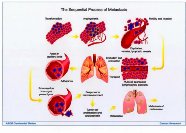
The Sequential Process of Metastasis Transformation Anglogenesls Responseto microenvironment Tumorcell proliferation and anglogonesla Mutticellaggregates (lymphocytes, platolots) Metastases 3
Motility and Invasion
Metastasla of metastases
Figure 2 'Cancer metastasis consists of sequential, interlinked, and selective steps. The outcome of each step is innuenced by the interaction of meta tatic cellular subpopulations with homeostatic factors. Each step of the metastatic cascade is potentially rate limiting such that failure of a tumour cell to complete any step effectively
impedes that portion of the process, th us, the interplay between the many factors is essential to understanding,
and perhaps identifying the factors most innuential in the process of benign tumours becoming cancerous'. (Tai madge & Fidler, 201 0)
1.1
Brain Turnours
Brain tu mours are a relative! y rare disorder, with an incidence of 2-10 cases per 100,000 people, globally. (Bondy et al., 2008) The majority of de nova brain tu mours are of neuroepithelial origin, and are called gliomas. These are graded according to their aggressiveness, from 1 to IV. Grade IV gliomas, or glioblastoma multiforme (GBM), are the most common glioma, and the most aggressive (Ohgaki & Kleihues, 2007) -GBM tumours represent only 1-2% of ali adult tumours however, they are ultimately untreatable. (Erpolat et al., 2009)(Wen & Kesari, 2008) This has led to the dire prognosis
4
of this pathology - treatment strategies are ineffective ( despite surgi cal intervention
extending li fe expectancy). The mean ti me from diagnosis to death with treatment is 14 months, and the 5 year survival rate is 10%. (Bhujbal de Vos, & Ni clou, 201 4) The ir histopathological hall marks include substantial vascularisation -GBMs being one of the
most highly vascularised neoplasms (Visted & Lund-Johansen, 2003). ln addition to
primary brain tumours, secondary metastases are a major challenge and common in
breast, colorectal, melanoma and Jung cancer. (Bhujbal et al., 201 4)
1.2
Glioblastomas
The aetiology of GBM is relatively unknown, with only the exception of
therapeutic irradiation being associated with an increased risk of malignancy. (Visted & Lund-Johansen, 2003) Associations with head injuries, foods containing N-nitroso compounds, occupational risk factors and electromagnetic fields have been inconclusive. (Wen & Kesari, 2008) However, there is sorne suggested evidence of immunological
factors contributing: atopic individuals having a reduced risk for glioma development; patients with GBMs who have elevated IgE Jevels having an improved prognosis.
lnterestingly, only 5% of GBM cases have familial association. (Wen & Kesari, 2008)
Brain tumours, and many cancers in general, can be characterised by aberrant receptor
tyrosine kinase (RTK) signalling. ln GBMs, malignant transformation can be attributed to growth factor signalling. Roughly 90% of GBM tumours have aberrant RTK signalling, with 45% involving the epidermal growth factor receptor (EGFR) family of RTKs. (McLendon et al., 2008) RTKs have been targeted in severa! clinical trials, but have ali ultimately fai led to show any benefit as a treatment. (Johansson et al., 201 3)
1.3
Treatment
Standard treatment of GBM involves, as much where possible, tumour resection followed by radiation therapy and temozolomide-based chemotherapy. (Kauer, Figueiredo, Hingtgen, & Shah, 2012) Radiation and chemotherapy does improve !ife expectancy - 18.9 vs 9.8 months without, (Erpolat et al., 2009) however this is still far
behind advances made in other cancer treatments, and the benefits of chemotherapy in
GBM treatment are debated. (Sarin, 2009) Despite resection of the tumour being effective, nearly 100% of GBM cases are fatal, (Lesniak & Brem, 2004) with 80-90%
recurrence of the tumour occurring within 2 cm of the resection cavity. (Bhujbal et al., 201 4) There are of course severa! factors that can be attributed to this phenomenon.
5
However, it is relatively weil accepted that the biggest barriers to effective treatment of
the residual, infiltrating cells, involves, among others: poor therapeutic access to CNS
because of the blood brain barrier (BBB)" (Muldoon et al., 2007) vascular dysfunction at
the tumour; (Jain, Tong, & Munn, 2007) and, a short half-life of therapeutic molecules
once in vivo. (Sarin, 2009) Pre-clinical studies have shown that the ineffectiveness of
many treatment strategies can be attributed to poor BBB penetration. For example, the use of monoclonal antibodies against EGFRvlll in a murine mode! led to tumour
shrinkage in subcutaneous melanomas but not in intracranial brain metastases due to poor BBB penetration. (J.H. Sampson, L.E. Crotty, S. Lee, G.E. Archer, D.M. Ashley, C.J.
Wikstrand, 2000; Lesniak & Brem, 2004)
1.4
Therapeutic
Barri
ers
Of
The CNS
One of the major obstacles in addressing pathologies of the CNS such as brain tumours; HIY encephalopathies; epilepsy; cerebrovascular disease; and
neurodegenerative diseases), is achieving therapeutic concentrations of pharmacological
agents within the CNS. (Misra, Ganesh, & Shahiwala, 2003) Severa! factors contribute to this phenomenon. Primarily, this is caused by the permeability of the BBB: due to its
specialized function in protecting the CNS, which in the ensuing text will be discussed, it is very difficult to select agents which are BBB-permeable. (Lesniak & Brem, 2004)
There are also other substantial factors, which are not limited to the CNS, that affect
therapeutic concentrations at the desired site. Especially with chemotherapeutic agents, active transport out from tumour cells and BBB endothelial cells by efflux pumps such as
P-glycoprotein results in diminished concentrations of the drug. (Schinkel et al., 1997) Furthermore, within the CNS there is a variance in drug volume distribution detennined by cellular uptake, interactions with lipids and proteins, as weil as accumulation in
subcellular compartments. (Muldoon et al., 2007) With systemic delivery of agents, there
is substantial plasma protein binding: for chemotherapeutic agents this can often be 90%, with only 10% free drug. However, this can be even higher: for example, the alkylating agent Chlormbucil is 99% plasma protein bound. (Muldoon et al., 2007) Another substantial barrier to effective targeting of cancer cells is the high tumoural interstitial pressure. This can often be above 50 mmHg; whereas the surrounding tissue will be at 2
mmHg. (Muldoon et al., 2007) This high interstitial pressure diminishes the transcapillary
6
blood- vesse! leaki ness; i nterstitial fi brosis; contraction of the i nterstitial matrix mediated by stroma! fibroblasts. (Heldin, Rubin, Pietras, & Ostman, 2004)
1.5
The
Blood Brain Barrier
(
BBB)
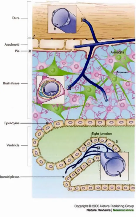
The BBB acts to protect the CNS from fluctuations in the composition of the blood (hormones, amino acids, glucose, ce11ain toxins etc.). lt tightly regulates the passage of materials across the CNS-cardiovascular interface through anatomical, physicochemical, and biochemical mechanisms. The neurovascular unit is the anatomical
feature forming the BBB, found at the endothelial cell - brain capillary site (Figure 3), deriving its properties from a specialized basal membrane, perycites and astrocytic endfeet. These features co-operate to produce the BBB, and to tightly control the passage of molecules a cross the interface. The conti nuous tight j unctions th at j oi n the endothelial
cells in the brain capillaries limit the diffusion of molecules across the BBB (the major proteins involved in this are summarised in Figure 4). The basal membrane provides structural support for the capillary and specifie proteins present in the basement membrane play a part in the development and establishment of the BBB. (Obermeier,
Da neman, & Ransohoff, 20 13) Astrocytic foot processes release specifie factors and are
necessary for the development of the BBB. Transport carriers for glucose and essential
amino acids facilitate the movement of these solutes into the brain - neuronal cells are unable to synthesize these essential amino acids, it is taken up from the blood. Secondary transport systems appear to cause eftlux of small molecules and non-essential amino acids from the brain to the blood. Sodium ion transporters on the luminal membrane and Na/K-ATPases and on the anti-luminal membrane account for the movement of sodium from the blood to the brain - the large number of mitochondria present in brain endothelial cells provide energy for the functionjng of these Na/K-ATPase. The major pathways for the movement of material into the CNS compartment (described in Figure 5) include: paracellular routes for small water-soluble molecules; transcellular for lipid soluble molecules, transport protein-mediated and receptor-mediated cellular internai ization and transport. 22-25
Dur"
A•achnoitl- . .
Ur ain tissue
-V nuicl
-Choroid plexus
-COpyright
e
2005 N:~ture Publistl ng Grou Nature Revlews 1 Ncurosclence7
Figure 3 Location of barrier sites in the CNS. Barriers are present at three main sites: the brain endothelium forming the blood-brain barrier (BBB) ( 1 ), the arachnoid epithelium (2) forming the middle layer of the meninges, and the choroid plexus epithelium (3), which secretes cerebrospinal Auid (CSF). At each site, the physical barrier is caused by tight junctions that reduce the permeability of the paracellular (intercellular cleft) pathway. Modified from. (Abbott, Réinnback,
8
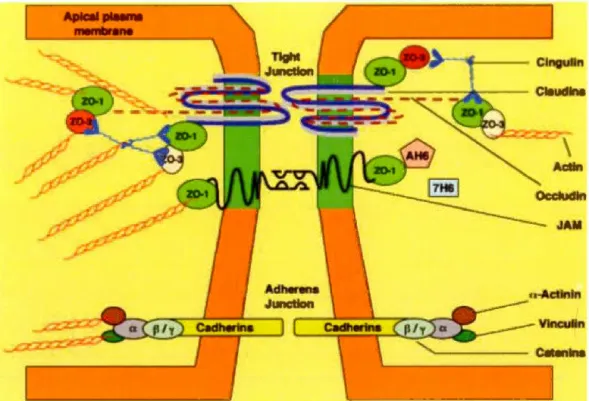
Figure 4 'The major proteins associated with tightjunctions (TJs) at the BBB are shown. The tight junction is embedded in a cholesterol-enriched region of the plasma membrane (shaded). Three integral proteins- claudin 1 and 2, occludin and junctional adhesion molecule (JAM)- form the tight junction. Claudins make up the backbone of the TJ strands f01·ming dimers and bind homotypically to claudins on adjacent cells to produce the primary seal of the TJ. Occludin
functions as a dynamic regulatory protein. The tight junction also consists of severa! accessary
proteins, which contribute to its structural support. The zonula occludens proteins (Z0-1 to 3) serve as recognition proteins for tight junctional placement and as a support structure for signal transduction proteins. AF6 is a Ras effector molecule associated with Z0-1. 7H6 antigen is a
phosphoprotein fou nd at tight junctions impermeable to ions and molecules. Cingulin is a
double-stranded myosin-like protein thal binds preferentially to ZO proteins at the globular head and to
other cingulin molecules at the globular tail. The primary cytoskeletal protein. actin, has known binding sites on ail of the ZO proteins·. (Nag, 2003)
9
: b c : e
P r ce~ ar~qu~ : Tr nsc ul.lr : Tr.an'f'O" prote : Ad~tlvc
~thwa : lop~>ph 1
.
!
.
f)tlth• lfllrl • 1 •too
0
go
0
Figure 5 'Pathways across the blood-brain barrier. A schematic diagram of the endothelial cells that form the blood- brain barrier (BBB) and their associations with the perivascular endfeet of astrocytes. The main routes for molecular traffic across the BBB are shown. A) Normally, the tight junctions severely restrict penetration of water-soluble compounds, including polar drugs. B) However, the large surface area of the lipid membranes of the endothelium offers an effective diffusive route for lipid-soluble agents. C) The endothelium contains transport proteins (carriers) for glucose, amino acids, purine bases, nucleosides, choline and other substances. Sorne transporters are energy-dependent (for example, P-glycoprotein) and act as efflux transporters. AZT, azidothymidine. D) Certain proteins, such as insulin and transferrin, are taken up by specifie receptor-mediated endocytosis and transcytosis. E) Native plasma proteins such as albumin are poorly transported, but cationization can increase their uptake by adsorptive-mediated endocytosis and transcytosis. Drug delivery across the brain endothelium depends on making use of pathways b-e; most CNS drugs enter via route b'. (Abbott et al., 2006)
10
1
.
6
The Cytoskeleton
The cytoskeleton's role is integral to almost every cellular function: it is the structure that gives cells their shape, mediates the controlled movement of subcellular structures such as organelles, as weil as facilitating the organisation of cellular processes. (Al bert, B., 201 0) The cytoskeleton consists of three major classes of molecules that differ in size and in prote in composition. Microtubules are the largest type of filaments and they are composed of a protein cal led tubulin. Actin filaments are the smallest type and they are made of a protein called actin. lntermediate filaments, as their name suggests, are mid-sized. (Albert, B., 201 0)
The actin cytoskeleton plays an integral role in the spatial order of interna!
structures, providing mechanical forces and mediating adherence of the cell, it provides the contractile forces as weil as linking mechanical stresses to biological responses. (Fischer & Fowler, 20 15) The actin cytoskeleton is also essential for the transport of cargos through the cel! cytosol, as weil as the mediation of the internalization of extracellular cargos, and the creation of membrane vesicles. (Albert, B., 2010)
These structures also function, to a certain extent, as detectors for mechanical
signais: crosstalk between the actin cytoskeleton and microtubule cytoskeleton plays a role in mediating the polarization of cells during division, morphological changes, as weil
as migration. This can be crucial for the development of tissues, as weil as the homeostatic balance between non-metastatic, and metastatic cells. ln migrating cells, growing microtubules that reach into the leading edge promote Rac activation and the formation of short, branched F-actin for lamellipodia formation. ln essence, the cel! can respond the extrinsic, physical eues that can be translated into biological responses, which lead to the formation ofthe required structures. (AkJ1shi, Wernike, & Piekny, 2014)
The cytoskeleton is also an integral component of vesicular trafficking: during endocytosis, transport within the cel! and, exocytosis. (Albert, B., 2010) The cytoskeleton also plays an integral part in modulating membrane curvature and tension. FUI1hermore, it is involved in the regulation of clathrin coated pit internalisation: GPCRs interacting with the actin cytoskeleton regulating the rate of the internalisation of cargos, as weil as perhaps, adding in the creation of forces during dynamin fission. ln addition, it has been
Il
suggested that the interplay between the cytoskeleton and the plasma membrane allows for mechanisms that facilitate internalisation of large volumes of membrane during, for example, pinocytosis or macropinocytosis.
Metastatic disease, or the movement of cancer cells from one site to another, is a complex process, as previously described, which can also involve dramatic remodelling of the cytoskeleton. The various components of the cytoskeleton are highly integrated and their functions are weil orchestrated in normal, physiological cells. (Albert, B., 201 0) ln contrast, in metastatic pathologies, mutations and abnormal expression of cytoskeletal and cytoskeletal-associated proteins can play an important role in the ability of cancer cells to metastasize, as weil as to resist treatment strategies. Studies on the role of actin and its interacting partners have highlighted key signalling pathways, such as the Rho GTPases (where they can mediate the formation of different types of F-actin that confer changes in cortical tension and contraction, and can be regulated by microtubules) and downstream effector proteins that, through the cytoskeleton, mediate tumour cel! migration, invasion and metastasis. lmproved understanding of how the cytoskeleton and its interacting partners influence tumour cel! migration and metastasis has led to the development of novel therapeutics against aggressive and metastatic disease. (Fife, McCarroll, & Kavallaris, 20 14)
1. 7
Ma tri
x Metalloproteinases (MMPs)
The plethora and complexity of the acquired phenotypes that enable cellular transformation to neoplastic tumours are exquisitely complex-in some cases dependent, and others independent of one another. The invasive and metastatic ability of tumour cells, especially in GBM cases, contributes significantly to their morbidity and mortality. This gained phenotype is often as a result of deregulation of the relationship between the cel! and the extracellular matrix (ECM). This is a complex interaction involving proteases (mainly MMPs), their inhibitors and a plethora of signalling molecules that orchestrate this delicate interaction. (Siemann, Dietmar, 2010)
The MMPs are a family of zinc-dependent endopeptidases that are able to degrade nearly ali the components of the ECM including, but not limited t,o fibrillar and nonfibrillar collagens, fibronectin, laminin and basement membrane proteoglycans.
12
(Uiasov, Yi, Guo, Sarvaiya, & Cobbs, 2014) as weil as other non-matrix substrates implicated in tumour establishment. The family consists of more than 23 members, both
secreted and membrane-anchored, that are synthesised as zymogens and many,
furin-activatable. (Siemann, Dietmar, 201 0) (Egeblad & Werb, 2002)
The MMPs play a significant role in the regulation of the tumour microenvironment. They are heavily involved in tissue remodelling, including specifie physiological processes such as cell migration and proliferation. (Siemann, 201 0) This involves the degradation of the ECM (the different MMPs being specifie for varying
substrates), and also the release of cytokines and growth factors from degraded basement membrane. Of the MMPs, MMP-2 and -9, as weil as MTI-MMP, have been studied extensively due to their involvement in migration, invasion and metastasis (Siemann, Dietmar, 201 0).
1.8
MTl-MMP
MTJ-MMP is a transmembrane MMP, and the most extensively studied, that plays a major role in cell motility. Among its functions, it is often found at the leading edge of migrating and invading cells - along with its inhibitor TIMP2, MTI-MMP, in a
multi-step process, is responsible for MMP-2 activation. (Sato et al., 1994, Ries et al., 2007)
Together, these MMPs are able to target many substrates in the ECM, including œ
il-adhesion molecules such as aV integrin subunit precursor (MTI-MMP), lamininY (both) and CD44 (MTI-MMP) - both MMPs playing integral roles in angiogenesis and cell invasion. (Egeblad & Werb, 2002; ltoh, 2006) To summarize, as shown in figure 6, MTI-MMP's major role in ECM proteolysis is amplified by its ability to activate MMP-2 (as weil as MMP-13). This is coupled with the processing and degradation of many œ il-adhesion molecules, with the release of ECM fragments that promote both growth and migration. (ltoh, 2006)
MTI-MMP has been shown to play an integral role in angiogenesis, and is likely linked to its role in tumorigenesis where its upregulation is involved in the formation of MTI-MMP-YEGFR2-Src complexes that result in the activation of Akt and mTOR (Eisenach et al., 201 0). However, adding to the complexity of its role is the result that in
13
MTI-MMP deficient mice, there is normal vascular formation. (Holmbeck et al., 1999) Whether the functions of MTI-MMP, during development, can be compensated by other systems, or that the function in vasulogensis of MTI-MMP become important following
development in unknown.
There are severa! other biological functions of MTl-MMP that merit mention. These
include MTI-MMP's modulation of the inflammatory response of macrophages.
MTI-MMP can trigger the expression and activation of a phosphoinositide 3-kinase d
(PJ3Kd)/Akt/GSK3b signaling cascade which in tum, MT!- MMP-dependent PI3Kd
activation regulates the immunoregulatory Mi-2/NuRD nucleosome remodeling complex that is responsible for controlling macrophage immune responses. (Shimizu-Hirota et al., 20 12) MTI-MMP also regulates Notch signal ling to maintain normal B-cell development
in bone marrow, which occurs through the cleavagee ofNotch ligand Delta-like 1, found on bone marrow stroma! cells cel! surface. (Jin et al., 2011)
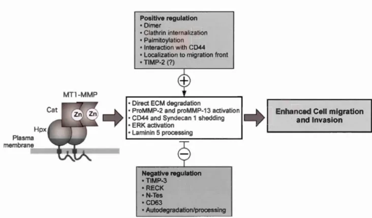
Plasma membrane MTl-MMP Positive regulation • Dîmer • Clathrin internalization • Palmitoylation • Interaction with CD44 • Localization to migration front • TIMP-2 (?)
• Direct ECM degradation
• ProMMP-2 and proMMP-13 activation • CD44 and Syndecan 1 shedding • ERK activation
• Laminin 5 processing Negative regulation • TIMP-3 • RECK • N-Tes •CD63 • Autodegradation/processing 14
Enhanced Cell migration and Invasion
Figure 6 'Biological activities of MTI-MMP (MTl-MMP) and their regulation. MTI-MMP enhances cel! migration and invasion by direct ECM degradation, activation of proMMP-2 and proMMP-13, CD44 and syndecan-1 shedding, ER.K activation, and laminin 5 processing. These activities are positively (+) and negative! y (-) regulated by a variety of processes. Disturbing one of the positive regulation pro cesses may be enough to inhibit MT 1-MMP-dependent cel!
migration'. (Itoh, 2006)
1.9
MTl-MMP Targets
As an MMP, MTI-MMP has a broad range of targets. ln it's function of pericellular proteolysis of ECM macromolecules. MTI-MMP degrades Collagen 1, Il, Il; Gelatin; Laminin 1 and IV; Fibronectin; Vitronectin; Aggrecan; Fibrin; Nidogen; Perlecan; Lumican. Furthermore, MTI-MMP is able to interact with cell surface proteins on nearby cells. Here, it is able interact with targets such as: CD44; Transglutaminase; LRP-1; Syndecan-1; Extracellular Signal Regulated Kinase (ERK) as weil as YEGF. (Uiasov et al., 2014) Due to the plethora of possible interactions that MTI-MMP, and the
15
MMPs, in general, have, a developed and integrated knowledge of these functions will help better direct MMP inhibitor therapies that have, thus far, failed. (Egeblad & Werb,
2002)
1.10 LRP-1
LRP-1 is a member of the LDL receptor family of endocytic receptors (Figure 7) and is formed of two subunits, one extracellular of 515 kDa; one cytoplasmic of 85 kDa.
The mature receptor is generated from its pro-form through cleavage of the 600 kDa peptide by Furin. (Willnow et al., 1996)
This family contains severa! functionally dynamic receptors involved in the cellular internalization of many circulating ligands. LJU>-1 is found predominantly in the CNS and liver where it plays a significant role: specifie for many ligands relevant to cholesterol homeostasis, it is also involved in the clearance of proteins, especially within the CNS. On binding to ligands, LJU>-1 and its ligands undergo clathrin-mediated endocytosis, where the ligand is further processed or degraded in lysosomes- LRP-1 is then recycled back to the cell surface or to other intracellular compartments. (Nubile, 2007)
LRP-1 is one of three LDLRs that contain NPXY motifs - it has two within its intracellular 85 kDa signalling subunit. LRP-1 plays a role in intra-cellular signalling and has been found to be implicated in many essential functions (KO in mice being embryonically lethal). (A. P. Lillis, Van Duyn, Murphy-Ullrich, & Strickland, 2008) For example, LJU>-1 plays an important role as a pro-survival signalling element; it has been implicated in varying fonns of Alzheimer 's disease (having both positive and negative effects). (Nubile, 2007)
LDLR NH2 LRPl LRPlB Megalin (also known as LRP2) MEGF7 (atso known as, LRP4)1 NHl 16 igand-binding rcp at GF rep <Jt 13-prop lier doma n 0-linked ugar dom
1n
VPSlOP domam
Fibronectin type Ill repeat Transm mbran do ain 1> NPx mo if
0 PPPSP motif
D GGA-binding motif
LRPS and LRP6 SORLA (als.o known as SORU and UR.11) NH2 APOE.R2 (also known VlDLR a.s. LRP8) NH2
Figure 7 The LDLR family of receptors. 'The structural organization of the low-density
lipoprotein receptor (LDLR) family members. Ali of the receptors are type 1 receptors that contain
a single membrane-spanning domain and a relatively short cytoplasmic tail. The extracellular regions ofthese receptors contain three characteristic modules: ligand-binding repeats (also called
complement-type repeats), epidermal growth factor (EGF) repeats and YWTD-containing ~
propeller domain s. The furin cleavage sites in LDLR-related protein 1 (LRP 1) and LRP 1 B are
indicated by arrows. The four clusters of ligand-binding repeats in LRP 1 are label led (l-I V).
1-lighlighted in blue are the two extra sequences in LRPIB (compared with LRPI). which are encoded by two extra exons: a ligand-binding repeat in the fourth ligand-binding domain and a 33
-amino acid insert in the cytoplasmic tail. LRP5, LRP6 and sortilin-related receptor with A-type
repeats (SORLA; also known as ORL 1 and LR Il) are di tant members of the family with
atypical structural arrangements. Severa! other LDLR family members with poorly defined
17
ln cancers, the literature suggests both positive and negative effects of LRP-1
status and function. lnitially, it appeared that LRP-1 had protective properties in the malignant phenotype, namely due to it's ability to clear the extracellular compartment of
proteases. LRP-1 expression has also been found to be downregulated in the most
malignant gliomas. However, in the past decades, studies have emerged suggesting that LRP-1 does in fact play a significant role in the malignant phenotype of cancer cells.
lt has been shown that LRP-1 is tethered to the actin network and focal adhesions sites; it was suggested that through this interaction, activating ERK signalling pathways
and inhibiting JNK pathways, LRP-1 contributes to the cancer cel! adhesive state
favouring invasion. Furthermore, it was demonstrated that LRP-1-dependent MAPK
signalling contributes to cytoskeleton architecture organisation, and the mediation of adhesive complex turnover. (Langlois et al., 201 0) An example of the LRP-1 paradox is, despite LRP-1 's role in clearing extracellular proteases that it has been shown to be involved in the induction of both MMP-2 and 9, albeit in endometrial explants and not
cancer cell models. (Sel vais et al., 2009)
LRP-1 is involved in cel! survival, proliferation and focal adhesion complex composition, and turnover (8 Langlois, Emonard, Martiny, & Dedieu, 2009) - LRP-1 being found on the invasive front of invading cancer cells. Furthermore novel interactions with the CD44 protein implicate LRP-1 in both its internalization and
recycling, with LRP-1 1 CD44 complexes being found at the migratory front of carcinoma
cells. (Perrot et al., 20 12) There are severa! studies demonstrating that LRP-1 blockade reduces the invasive phenotype of cancer cel! models. ln both carcrinoma and GBM cells
LRP-1 si lencing has shown to reduce ce li invasion and migratory capacity, des pite
elevated levels of MMP-2 in the extracellular compartment. (Dedieu et al., 2008)
Another interesting function of LRP-1, which may have implications in the malignant phenotype, is its roles in cell survival: LRP-1, in primary neurones, was
demonstrated to have an anti-apoptopic function where it is able to regulate the insulin
receptor, as weil as the Akt survival pathway (Fuentealba, Liu, Kanekiyo, Zhang, & Bu,
2009) (an affected target in ConA mediated cel! death). LRP-1 also plays a significant
role in the vascular system, where it is weil studied. Recently a study has shown the potential of LRP-1 to contribute to the recruitment of monocytes to the tumour
18
compartment, th at in tu rn are attributed with the a bi 1 ity to promote vascularisation of the tumour. (Staudt et al., 201 3)
ln ali, LRP-1 's roles are yet to be fully characterised and understood. However, it is evident that due to the plethora of rotes it has, and can have, in a potentially environment 1 compartment specifie manner, further understanding of its regulation have far-reaching implications. One can speculate that LRP-1 does play a role in the transition from benign to malignant tumour, and that it contributes to this phenotype in multiple ways.
1.11 Na tura! and Synthetic Ligands Of LRP-1
; Vectorized Drug
Delivery
As described previously, LRP-1 has many functions including lipoprotein metabolism, degradation of proteases, activation of lysosomal enzymes and cellular entry of bacterial toxins and viruses. This broad role is reflected in the number of ligands it binds, with LRP-1 being reported to bind up to 60 ligands, including: apolipoprotein E-enriched lipoproteins (chylomicron and YLDL remnants), a2Macroglobulin, uPA uPA/PAI-1, neuroserpin, neuroserpin/tPA complexes, MMP-9, MMP-13, MMP-2, HIV
Tat protein, as weil as severa! growth factors, to name a few. (Lillis, Mikhailenko, & Strickland, 2005)
Synthetic ligands of LRP-1 have been developed for the vectorized delivery of drugs. Angiopep-2 is a 19 amino acid peptide whose sequence is derived from the kunitz domain of aprotinin (and other peptides that are substrates for BBB tran cytosis) that is selectively transported from the blood to the CNS compartment. lt was developed with the aim of providing a delivery vehicle for pharmacological agents that are otherwise
excluded from the CNS compartment by BBB selectivity. (Demeule et al., 2008) Angiopep-2 has been shawn to be transcytosed and internalized in a LRP-1 dependent manner, a receptor that is significantly expressed in brain endothelial cells, as weil as
some brain tumours. Furthermore, it is not a substrate for the P-glycoprotein efflux pump,
making it a highly attractive vehicle for molecules that are otherwise ejected from the CNS and tumour compartments. (Demeule et al., 2008) Chemotherapeutic conjugates of Angiopep-2 have been demonstrated, both in vitro and in vivo, to be as effective in killing
19
tumour cells - as weil as showing improved BBB penetration and increased tumour penetration -as un-conjugated forms. This includes species conjugated to Doxorubicin, Nocodazole and Paclitaxel. (Bertrand et al., 20 Il; Ché et al., 201 0) Angiopep conjugates are currently in clinical-phase trials, and appearing to be efficacious: the paclitaxel
conj ugate showing promising results for breast cancer patients with secondary brain metastases.
1.12 The Proprotein Convertase Furin
Furin is a ubiquitously expressed protein that mediates the proteolytic maturation, by cleavage, of proprotein substrates in the secretory pathway. Though not a substantial topic in this thesis, it is germane to discuss as both MTI-MMP and LRP-1, who both
contain the consensus site that furin cleaves (positioned after the carboxy-terminal arginine residue in the sequence -Arg-X-Lys/Arg-Arg -), undergo Furin-mediated proteolytic maturation. On its discovery, Furin was thought to be a housekeeping protein, but has now been shown to have many roles and play an integral role in both normal physiology and pathology. Furin is a 794 amino acid peptide that resides predominantly
in the trans-golgi network- its location being determined by signalling sequences in its cytoplasmic domain. The 83 amino acid pro-domain aids the peptide in its folding and
activation. This process of maturation occurs in a similar way to that ofFurin's actions on
other proproteins- it is autoactivated in a compartment and pH specifie manner, using its
'measure once, eut twice' rule. (Thomas, 2002) Furin is an essential protein in
embryogenesis and homeostasis, as weil as being implicated in some major pathologies. For example, Furin is the principal endoprotease for the 16 kDa ~-nerve growth factor, a critical player in neuronal cel! death 1 survival balance, as weil as the transmembrane
receptor Notch. Furin has a Iso implications in neurodegenerative diseases su ch as
Alzheimer's where it is involved in APP processing and the activation of both a-and ~ secretase (members of the ADAM family of metaloproteiases). (Thomas, 2002) Furthermore, Furin has been implicated in severa! cancers: upregulated in GBMs as weil
as non-small cel! lung carcinomas and squamous-cell carcinomas of the head and neck. (Mbikay et al., 1997) Furin is able to increase the mal ignancy of tu mours through its
ability to activate MTI-MMP, whjch, in turn, via MMP-2, increases degradation of the ECM (depicted in Figure 8). Furin, in vitro, has been targeted pharmacologically, where
- - - -~--- - - -- - - -- - - -- -
-20
its inhibition resulted in decreased cel! motility and invasiveness in CHO and HTI080 cel! li nes. (Coppola et al., 2008)
FURJN
pro
MT
1
·
MP
MM
2
~
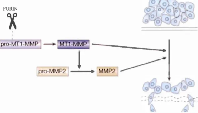
Figure 8 Furin (scissors in figure) is involved in the maturation of MTI-MMP, leading to a subsequent increase in MMP-2 activation, contributing to the metastatic phenotype of tumours. Adapted from. (Thomas, 2002)
1.13 Plant Lectins
Plant lectins are a family ofCa2
+/Mn2
+- dependent carbohydrate binding proteins that are able to bind selectively and reversibly to free sugars on glycoproteins and glycolipids. (Vijayan & Chandra, 1999) The specificity of the binding has lead to the classification of the lectins into 12 families. Over the past 20 years the lectins have been used to label and identify malignant vs benign tumours, evaluating the glycosylation state of malignancies (Mody, Joshi, & Chaney, 1995) and recently, their introduction into microarrays for high throughput analysis of protein glycosylation. (Z. Liu, Luo, Zhou, &
Zhang, 20 13) Furthermore, increased interest has been placed in the anti-tumour properties exhibited by these lectins. For example, they have apoptopic and autophagie inducing properties (the programmed cel! death signalling network illustrated in figure 9)
21
, (Zhang, Chen, Ouyang, Cheng, & Liu, 20 12) as weil as anti-angiogenic. (Li, Yu, Xu, & Bao, 20 1 1 )'(Z. Liu et al., 20 13)
There is a substantial volume of literature reporting the multiple effects of the plant lectins on cancers throughout the tissues of the body. This includes severa! reports on the induction of apoptopic cel! death induced by the lectins ConA, Polygonatum
cyrtonema lectin and Mistletoe lectins.
1.14
Concanavalin
A
ConA has been reported to induce both the extrinsic (Fas family of death
receports dependent) and the intrinsic (mitochondrial dependent) pathways of apoptopic cel! death. (Z. Liu et al., 2013) For example, apoptosis via the intrinsic pathway has been reported in both human melanoma A375 cells (B. Liu et al., 2009) and hepatocellular carcinoma HepG2 cells; (Zhongyu Liu, Li, Ding, & Yang, 2010). Furthermore, in U87 GBM cells, it has been shown to up-regulate COX-2 expression, as weil as down-regulate Akt expression via lKK!NFkB-dependent pathways. (Pratt et al., 2012) ConA induces apoptosis by inhibiting the Akt survival pathways as weil as activating FoxOia-Bim signalling in both ovarian and Li-Fraumeni syndrome cells. (Z. Liu et al., 2013)
ConA has also been shown to induce autophagie cell death via a BCL
-2/adenovirus EIB 19kDa-interacting protein 3 (BNIP-3) - mediated pathway. (Lei & Chang, 2009a) The mechanisms of ConA-induced cell death are varied (ConA's
mutli-faceted effects resulting from various interactions, and modulations of a host of pathways). (Li et al., 20 Il) One mechanism includes the association of ConA with mannose moieties at the plasma membrane, clathrin-dependent internalization to mitochondria and the initiation of autophagie cel! death. (Lei & Chang, 2009b)
Furthermore, autophagy induced by ConA can be abrogated through the silencing of MTl-MMP; though not by catalytic inhibition. (Pratt et al., 2012) This suggests that
MTI-MMP-mediated ConA-induced autophagy is signalled, in the case of MTl-MMP, via the non-catalytic, cytoplasmic domain. (Pratt et al., 20 12) Another interesting property of the plant lectins, especially ofConA, is the ability to perturb the cytoskeleton. (Vijayan & Chandra, 1999)
22
Interestingly, severa! of the cellular effects induced by Concanavalin A (ConA)
are mediated, in part via MTI-MMP. This includes MTI-MMP-mediated-MMP-2 activation; COX-2 induction, independent of the MTI-MMP catalytic domain;
MTI-MMP activation and transcription; the induction of autophagy biomarkers via MT
I-MMP's cytoplasmic signalling domain. (Akla, Pratt, & Annabi, 20 12; Annabi et al.,
2009; Annabi et al., 20 14; Pratt, Roy, & Annabi, 20 12; Si na et al., 201 0) Furthermore, of
interest, is the ability of MTI-MMP to cleave LRP-1 in malignant cells- MTI-MMP
acting as a 'sheddase', leading to the N-terminal being shed into the extracellular milieu.
(Rozanov et al., 2004) Certain sugar-contalning reoeptor TNFR112 MTI·MMP EGFR Fasl Fa ~--~ A
...
~ t====:::~ SHPS-1,MMP-2J9-==-::::::r.
16
Pr<>-dealh - A Rlcin-B ramily
Pro-survival A Proteins wilh legume lectin domalns
Other GNA famlly
Q Autophagosome Apoptosome Vis leie
---@ Mitochondrla Receptor Ribosome Activation ~ lnactivalion AbsenceFigure 9 The programmed cell death (PCD) signalling network which can be regulated by plant
lectin , especially CanA. ln particular, the pathways regulated by the fas family of death receptors
and MTI-MMP. Modified from (Fu et al., 2011)
23
2 Hypothesis
LRP-1 is a crucial player in GBM biology and a target for GBM treatments (as
weil as other CNS disorders). lt also plays an integral role in both physiology, and the
pathology of severa! diseases. As it has begun to be studied in more depth, reports are emerging showing a direct link to the cytoskeleton. Given this, further understanding of its regulation is certainly pertinent for our collective understanding. Within our group, MTI-MMP has been extensively studied, and has been shown in severa! other groups to be able to regulate LRP-1 expression. The plant lectin ConA presents as an entity with therapeutic potential, but its ability to induce the activation and transcription of MTl-MMP (alongside other weil characterised effects, such as its ability to disrupts the
ctyoskeleton), presents it as a useful tool to explore and investigate severa! potential
cellular events that may impact on LRP-1. Thus, whether ConA is able to modulate LRP-1 in GBM cells, and affect the internalization of LRP-1 ligands, is of interest.
Furthermore, given MTl-MMP's role in the GBM phenotype (as weil as other
malignancies), further probing its effects, linked to the malignant processes that can be
modelled with ConA, will contribute to our understanding of the phenotypic
transformation essential for transition to malignancy.
Hypothesis: Treating U87 cells with ConA at a concentration able to induce
MTl-MMP activation will lead to MT 1-MMP mediated proteolytic processing of LRP-1.
The processing of LRP-1 will in turn lead to a reduced capacity of LRP-1-Iigand
internai ization.
2.1
Aims and Objectives
• Further characterise ConA mediated effects in U87 cells - specifically
MTl-MMP processing and MTI-MMP mediated events
• Explore the role of ConA and its effects on MTI-MMP 111 LRP-1-dependent
Chapter II
3 Results
Publi hed as: Nanni et al. Impact of Concanavalin-A-Mediated Cytoskeleton Disruption on Low-Density Lipoprotein Receptor-Related Protein-1 lnternalization and Cell Surface Expression in Glioblastomas. Biomarkers in Cancer 2016:8 77-87
doi: 1 0.4137/BIC.S38894. Published April 26 2016.
Impact of Concanavalin-A-mediated cytoskeleton disruption on low-density
lipoprotein receptor-related protein-1 internalization and cel! surface expression in gl ioblastomas
Samuel Burke Nanni, Jonathan Pratt, David Beauchemin, Khadidja Haidara and Borhane Annabi
From the Laboratoire d'Oncologie Moléculaire, Centre de recherche BIOMED,
Département de Chimie, Université du Québec à Montréal, Quebec, Canada
Running title : LRP-1 expression is decreased upon cytoskeleton disruption Correspondence should be directed to : Borhane Annabi, Laboratoire d'Oncologie Moléculaire, Université du Québec à Montréal, C.P. 8888, Suce. Centre-ville, Montréal,
Québec, Canada, H3C 3P8; Phone: (514) 987-3000 ext 7610; Fax: (514) 987-0246; E -mail : annabi.borhane@uqam.ca
Keywords: Glioblastoma, Brain cancer, LRP-1, Concanavalin-A, Cytoskeleton
3.1
Summary
The low-density lipoprotein receptor-related protein 1 (LRP-1) is a multiligand
endocytic receptor which plays a pivotai role in controlling cytoskeleton dynamics during
cancer cell migration. lts rapid endocytosis further allows efficient clearance of extracellular ligands. Concanavalin-A (ConA) is a lectin used to trigger in vitro
physiological cellular processes including cytokines secretion, nitric oxide production, and T lymphocytes activation. Given that ConA exe1is pa1i of its effects through cytoskeleton remodeling, we questioned whether it affected LRP-1 expression,
intracellular trafticking, and cell surface function in grade IV U87 glioblastoma cells.
25
600-kDa mature form of LRP-1 occurs upon ConA treatment. Consequently,
internalization of the physiological 2-Macroglobulin and of the synthetic Angiopep-2
ligands of LRP-1 was a Iso decreased. Silencing of known mediators of ConA, such as the
membrane type-! matrix metalloproteinase, and the Toll-like receptors (TLR)-2 and
TLR-6, was unable to rescue ConA-mediated LRP-1 expression decrease, implying that the loss of LRP-1 was independent of cel! surface relayed signaling. The ConA-mediated
reduction in LRP-1 expression was emulated by the actin-cytoskeleton disrupting agent
Cytochalasin-D, but not by the microtubule inhibitor Nocodazole, and required both
lysosomal- and ubiquitin-proteasome system-mediated degradation. Our study implies
that actin cytoskeleton integrity is required for proper LRP-1 cel! surface functions, and
that impaired trafficking leads to specialized compartmentation and degradation. Our data
also strengthen the biomarker role of cel! surface LRP-1 functions in the vectorized
transport of therapeutic Angiopep bioconjugates into brain cancer cells.
3.2
Introduction
Low density lipoprotein receptor-related protein 1 (LRP-1) is a member of the LDL
receptor family of endocytic receptors formed of one extracellular 515 kDa subunit, and
one cytoplasmic 85 kDa subunit; 1 the mature receptor having been generated by the
cleavage of a 600 kDa propeptide by Furin.2 The LRP family contains severa! functionally dynamic receptors involved in the cellular internalization of more than 40 circulating physiological ligands, including apolipoprotein E3, a2-Macroglobulin4, factor
Vlll5, lipoproteins6 and Amyloid-~.7 Once ligand is bound, the LRP-1/Iigand complex
undergoes clathrin-mediated endocytosis in order that the ligand be further targeted
within specialized intracellular compartments.8 Disruption of the LRP-1 gene in mice was found to be embryonically lethal, presumably because LRP-1 transduces intracellular
signal ling and is involved in many essential functions.9
Over the past few decades, it has emerged that LRP-1 plays a significant role in the
malignant phenotype of brain cancer cells, where it is tethered to the actin network and
focal adhesion sites.10 Through LRP !-dependent ac tin network remodel ing, the activating ERK and inhibiting JNK signal ling pathways contribute to the adhesive states of cancer
ce lis which favor invasion.11 lntriguingly, both positive and negative effects of the LRP-1
26
properties in the CNS malignant phenotype, due to its ability to clear the extracellular
compartment of proteases.12 The status of LRP-1 expression was also assessed in human
glioma celllines13
, in in vivo glioblastomas1\ and was found to be particularly elevated in U87 glioblastoma cells15
as weil as CDI33+ pediatrie brain tumor cells.4 Severa! studies
have also demonstrated that LRP-1 blockade reduced the invasive phenotype in numerous cancer cell models.16
In glioblastoma cells, LRP-1 silencing reduced cel! invasion and
migration abilities, despite elevated levels of MMP-2 in the extracellular compar1ment.16 Furthermore, its cel! surface interactions with the CD44 protein implicated LRP-1 in both internalization and recycling, with LRP-1 /CD44 complexes being fou nd at the migra tory
front of carcinoma cells.17
This association of LRP-1 compartmentation at the leading
edge of migrating/invading cancer cells is relevant for its role in brain tumor development, and understanding of its cel! surface expression will be crucial for the development of future therapeutic strategies. lnterestingly, both LRP-1 and CD44 are cleaved by MT1-MMP,18•19 a transmembrane matrix metalloproteinase that plays a fundamental role in cel! motility.20 Regulation of the invasive phenotype of glioma cells
involving a MT1-MMP/CD44/Caveolin-l interaction has been described21'22 through, in
part, its rapid trafficking/recycling to the plasma membrane from trans-Golgi network!endosome storage compartments.23
Recently, the ligand internalization functions and recycling of LRP-1 to the cell
surface have been exploited for the vectorized transport of synthetic cargo peptides, termed Angiopep, through the blood-brain barrier (BBB) and to the brain.24•25 This
successful strategy led to the design of receptor-mediated internalization strategies
through high brain permeable anticancer drugs such as paclitaxei-Angiopep bioconjugates
to gliomas?6
-29 How cytoskeletal remodeling alters LRP-1 cel! surface availability and
functions in ligand internalization have not yet been explored. Here, we used
Concanavalin-A (ConA), a lectin regulating MTI-MMP cel! surface proteolytic
functions30
•31 as weil as MTl-MMP catalytic independent inflammation and autophagy cel! signaling/2
·33 to trigger molecular alterations of the cytoskeleton34'35 and assessed its
impact on LRP-1 1 igand i nternal ization functions.
3
.
3
Exper
imental
Procedures
Materials Electrophoresis reagents were purchased from Bio-Rad
27
were from Denville Scientific lnc. (Rockford, IL). Micro bicinchoninic acid
protein assay reagents were from Pierce (USA). The MMP inhibitor Jlomastat and
the anti-LRP-1 light chain mAb (5A6) were purchased from EMD Millipore (Etobicoke, ON). Angiopep-2 and a2-Macroglobulin were gifts from Angiochem
Inc (Montreal, QC). The antibody against murine LRP Heavy Chain (8G 1) was
from Calbiochem (San Diego, CA), the anti-COX-2 antibody (61 0203) was from BD Biosciences (San Jose, CA), and the anti-GAPDH (Ab8245) and a nti-Ubiquitin (Ab7780) antibodies were from Abcam (Toronto, ON). The R-phycoerythrin (PE)-conjugated mouse antibodies against human CD91 and IgG 1
K Isotype were from BD Biosciences (Mississauga, ON). Horseradish peroxidase
-conjugated donkey anti-rabbit and anti-mouse IgG secondary antibodies were
from Jackson lmmunoResearch (West Grave, PA). The anti-MT1-MMP hinge region antibody (M3927), Concanavalin-A, Cytochalasin-0, Nocodazole, Furin inhibitor ll, Tofacitinib, SB203580, PP2, UO 126, Acetyl- 1 1-keto-beta-boswellic acid, sodium dodecylsulfate (SOS) and bovine serum albumin (BSA) were from Sigma-Aldrich (Oakville, ON).
Cel/ culture : The human U87 glioblastoma cell line (American Type Culture
Collection, HTB-14) was maintained in Eagle's Minimum Essential Medium
(EMEM, Wisent, 320-006CL) containing 10% (v/v) calf serum (HyCione Laboratories, SH30541.03), 1 mM sodium pyruvate (Sigma-Aldrich Canada,
P2256), 100 units/ml penicillin and lOO mg/ml streptomycin (Wisent, 250-202
-EL). Cells were incubated at 37°C with 95% air and 5% C02.
Total RNA isolation, eDNA synthesis and real-time quantitative RT-PCR :
Total RNA was extracted from cell monolayers using TriZol reagent (Life
Technologies, 15596-018). For eDNA synthesis, 2 1-lg of total RNA were reverse
-transcribed using a high capacity eDNA reverse transcription kit (Applied
Biosystems, 4368814). eDNA was stored at -80°C prior to PCR. Gene expression
was quantified by real-time quantitative PCR using iQ Sso Fast EvaGreen
28
Reai-Time System (Bio-Rad) and product detection was performed by measuring
binding of the fluorescent dye EvaGreen to double-stranded DNA. The
QuantiTect primer sets were provided by QIAGEN: LRP1 (Hs_LRP1_1_SG QT00025536), MTI-MMP (Hs_Mmp14_1_SG QT00001533), TLR-2 (Hs_ TLR2_1_SG, QT00236131), TLR-6 (Hs_TLR6_1_SG, QT00216272),
GAPDH (Hs_GAPDH_2_SG QT01192646) and ~-actin (Hs_Actb_2_SG
QTOI680476). The relative quantities oftarget gene mRNA compared against two
internai controls, GAPDH and ~-actin RNA, were measured by following a L'.CT
method employing an amplification plot (fluorescence signal vs. cycle number).
The difference (L'.CT) between the mean values in the triplicate sam pies of target
gene and those of GAPDH and ~-actin mRNAs were calculated by the CFX
manager Software version 2.1 (Bio-Rad) and the relative quantified value (RQV)
was expressed as 2-ôCT·
Transfection method and RNA interference : Cells were transiently
transfected with 10 nM siRNA against MT1-MMP (Hs_MMPI4_6 HP validated
siRNA; QIAGEN SI03648841), TLR-2 (HS_TLR2_1_SG Q1AGEN
QT00236131), TLR-6 (HSTLR6_1_SG QLAGEN QT00216272), or scrambled
sequences (AIIStars Negative Control siRNA; QIAGEN, 1027281) using
Lipofectamine 2000 (Invitrogen, 1 1 668). Every specifie gene knockdown was evaluated by qRT-PCR as described above.
Ge/afin zymograplty : Gelatin zymography was used to assess the extracellular
levels of proMMP-2 and MMP-2 activities. Briefly, an aliquot (20 ) .. tl) of the
culture medium was subjected to SDS-polyacrylamide gel electrophoresis (SOS
-PAGE) in a gel containing 0.1 mg/m 1 ge latin (Sigma-Aldrich Canada, G2625).
The gels were then incubated in 2.5% Triton X-1 00 (Bioshop, TRX506.500) and
rinsed in deionized distilled water. Gels were further incubated at 37°C for 20 hours in 20 mM NaCI, 5 mM CaCI2, 0.02% Brij-35, 50 mM Tris-HCI buffer, pH 7.6 and then stained with 0.1% Coomassie Brilliant blue R-250 (Bioshop,
29
CBB250) and destained in 10% acetic acid, 30% methanol in water. Gelatinolytic
activity was detected as unstained bands on a blue background.
Immunoblotting procedures : Following treatments or transfection, U87 cells
were washed with PBS and lysed with lysis buffer (50 mM Tris-HCI, pH 7.4, 120 mM NaCI, 5 mM EDTA, 0.5% Nonidet P-40, 0.1% Triton) in the presence of
phosphatase and protease inhibitors on ice for 30 minutes. Cell debris was
pelleted by centrifugation for 10 min at high speed. Prote in concentration was
quantified using a micro bicinchoninic acid protein assay kit (Thermo Fisher Scientific Inc). Proteins (30 Dg) from control and treated cells were separated by
SDS-PAGE. After electrophoresis, proteins were electrotransferred to polyvinylidene difluoride membranes which were then blocked for 1 hour at room
temperature with 5% non-fat dry milk in Tris-buffered saline (150 mM NaCI, 20
mM Tris-HCI, pH 7.5) containing 0.3% Tween-20 (TBST; Bioshop, TWN51
0-500). Membranes were further washed in TBST and incubated with the indicated primary antibodies (1/1,000 dilution) in TBST containing 3% bovine serum
album in and 0.1% sodium azide (Sigma-Aldrich Canada, S2002), followed by a 1 hour incubation with horseradish peroxidase-conjugated donkey anti-rabbit (Jackson lmmunoResearch Laboratories, 711-035-152) or goat anti-mouse IgG (Jackson lmmunoResearch Laboratories, 115-035-062) at 1/2,500 dilutions in
TBST containing 5% non-fat dry milk. Immunoreactive material was visualized
by enhanced chemiluminescence (Amersham Pharmacia Biotech, RPN3004, Baie d'Urfé, QC).
Binding and uptake assays of Angiopep-2 and a2-Macroglobulin: Cells were
incubated with 250 nM Alexa488-a2-Macroglobulin 1 Ringer HEPES or Alexa488
-Angiopep-2 1 Ringer-HEPES or Ringer-HEPES alone for 1 hour at 37°C in the dark and washed 3 times with PBS 1 BSA (5%) 1 EDTA (2 nM). Fluorescence was then measured in the FLI-A channel using a C6 Accuri flow cytometer (BD