S
ELECTIVE ALTERATIONS OF BRAIN OSMOLYTES IN
ACUTE LIVER FAILURE
:
PROTECTIVE EFFECT OF MILD
HYPOTHERMIA
Claudia Zwingmann
a, b, Nicolas Chatauret
a, Christopher Rose
a, Dieter Leibfritz
b,
Roger F. Butterworth
a Neuroscience Research Unit, Hôpital Saint-Luc (CHUM), 1058 St.-Denis Street, Montreal, Quebec, Canada H2X 3J4b
Department of Organic Chemistry, University of Bremen, Bremen, Germany
A
BSTRACT
The principal cause of mortality in patients with acute liver failure (ALF) is brain herniation resulting from intracranial hypertension caused by a progressive increase of brain water. In the present study, ex vivo high-resolution 1H-NMR spectroscopy was used to investigate the effects of ALF, with or without superimposed
hypothermia, on brain organic osmolyte concentrations in relation to the severity of encephalopathy and brain edema in rats with ALF due to hepatic devascularization. In normothermic ALF rats, glutamine concentrations in frontal cortex increased more than fourfold at precoma stages, i.e. prior to the onset of severe encephalopathy, but showed no further increase at coma stages. In parallel with glutamine accumulation, the brain organic osmolytes
myo-inositol and taurine were significantly decreased in frontal cortex to 63% and 67% of control values,
respectively, at precoma stages (p<0.01), and to 58% and 67%, respectively, at coma stages of encephalopathy (p<0.01). Hypothermia, which prevented brain edema and encephalopathy in ALF rats, significantly attenuated the depletion of myo-inositol and taurine. Brain glutamine concentrations, on the other hand, did not respond to hypothermia. These findings demonstrate that experimental ALF results in selective changes in brain organic osmolytes as a function of the degree of encephalopathy which are associated with brain edema, and provides a further rationale for the continued use of hypothermia in the management of this condition.
Keywords
Disorders of the nervous system, Neuropsychiatric disorder Acute liver failure; Brain edema, Encephalopathy; Glutamine;Hypothermia;Organic osmolytesI
NTRODUCTION
Brain edema and encephalopathy are severe central nervous system complications of acute liver failure (ALF). Brain edema frequently results in increased intracranial pressure leading to death by brain herniation. Despite several decades of investigation, the precise mechanisms responsible for brain edema in ALF are not completely understood. Both biochemical and spectroscopic studies suggest that reductions of osmosensitive substances such as myo-inositol [9] and [11] and taurine [9] are compensatory mechanisms to reduce the increased intracellular osmolarity caused by increased brain concentrations of glutamine [2] and [3] in both human and experimental liver failure. However, these findings occur both in ALF and chronic liver failure whereas brain edema sufficient to cause increased intracranial pressure occurs primarily in ALF.
Increased brain glutamine in animal models [2], [12], [17], [20], [25], [26], [31] and [25] as well as autopsied brain tissue from patients [28] with ALF have long been suggested to be a major factor determining brain edema and encephalopathy in this condition. However, an accumulating body of evidence suggests that other factors such as hyperemia and/or increased brain lactate production are also involved [6], [7], [15], [16] and [35]. Mild hypothermia
prevents brain edema and is currently undergoing evaluation in the management of patients with ALF [14]. It has been shown that hypothermia prevents the increase of brain lactate synthesis, but not glutamine accumulation in experimental ALF [6]. Given the proposed role of changes in intracellular osmolarity caused by glutamine
accumulation in the pathogenesis of brain edema in ALF, the present study was designed to evaluate the effects of ALF due to hepatic devascularization on the concentrations of brain glutamine and other osmolytes in relation to the severity of encephalopathy and brain edema. In addition, the effects of mild hypothermia (35 °C) on brain organic osmolyte concentrations in relation to the severity of encephalopathy and presence of brain edema were
investigated. 1H-NMR spectroscopy was employed for the simultaneous measurement of glutamine, myo-inositol and
taurine.
M
ATERIALS AND METHODS
Experimental animal model of acute liver failure (ALF)/surgical procedures
ALF was induced in adult male Sprague–Dawley rats (175–200 g) by portacaval anastomosis (PCA) followed by hepatic artery ligation (HAL) [35]. In brief, rats were anesthetized with halothane, the inferior vena cava and portal vein were isolated, the inferior vena cava was partially clamped (anastomosis clamp, Roboz Instruments,
Washington, DC), and an elliptical piece of vein 1.5 times the portal vein diameter was removed. The portal vein was ligated and cut, and an end-to-side anastomosis performed under a dissecting microscope. Total surgery time was <15 min. In sham-operated control rats, the inferior vena cava and portal vein were occluded for 15 min. Following surgery, all animals were housed individually under constant conditions of temperature, humidity, and light cycles and were allowed free access to standard laboratory chow and water. Overall mortality for shunted rats was less than 5%. Forty-eight hours after PCA surgery, animals were anesthetized with halothane and subjected to hepatic artery ligation (HAL) or laparotomy (controls). Following HAL, body temperatures were monitored continuously and maintained at 37 or 35 °C by means of thermal pads and heating lamps (hypothermia occurred spontaneously in the absence of external heating). Animals were assessed neurologically every 30 min during progression of ALF. Animals that could no longer right themselves after being placed on their backs were considered to be in precoma stage of encephalopathy; animals in which both righting ability and corneal reflex could not be elicited were considered to be in coma. Hypothermic rats were sacrificed post HAL at times paired to comatose normothermic rats. All animals received humane care according to the criteria outlined in the “Guide for the Care and Use of Laboratory Animals” prepared by the National Academy of Sciences and published by the National Institutes of Health (NIH publication 6-23 (revised), 1985) and the Animal Ethic's committee of the University.
Preparation of brain extracts
Groups of sham-operated control rats, and hepatic devascularized rats maintained at 37 °C (normothermic ALF rats) or at 35 °C (hypothermic ALF rats) were sacrificed by decapitation. Brain tissue was immediately frozen in liquid nitrogen. The samples were powdered over liquid nitrogen. Blood and tissue samples were extracted with 12% perchloric acid as previously described [35].
NMR spectroscopy
Lyophilized extracts of brain tissue and blood plasma were dissolved in 0.6 ml D2O and centrifuged. The pH was
adjusted to 7.0 with DCl and NaOD. 1H-NMR spectra were recorded on a Bruker spectrometer DRX 600, operating at
a frequency of 600 MHz. Spectra were recorded with a 5-mm H,C,N inverse triple resonance probe, 400
accumulations, repetition time 15 s, spectral width 7183 Hz (DRX 600) or 3623 Hz (AM/AMX 360). Chemical shift values indicated are with reference to lactate at 1.33 ppm.
Quantification of metabolite concentrations
The concentrations (μmol/g tissue) of metabolites were determined from fully relaxed 1H-NMR spectra of brain
for measurement in 96-well microtiter ELISA plates) using a commercially available kit (Roche Molecular
Biochemicals, Mannheim, Germany). Postmortem changes were evaluated by measurement of the metabolites after varying intervals of the time between decapitation and freezing in liquid nitrogen. All metabolites, measured in this study, were stable for at least 30 min at 0 °C.
Statistical analysis
NMR studies were carried out in 9 sham-operated controls and 27 hepatic devascularized rats (normothermic ALF rats at precoma stage: n=8, normothermic ALF rats at coma stages: n=9, and hypothermic ALF rats: n=10). Data are expressed as mean±S.D. Data between individual groups were analyzed using two-way ANOVA and post hoc Tukey test. Differences were considered significant when p<0.05 (†significantly different from sham-operated controls; ‡significantly different between hypothermic and normothermic ALF groups; *p<0.05).
Brain water measurement
Water content of the brain (percentage) was measured gravimetrically using a density gradient of bromobenzene– kerosene precalibrated with K2SO4 as previously described [18]. Eight measurements were made per animal, and
values were arithmetically averaged.
R
ESULTS
Hepatic devascularization resulted in a reproducible time-dependent appearance of encephalopathy defined by loss of righting reflex (precoma stage), progressing to loss of both righting and corneal reflexes (coma stage) and a sequential increase in brain water content as shown in Table 1. Mild hypothermia completely prevented both the encephalopathy and brain edema associated with hepatic devascularization.
Table 1. Encephalopathy and brain edema in acute liver failure: effect of mild hypothermia Sham-operated
controls
Acute liver failure normothermic (precoma stage)
Acute liver failure normothermic (coma stage)
Acute liver failure (hypothermic)
n(9) (8) (9) (10)
Encephalopathy
grade 0 1–2 4 0
Brain water (%) 80.22±0.12 ns 81.74±0.13* 80.48±0.15†
Number of animals indicated in parenthesis; ns: not studied. These data were previously published [29].
*Significantly different from controls (p<0.05), by two-way ANOVA and post hoc Tukey test.† Significantly different between hypothermic and normothermic ALF rats (p<0.05), by two-way ANOVA and post hoc Tukey test.
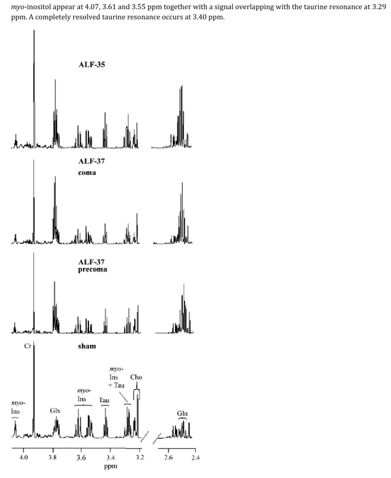
Segments of typical 1H-NMR spectra of brain extracts from a sham-operated control rat, a hepatic devascularized rat
maintained at 37 °C sacrificed at precoma stage of encephalopathy (ALF-37, precoma), and a hepatic devascularized rat maintained at 37 °C and sacrificed at coma stage of encephalopathy (ALF-37, coma), compared to a hepatic devascularized rat free from encephalopathy due to its being maintained at 35 °C (ALF-35) are shown in Fig. 1. Glutamine (Gln) resonances occur at 2.46 ppm (β-1H) together with two unresolved series of signals attributed to a
myo-inositol appear at 4.07, 3.61 and 3.55 ppm together with a signal overlapping with the taurine resonance at 3.29
ppm. A completely resolved taurine resonance occurs at 3.40 ppm.
Fig. 1. 1H-NMR spectra of brain extracts. Representative segments of 1H-NMR spectra of brain extracts from a
(ALF-hypothermia). Peak assignments: Cr: creatine; Gln: glutamine; Glx: glutamine+glutamate; myo-Ins: myo-inositol; Tau: taurine.
Mean values from 8 to 10 animals per group of concentrations of glutamine, myo-inositol and taurine are shown in Table 2. ALF under normothermic conditions (ALF-37) results in substantial increases of the Gln resonances, whereas myo-inositol and taurine signals are concomitantly decreased in the brains of these animals. While Gln remains unchanged at coma stages compared to precoma stages of encephalopathy, further decreases of myo-inositol and taurine occurs at coma stages. Mild hypothermia (ALF-35) led to normalization of the taurine resonances, and near-normalization of myo-inositol (p<0.05 compared to ALF-37), but had no effect on Gln concentrations.
Table 2. Concentrations of organic osmolytes in brain in acute liver failure: effect of mild hypothermia Sham-operated
controls
Acute liver failure normothermic (precoma stage)
Acute liver failure normothermic (coma stage)
Acute liver failure (hypothermic) n (9) (8) (9) (10) Glutamine 5.20±0.31 24.61±1.83* 23.03±1.61* 26.12±2.40* myo-Inositol 6.32±0.48 3.99±0.30* 3.66±0.30* 5.27±0.44* and † Taurine 5.17±0.31 3.91±0.35* 3.45±0.27* 5.29±0.51†
Concentrations of metabolites were calculated by integration of the respective peaks in 1H-NMR spectra (Fig. 1) of brain extracts obtained from sham-operated control rats, rats with ALF maintained at 37 °C at precoma or coma stages, and rats with ALF maintained at 35 °C (ALF-hypothermia time matched to ALF-37 coma). Values are given in μmol/g wet weight and represent means±S.D. Number of animals indicated in parenthesis.*Significantly different from controls (p<0.05), by two-way ANOVA and post hoc Tukey test). †Significantly different between hypothermic and normothermic ALF rats (p<0.05), by two-way ANOVA and post hoc Tukey test).
Changes in the sum of brain organic osmolytes are summarized in Fig. 2. The sum of organic osmolytes was increased in the brain of ALF rats at precoma stages to 200% of sham-operated controls (p<0.05), but was not increased further at coma stages in normothermic ALF rats. Mild hypothermia caused a further increase in the sum of organic osmolytes (p<0.05 compared to normothermic ALF rats at coma stages).
Fig. 2. Sum of organic osmolytes in the brain in ALF. The sum of the concentrations of organic osmolytes (glutamine+myo-inositol+taurine) was obtained from 1H-NMR spectra of brain extracts from sham-operated
controls, and rats with ALF maintained at either 37 °C (ALF-precoma and ALF-coma) or 35 °C (ALF-hypothermia). Values are given in μmol/g wet weight and represent means±S.D. (number of animals as shown in Table 1; *significantly different from sham-operated controls; †significantly different between hypothermic (ALF-35) and normothermic (ALF-37 coma) groups; by two-way ANOVA and post hoc Tukey test).
D
ISCUSSION
It is well established that hyperammonemia plays a key role in the pathogenesis of ALF. For example, arterial ammonia concentrations show a significant positive correlation with the encephalopathy in patients with ALF [8]. Furthermore, both human [28] and experimental ALF resulting from hepatectomy [11], hepatic devascularization [17], [20], [31] and [35] or toxic liver injury [26] are all associated with increased brain concentrations of glutamine, a finding which has been attributed to increased removal of ammonia by brain via the activity of glutamine
synthetase, a predominantly astrocytic enzyme. It has been proposed that the intracellular accumulation of glutamine is a major cause of cell swelling leading to brain edema in ALF [3]. In favour of this mechanism,
pretreatment of the animals with the glutamine synthetase inhibitor methionine sulfoximine (MSO) prior to hepatic desvascularization significantly diminished brain water accumulation [3]. Based upon these findings, preliminary 1H-NMR studies in patients with ALF suggest that monitoring of the α-proton of glutamine could be useful for assessment of encephalopathy in these patients [21]. However, despite several experimental data supporting glutamine as accumulating osmolyte in HE, whether or not glutamine accumulation is a major cause for the development of brain edema in ALF is uncertain Furthermore, a previous study [35] did not reveal any significant correlations between either brain glutamine concentrations or glutamine synthesis with the severity of
encephalopathy or occurrence of brain edema in experimental ALF. Results of the present study confirm and extend these negative findings. Furthermore, they demonstrate that mild hypothermia sufficient to prevent encephalopathy and brain edema in rats with ALF due to hepatic devascularization does not result in significant reductions of brain glutamine, confirming that glutamine accumulation does not play a major role in the development of brain edema in this model of ALF [6] and [35].
It has also been proposed that reductions in brain concentrations of myo-inositol, an important organic osmolyte [13], which is localized predominantly in glia [5], are implicated in the pathogenesis of ammonia-induced brain edema [9]. Indeed, both biochemical and spectroscopic studies in experimental liver failure [9] and [11] as well as in ammonia-treated cultured astrocytes [23] provide evidence for decreased brain myo-inositol concentrations concomitant with cell swelling. Results of the present study are consistent with these findings. McConnell et al. [19] reported decreased myo-inositol in brain of patients with ALF, and normalization of myo-inositol decrease was observed in the brain of a patient with ALF after clinical recovery [11]. More convincing evidence for a role of myo-inositol in the pathogenesis of brain edema in ALF is provided by results of the present study showing a significant attenuation of brain myo-inositol concentrations following the prevention of brain edema by mild hypothermia. Studies in cultured cortical astrocytes [25] as well as in vivo perfusion studies [32] suggest that taurine is released from astrocytes during regulatory volume decrease. By this means, taurine release by astrocytes into the
extracellular fluid may represent an osmoregulatory mechanism whereby the brain attempts to compensate for cell swelling in ALF [31]. Results of the present study suggest that taurine may play an important role in the
pathogenesis of both encephalopathy and brain edema in ALF, given the finding that mild hypothermia sufficient to prevent both encephalopathy and brain edema in these animals is accompanied by a complete normalization of brain taurine concentrations. However, in addition to its role in astrocytic volume regulation, taurine is also highly
concentrated in neurons [22], and is released from these cells during hypoosmotic stress [22]. Thus, the cellular origin of taurine release in brain in ALF is still unclear. In addition, ammonia-induced taurine release is not necessarily related to a cell volume regulatory response [33] and [34]. It remains to be determined whether previously reported taurine release into the extracellular space [4] or changes in cerebrospinal fluid (CSF) taurine concentrations [31] are implicated in the pathogenesis of encephalopathy or brain edema in ALF.
The lack of correlation between changes in the sum of organic osmolytes with severity of encephalopathy in ALF suggests that the decrease of brain myo-inositol and taurine may not adequately compensate for the increased intracellular osmolarity caused by glutamine accumulation. Other mechanisms which have been proposed include impaired glucose (pyruvate) oxidation leading to brain lactate accumulation [6] and [35] and cerebral hyperemia [7], [15] and [16]. For example, a significant correlation exists between brain lactate concentrations and both
encephalopathy grade [24] and [35] and EEG changes [10] in ALF. 13C-NMR studies show that mild hypothermia (35 °C), which prevents the encephalopathy and brain edema in experimental ALF, selectively normalizes brain lactate synthesis, but has no effect on glutamine synthesis. Furthermore, results of in vitro studies confirm that lactate exposure leads to astrocyte swelling [30]. A role of hyperemia in the pathogenesis of ALF, on the other hand, is supported by the measurement of hemodynamic changes in comatose patients with ALF [15], and studies by Chung et al. [7] demonstrate that indomethacin prevents brain edema, as well as the increase in cerebral blood flow and intracranial pressure in experimental ammonia-induced edema in liver failure.
Although glutamine may have only a limited role for the development of brain edema in ALF, glutamine has the potential to exert secondary effects, which may aggravate ammonia-induced impairment of mitochondrial brain energy metabolism [1]. For example, like ammonia, glutamine also induces the mitochondrial permeability transition [27], and mediates the generation of free radicals in cultured astrocytes [21].
In summary, the present study using high-resolution 1H-NMR spectroscopy demonstrates that experimental ALF results in selective changes in brain organic osmolytes as a function of the degree of encephalopathy and brain edema. Moreover, the findings of significant reductions in severity of brain edema and encephalopathy in ALF by hypothermia, together with significant attenuation of the decreases of myo-inositol and taurine, suggest a role of these osmolytes in the pathogenesis of these central nervous system complications of ALF and provide a rationale for the continued use of hypothermia in its management.
Supported by a grant from The Canadian Institutes of Heath Research. Dr. Zwingmann is a recipient of research awards from the Quebec Ministry of Education and the Deutsche Forschungsgemeinschaft, Germany.
R
EFERENCES
[1]J. AlbrechtGlucose-derived osmolytes and energy impairment in brain edema accompanying liver failure: the role of glutamine reevaluated Gastroenterology, 125 (2003), pp. 976–977
[2] J. Albrecht, M. Dolinska Glutamine as a pathogenic factor in hepatic encephalopathy J. Neurosci. Res., 65 (2001), pp. 1–5
[3] A.T. Blei, S. Olafsson, G. Therrien, R.F. Butterworth Ammonia-induced brain edema and intracranial hypertension in rats after portacaval anastomosis Hepatology, 19 (1994), pp. 1437–1444
[4] D.K. Bosman, N.E. Deutz, A.A. De Graaf, R.W. vd Hulst, H.M. Van Eijk, W.M. Bovee, M.A. Maas et al. Changes in brain metabolism during hyperammonemia and acute liver failure: results of a comparative 1H-NMR spectroscopy and
biochemical investigation Hepatology, 12 (1990), pp. 281–290
[5] A. Brand, C. Richter-Landsberg, D. Leibfritz Multinuclear NMR studies on the energy metabolism of glial and neuronal cells Dev. Neurosci., 15 (1993), pp. 289–298
[6] N. Chatauret, C. Zwingmann, C. Rose, D. Leibfritz, R.F. Butterworth Effects of hypothermia on brain glucose metabolism in acute liver failure: a H/C-nuclear magnetic resonance study Gastroenterology, 125 (2003), pp. 815– 824
[7] C. Chung, J. Gottstein, A.T. Blei Indomethacin prevents the development of experimental ammonia-induced brain edema in rats after portacaval anastomosis Hepatology, 34 (2001), pp. 249–254
[8] O. Clemmesen Splanchnic circulation and metabolism in patients with acute liver failure Dan. Med. Bull., 49 (2002), pp. 177–193
[9]J. Cordoba, J. Gottstein, A.T. Blei Glutamine, myo-inositol, and organic brain osmolytes after portocaval anastomosis in the rat: implications for ammonia-induced brain edema Hepatology, 24 (1996), pp. 919–923 [10] N.E.P. Deutz, A.A. De Graaf, J.G. De Haan, W.M.M.J. Bovée, R.A.F.M. Chamuleau In: vivo brain 1H-NMR
spectroscopy (1-NMRS) during acute hepatic encephalopathy (HE) P.B. Soeters, J.H.P. Wilson, A.J. Meijer, E. Holm
(Eds.), Advances in Ammonia Metabolism and Hepatic Encephalopathy, Excerpta Media, Amsterdam (1988), pp. 439–446 Chap. 57
[11] R.K. Gupta, V.A. Saraswat, H. Poptani, R.K. Dhiman, A. Kohli, R.B. Gujral, S.R. Naik Magnetic resonance imaging and localized in vivo proton spectroscopy in patients with fulminant hepatic failure Am. J. Gastroenterol., 88 (1993), pp. 574–670
[12] T. Holmin, C.D. Agardh, G. Alinder, P. Herlin, B. Hultberg The influence of total hepatectomy on cerebral energy state, ammonia-related amino acids of the brain and plasma amino acids in the rat Eur. J. Clin. Investig., 13 (1983), pp. 215–220
[13]R.E. Isaacks, A.S. Bender, C.Y. Kim, N.M. Prieto, M.D. Norenberg Osmotic regulation of myo-inositol uptake inprimary astrocyte cultures Neurochem. Res., 19 (1994), pp. 331–338
[14] R. Jalan, S.W. Damink, N.E. Deutz, A. Lee, P.C. Hayes Moderate hypothermia for uncontrolled intracranial hypertension in acute liver failure Lancet, 354 (1999), pp. 1164–1168
[15] F.S. Larsen, B.A. Hansen, L.G. Jorgensen, N.H. Secher, S. Bondesen, P. Linkis, A. Hjortrup, P. Kirkegaard, N. Agerlin, J. Kondrup et al. Cerebral blood flow velocity during high volume plasmapheresis in fulminant hepatic failure Int. J. Artif. Organs, 17 (1994), pp. 353–361
[16] F.S. Larsen, J. Gottstein, A.T. Blei Cerebral hyperemia and nitric oxide synthase in rats with ammonia-induced brain edema J. Hepatol., 34 (2001), pp. 548–554
[17] A.M. Mans, M.R. DeJoseph, R.A. Hawkins Metabolic abnormalities and grade of encephalopathy in acute hepatic failure J. Neurochem., 63 (1994), pp. 1829–1938
[18] A. Marmarou, W. Poll, H. Bhagavan A simple gravimetric technique for measurement of cerebral edema J. Neurosurg., 49 (1978), pp. 530–537
[19] J.R. McConnell, D.L. Antonson, C.S. Ong, W.K. Chu, I.J. Fox, T.G. Heffron, A.N. Langnas, B.W. Shaw Jr. Proton spectroscopy of brain glutamine in acute liver failure Hepatology, 22 (1995), pp. 69–74
[20] A. Michalak, C. Rose, J. Butterworth, R.F. Butterworth Neuroactive amino acids and glutamate (NMDA) receptors in frontal cortex of rats with experimental acute liver failureHepatology, 24 (1996), pp. 908–913
[21] C.R. Murthy, K.V. Rama Rao, G. Bai, M.D. Norenberg Ammonia-induced production of free radicals in primary cultures of rat astrocytes J. Neurosci. Res., 66 (2001), pp. 282–288
[22] E.A. Nagelhus, A. Lehmann, O.P. Ottersen Neuronal–glial exchange of taurine during hypo-osmotic stress: a combined immunocytochemical and biochemical analysis in rat cerebellar cortex Neuroscience, 54 (1993), pp. 615– 631
[23] M.D. Norenberg, A.S. Bender Astrocyte swelling in liver failure: role of glutamine and benzodiazepines Acta Neurochir., Suppl. (Wien), 60 (1994), pp. 24–27
[24] S.L. Nyberg, F.B. Cerra, R. Gruetter Brain lactate by magnetic resonance spectroscopy during fulminant hepatic failure in the dog Liver Transplant. Surg., 4 (1998), pp. 158–165
[25] H. Pasantes-Morales, O. Quesada, J. Moran Taurine: an osmolyte in mammalian tissues Adv. Exp. Med. Biol., 442 (1998), pp. 209–217
[26] J. Peeling, L. Shoemaker, T. Gauthier, A. Benarroch, G.R. Sutherland, G.Y. Minuk Cerebral metabolic and histological effects of thioacetamide-induced liver failure Am. J. Physiol., 265 (1993), pp. G572–G578
[27] K.V. Rama Rao, A.R. Jayakumar, M.D. Norenberg Induction of the mitochondrial permeability transition in cultured astrocytes by glutamine Neurochem. Int., 43 (2003), pp. 517–523
[28] C.O. Record, B. Buxton, R.A. Chase, G. Curzon, I.M. Murray-Lyon, R. Williams Plasma and brain amino acids in fulminant hepatic failure and their relationship to hepatic encephalopathy Eur. J. Clin. Investig., 6 (1976), pp. 387– 394
[29] C. Rose, A. Michalak, M. Pannunzio, N. Chatauret, A. Rambaldi, R.F. Butterworth Mild hypothermia delays the onset of coma and prevents brain edema and extracellular brain glutamate accumulation in rats with acute liver failure Hepatology, 31 (2000), pp. 872–877
[30] F. Staub, A. Baethmann, J. Peters, H. Weigt, O. Kempski Effects of lactacidosis on glial cell volume and viability J. Cereb. Blood Flow Metab., 10 (1990), pp. 866–876
[31] M. Swain, R.F. Butterworth, A.T. Blei Ammonia and related amino acids in the pathogenesis of brain edema in acute ischemic liver failure in rats Hepatology, 15 (1992), pp. 449–453
[32] J.V. Wade, J.P. Olson, F.E. Samson, S.R. Nelson, T.L. Pazdernik A possible role for taurine in osmoregulation within the brain J. Neurochem., 51 (1988), pp. 740–745
[33] C.E. Wright, H.H. Tallan, Y.Y. Lin, G.E. Gaull Taurine: biological update Ann. Rev. Biochem., 55 (1986), pp. 427– 453
[34] M. Zielinska, W. Hilgier, R.O. Law, P. Gorynski, J. Albrecht Effects of ammonia in vitro on endogenous taurine efflux and cell volume in rat cerebrocortical minislices: influence of inhibitors of volume-sensitive amino acid transportNeuroscience, 91 (1999), pp. 631–638
[35] C. Zwingmann, N. Chatauret, D. Leibfritz, R.F. Butterworth Selective increase of brain lactate synthesis in experimental acute liver failure: results of a [1H–13C] nuclear magnetic resonance study Hepatology, 37 (2003), pp.
420–428