11
POSTEMBRYONIC DEVELOPMENT OF THE CEPHALIC
SKELETON IN DICENTRARCHUS LABRAX (PISCES,
PERCIFORMES, SERRANIDAE)
Isabelle Gluckmann, Françoise Huriaux,
Bruno Focant and Pierre Vandewalle
ABSTRACT
At hatching, Dicentrarchus labrax larvae are 3.0 mm long and devoid of any cephalic skeleton. At 3.6 mm, the Meckelian cartilage appears, after which the whole skeleton develops so slowly and gradually that clear-cut stages are impossible to define. Some cephalic elements, however, develop faster than others. Skeletal development is subject to constraints imposed by vital functions such as aquatic respiration and feeding. As the yolk sac shrinks, the branchial parts develop. By the time the vitellus is completely ex-hausted, the mandible, pharyngeal jaws, hyoid bar, and parts of the suspensorium and operculum are present. Though still incomplete, these structures are probably sufficient to allow ingestion of exogenous food. Further development should enable the larvaes to perform suction feeding, as is typical of perciforms. Before the shift to exogenous feed-ing, the cartilaginous floor of the skull remains open, but the opening is then closed by the parasphenoid and basioccipital, so the brain is completely isolated from the buccal cavity. The cranial vault and ethmoid region develop later: these structures are probably less essential to fry survival than the earlier and more rapidly developing structures. Throughout their postembryonic development, i.e., until the appearance of the first adult characters (Krupka, 1988; Haylor, 1992), fish must be equipped to survive, notably at certain critical times such as hatching and the shift to exogenous feeding. Upon hatch-ing, the embryo comes in contact with the environment. It feeds on the nutrients con-tained in the yolk sac. The latter also performs a respiratory function, being highly vascularised. Within a few days of hatching, development must enable the larvae to breathe in and collect food from its environment. To perform these functions, the larvae must at least have developed an effective means of locomotion, a system for capturing and digest-ing prey, a new respiratory system, and nervous and sensory systems.
The prey-capture and respiratory systems are located in the head and consist notably of skeletal structures and muscle. The present work concerns the post-embryonic develop-ment of Dicentrarchus labrax, a teleost species of the order Perciforms.
Investigators have long shown interest in the post-embryonic development of the head region (i.e., Stöhr, 1882; Tischomiroff, 1885; Winslow, 1897; Swinnerton, 1902; Gaupp, 1903). Not all of them, however, have pursued the same goals. Many studies have focused solely on external development: recent examples include Able et al. (1986), Munehara and Mishima (1986), Nishikawa (1987), Fukuhara (1988), Krupka (1988), Baldwin et al. (1991), Kovac (1993,1994), Ditty et al. (1994). Others have dealt solely with the osteoc-ranium (Morris and Gaudin, 1982; Jollie, 1984; Potthoff et al., 1988; Vandewalle et al., 1995) or only with the chondrocranium (Wells, 1923; Bhargava, 1958; Bertmar, 1959). Sometimes the focus has been the whole cephalic skeleton (Kindred, 1919; Bamford, 1948; Elman and Balon, 1980; Vandewalle et al., 1992, 1997), sometimes it has been only certain cranial elements (Haines, 1937; Devillers, 1944; Corsin, 1961; Francillon, 1974; Arratia and Schultze, 1990; Potthoff and Tellock, 1993), occasionally with emphasis on
12 BULLETIN OF MARINE SCIENCE, VOL. 65, NO. 1, 1999
functional aspects (Verraes, 1977). Several authors have described only a few develop-mental stages (Srinivasachar, 1959; Howes and Sanford, 1987; Surlemont and Vandewalle, 1991; Kobayakawa, 1992). A few, finally, have tried to get a synthetic or general picture (De Beer, 1937; Daget, 1964).
Few studies devoted to the bony and cartilaginous skeleton provide a basis for accu-rately timing the events that mark a teleost’s post-embryonic development. Yet knowledge of this timing is needed in order to understand the evolution of respiration and feeding. On a chronological basis alone it is possible to establish many correlations between the development of structures and vital requirements. This chronology is based upon the size of larvae instead of time (i.e., days after hatching). This is the first aim of the present study. The second is to increase our currently very fragmentary knowledge concerning the development of the cephalic skeleton in perciforms.
The nomenclature used here to designate developing stages and skeletal structures is based principally on the work of De Beer (1937), Daget (1964), Patterson (1977) and Kendall et al. (1984).
MATERIALAND METHODS
Dicentrarchus labrax specimens were from the hatcheries of the “Ecloserie marine Sepia
Exploi-tation” located at the site of the Graveline nuclear reactors (France). The larvae were raised in water ranging in temperature from 14.3°C (at hatching) to 20.6°C (latest stage). Twelve batches of 50 larvae were sampled from hatching (3.0-mm fry) to day 18 post-hatching (8.1-mm fry) and five batches of 30 larvae from day 22 (8.3 mm) to day 34 (11.2 mm). The larvae were fixed in buffered 10% formalin and trypsin-cleared. Some were stained with alcyan blue to reveal the cartilages, others with alizarin to reveal the bones. The staining techniques were derived from those of Dingerkus and Uhler (1977), Potthoff (1984) and Taylor and Van Dyke (1985). The stained larvae were then observed under a Wild M5 binocular magnifier. The drawings representing the cartilaginous or bony structures were made using a camera lucida mounted on the magnifier. Each stage is repre-sented by a lateral view, a ventral view, and when necessary, a dorsal view.
Although our presentation of results begins at hatching, we in fact had to work backward to establish homologies and recognise structures. We compared the structures of adult perciforms (Liem, 1970; Vandewalle, 1972; Benmouna et al., 1984) with those of the oldest larvae and pro-ceeded by successive comparisons from the oldest larvae to the youngest.
RESULTS
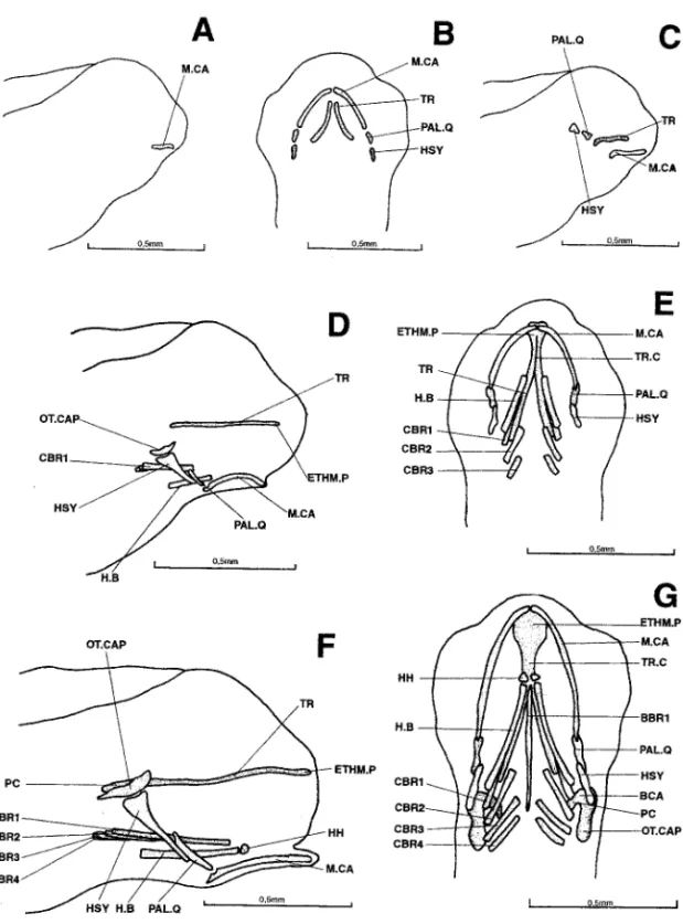
3.0 mm (hatching).—No cephalic skeletal structure is visible.
3.6 mm (Fig. 1A).—The mandibular arch is represented by Meckel’s cartilages. 4.0 mm (Fig. 1B,C).—Several cartilaginous elements have appeared: the trabeculae,
first parts of the neurocranium, and parts of the palato-quadrates, added to the mandibu-lar arch where Meckel’s cartilages have considerably developed. The hyoid arch is repre-sented by the hyosymplectics.
4.3 mm (Fig. 1D,E).—The trabecular bars have lengthened posteriorly and fused
ante-riorly to constitute the beginnings of a trabecula communis and ethmoid plate. A pair of lateral cartilages has appeared, probably the first signs of the latero-dorsal walls of the otic capsules. Below these cartilages, the hyosymplectics are already well developed, particularly broad in their upper parts and touching the palato-quadrates with their lower
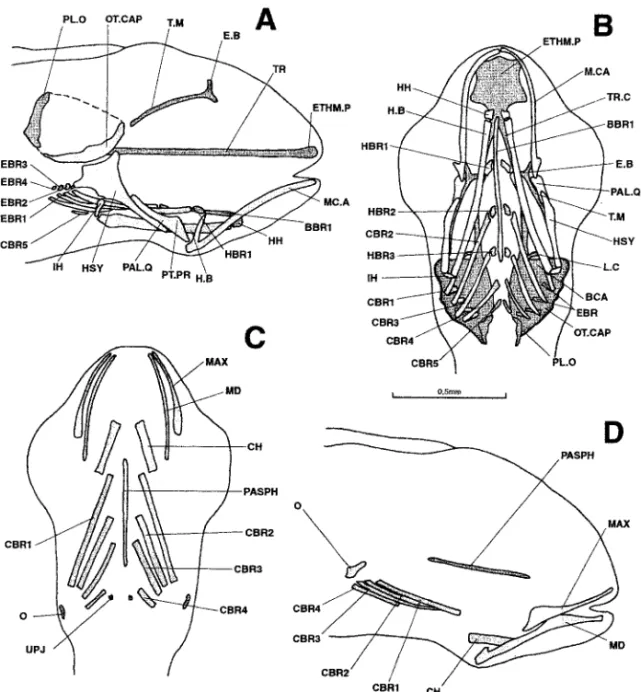
Figure 1. Dicentrarchus labrax : Lateral view (a) of the chondrocranium in a 3.6-mm larvae; ventral (b) and lateral (c) views of the chondrocranium in a 4.0-mm larvae; lateral (d) and ventral (e) views of the chondrocranium in a 4.3-mm larvae; lateral (f) and ventral (g) views of the chondrocranium in a 4.8-mm larvae. BBR 1: basibranchial 1; BCA: commissura bicapsularis anterior; CBR 1-4: ceratobranchial 1-4; ETHM.P: ethmoid plate; H.B: hyoid bar; HH: hypohyal; HSY: hyosymplectic: M.CA: Meckel’s cartilage; OT.CAP: otic capsule; PAL.Q: palato-quadrate: PC: parachordal plate; TR: trabecular bar; TR.C: trabecula communis.
14 BULLETIN OF MARINE SCIENCE, VOL. 65, NO. 1, 1999
tips. The palato-quadrates point towards the rear portions of Meckel’s cartilages, which exhibit a cavity already shaped like a joint socket. Meckel’s cartilages, markedly curved, touch each other anteriorly. Additions to the splanchnocranium include two already well-developed hyoid bars and the first three pairs of ceratobranchials.
4.8 mm (Fig. 1F,G).—To the front of the neurocranium, the ethmoid plate has
broad-ened. To the rear, the parachordal plates are in place. They are connected to the latero-dorsal walls of the otic capsules, probably by the almost-horizontal commissurae bicapsulares anteriores.
A pair of hypohyals has appeared in front of the hyoid bars. The branchial basket now possesses a fourth pair of ceratobranchials and a long basibranchial bar. Meckel’s cartilages are straightening.
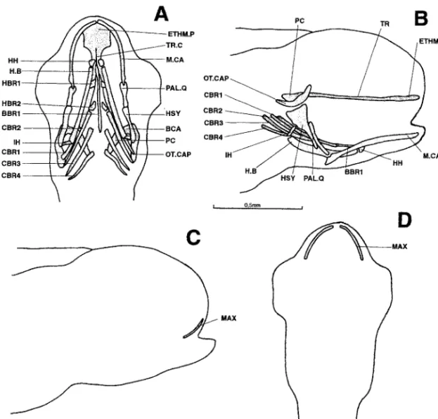
5.2 mm (Fig. 2).—The trabecular bars have lengthened posteriorly but remain separate
from the parachordal plates.
Figure 2. Dicentrarchus labrax : ventral (a) and lateral (b) views of the chondrocranium and lateral (c) and ventral (d) views of the osteocranium in a 5.2-mm larvae. BBR 1: basibranchial 1; BCA: commissura bicapsularis anterior; CBR 1-4: ceratobranchial 1-4; ETHM.P: ethmoid plate; H.B: hyoid bar; HBR 1-2: hypobranchial 1-2; HH: hypohyal; HSY: hyosymplectic; IH: interhyal; MAX: maxillary; M.CA: Meckel’s cartilage; OT.CAP: otic capsule; PAL.Q: palato-quadrate; PC: parachordal plate; TR: trabecular bar; TR.C: trabecula communis.
The hyoid bars are connected to the hyosymplectics by a pair of independent interhyals. The branchial basket now possesses its first two pairs of hypobranchials. Meckel’s cartilages are long and almost straight.
The maxillaries are present on the edge of the upper lip in the form of two small, bony rods.
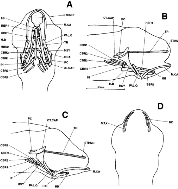
5.3 mm (Fig. 3A,B).—The trabecular bars and parachordal plates are united. Anteriorly
and laterally they limit the hypophyseal fenestra, which remains open in the rear. A third pair of hypobranchials has appeared in the branchial basket. The maxillaries are still the only discernible ossifications.
5.7 mm (Fig. 3C,D).—No new neurocranial element has appeared, but the existing
components have enlarged.
Figure 3. Dicentrarchus labrax : Ventral (a) and lateral (b) views of the chondrocranium in a 5.3-mm larvae; lateral view (c) of the chondrocranium and ventral view (d) of the osteocranium in a 5.7-mm larvae. BBR 1: basibranchial 1; BCA: commissura bicapsularis anterior; CBR 1-4: ceratobranchial 1-4; ETHM.P: ethmoid plate; H.B: hyoid bar; HBR 1-3: hypobranchial 1-3; HH: hypohyal; HSY: hyosymplectic; IH: interhyal; MAX: maxillary; M.CA: Meckel’s cartilage; MD: mandible; OT.CAP: otic capsule; PAL.Q: palato-quadrate; PC: parachordal plate; TR: trabecular bar; TR.C: trabecula communis.
16 BULLETIN OF MARINE SCIENCE, VOL. 65, NO. 1, 1999
A pointed processus dorsalis has appeared behind Meckel’s cartilages.
A second pair of ossifications has developed, constituting the dentaries. The maxillaries have enlarged posteriorly.
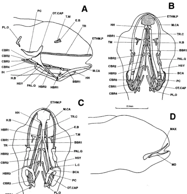
6.3 mm (Fig. 4).—Dorsally, the neurocranium now includes the primordia of the
ante-rior portions of the taeniae marginales and epiphyseal bridge. On each side, a small carti-laginous column has appeared at the posterior tip of the trabecular bar, linking the junc-tion between the trabecula and parachordal plate with the front of the otic capsule. These columns are probably the commissurae laterales. Finally, the pilae occipitales are present at the rear of the braincase; they are already almost in contact with the parachordal plates. The composition of the splanchnocranium is unchanged. The two ossifications previ-ously present have developed further.
Figure 4. Dicentrarchus labrax: Lateral (a), ventral (b), and dorsal (c) views of the chondrocranium and lateral (d) view of the osteocranium in a 6.3-mm larvae. BBR : basibranchial 1 : basibranchial 1; BCA: commissura bicapsularis anterior; CBR 1-4 : ceratobranchial 1-4; E.B: epiphyseal bridge; ETHM.P: ethmoid plate; H.B: hyoid bar; HBR 1-3: hypobranchial 1-3; HH: hypohyal; HSY: hyosymplectic; IH: interhyal; MAX: maxillary; M.CA: Meckel’s cartilage; MD: mandible; OT.CAP: otic capsule; PAL.Q: palato-quadrate; TR: trabecular bar; TR.C: trabecula communis.
6.7 mm (Fig. 5).—The taeniae marginales have extended towards the otic capsules and
the pilae orbitales are practically attached to the parachordal plates. The floor of each otic capsule has broadened, but without closing the hypophyseal fenestra to the rear. The lat-eral walls of these capsules are forming but their outlines are not clear.
The palato-quadrates bear a “budding” pterygoid process. To the branchial basket have been added the fifth pair of ceratobranchials and four pairs of epibranchials.
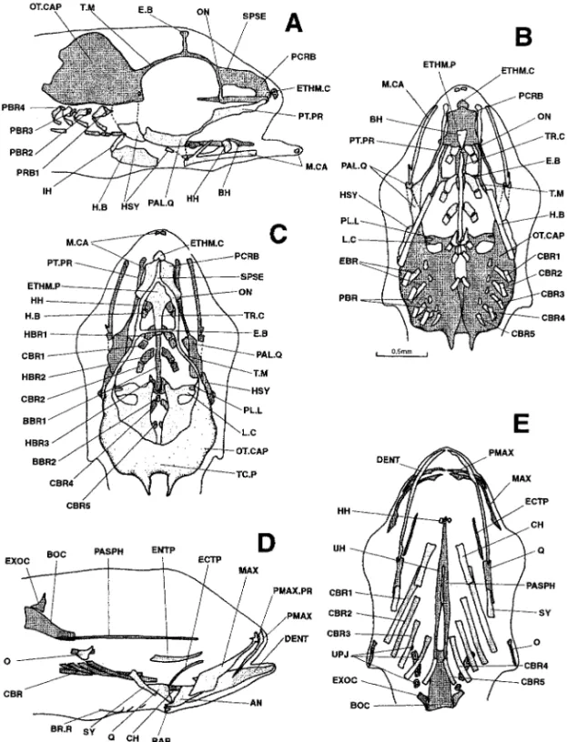
Figure 5. Dicentrarchus labrax: lateral (a) and ventral (b) views of the chondrocranium and ventral (c) and lateral (d) views of the osteocranium in a 6.7-mm larvae. Interrupted lines indicate the probable limit of the otic capsule. The buccal teeth are not represented in Figs. c and d. BBR : basibranchial; BCA: commissura bicapsularis anterior ; CBR 1-5: ceratobranchial 1-5; CH: ceratohyal; EBR: epibranchial; E.B: epiphyseal bridge; ETHM.P: ethmoid plate; H.B: hyoid bar; HBR 1-3: hypobranchial 1-3; HH: hypohyal; HSY: hyosymplectic; IH: interhyal; L.C: commissura lateralis; MAX: maxillary; M.CA: Meckel’s cartilage; MD: mandible; O: opercular; OT.CAP: otic capsule; PAL.Q: palato-quadrate; PASPH: parasphenoid; PL.O: pila occipitalis; PT.PR: pterygoid process; T.M: taenia marginalis; TR: trabecular bar; TR.C: trabecula communis; UPJ: upper pharyngeal jaw.
18 BULLETIN OF MARINE SCIENCE, VOL. 65, NO. 1, 1999
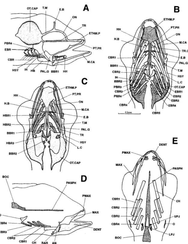
Figure 6. Dicentrarchus labrax : Lateral (a), ventral (b), and dorsal (c) views of the chondrocranium and lateral (d) and ventral (e) views of the osteocranium in a 7.6-mm larvae. The dotted lines indicate a regressing cartilaginous region. The buccal teeth are not represented in Figs. d and e. AN: angular; BBR 1-2: basibranchial 1-2; BOC: basioccipital; CBR 1-5: ceratobranchial 1-5; CH: ceratohyal; DENT: dentary; EBR 1-4: epibranchial 1-4; E.B: epiphyseal bridge; ETHM.P: ethmoid plate; H.B: hyoid bar; HBR 1-3: hypobranchial 1-3; HH: hypohyal; HSY: hyosymplectic; IH: interhyal; L.C: commissura lateralis; LPJ: lower pharyngeal jaw; MAX: maxillary; M.CA: Meckel’s cartilage; O: opercular; ON: lamina orbitonasalis; OT.CAP: otic capsule; PAL.Q: palato-quadrate; PASPH: parasphenoid; PBR 3-4: pharyngobranchial 3-4; PMAX: premaxillary; PT.PR: pterygoid process; RAR: retroarticular; T.M: taenia marginalis; TR: trabecular bar; TR.C: trabecula communis; UPJ: upper pharyngeal jaw.
The parasphenoid, first ossification of the neurocranium, has appeared in the middle of the hypophyseal fenestra. The bony splanchnocranium displays the operculars, ceratohyals, four pairs of well-developed ceratobranchials, and a pair of small dorsal toothed plates. The latter are apparently located above the fifth pair of cartilaginous ceratobranchials, i.e., at the position corresponding to the toothed plates of the upper pharyngeal jaws. The composition of the latter is hard to determine at this stage.
7.6 mm (Fig. 6).—On one hand, the cartilaginous skull continues to develop. The left
and right parts of the braincase have broadened ventrally, laterally, and dorsally without actually joining. The taeniae marginales are in contact posteriorly with the otic capsules; anteriorly, they are extended by the laminae orbitonasales (perhaps connected to the pre-orbital roots). The laminae orbitonasales are attached to the latero-posterior tips of the ethmoid plate. The pilae occipitales are fused with the otic capsules and the epiphyseal bridge is complete. The branchial basket now displays two posterior pairs of pharyngobranchials and a small basibranchial. Lastly, the pterygoid processes have length-ened and now reach the front of the ethmoid plate.
On the other hand, certain cartilages have begun to regress. The otic capsules have separated from the trabecular bars. Meckel’s cartilages are also resorbing, isolating two small, anterior cartilaginous masses.
The osteocranium now possesses a basioccipital and small premaxillaries; the lower jaw consists of distinct dentaries, angulars, and retroarticulars. The parasphenoid has broad-ened and bears two posterior points surrounding the basioccipital. The pharyngeal jaws, formed by the 5th ceratobranchials bearing toothed plates, lie opposite a pair of upper jaws whose composition is still hard to determine.
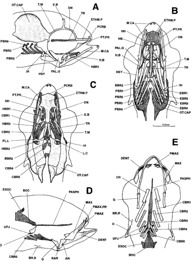
8.1 mm (Fig. 7).—At this stage the chondrocranium displays the first signs of a lamina
precerebralis and four pairs of pharyngobranchials. Two small protuberances appear on the posterior rims of the commissurae laterales: they constitute the first signs of the pilae laterales (see 8.9-mm larvae).
Cartilage regression continues with that of the hyoid bars, which have begun to split up. The occipital region of the neurocranium displays dorsally a pair of small ossified exoccipitals, one to each side of the basioccipital. The upper pharyngeal jaws are repre-sented by three pairs of toothed plates which probably correspond with ossification of the three posterior pairs of pharyngobranchials.
Ossification of each quadrate has begun at the joint with the mandible. The premaxillaries curve upward to constitute the first signs of the processus ascendentes. Like the lower jaw, they are toothless or sometimes bear a single tooth. The first pair of branchiostegal rays has appeared.
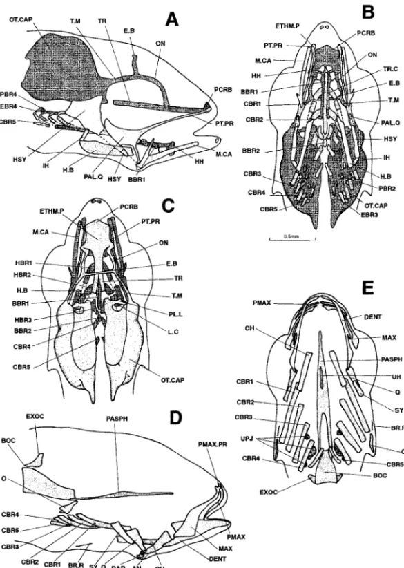
8.3 mm (Fig. 8).—No new element appears in the chondrocranium, which continues to
regress. Ventrally, the ethmoid and otic regions have completely separated. A major part of the central region of each ceratobranchial is resorbing, isolating the two cartilaginous extremities. The same applies to the hyosymplectics, where a hyomandibular and a symplectic region are distinguishable. The regions where the palato-quadrates articulate with the lower jaw are also disappearing.
The osteocranium now displays symplectics and an increased number of buccal teeth (4–5 per half-jaw). The parasphenoid is in contact with the basioccipital. A long urohyal has appeared between the ceratohyals.
8.9-mm (Fig. 9).—The cartilaginous braincase is beginning to close dorsally with the
para-20 BULLETIN OF MARINE SCIENCE, VOL. 65, NO. 1, 1999
Figure 7. Dicentrarchus labrax: Lateral (a), ventral (b), and dorsal (c) views of the chondrocranium and lateral (d) and ventral (e) views of the osteocranium in an 8.1-mm larvae. The dotted lines indicate a regressing cartilaginous region. The buccal teeth are not represented in Figs. d and e. AN: angular; BBR 1-2: basibranchial 1-2; BOC: basioccipital; BR.R: branchiostegal ray; CBR 1-5: ceratobranchial 1-5; CH: ceratohyal; DENT: dentary; EBR 1-4: epibranchial 1-4; E.B: epiphyseal bridge; ETHM.P: ethmoid plate; EXOC: exoccipital; H.B: hyoid bar; HBR 1-3: hypobranchial 1-3; HH: hypohyal; HSY: hyosymplectic; IH: interhyal; L.C: commissura lateralis; MAX: maxillary; M.CA: Meckel’s cartilage; O: opercular; ON: lamina orbitonasalis; OT.CAP: otic capsule; PAL.Q: palato-quadrate; PASPH: parasphenoid; PBR 1-4: pharyngobranchial 1-4; PCRB: lamina precerebralis; PL.L: pila lateralis; PMAX: premaxillary; PMAX.PR: premaxillary process; PT.PR: pterygoid process; Q: quadrate; RAR: retroarticular; T.M: taenia marginalis; TR: trabecular bar; TR.C: trabecula communis; UPJ: upper pharyngeal jaw.
Figure 8. Dicentrarchus labrax : Lateral (a), ventral (b), and dorsal (c) views of the chondrocranium and lateral (d) and ventral (e) views of the osteocranium in an 8.3-mm larvae. The dotted lines indicate a regressing cartilaginous region. The buccal teeth are not represented in Figs. d and e. AN: angular; BBR 1-2: basibranchial 1-2; BOC: basioccipital; CBR 1-5: ceratobranchial 1-5; CH: ceratohyal; DENT: dentary; EBR 1-4: epibranchial 1-4; E.B: epiphyseal bridge; ETHM.P: ethmoid plate; EXOC: exoccipital; H.B: hyoid bar; HBR 1-3: hypobranchial 1-3; HH: hypohyal; HSY: hyosymplectic; IH: interhyal; L.C: commissura lateralis; MAX: maxillary; M.CA: Meckel’s cartilage; O: opercular; ON: lamina orbitonasalis; OT.CAP: otic capsule; PAL.Q: palato-quadrate; PASPH: parasphenoid; PBR 1-4: pharyngobranchial 1-4; PCRB: lamina precerebralis; PL.L: pila lateralis; PMAX: premaxillary; PMAX.PR: premaxillary process; PT.PR: pterygoid process; Q: quadrate; RAR: retroarticular; T.M: taenia marginalis; TR: trabecular bar; TR.C: trabecula communis; UH: urohyal; UPJ: upper pharyngeal jaw.
22 BULLETIN OF MARINE SCIENCE, VOL. 65, NO. 1, 1999
Figure 9. Dicentrarchus labrax: Lateral (a), ventral (b), and dorsal (c) views of the chondrocranium and lateral (d) and ventral (e) views of the osteocranium in an 8.9-mm larvae. The dotted lines indicate a regressing cartilaginous region. The buccal teeth are not represented in Figs. d and e. AN: angular; BBR 1-2 : basibranchial 1-2; BH: basihyal; BOC: basioccipital; CBR 1-5 : ceratobranchial 1-5; CH: ceratohyal; DENT: dentary; EBR 1-4: epibranchial 1-4; ECTP: ectopterygoid; ENTP: entopterygoid; E.B: epiphyseal bridge; ETHM.C: ethmoid cartilage; ETHM.P: ethmoid plate; EXOC: exoccipital; H.B: hyoid bar 1-3: hyoid bar; HBR 1-3: hypobranchial 1-3; HH: hypohyal; HSY: hyosymplectic; IH: interhyal; L.C: commissura lateralis; MAX: maxillary; M.CA: Meckel’s cartilage; O: opercular; ON: lamina orbitonasalis; OT.CAP: otic capsule; PAL.Q: palato-quadrate; PASPH: parasphenoid; PBR: pharyngobranchial; PCRB: lamina precerebralis; PL.L: pila lateralis; PMAX: premaxillary; PMAX.PR: premaxillary process; PT.PR: pterygoid process; Q: quadrate; RAR: retroarticular; SPSE: commissura sphenoseptalis; SY: symplectic; TC.P: tectum posterius; T.M: taenia marginalis; TR: trabecular bar; TR.C: trabecula communis; UH: urohyal; UPJ: upper pharyngeal jaw.
chordal plates have come closer together but do not yet form a basal plate. The protuber-ances on the commissurae laterales have continued to develop, attaching to the bases of the taeniae marginales to form the pilae laterales. The lamina precerebralis has developed upwardly and to the rear and is connected to the laminae orbitonasales and taeniae marginales via the commissurae sphenoseptales. A new cartilaginous element, single and small, has appeared in front of the ethmoid region: this is the cartilaginous pad or eth-moid cartilage, situated under the processus ascendentes of the premaxillaries in Acanthopterygii. A basihyal is also present.
The trabecular bars are now no more than the posterior tips of the ethmoid plate. The upper parts of the hyomandibulars are regressing. So are the palato-quadrates, which form an isolated cartilaginous island at the site of the joint with Meckel’s cartilage. The central portion of each cartilaginous ceratobranchial has totally disappeared, leaving a very scattered-looking branchial basket.
The bony splanchnocranium now includes pairs of ecto- and entopterygoids and two small hypohyals. There are two additional pairs of branchiostegal rays and the number of buccal teeth has increased. The median pharyngobranchial toothed plates have length-ened.
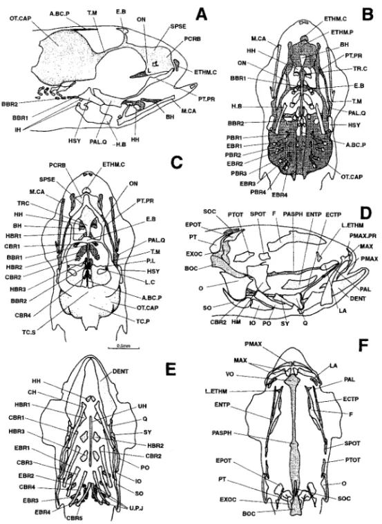
9.9 mm (Fig. 10).—The cartilaginous neurocranium is continuing to close dorsally,
from rear to front, with the formation of the tectum synoticum. A thin processus anteromedialis appears at this site, pointing towards the epiphyseal bridge. The lamina precerebralis, commissurae sphenoseptales, and laminae orbitonasales constitute a whole forming the dorso-medial part of the ethmoid region. This whole is pierced by the fo-ramina for the olfactory nerves. In the splanchnocranium, the first pair of hypobranchials and the interhyals have begun to resorb centrally.
Most characteristic of this stage is the abundance of new bony parts in the neurocra-nium. In front there are a vomer, a pair of lateral ethmoids, and a pair of lacrymals. In the orbital region, the frontals have appeared. The otic region includes sphenotics, pterotics, and epiotics. Finally, the occipital region now displays a supraoccipital and a pair of posttemporals.
The bony splanchnocranium now includes the palatines, the interhyals, the preoperculars, the inter- and suboperculars, three pairs of hypobranchials, and all four pairs of epibranchials. The first, second, and third pairs of ceratobranchials bear gillrakers. The eight pairs of branchiostegal rays that characterise the adult are well developed.
11.2 mm (Fig. 11).—The cartilaginous braincase still has no basal plate. Dorsally, a
taenia tecta medialis is developing from the epiphyseal bridge towards the rear, in the direction of the anterior process of the braincase.
All the bony parts previously present have enlarged. This is especially true of the pterotics, supraoccipital, preoperculars, maxillaries, and premaxillaries. The pterotics consist of two parts; the anterior part bears a portion of the bony tube of the pterotic sensory canal. The parasphenoid exhibits the lateral wings which in the adult limit the trigemino-facial chamber.
The ethmoid region now displays a pair of nasals and a mesethmoid, pleurosphenoids are present in the orbital region, a pair of parietals reinforces the otico-occipital region, and the epihyals have finally appeared. The first palatine and vomerine teeth are present.
24 BULLETIN OF MARINE SCIENCE, VOL. 65, NO. 1, 1999
Figure 10. Dicentrarchus labrax : Lateral (a), ventral (b), and dorsal (c) views of the chondrocranium (d), lateral view of the osteocranium (d), ventral view (e) of the bony splanchnocranium and part of the neurocranium, and dorsal view (f) of the neurocranium in a 9.9-mm larvae. The dotted lines indicate a regressing cartilaginous region. The branchiostegal rays, gillrakers, and buccal teeth are not represented in Figs. d, e, and f. A.BC.P: anterior process of the braincase; BBR 1-2: basibranchial 1-2; BH: basihyal; BOC: basioccipital; CBR 1-5: ceratobranchial 1-5; CH: ceratohyal; DENT: dentary; EBR 1-4: epibranchial 1-4; ECTP: ectopterygoid; ENTP: entopterygoid; E.B: epiphyseal bridge; EPOT: epiotic: ETHM.C: ethmoid cartilage; ETHM.P: ethmoid plate; EXOC: exoccipital; F: frontal; H.B: hyoid bar; HBR 1-3: hypobranchial 1-3; HH: hypohyal; HM: hyomandibular; HSY: hyosymplectic; IH: interhyal; IO: infraopercular: LA: lacrymal; L.C: commissura lateralis; L.ETHM: lateral ethmoid; MAX: maxillary; M.CA: Meckel’s cartilage; O: opercular; ON: lamina orbitonasalis; OT.CAP: otic capsule; PAL: palatine; PAL.Q: palato-quadrate; PASPH: parasphenoid; PBR 1-4: pharyngobranchial 1-4; PCRB: lamina precerebralis; PL.L: pila lateralis; PL.O: pila occipitalis; PMAX: premaxillary; PMAX.PR: premaxillary process; PO: preopercular; PT: posttemporal; PTOT: pterygoid; PT.PR: pterygoid process; Q: quadrate; RAR: retroarticular; SO: subopercular; SOC: supraoccipital; SPSE: commissura sphenoseptalis; SPOT: sphenotic; SY: symplectic; TC.P: tectum posterius; TC.S: tectum synoticum; T.M: taenia marginalis; TR: trabecular bar; TR.C: trabecula communis; UH: urohyal; UPJ: upper pharyngeal jaw.
Figure 11. Dicentrarchus labrax: Lateral (a) and dorsal (b) views of the chondrocranium, lateral (c) view of the osteocranium, ventral view (d) of the bony splanchnocranium and part of the neurocranium, and dorsal view (e) of the bony neurocranium in an 11.2-mm larvae. The dotted lines indicate a cartilaginous region that is regressing. The branchiostegal rays, gillrakers, and buccal teeth are not represented on Figs. d, e, and f. A.BC.P: anterior process of the braincase; AN: angular; BBR 1-2: basibranchial 1-2; BH: basihyal; BOC: basioccipital; CBR 1-5 : ceratobranchial 1-5; CH: ceratohyal; DENT: dentary; EBR 1-4: epibranchial 1-4; ECTP: ectopterygoid; ENTP: entopterygoid; E.B: epiphyseal bridge; EPOT: epiotic; ETHM.C: ethmoid cartilage; ETHM.P: ethmoid plate; EXOC: exoccipital; F: frontal; H.B: hyoid bar; HBR 1-3: hypobranchial 1-3; HH: hypohyal; HM: hyomandibular; HSY: hyosymplectic; IH: interhyal; IO: infraopercular; LA: lacrymal; L.C: commissura lateralis; L.ETHM: lateral ethmoid; MAX: maxillary; M.CA: Meckel’s cartilage; METHM: mesethmoid; NA: nasal; O: opercular; ON: lamina orbitonasalis; OT.CAP: otic capsule; PA: parietal; PAL: palatine; PAL.Q: palato-quadrate; PASPH: parasphenoid; PBR: pharyngobranchial; PCRB: lamina precerebralis; PL.L: pila lateralis; PLSPH: pleurosphenoid; PMAX: premaxillary; PMAX.PR: premaxillary process; PO: preopercular; PT: posttemporal; PTOT: pterygoid; PT.PR: pterygoid process; Q: quadrate; RAR: retroarticular; SO: subopercular; SOC: supraoccipital; S.PTOT: dermopterotic; SPSE: commissura sphenoseptalis; SPOT: sphenotic; SY: symplectic; TC.P: tectum posterius; TC.S: tectum synoticum; T.M: taenia marginalis; TR: trabecular bar; T.TM: tecta taenia medialis; TC.C: trabecula communis; UH: urohyal; UPJ: upper pharyngeal jaw.
26 BULLETIN OF MARINE SCIENCE, VOL. 65, NO. 1, 1999 DISCUSSION
D. labrax is a species that usually develops at a moderately high temperature. Species
reared at higher temperatures develop faster (Pelosi et al., 1993; Regner and Dulcic, 1994; Vandewalle et al., 1997).
The order in which the different structures appear is presented in Table 1.
Chondrocranium.—In teleosts, the chondrocranium may begin to develop just before
hatching, as in Salmo trutta, be partially present at hatching, as in Heteropneustes fossilis and Barbus barbus, or not form until later as in the Siluriforms Clarias gariepinus,
Heterobranchus longifilis, the Cypriniforms Leuciscus rutilus, Catostomus commersonii, Danio rerio, the Lophiiforms Lophius gastrophysus or the Perciforms Lutjanus campechanus, Bette splendens, Anisotremus davidsonii and Xenistius californiensis (De
Beer, 1937; Hubendick, 1942; Srinivasachar, 1959; Elman and Balon, 1980; Matsuura and Yomeda, 1987; Potthof et al., 1988; Surlemont and Vandewalle, 1991; Watson and Walker, 1992; Vandewalle et al., 1992, 1997; Cubbage and Mabee, 1996; Mabee and Trendler, 1996). D. labrax falls into the third category, since the first structures begin to appear in 3.6 mm larvae (a day after hatching).
Usually, the structures of the mandibular and hyoid arches are the first cranial elements to appear with the Meckel’s cartilage followed by the palato-quadrates, the hyosymplectics, and the hyoid bars in D. labrax. This timing is very similar to that observed in other perciforms (Potthoff et al., 1988; Watson and Walker, 1992; Potthoff and Tellock, 1993; Cubbage and Mabee, 1996). In most teleosts, these elements of the splanchnocranium are independent (Hubendick, 1942; Elman and Balon, 1980; Badenhorst, 1989b; Vandewalle et al., 1992). In most siluriforms, on the other hand, all or most of them are fused together (Kindred, 1919; Srinivasachar, 1959; Surlemont et al., 1989; Surlemont and Vandewalle, 1991; Vandewalle et al., 1997). The latter situation also arises in Clupea harengus (Wells, 1923). In Cypriniforms as L. rutilus and C. commersonii, the first structures to appear are the trabeculae and parachordal plates forming the base of neurocranium (Hubendick, 1942; Elman and Balon, 1980).
In most teleosts, the elements of the branchial basket are the next to complement the first structures of the splanchnocranium. The ceratobranchials develop first, generally from front to rear as in H. fossilis and C. gariepinus (Srinivasachar, 1959; Surlemont and Vandewalle, 1991), or else the first four develop simultaneously, followed by the fifth, as in B. barbus, H. longifilis and Engraulis japonicus (Balart, 1985; Vandewalle et al., 1992, 1997). Sometimes, all five pairs of ceratobranchials seem to appear at once, as in
Gasterosteus aculeatus, L. rutilus, C. commersonii, A. davidsonii, and X. californiensis
(Swinnerton, 1902; Hubendick, 1942; Elman and Balon, 1980; Watson and Walker, 1992). In D. labrax, the first ceratobranchial appears at the same time as the hyoid bar and the fifth does not appear until later, along with the epibranchials but after the hypobranchials. Then f inally, in both D. labrax and B. barbus (Vandewalle et al., 1992), the pharyngobranchials appear from rear to front. In other species, the first epibranchial may appear before the rear ceratobranchials (Srinivasachar, 1959). Both pairs of pharyngobranchials appear at 7.6 mm in D. Labrax. They likewise do not appear until development is quite advanced in H. fossilis, C. batrachus, and H. longifilis (Srinivasachar, 1959; Vandewalle et al., 1997).
n i s e r u t c u r t s l a t e l e k s s u o i r a v e h t f o e c n a r a e p p a e h t f o g n i m i T . 1 e l b a T Dicentrarchuslabrax. s e g a t S ) m m ( s e g a l i t r a C Ossifications g n i h c t a H noskeletalstructureisvisible 6 . 3 appear:Meckel’scartilage 0 . 4 appear:trabecularbars,palato-quadrates, s c i t c e l p m y s o y h 3 . 4 appear:basesofthedorso-lateralwalls , s i n u m m o c a l u c e b a r t , s e l u s p a c c i t o e h t f o , e t a l p d i o m h t e , s r a b d i o y h 3 -2 -1 s l a i h c n a r b o t a r e c 8 . 4 appear:parachordalplates,commissurae , s l a y h o p y h , s e r o i r e t n a s e r a l u s p a c i b 1 l a i h c n a r b i s a b , 4 s l a i h c n a r b o t a r e c 2 . 5 appear:interhyals,hypobranchials1-2 maxillaries 3 . 5 appear:hypobranchials3 7 . 5 appear:processusmediodorsalesof s e g a l i t r a c s ’ l e k c e M w a j r e w o l 3 . 6 appear:commissuraelaterales,pilae e g d i r b l a e s y h p i p e e h t f o s e s a b , s e l a t i p i c c o s e l a n i g r a m e a i n e a t d n a * 7 . 6 appear:firstsignsofthepterygoid , 5 s l a i h c n a r b o t a r e c , s e s s e c o r p 4 -3 -2 -1 s l a i h c n a r b i p e s l a i h c n a r b o t a r e c , s r a l u c r e p o , d i o n e h p s a r a p -r e p p u f o r i a p t s r i f , s l a y h o t a r e c , 4 -3 -2 -1 s e t a l p d e h t o o t w a j -l a e g n y r a h p * * 6 . 7 appear:completelateralwallsoftheotic l a t i b r o e r p ( s e l a s a n o t i b r o e a n i m a l , s e l u s p a c l a e s y h p i p e , ) s e l a n i g r a m e a i n e a t e h t f o s t o o r , 4 -3 s l a i h c n a r b o g n y r a h p , e g d i r b ; 2 l a i h c n a r b i s a b r a l u c e b a r t e h t f o s t r a p r o i r e t s o p : s s e r g e r s e g a l i t r a c s ’ l e k c e M f o s t r a p l a r t n e c , s r a b , s r a l u g n a , s e i r a l l i x a m e r p , l a t i p i c c o i s a b r e w o l , s r a l u c i t r a o r t e r , s e i r a t n e d s w a j l a e g n y r a h p 1 . 8 appear:laminaprecerebralis, ; 2 -1 s l a i h c n a r b o g n y r a h p ; s e l a r e t a l e a l i p e h t f o s g n i n n i g e b s r a b d i o y h e h t f o s t r a p l a r t n e c : s s e r g e r , 5 s l a i h c n a r b o t a r e c , s e t a r d a u q , s l a t i p i c c o x e d e h t o o t w a j -l a e g n y r a h p -r e p p u f o s r i a p 2 , s y a r l a g e t s o i h c n a r b f o r i a p t s 1 , s e t a l p h c a e n o s n e d n e c s a s u s s e c o r p s e i r a l l i x a m e r p e h t n o h t e e t , y r a l l i x a m e r p s e i r a t n e d d n a 3 . 8 regress:centralpartsofceratobranchials d n a r a l u b i d n a m o y h e h t n e e w t e b , 4 -3 -2 -1 e h t e r e h w s n o i g e r , s n o i g e r c i t c e l p m y s s ’ l e k c e M h t i w e t a l u c i t r a s e t a r d a u q s e g a l i t r a c l a y h o r u , s c i t c e l p m y s 9 . 8 appear:tectumposterius,pilaelaterales , l a y h i s a b , s e l a t p e s o n e h p s e a r u s s i m m o c e g a l i t r a c d i o m h t e r a l u b i d n a m o y h e h t f o s t r a p r e p p u : s s e r g e r s n o i g e r , s l a y h o p y h , s d i o g y r e t p o t n e , s d i o g y r e t p o t c e s y a r l a g e t s o i h c n a r f o s r i a p l a n o i t i d d a 2 9 . 9 appear:tectumsynoticum,extensionof e s a c n i a r b e h t , 1 s l a i h c n a r b o p y h f o s t r a p l a r t n e c : s s e r g e r s l a y h r e t n i e h t f o l a r e t a l , r e m o v , s l a r o p m e t t s o p , l a t i p i c c o a r p u s , s c i t o n e h p s , s l a m y r c a l , s l a t n o r f , s d i o m h t e , s r a l u b i d n a m o y h , s c i t o i p e , s c i t o r e t p , 3 -2 -1 s l a i h c n a r b o p y h , s l a y h r e t n i , s e n i t a l a p , s r a l u c r e p o b u s , 4 -3 -2 -1 s l a i h c n a r b i p e n o s r e k a r l l i g , s r a l u c r e p o e r p , s r a l u c r e p o r e t n i f o s r i a p 5 , 3 -2 -1 s l a i h c n a r b o t a r e c s y a r l a g e t s o i h c n a r b 2 . 1 1 appear:taeniatectamedialis s l a y h r e t n i e h t f o s t r a p l a r t n e c : s s e r g e r , s d i o n e h p s o r u e l p , s d i o m h t e s e m , s l a s a n e n i r e m o v d n a e n i t a l a p , s l a y h i p e , s l a t e i r a p . h t e e t e b u t e v i t s e g i d e h t n i d o o f f o e c n e s e r p d n a c a s k l o y e h t f o n o i t c u d e r d e k r a m y r e v * c a s k l o y e h t f o e c n a r a e p p a s i d * *
28 BULLETIN OF MARINE SCIENCE, VOL. 65, NO. 1, 1999
The appearance of the trabecular bars as the first structures of the neurocranium floor, before the rest of the neurocranium, appears as a general rule in teleosts. They are fol-lowed by the parachordal plates.
As development continues, the order in which the elements of the cartilaginous neuro-cranium appear and the rate at which they develop are variable. In Anguilla vulgaris, the ethmoid and orbital regions develop before the otic capsules (De Beer, 1937), while in some Siluriforms, the braincase floor is the first to develop, followed by the anterior portion (Srinivasachar, 1959; Surlemont et al., 1991; Vandewalle et al., 1997). In D. labrax, and in more Perciforms and B. barbus, both parts develop more or less simultaneously (Vandewalle et al., 1992). In Perciforms, notably D. labrax, the braincase is not isolated from the splanchnocranium by formation of a basal plate in front of the notochord. In Siluriforms, the basal plate develops early in the chondrocranium, by fusion of the ante-rior parts of the parachordal plates (Bamford, 1948; Srinivasachar, 1959; Surlemont and Vandewalle, 1991; Vandewalle et al., 1996). The basal plate appears late in Cypriniforms and Ameirus nebulosus (Kindred, 1919; Badenhorst, 1989a; Vandewalle et al., 1992; Cubbage and Mabee, 1996).
In its general outline, the structure of the chondrocranium may appear fairly constant, but variations do exist. In D. labrax, there appears an independent cartilage, also present in others perciforms, at the front of the ethmoid region (in the adult, this special part, called the ethmoid cartilage, plays a role in enabling the premaxillary to move with re-spect to the neurocranium). These parts appear to be absent in the Perciforms L.
campechanus, C. undecimalis, and B. splendens (Potthoff et al., 1988; Potthoff and Tellock,
1993; Mabee and Trendler, 1996). In the anterior region, the shape of the ethmoid plate is variable. In D. labrax and B. barbus, it is formed by anterior fusion of the two trabecular bars (Vandewalle et al., 1992). It then develops into an actual platform as in A. nebulosus,
L. rutilus, N. aequoreus, and C. commersonii (Kindred, 1919; Hubendick, 1942; Kadam,
1961; Elman and Balon, 1980). In H. longifilis, the ethmoid plate remains narrow and appears merely as the base of the lamina cerebralis (Vandewalle et al. 1997).
The taeniae marginales and epiphyseal bridge also appear to develop differently in different species. In the Siluriforms H. fossilis and H. longifilis, the Cypriniforms B.
barbus and Merluccius capensis, the taeniae marginales develop very early, from the
anterodorsal parts of the otic capsules (Srinivasachar, 1959; Badenhorst, 1989a; Vandewalle et al., 1992, 1997; Cubbage and Mabee, 1996). They then extend forward to form the base of the epiphyseal bridge. In D. labrax, parts of the epiphyseal bridge and taeniae marginales appear dorsally, isolated in the middle of the orbit. Each taenia marginalis then extends toward the rear to join with the front of the otic capsule. This mode of development is also seen others Perciformes. H. fossilis displays a peculiarity at this level: the taeniae marginales are not linked dorsally by formation of the epiphyseal bridge (Srinivasachar, 1959). The lamina precerebralis, taeniae marginales, and trabecular bars are connected via the lami-nae orbitonasales, apparently directly linked to the preorbital roots of the taeniae marginales in both D. labrax and B. barbus (Vandewalle et al., 1992). In H. longifilis, on the other hand, the preorbital roots develop later, independently of the laminae orbitonasales (Vandewalle et al., 1997).
In D. labrax, the latero-ventral sphenoid fenestrae are each divided in three by the pila lateralis and commisura lateralis; the two latter structures do not appear in H. fossilis, C.
gariepinus, and H. longifilis (Srinivasachar 1959; Surlemont and Vandewalle 1991;
In D. labrax , a tectum synoticum completes the braincase vault in front of the tectum posterius, and a taenia tecta medialis is formed by mediodorsal extensions issuing poste-riorly from the cranial vault and anteposte-riorly from the epiphyseal bridge. A similar situation is seen in others Perciformes (Watson and Walker, 1992). In M. capensis and B. barbus, a taenia tecta medialis also develops, but it seems to issue only from the epiphyseal bridge (Badenhorst, 1989a; Vandewalle et al., 1992). In H. longifilis there appears no primor-dium of a taenia tecta medialis (Vandewalle et al., 1997). In D. labrax, the hypophyseal fenestra is narrow and open posteriorly. This contrasts with the situation in B. barbus and
H. longifilis, where it is broad, long, and limited by the parachordal plates and trabecular
bars (Vandewalle et al., 1992; Vandewalle et al., 1997). D. labrax thus displays a tropitrabic skull.
Regression of the cartilaginous parts exists in many teleosts, but it begins at different developmental sizes in different species (Wells, 1923; De Beer, 1937; Vandewalle et al., 1995, 1997). In D. labrax, the phenomenon starts when all parts of the chondroneurocranium are not yet present and the splanchnocranium lacks at least the basihyal. The posterior part of the trabecular bars regresses first, ventrally isolating, very rapidly, the anterior and posterior parts of the neurocranium as in M.capensis and B.
barbus (Badenhorst, 1989a; Vandewalle et al., 1992). In H. longifilis, regression starts
when the chondrocranium is practically complete, beginning with the central parts of the ceratobranchials and hyoid bars (Vandewalle et al., 1997).
Meckel’s cartilages regress differently according to the species studied. In D. labrax, they split in two, one part being small and isolated in front. In H. longifilis, Meckel’s cartilage splits into three parts, one of which is again situated at the front end (Vandewalle et al., 1997). Isolation of a cartilaginous element at the front of the mandible may shed some doubt as to the purely dermal origin of the dentary bones. These might have ab-sorbed the small, anterior cartilaginous elements and thus have a dual origin, like the palatines, for instance, which each include an auto- and a dermo-palatine. In B. barbus, Meckel’s cartilage remains whole beyond the larval stage (Vandewalle et al., 1992).
Regression of the neurocranium, which begins very early in D. labrax with the separa-tion of the trabecular bars from the parachordal plates, does not progress any further during the larval stage in this species or B. barbus (Vandewalle et al., 1992). In H. longifilis, on the other hand, regression of the neurocranium simultaneously affects the otic cap-sules, trabecular bars, and parachordal plates (Vandewalle et al., 1997).
Osteocranium.—The development of the bony skull varies considerably among
teleo-sts (De Beer, 1937; Bamford, 1948; Jollie, 1984; Matsuura and Yomeda, 1987).
At hatching, there is usually no ossified element. D. labrax follows this rule. G. felis and P. antarticum appears as an exception, since at hatching they already possess opercu-lars, dentaries, and premaxillaries (Tilney and Hecht, 1993; Voskoboinikova, 1994).
The dermal splanchnocranium appears to ossify first. The operculars and the parts forming the buccal jaws (maxillary, premaxillary and dentary) develop earliest, followed by the pharyngeal jaws or their toothed plates. In D. labrax the maxillaries and premaxillaries are superposed at the outset, as in the others perciformes (Otten, 1982; Kohno et al., 1983; Potthoff et al., 1988; Oozeki et al., 1992; Watson and Walker, 1992; Potthoff and Tellock, 1993, Voskoboinikova, 1994; Mabee and Trendler, 1996) as well as
L. gastrophysus and B. barbus (Matsuura and Yomeda, 1987; Vandewalle et al., 1992), all
which species have a protractile mouth at the adult stage. There is thus no transient, “primi-tive” state in which the premaxillaries and maxillaries are adjacent. This situation does
30 BULLETIN OF MARINE SCIENCE, VOL. 65, NO. 1, 1999
arise in E. lucius and O. kisutch (Jollie, 1975, 1984). In D. rerio, The first structures to appear are those of neurocranium (parasphénoïde, exoccipital and basioccipital) (Cubbage and Mabbee, 1996).
Next there appear the first deep ossifications of the splanchnocranium, which progres-sively replace the existing cartilaginous skeleton. In D. labrax, the quadrates appear first, followed by the symplectics. The hyomandibulars and palatines appear later, after the dermal bones of the suspensorium, i.e., the entopterygoids and ectopterygoids. In L.
gastrophysus, the palatines and quadrates come first, the hyomandibulars appearing at
the next stage (Matsuura and Yomeda, 1987). B. barbus also shows delayed development of the hyomandibular (Vandewalle et al., 1992). In N. aequoreus, the quadrates appear at the same time as the first dermal structures of the splanchnocranium (Kadam, 1961). In
E. lucius the hyomandibulars, symplectics, and quadrates appear simultaneously but
some-what later than the other structures, notably the ectopterygoids (Jollie, 1975).
The ectopterygoids and entopterygoids are usually considered to be dermal ossifica-tions and the metapterygoid to be an endochondral one (Devillers, 1958; Daget, 1964). Watson (1987), surprisingly, describes the metapterygoid as the only dermal bone of the suspensorium in Enchelyurus brunneolus, although this bone is usually considered endo-chondral and although Watson and Walker (1992) report a cartilaginous stage for the ectopterygoid and entopterygoid in two Perciformes Haemulidae species. Matsuoka (1987) believes that in Sparidae, the quadrato-pterygoido-palatine complex derives from two cartilaginous primordia, while Potthoff and Tellock (1993) report that only the ectopterygoid is dermal in C. undecimalis. If we compare, in D. labrax, the positions of the cartilaginous and bony elements, only the ectopterygoid might be considered to have an endochondral origin, while the entopterygoid is clearly dermal.
Ossification of the hyoid system begins, in all known cases, with the center of the hyoid bar, which becomes the ceratohyal. Next to appear are the two hypohyals, before or along with the posterior ossification of the hyoid bar or epihyal. The branchiostegal rays appear gradually, always from rear to front; the first ray is present well before the first ossifica-tion of the hyoid bar (Potthoff et al., 1988; Surlemont and Vandewalle, 1991; Watson and Walker, 1992; Vandewalle et al., 1992; Potthoff and Tellock, 1993; Vandewalle et al., 1995). In some cases there appears a third anterior ossification of the hyoid bar (Tilney and Hecht, 1993). In other cases, only a hypohyal seems to ossify (Tilney and Hecht, 1993; Vandewalle et al., 1995, 1997; Cubbage and Mabee, 1996). Finally, the cartilagi-nous interhyal may in some cases never ossify; it may even disappear completely (Adriaens and Verraes, 1994). In D. labrax, the only peculiarity is the absence of an interhyal ossifi-cation at 40 d; it will appear later, since adult serranids possess an interhyal bone (Benmouna et al., 1984).
Ossification of the branchial basket is somewhat variable among teleosts. In several perciformes, all five ceratobranchials appear together, along with the first pharyngeal toothed plates; then come the epibranchials, pharyngobranchials, and hypobranchials. Finally, the axes of the basibranchials also ossify. The gillrakers appear first on the first bony ceratobranchial, then towards the rear on the epibranchials (Potthoff et al., 1988; Watson and Walker, 1992; Potthoff and Tellock, 1993; Mabee and trendler, 1996). In D.
labrax, the timing is somewhat different: the first four ceratobranchials ossify as the
up-per toothed plates appear, a little before the bony tooth-bearing fifth ceratobranchials and before the rest of the branchial basket. In the Ostariophysi whose development is known in this respect, the pharyngeal teeth and jaws always appear well before the other bony
branchial elements (Surlemont and Vandewalle, 1991; Vandewalle et al., 1992, 1995, 1997; Cubbage and Mabee, 1996). In all cases, the pharyngeal tooth-bearing elements appear at the same time as the buccal teeth, or nearly so.
Development of the neurocranium begins rather early, almost always with the appear-ance of the parasphenoid just after the first dermal structures of the splanchnocranium. This bone’s growth seems related to the shape of the hypophyseal fenestra, which it gradu-ally closes. The parasphenoid remains narrow in D. labrax. It rapidly broadens in O.
kisutch, B. barbus, A. davidsonii and in Siluriformes (Jollie, 1984; Surlemont and
Vandewalle, 1991; Vandewalle et al., 1992; Watson and Walker, 1992; Tilney and Hecht, 1993; Vandewalle et al., 1995, 1997; Cubbage and Mabee, 1996).
The second braincase bone to appear very early in development is the basioccipital. In
B. barbus, it even appears just before the parasphenoid, and in X. californiensis, both
bones appear simultaneously (Vandewalle et al., 1992; Watson and Walker, 1992). Shortly after the basioccipital, the exoccipitals appear in D. labrax , B. barbus, and C. auratus (Vandewalle et al., 1992, 1995). In N. aequoreus (Kadam, 1961) and E. lucius (Jollie, 1975), the exoccipitals and basioccipital appear simultaneously, but later in development. In O. kisutch and D. rerio, the exoccipitals develop along with the parasphenoid, after the first dermal parts of the splanchnocranium (Jollie, 1984; Cubbage and Mabee, 1996). In
H. longifilis , the first step in bony development is the simultaneous appearance of the
parasphenoid, basioccipital, and exoccipitals (Vandewalle et al., 1997).
The neurocranium then continues to develop in D. labrax, but more slowly than the splanchnocranium. The usual pattern is for the braincase vault to develop later, with the appearance of the supraoccipital, posttemporals, and epiotics, together with the pterotics and sphenotics. Then the front of the neurocranium, notably the frontals, is formed. The order in which these parts appear differs according to the species. In A. davidsonii, the frontal appears first, followed by the pterotic; in X. californiensis, the opposite occurs (Watson and Walker, 1992). In three Silurus species studied, B. barbus, and C. auratus, the supraoccipitals, pterotics, and sphenotics appear at the same time as the frontals (Kobayakawa, 1992; Vandewalle et al., 1992,1995). In H. longifilis , the frontals develop first, then the rear of the neurocranium (Vandewalle et al., 1997). In O. kisutch and G.
felis , the posttemporals and frontals appear at the same time as the parasphenoid (Jollie,
1984; Tilney and Hecht, 1993), while in E. lucius, they appear just after it (Jollie, 1975). The bony cephalic skeleton then continues its development with the appearance of the front parts of the neurocranium and further development of the parts already present.
It is easy to see, by comparing alcyan-blue- and alizarin-stained individuals at the same developmental stage, that the deeper skeletal ossifications appear where cartilage regres-sion is to occur. Ossification begins before regresregres-sion. For the splanchnocranium, this correspondence is seen with the ceratobranchials, the ceratohyals, and parts of the sus-pensorium. The dentary is well formed by the time Meckel’s cartilage begins to regress. According to Huyssene (1990), this regression occurs only at the level of the tooth-bear-ing parts. Construction of the cartilaginous neurocranium is still under way and only the trabecular bars show signs of regression. But in this particular case, no bony structure of endochondral origin is yet in place. The parasphenoid, early-appearing dermal bone, is ventral with respect to such structures.
General consideration.—In teleosts, the different parts of the skull develop at
32 BULLETIN OF MARINE SCIENCE, VOL. 65, NO. 1, 1999
Whatever the variations among teleosts, development of the skull must always meet the survival requirements of the larvae. In D. labrax hatchings, respiration is cutaneous and the highly vascularised yolk sac plays an important role in this process. Feeding, more-over, is endogenous. No skeletal structure is a priori necessary to support these two es-sential functions. The yolk sac resorbs slowly in D. labrax , in accordance with the gradual appearance of the skeletal structures. As the sac shrinks, the branchial system develops from front to rear, with the appearance of the first four cartilaginous ceratobranchials, the main bearers of respiratory filaments in the adult. The fifth ceratobranchials, having no respiratory function, form later and participate in feeding. Once the yolk sac has disap-peared, cutaneous respiration is probably much reduced, but by then the branchial basket also possesses epibranchials (Table 1). Respiration in an aquatic medium requires the creation of a water current from front to rear (Hughes and Shelton, 1958; Ballintijn, 1969; Osse, 1990). This is possible in D. labrax , since 7.6-mm larvae possess a suspensorium articulating with the neurocranium, a hyoid bar hanging from the hyosymplectic, and the beginnings of an operculum. The very gradual ossification of the branchial basket sug-gests that cartilaginous structures can adequately carry out respiration.
Total resorption of the vitellus makes it necessary for the fry to switch from endog-enous to exogendog-enous feeding. This means the buccal cavity must be able to take up food from the external environment. First of all, this cavity must be limited, to avoid damage to other parts of the head such as the brain. In 7.6-mm larvae, the cartilaginous braincase is not yet closed ventrally, but the presence of the parasphenoid and basioccipital bones complements it and makes it rigid, dorsally limiting the buccal cavity. This should reduce the mechanical relations between the buccal cavity and the brain. Next, the mandible and the strong tooth-bearing pharyngeal jaws should be able to ensure food processing. A protractile mouth typical of perciforms (Liem, 1970, 1979; Lauder and Liem, 1981) does not develop until the larvae reach 8.1 mm, with the formation of a ascending process on the premaxillary. To be functional, such a mouth should also possess an ethmoid carti-lage, indispensable to enable the premaxillary to slide along the neurocranium. A suspen-sorium (even cartilaginous), a hyoid bar, and an opercular are probably sufficient both for aquatic respiration and to constitute the beginnings of a suction feeding typical of evolved fish (Muller and Osse, 1984; Lauder, 1985), but one would expect the efficiency of this system to be far from optimal. This suggests that exogenous feeding probably begins at 7.6 mm with prey seizing and that efficient suction feeding appears gradually, with ossi-fication of the endochondral structures and the appearance of all the bony elements of the suspensorium and operculum.
Yet other functional constraints govern construction of the cephalic skeleton. Late clo-sure of the cranial vault probably reflects the need for the brain to grow. On the other hand, the development of certain cephalic structures such as the ethmoid region might not be subject to such crucial or obvious constraints, and their late appearance should not compromise fry survival.
Differences between teleost species in the development of the cephalic skeleton may reflect different survival requirements. Yet as observed by Vandewalle et al. (1997), one general rule does appear to emerge: the parasphenoid, the basioccipital, and the first der-mal ossifications of the splanchnocranium appear at the time the yolk sac disappears or just after. All species must indeed solve the same survival problem linked with the switch from endogenous to exogenous feeding.
ACKNOWLEDGEMENTS
The authors wish to thank Sepia International (Saint Quentin en Yvelines, France) for rearing and selecting the specimens studied and K. Broman for translating the text into English. This work was funded by grant no. 2.9006.90 of the Fonds de la Recherche Fondamentale Collective (Bel-gium). B. F. is Chercheur Qualifié of the Fonds National de la Recherche Scientifique (Bel(Bel-gium).
LITERATURE CITED
Able, K. W., M. P. Fahal and D. F. Markle. 1986. Development of larval snailfishes (Pisces, Cyclopteridae, Liparidinae) from the western north Atlantic. Can. J. Zool. 64: 2294–2316. Adriaens, D. and W. Verraes. 1994. On the functional significance of the loss of the interhyal during
ontogeny in Clarias gariepinus Burchell, 1822 (Teleostei: Siluroidei). Belg. J. Zool. 124: 139– 155.
Arratia, G. and H. P. Schultze. 1990. The urohyal development and homology within osteichthyans. J. Morph. 203: 247–282.
Badenhorst, A. 1989a. Development of the chondrocranium of the shallow-water cape hake
Merluccius capensis (Cost.), part 1: neurocranium. S. Afr. J. Zool. 24: 33–48.
____________. 1989b. Development of the chondrocranium of the shallow-water cape hake
Merluccius capensis (Cost.), Part 2: viscerocranium. S. Afr. J. Zool. 24: 49–57.
Baldwin, C. C., G. D. Johnson and P. L. Colin. 1991. Larvae of Diploprion bifasciatum, Belonoperca
chabanaudi and Grammistes sexlineatus (Serranidae: Epinephelinae) with a comparison of known
larvae of other epinephelines. Bull. Mar. Sci. 48: 67–93.
Ballintijn, C. M. 1969. Functional anatomy and movements co-ordination of the respiratory pump of the carp (Cyprinus carpio L.). J. Exp. Biol. 50: 547–567.
Bamford, T. W. 1948. Cranial development of Galeichthys felis. Proc. Zool. Soc. Lond. 118: 364– 391.
Benmouna, H., I. Trabert, P. Vandewalle and M. Chardon. 1984. Comparaison morphologique du neorocrâne et du splanchnocrâne de Serranus scriba (Linné 1758) et de Serranus cabrilla (Linné 1758), (Pisces, Serranidae). Cybium 8: 71–93.
Bertmar, G. 1959. On the ontogeny of the chondral skull in Characidae, with a discussion on the chondrocranial base and visceral chondrocranium in fishes. Acta Zool. 40. Stockholm. 162 p. Bhargava, H. N. 1958. The development of the chondrocranium of Mastacembelus armatus (Cuv.
Et Val.). J. Morph. 102: 401–426.
Corsin, J. 1961. Etude de quelques corrélations morphogénétiques dans le développement du chondrocrâne de Salmo. Bull. Soc. Zool. de France 86: 772–785.
Cubbage, C. C. and P. M. Mabee. 1996. Development of the cranium and paired fins in the zebrafish
Danio rerio (Ostariophysi, Cyprinidae). J. Morph. 229: 121–160.
Daget, J. 1964. Le crâne des téléostéens. Mém. Mus. Natn. Hist Nat., Sér.A, 31: 167–340. De Beer, G. R. 1937. The development of the vertebrate skull. Oxford: Clarendon Press., London. Devillers, C. 1944. Morphogenèse de quelques os crâniens chez la truite arc-en-ciel, Salmo irideus
(Gibb.). Ann. Sc. Nat., Zoo. et Biol. animales, Série II 6: 25–31.
Dingerkus, G. and L. D. Uhler. 1977. Enzyme clearing of alcian blue stained whole small verte-brates for demonstration of cartilage. Stain Technology 52: 229–232.
Ditty, J. G., E. D. Houde and R. F. Shaw. 1994. Egg and larval development of spanish sardine,
Sardinella aurita (family Clupeidae), with a synopsis of characters to identify clupeid larvae
from the nothern gulf of Mexico. Bull. Mar. Sci. 54: 367–380.
Elman, J. F. and E. K. Balon. 1980. Early ontogeny of white sucker Catostomus commersonii, with steps of saltatory development. Env. Biol. Fish 5: 191–224.
Francillon, H. 1974. Développement de la partie postérieure de la mandibule de Salmo trutta fario L. (Pisces, Teleostei, Salmonidae). Zool. Scripta 3: 41–51.
34 BULLETIN OF MARINE SCIENCE, VOL. 65, NO. 1, 1999
Fukuhara, O. 1988. Morphological and functionnal development of the larvae and juvenile Limanda
yokohamae (Pisces Pleuronectidae) reared in the laboratory. Mar. Biol.. 99: 271–281.
Gaupp, E. 1903. Zür entwicklung der schädelknocken bei den teleostiern. Verhand. anat. gesellschaft: 113–123.
Haines, R.W. 1937. The posterior end of Meckel’s cartilage and related ossifications in bony fishes. Quart. J. Microsc. Sci. 80: 1–38.
Haylor, G. S. 1992. Terminology for the early developmental stages of the African catfish, Clarias
gariepinus (Burchell): working definitions for aquaculture. Aquacult. Fish. Manage. 23: 511–
514.
Howes, G. J. and C. P. J. Sanford. 1987. Oral ontogeny of the ayu, Plecoglossus altivelis, and com-parisons with other salmoniform fishes. Zool. J. Linn. Soc. 89: 133–169.
Hubendick, B. 1942. Zür kenntnis der entwicklung des primordialcraniums bei Leuciscus rutilus. Ark. Zool. 34A: 1-35.
Hughes, G. M. and G. Shelton. 1958. Pressure changes during the respiratory movements of teleo-stean fishes. Nature 179: 255.
Huyssene, A. 1990. Development of the anterior part of the mandible and the mandibular dentition in two species of Cichlidae (Teleostei). Cybium 14: 327–344.
Jollie, M. 1975. Development of the head skeleton and pectoral girdle in Esox. J. Morph. 147: 61– 88.
_______. 1984. Development of the head skeleton and pectoral girdle of salmons, with a note on the scales. Can. J. Zool. 62: 1757–1778.
Kadam, K. M. 1961. The development of the skull in Nerophis (Lophobranchii). Acta Zool., Stockholm 42: 1–42.
Kendall, A. W., E. H. Ahlstrom and H. G. Moser. 1984. Early life history stages of fishes and their characters. Pages 11-22 in H. G. Moser, W. J. Richards, D. M. Cohen, M. P. Fahay, A. W. Kendall and S. L. Richardson, eds. Ontogeny and systematics of fishes. Amer. Soc. Ichthyol. Herptol. Spec. Publ. No. 1
Kindred, J. E. 1919. Development of skull in Ameirus nebulosus. Illinois Biol. Monogr. 5: 7–121. Kobayakawa, M. 1992. Comparative morphology and development of bony elements in the head region in three species of japanese catfishes (Silurus: Siluridae: Siluriformes). Jap. J. Ichthyol. 39: 25–36.
Kohno, H., Y. Taki, Y. Ogasawara, Y. Shirojo, M. Taketomi and M. Inoue. 1983. Development of swimming and feeding functions in larval Pagrus major. Jap. J. Ichthyol. 30: 47–60.
Kovac, V. 1993. Early development of the balon’s ruff, Gymnocephalus baloni Holcik et Hensel, 1974. Folia Zool. 42: 349–360.
_______. 1994. Early ontogeny of three Gymnocephalus species (Pisces Percidae): reflections on the evolution of the genus. Env. Biol. Fish. 40: 241–253.
Krupka, I. 1988. Early development of the barbel Barbus barbus (Linneaus, 1758) Prace Ustavu rybarstua a hydrobiologia 6: 115–138.
Lauder, G. V. 1985. Aquatic feeding in lower vertebrates. Pages 210–229 in Hildebrand, Bramble, Liem and Wake, eds. Functional vertebrate morphology. Harvard Univ. Press, Cambridge and London.
___________ and K. F. Liem. 1981. Prey capture by Luciocephalus pulcher: implications for mod-els of jaw protrusion in teleost fishes. Env. Biol. Fish. 6: 257–268.
Liem, K. F. 1970. Comparative functional anatomy of the Nandidae (Pisces: Teleostei). Field. Zool. 56: 1-166.
________. 1979. Modulatory multiplicity in the feeding mechanism in cichlid fishes, as exempli-fied by the invertebrate pickers of lake Tanganyika. J. Zool. London 189: 93–125.
Mabee, P. M. and T. A. Trendler. 1996. Development of the cranium and paires fins in Betta splendens (Teleostei: Percomorpha): intraspecific variation and interspecific comparisons. J. Morph. 227: 249–287.
Matsuoka, M. 1995. Osteological development in the red sea bream, Pagrus major. Japan. J. Ichthyol. 32: 35–51.
Matsuura, Y. and N. T. Yomeda. 1987. Osteological development of the lophiid anglerfish, Lophius
gastrophysus. Japan. J. Ichthyol. 33: 360–367.
Morris,S. L. and A. J. Gaudin. 1982. Osteocranial development in the viviparous surfperch
Amphistichus argenteus (Pisces: Embiotocidae). J. Morph. 174: 95–120.
Munehara, H. and S. Mishima. 1986. Embryonic development, larval and juvenile of elkhorn sculpin,
Aleichthys alcicornis. Japan. J. Ichthyol. 33: 46–50.
Muller, M. and J. W. M. Osse. 1984. Hydrodynamics of suction feeding in fish. Trans. Zool. Soc. Lond. 37: 51–135.
Nawar, G. 1954. On the anatomy of Clarias lazera. 1. Osteology. J. Morph.. 94 : 551–585. Nishikawa, Y. 1987. Larval morphology and occurrence of the louvar Luvarus imperialis (Luvaridae).
Japan. J. Ichthyol. 34: 215–221.
Oozeki, Y., P. P. Hwang, and R. Hirano. 1992. Larval development of the japanese whiting, Sillago
japonica. Japan. J. Ichthyol. 39: 59–66.
Osse, J. W. M. 1990. Form changes in fish larvae in relation to changing demands of function. Neth. J. Zool. 40: 362–385.
Otten, E. 1982 The development of a mouth-opening mechanism in generalized Haplochromis species: H. elegans Trewawas, 1933 (Pisces, Cichlidae). Neth. J. Zool. 32: 31–48.
Patterson, C. 1977. Cartilage bones, dermal bones and membrane bones, or the exoskeleton versus the endoskeleton. Page 77–121 in A. D. Mahala, R. S. Miles and A. D. Walker, eds. Problems in vertebrate evolution. Linnean Soc. Symp. 4: 77–121.
Pelosi, S., P. Villani and G. C. Cozzolino. 1993. The effects of temperature on the eggs and larval development of Dicentrarchus labrax L. Special publication of the European Aquaculture Soci-ety 18: 205–212.
Potthoff, T. 1984. Clearing and staining techniques. In Ontogeny and systematics of fisches, spec. publ. no.1, Annals Amer. Soc. Ichthyol. Herptol.: 35–37.
________, S. Kelley and L.A. Collins. 1988. Osteological development of the red snapper, Lutjanus
campechanus (Lutjanidae). Bull. Mar. Sci. 43: 1–40.
________ and J. A. Tellock. 1993. Osteological development of the snook, Centropomus undecimalis (Teleostei, Centropomidae). Bull. Mar. Sci. 52: 669–716.
Regner, S. and J. Dulcic. 1994. Growth of sea bass, Dicentrarchus labrax, larval and juvenile stages and their otoliths under quasi-steady temperature conditions. Mar. Biol. 119: 169–177. Srinivasachar, H. R. 1959. Development of the skull in catfishes: part III: the development of the
chondrocranium in Heteropneustes fossilis (Bloch) (Heteropneustidae) and Clarias batrachus (Linn.) (Clariidae). Morph. Jb. 101: 373–405.
Stöhr, P. 1882. Zür entwicklungsgeschichte des kopfskelettes der teleostier.. in Feschrift 3 säkulafeier Alma Julia Maximiliana, Univesitäts Wunzburg, Leipzig, 2: 1–23.
Surlemont C. and P. Vandewalle. 1991. Développement postembryonnaire du squelette et de la musculature de la tête de Clarias gariepinus (Pisces, Siluriformes) depuis l “éclosion jusqu” à 6.8 mm. Can. J. Zool. 69: 1094–1103.
Swinnerton, H. H. 1902. A contribution to the morphology of the teleostean head skeleton, based upon a study of the developping skull of the three-spined stickelback (Gasterosteus aculeatus). Quart.J. Microsc. Sci. 45: 503–593.
Taylor, W. R. and G. C. Van Dyke. 1985. Revised procedures for staining and cleaning small fishes and other vertebrates for bone and cartilage study. Cybium, 9: 107–121.
Tilney, R. L. and T. Hecht. 1993. Early ontogeny of Galeichthys feliceps from the south east coast of South Africa. J. Fish. Biol. 43: 183–212.
Tischomiroff, A. 1885. Zür entwicklung dez schädels bei der teleostieren. Zool. Anz. 8: 533–537. Vandewalle, P. 1972. Ostéologie et myologie de Tilapia guinensis. Mus. r. Afr. Centr. Tervuren,
36 BULLETIN OF MARINE SCIENCE, VOL. 65, NO. 1, 1999
___________, B. Focant, F. Huriaux and M. Chardon. 1992. Early development of the cephalic skeleton of Barbus barbus (Teleostei, Cyprinidae). J. Fish Biol. 41: 43–62.
___________, I. Gluckmann, E. Baras, F. Huriaux, and B. Focant. 1997. Postembryonic develop-ment of the cephalic region in Heterobranchus longifilis. J. Fish Biol. 50: 227–253.
___________, P. Laleye and B. Focant. 1995. Early development of cephalic bony elements in
Chrysichthys auratus (Pisces, Siluriformes, Bagriidae). Belg. J. Zool. 125: 329–347.
___________, C. Surlemont and M. Chardon. 1993. About the early larval development of the anterior suspensorial ossifications of Clarias gariepinus (Burchell, 1822). Zool. Anz. 231: 11– 19.
Verraes, W. 1977. Postembryonic ontogeny,and functionnal anatomy of the ligamentum mandibulo-hyoideum and the ligamentum interoperculo-mandibulare, with notes on the opercular bonesand some other cranial elements in Salmo gaiderni Richardson, 1836. (Teleostei: Salmonidae). J. Morph. 151: 11–120.
Voskoboinikova, O. S., O. Yu Tereshchuk and A. Kellermann. 1994. Osteological development of the antartic silverfish Pleuragramma antarticum (Nototheniidae). Cybium 18: 251–271. Watson, W. 1987. Larval development of the endemic Hawaiian blenniid, Enchelyurus brunneolus
(Pisces: Blenniidae: Omobranchini). Bull. Mar. Sci. 41: 856–888.
_________ and H. J. Walker. 1992. Larval development of sargo (Anisotremus davidsonii) and salema (Xenistius californiensis) (Pisces, Haemulidae) from the Southern California bight. Bull. Mar. Sci. 51: 360–406.
Wells, F. R. 1923. On the morphology of the chondrocranium of the larval rearing (Clupea harengus). Proc. Zool. Soc., Lond. 1213–1229.
Winslow, G. W. 1897. The chondrocranium of the Ichthyopsida. Tufts College Studies 5: 147–201. DATE SUBMITTED: August 19, 1997. DATE ACCEPTED: October 28, 1997.
ADDRESSES: (I.G. and P.V.) Université de Liège, Institut de Zoologie, Laboratoire de Morphologie
Fonctionnelle, 22 quai Van Beneden, B-4020 Liège, Belgium; Phone: (32) 43665040, Fax: (32) 43665010, E-mail: P.Vandevalle@ulg.ac.be; (F.H. and B.F.) Université de Liège, Institut d’Anatomie, Laboratoire de Biologie cellulaire et tissulaire, 20, rue de Pitteurs, B-4020 Liège, Belgium.