The dppA gene of Bacillus subtilis encodes a new
D
-aminopeptidase
Abdelatif Cheggour,1²Laurence Fanuel,1²Colette Duez,1Bernard Joris,1Fabrice Bouillenne,1Bart Devreese,2Gonzales Van Driessche,2Jozef Van Beeumen,2Jean-Marie FreÁre1and Colette Goffin1* 1Centre for Protein Engineering, University of LieÁge, Institut de Chimie B6, Sart Tilman, 4000 LieÁge, Belgium. 2Laboratorium voor Eiwitbiochemie en Eiwitengineering, Fysiologie en Microbiologie, Universiteit Gent, 9000 Gent, Belgium.
Summary
Different strains of Bacillus were screened for their ability to hydrolyse D-alanyl-p-nitroanilide. Activity
was detected in Bacillus pumilus, Bacillus brevis, Bacillus licheniformis 749I and Bacillus subtilis 168. The last strain was the best producer and was selected for the production and purification of the enzyme. The determination of the N-terminal sequence identified the enzyme as the product of the dppA gene (previously named dciAA) belonging to the dipeptide ABC transport (dpp) operon expressed early during sporulation. Open reading frames (ORFs) encoding putative related proteins were found in the genomes of a variety of Archaea and both sporulating and non-sporulating bacteria. The enzyme behaves as a D-aminopeptidase and
represents the prototype of a new peptidase family. Among the tested substrates, the highest activities were found with D-Ala-D-Ala and D-Ala-Gly-Gly. The
active enzyme behaves as an octamer of identical 30 kDa subunits. It exhibits a broad pH optimum, extending between pH 9 and 11. It is reversibly inhibited in the presence of Zn21 chelators, and the sequence comparisons highlight the conservation of potential Zn-binding residues. As it has been shown by others that null mutations in the dpp operon do not inhibit spore formation, the physiological role of DppA is probably an adaptation to nutrient deficiency. Introduction
Stereospecific peptidases that recognize the amide bond involving one or two D-amino acid residues are mainly
associated with the biosynthesis and remodelling of bacterial wall peptidoglycan. Several of these LD- and DD-carboxy- and transpeptidases have been studied in
detail (for reviews, see FreÁre et al., 1992; Holtje, 1998). Most DD-peptidases are active-site serine enzymes,
whose active serine also reacts with the b-lactam ring of penicillins to form a relatively long-lived penicilloyl enzyme. In consequence, these DD-peptidases are also
called penicillin-binding proteins or PBPs (Goffin and Ghuysen, 1998).
Conversely, a penicillin-insensitive DD
-carboxypepti-dase secreted by Streptomyces albus G (Dpd) was found to be a metallo(Zn21)protein (Dideberg et al., 1982). Another unusual DD-carboxypeptidase is VanY,
an essential component of the vancomycin resistance system (Arthur et al., 1998), whose activity is also dependent on divalent cations. It hydrolyses Na-acetyl-L -Lys-D-Ala-D-Ala and, less efficiently, Na-acetyl-L-Lys-D
-Ala-D-lactate. VanX, another essential component of the
vancomycin resistance system, hydrolyses D-alanyl-D
-alanine, thus preventing its incorporation in the peptido-glycan precursor (Reynolds et al., 1994). VanX, VanY and Dpd would belong to a new Zn21 protease family characterized by the presence of the SXHXXGXAXD and EXXH motifs. The VanX and Dpd folds are similar, probably resulting from convergent evolution (Lessard and Walsh, 1999a). Curiously, a VanX homologue (36% identity) is present in the periplasmic space of Salmonella enterica. This enzyme exhibits a similar substrate specificity and hydrolyses (D-Ala)2, but not (L-Ala)2or (D
-Ala)3(Hilbert et al., 1999).
Only a few D-amidohydrolases have been described
that do not specifically release a C-terminal residue. Four enzymes were isolated by Asano and colleagues from soil microorganisms in a search for enzymes potentially useful in the synthesis ofD-amino acid derivatives (Kato et al.,
1989). The first one is specific for amides ofD-amino acids
with bulky side-chains (Asano et al., 1989a). The second, DAP, is specific for peptides exhibiting an N-terminal D
-residue. Its best substrates are D-alanyl derivatives [D
-alanyl-amide, (D-Ala)3, (D-Ala)4 and D-Ala-L-Ala-L-Ala,
whereas (D-Ala)2 is hydrolysed less efficiently; Asano
et al., 1989b; 1992]. The third enzyme is an alkaline
D-stereospecific endopeptidase (ADP), discovered in
screening for (D-Phe)4-degrading enzymes. It hydrolyses (D-Phe)4 into (D-Phe)2, and D-Tyr-D-Phe-D-Phe into D
-phenylalanine andD-Tyr-D-Phe (Asano et al., 1996). The
Accepted 21 July, 2000.²The first two authors contributed equally to
this work. *For correspondence. E-mail [email protected]; Tel. (132) 4 366 3397; Fax (132) 4 366 3364.
fourth enzyme, DaaA is a strict D-amino acid amidase
devoid of activity on di- and tripeptides. The last three enzymes, DAP, DaaA and ADP, present 27%, 29% and 35% identical residues, respectively, to the penicillin-sensitive DD-carboxypeptidase from Streptomyces R61
and are thus related to the PBP family.
Finally, DmpA, an N-terminal nucleophile hydrolase, exhibits different stereospecificities according to the nature of the substrates. It hydrolysesDalanylamide,
-p-nitroanilide and -methylester more efficiently than their
L-counterparts but is a strict L-aminopeptidase with
peptides (Fanuel et al., 1999).
Among all these enzymes, only Asano et al.'s first two enzymes can strictly be considered asD-aminopeptidases.
In order to find newD-aminopeptidases, various strains of
Bacillus were screened with the chromogenic compoundD
-alanyl-paranitroanilide (D-Ala-pNa). In this paper, we
describe the cloning and some properties of a new D
-alanyl-aminopeptidase from Bacillus subtilis 168. This protein is not homologous to any of the enzymes mentioned above and is encoded by the first open reading frame (ORF) of the dipeptide transport operon, a gene called dppA whose product has not been biochemically characterized before. Results
Production and purification of the enzyme
Hydrolytic activity versusD-Ala-p-Na was detected in culture
supernatants of Bacillus pumilus, Bacillus brevis, Bacillus licheniformis 749I and B. subtilis 168. The last strain was the best producer and was therefore selected for further study, another advantage being that the complete sequence of the B. subtilis chromosomeis known. Various culture media were tested, and activity was determined in the culture super-natants and in cell sonicates. In all cases, the appearance of activity in the culture supernatants was correlated with a strong decrease in the A600value, corresponding to cell lysis of ageing cultures. The highest activity was obtained after 60±80 h of culture in an optimized medium (medium C; see Experimental procedures). After the final non-denaturing PAGE, enzymatic activity was located by overlaying the gel with a Whatman paper previously soaked in 10 mMD
-Ala-p-Na. The corresponding protein (about 80 mg) was eluted from the gel; its Mrwas estimated to be 31 000 by SDS± PAGE and its isoelectric pH to be 5.0. A sample of 200 pmol of enzyme was submitted to N-terminal sequencing, yielding the following result: MKLYMSVDMEGISG±. Exploration of the B. subtilis database revealed that this sequence was identical to that of the protein encoded by the dppA gene (Mathiopoulos et al., 1991). Both the theoretical Mr and isoelectric pH of DppA (30 184 and 5.12 respectively) were in good agreement with those determined for theD
-Ala-p-Na-hydrolysing enzyme.
Cloning of the gene and production of the recombinant protein in Escherichia coli
Only the coding region of the dppA gene was amplified by polymerase chain reaction (PCR) from genomic DNA of B. subtilis, with the two primers described in Experimental procedures. The PCR product with a size of about 850 bp corresponded to the structural dppA gene with, at the 50
end, the Asp718 and NdeI restriction sites and, at the 30
end, a SacI restriction site. It was subcloned in pUC19 between the Asp718 and SacI sites. Its coding sequence was identical to that of the dppA gene, with a single discrepancy resulting in the Pro199Ala substitution, in agreement with more recent nucleotide sequence data (Kunst et al., 1997). The PCR product was then subcloned in the expression plasmids pET28a and pET22bKr between the NdeI and SacI sites, yielding plasmids pDML1111 and pDML1112, respectively, which were used to transform E. coli BL21(DE3) cells.
When carried out at 378C, the overproduction attempts resulted in the formation of inclusion bodies. Soluble and active enzyme was obtained in the cytoplasm by cooling the culture to 188C just before induction performed at an A600value of 0.8 by 1 mM IPTG and further growth of the cells at the same temperature for 24 h.
The His6-tagged protein overproduced with the pDML1111 plasmid was purified on a Ni21-NTA agarose column. About 2 mg of enzyme was obtained, and an electrospray ionization mass spectrometry (ESMS) ana-lysis revealed the presence of two distinct species exhibiting Mr values of 30 909 ^ 4.4 (60%) and 30 444 ^ 3.6 (40%), well below the expected value of 32 322. The determination of the C-terminal sequence (three residues) gave the expected result, thus excluding the action of a carboxypeptidase. The N-terminal sequence revealed a heterogeneity with a loss of 13 or 17 residues from the N-terminal extension as a result of the genetic construction. The theoretical Mr values for these truncated recombinant proteins, 30 906 and 30 440, respectively, were in good agreement with those measured by ESMS. In both cases, the six histidines had been eliminated. The kcat/Km value of 80 000 M21s21was determined with the heterogeneous protein and 120 mM D-Ala-pNa at a concentration well
below the Kmvalue (see the next section).
To avoid the N-terminal heterogeneity, PDML1112 was used to overproduce a protein identical to that found in the original B. subtilis strain. The purification was performed as described in Experimental procedures. Owing to the rather low capacity of the MonoQ column, the last step was repeated several times, each run yielding about 1.2 mg of enzyme exhibiting a purity of more than 95%. Fifty milligrams of pure enzyme was thus obtained starting from 1 l of culture. The enzyme solution was dialysed
against 50 mM KPi, pH 8 (buffer A), concentrated to 3.5 mg ml21 for preliminary characterization and kinetic studies, or to 15 mg ml21for crystallographic studies, and stored at 2208C. The Mrvalue determined by ESMS was 30 152.7 ^ 6, in good agreement with that calculated from the sequence (30 158.7).
Characterization of the protein
The hydrolysis of 120 mM D-Ala-p-Na remained strictly
first order throughout complete hydrolysis time courses, from which a kcat/Kmvalue of 100 000 ^ 10 000 M21s21 was deduced. Accordingly, the rate of hydrolysis of the same substrate remained proportional to its concentration up to 1 mM, indicating that the Kmvalue was significantly higher than the latter concentration. With 1 mMD-Ala-pNa
in 50 mM KPi, pH 8, with 40 mM ZnSO4added (buffer B), the specific activity of the enzyme was 80 mmol min21 (IU) mg21pure protein.
Dialysis of the purified preparation (116 mM) against 50 mM HEPES buffer, pH 8.0, resulted in a significant decrease in activity, but the addition of Zn21 concentra-tions . 10 mM to diluted samples restored the initial activity. Other metal cations (Fe31, Mn21, Cu21 and Co21) were less efficient or showed no effect. The activity was inhibited as expected in the presence of 8 mM 1, 10-phenanthroline or 50 mM EDTA.
The activity also decreased in the presence of mono-valent cations. Residual activities of 80%, 40% and 15% were found in the presence of 0.125, 1.0 and 4.0 M NaCl respectively. Similar results were obtained with KCl.
After 2 h at 508C or 10 min at 608C, the specific activity remained unchanged, but decreased to 20% after 10 min at 758C. Incubation of 1 nM enzyme for 120 min or of 120 nM for 1 min at 308C with 120 mMD-Ala-pNa yielded
the same quantity of product (Selwyn's test), indicating that the enzyme was stable for at least 2 h at the lowest
concentration in the presence of a substrate concentration well below the Kmvalue.
Finally, the enzyme exhibited a rather broad optimum in the pH range 9±11. At pH values of 8, 7 and 6, the activities corresponded to 60%, 20% and 2% of the maximum respectively.
DppA activity on oligopeptides ± qualitative analysis To characterize the catalytic specificity of DppA further, various oligopeptides containing L- or D-amino acid
residues were tested as possible substrates. The pro-ducts were analysed by thin-layer chromatography (TLC) on silica gel plates.
After a 24 h incubation with 11.6 mM pure DppA (specific activity versusD-Ala-pNa, 80 IU mg21) at 308C,
no hydrolysis was observed with the following peptides at concentrations of 18 or 9 mM (see Experimental proce-dures): (L-Ala)2,L-Ala-D-Ala,L-Ala-Gly, Gly-L-Leu,L-Arg-L
-Phe, (L-Phe)2, (L-Phe)3,L-Phe-Gly-Gly, (L-Ser)3,L
-Ala-Gly-Gly, (Gly)3, (Gly)4, (Gly)5. Only faint spots corresponding toD-Ala and/orL-Ala were observed upon incubation ofD
-Ala-L-Ala, L-Ala-L-Ala or (L-Ala)4 under the same condi-tions. In contrast, theD-alanine release fromD-Ala-D-Ala
andD-Ala-Gly-Gly was complete after a 2 h incubation. In
the second case, the resulting Gly-Gly dipeptide was only partially hydrolysed after a much longer incubation (24 h).
Activity on oligopeptides ± quantitative analysis
The release of D-alanine from D-Ala-D-Ala, (D-Ala)3, (D
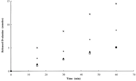
-Ala)4, D-Ala-Gly and D-Ala-Gly-Gly, monitored with the help of theD-amino acid oxidase test, revealed thatD -Ala-D-Ala andD-Ala-Gly-Gly were the best substrates (Fig. 1)
with turnover numbers of about 5 min21 (at an 8 mM substrate concentration).
Fig. 1 Time courses of the hydrolysis ofD
-alanyl-R oligopeptides by DppA. Samples of 0.8 mmol of (D-Ala)2(X),D-Ala-Gly-Gly (1),
(D-Ala)3(A) or (D-Ala)4(O) were incubated at
308C with 0.28 nmol of pure DppA in a total volume of 100 ml of buffer B with 0.1% BSA added. The releasedD-alanine was quantified
by theD-amino acid oxidase assay (FreÁre
et al., 1976). SD values did not exceed 10%. WithD-Ala-Gly and under the same
conditions, about 2 nmol ofD-alanine was
Molecular properties
After electrophoresis on a 7.5% non-denaturing polyacryla-mide gel, Coomassie blue staining revealed one single protein band, but the migration velocity appeared to be very low for a 30 kDa protein with an isoelectric pH of 5.1. Accordingly, chromatography on an analytical Superdex 200 column in buffer B indicated an Mrvalue of 230 ^ 15 kDa. In consequence, the native enzyme might be an octamer.
SDS±PAGE analysis revealed one major band with the expected Mr of about 30 kDa, but also two minor bands (Fig. 2A, lane 1). The larger one, with a slightly lower Mr than that of the major protein, represented at most 5±10% of the amount of the latter, and the smaller one exhibited a Mrvalue of about 6 kDa. The N-terminal sequences of the three peptides were determined. The major protein and the 6 kDa peptide had the same 10 N-terminal residues. The N-terminal sequence of the larger minor peptide corresponded to an internal sequence beginning at Ser-61. As the active enzyme is a multimeric protein that migrates in a non-denaturing PAGE as a homogeneous single species, it can be concluded that the two polypep-tide chains generated by the His-60/Ser-61 cleavage remain associated and retain the same folding.
Western blot analysis
To study the protein produced by the original strain of B. subtilis, samples of supernatants from 36 h and 72 h cultures were analysed by Western blotting after SDS± PAGE. The results are shown in Fig. 2.
Coomassie blue staining of the gel revealed, as
expected, that many proteins were produced in large quantities (Fig. 2A, lanes 2 and 3), but the amounts of DppA estimated by measuring the activity with 1 mM D
-Ala-pNa were about 1 and 15 ng in lanes 2 and 3, respectively, and thus well below the detection limit of Coomassie blue staining.
The Western blot analysis revealed two bands both in a 10 ng sample of purified DppA (produced in E. coli) and in the 72 h B. subtilis culture supernatant (Fig. 2B, lane 1 and 3 respectively). In the former, the positions and intensities of the two bands corresponded to those of the 30 kDa and 24 kDa peptides visualized by Coomassie blue staining and, in the latter, the major species remained the 30 kDa protein, but a smaller peptide of about 24 kDa was visible (Fig. 2B, arrow). In contrast, in the 36 h culture supernatant, the Western blot revealed a single, but much less intense, 30 kDa DppA species, so that the smaller peptide might be present but remain undetectable. So, when produced by the original strain, a small amount of the 30 kDa DppA was also cleaved, and the sizes of the fragments were similar to those observed with the enzyme produced with E. coli.
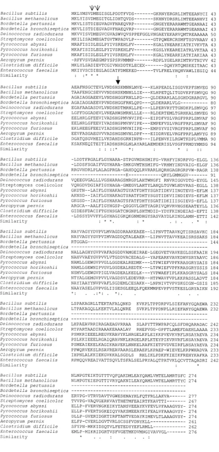
Alignment analysis
An exploration of the databases revealed 11 sequences related to the DppA sequence. Figure 3 shows the multiple sequence alignments. The similarity between the 12 sequences is particularly significant in the first 200 amino acid residues with several highly conserved residues, even when one includes the much shorter (incomplete?) Bordetella sequences. Among the strictly conserved residues, Asp-8, Glu-10 and His-60 could be involved in the interactions with Zn21ions, although these conclusions might be questioned if the 24 kDa form was found to be active. It is interesting to note that a DppA-like enzyme seems to exist in very different genera (Gram1, Gram±, Streptomyces, Archaea), both sporulating (Bacillus, Clos-tridium,¼) and non-sporulating (Deinococcus, Archaea). Discussion
While screening different bacterial species for their ability to hydrolyse D-alanyl-para-nitroanilide, we found that
several members of the Bacillus genus produced such an activity, which was later shown to be associated with a
D-aminopeptidase. The enzyme was purified from B.
subtilis; the corresponding gene was cloned, sequenced and found to be dppA, the first ORF of the dpp operon that is expressed early during sporulation according to Mathiopoulos et al. (1991). To facilitate the purification, the enzyme was first produced with an N-terminal extension containing a (His)6tag. However, this resulted in the isolation of two proteolytic products in which only
Fig. 2. SDS±PAGE analysis of purified DppA and of culture supernatants of B. subtilis.
A. Coomassie blue staining.
B. Western blot.Lane M, molecular mass standards; lanes 1A and 1B, DppA purified from E. coli: 5 mg (A) or 10 ng, i.e. 0.8 mIU (B); lanes 2A and 2B, 20 ml of sevenfold concentrated supernatant from a 36 h B. subtilis culture (about 1 mg of total protein and 0.08 mIU); lanes 3A and 3B,15 ml of supernatant from a 72 h B. subtilis culture (about 2 mg of total protein and 1.2 mIU).The arrow indicates the position of the 24 kDa polypeptide (see text).
Fig. 3. Alignment of the DppA sequence (B. subtilis) with those of related sequences found in the databases. (*) indicates conserved residues, (:) conservative substitutions and (.) semi-conservative substitutions in all sequences excluding the incomplete Bordetella pertussis and B. bronchiseptica sequences. The heavy arrow indicates the cleavage site observed in DppA purified from E. coli (see text). The light arrows indicate the conserved residues that might be involved in the interaction with Zn21ions.
parts of the extension had been retained. The kcat/Km value versus D-Ala-pNa measured with this
heteroge-neous preparation was 80 000 M21s21. Thus, these N-terminal alterations have little effect onD-Ala-pNa
hydro-lysis. As already mentioned in the literature (Ledent et al., 1997), this type of protein engineering, although poten-tially simplifying the purification procedure, may have some unexpected consequences on the biochemical characteristics of the target protein and must be used with care. In consequence, a protein identical to the original B. subtilis one was produced in E. coli.
Molecular sieve chromatography suggested that the active enzyme was an octamer composed of identical subunits. In contrast to the D-aminopeptidase from
Ochrobactrum anthropi (DAP), DppA does not hydrolyse
D-Ala-L-Ala and has a poor efficiency on peptide
sub-strates such asD-Ala-Gly-Gly. VanX and the periplasmic D-alanyl-D-alanine dipeptidase from Salmonella enterica
do not hydrolyse (D-Ala)3, one of the good substrates of
DppA. DmpA, theL-aminopeptidase D
-Ala-esterase/ami-dase from O. anthropi hydrolysesL-Ala-pNa and variousL
-dipeptides, whereas DppA is devoid of activity versusL
-derivates and behaves as a strict D-stereospecific
amidohydrolase far more active on D-Ala-pNa (kcat/ Km 100 000 M21s21 with a kcat value of at least 100 s21 as K
m is . 1 mM) than on its best peptide substrates (D-Ala)2andD-Ala-Gly-Gly (turnover numbers
of about 5 min21). WithD-Ala-pNa, the pH dependence of DppA is bell-shaped with a broad maximum extending from pH 9 to pH 11. A 90% inhibition was observed in the presence of Zn21 chelators. The initial activity could be recovered by the addition of 10 mM Zn21.
An exploration of the databases showed that the genomes of Bacillus methanolicus, Deinococcus radio-durans and Streptomyces coelicolor contained ORFs encoding putative proteins very similar to DppA. Other genomes mentioned in Fig. 3 (Pyrococcus abysii, Pyro-coccus furiosus, PyroPyro-coccus horikoshii, Aeropyrum pernix, Enterococcus faecalis, Bordetella pertussis, Bordetella bronchiseptica and Clostridium difficile) also contained ORFs encoding putative, probably related proteins. Some residues, often involved in Zn21 binding in metalloen-zymes, are conserved in these DppA-related sequences.
To date, none of the DppA-like proteins mentioned above has been biochemically characterized. The amino acid sequences of the knownD-aminopeptidases are not
related to that of DppA. The catalytic specificity of DppA is original. In consequence, DppA might be the prototype of a new family of D-aminopeptidases, although the exact
function of some homologous proteins might have evolved in a somewhat different direction.
The gene encoding DppA was described by Mathio-poulos et al. (1991) as the first ORF of the decoyinine-inducible or dciA operon. Decoyinine, an inhibitor of GMP
synthetase, induces sporulation of B. subtilis (Mitani et al., 1977). The dciA operon is also called the dipeptide transport operon or dpp because the four genes that follow the dciAA/dppA gene encode the elements of an ATP-binding cassette (ABC) transporter (Kuan et al., 1995; Saurin et al., 1999), and disruption of the dciAE gene renders a proline auxotroph unable to grow on Pro-Gly as sole proline source (Mathiopoulos et al., 1991).
The dpp operon is negatively regulated by two trans-acting factors, AbrB and CodY (control of dciA/dpp operon) (Slack et al., 1991; 1993; 1995; Serror and Sonenshein, 1996). AbrB is a well-known repressor of some genes expressed early during sporulation (Robert-son et al., 1989), and CodY would belong to a nutrient-sensing mechanism. CodY competes with AbrB for the promoter region of the dpp operon, but the repressor activity of CodY requests the presence of an unidentified co-repressor. Nutritional adaptation and spore formation are usually considered as competitive and mutually exclusive processes (Mathiopoulos et al., 1991), but the choice between the two pathways depends on an integration of multiple signals. The identification of the enzymatic reactions catalysed by the first ORF of this dciA/dpp operon might contribute to a better under-standing of these phenomena.
The physiological role of DppA, aD-aminopeptidase that
also hydrolyses (D-Ala)2 and is expressed early during
sporulation, is probably an adaptation to nutrient deficiency rather than an element favouring sporulation by hydrolys-ing the D-Ala-D-Ala dipeptide required in peptidoglycan
biosynthesis. Indeed, DppA-like proteins are found in microorganisms that do not sporulate (Archaea, Deinococ-cus radiodurans, Bordetellae), and Mathiopoulos et al. (1991) have already mentioned that null mutations in the dciA operon do not inhibit spore formation. The releasedD
-alanine could then be used as a metabolic fuel. A similar role has been proposed for the VanX homologue (DdpX) in E. coli (Lessard and Walsh, 1999b). Interestingly, the genes encoding DdpX and the DaaA amidase produced by O. anthropi and mentioned in the Introduction are also parts of peptide transport operons (Lessard and Walsh, 1999b; Komeda and Asano, 2000).
The present study highlights the complementarity of genome sequencing and biochemical analysis. The availability of the complete B. subtilis genome sequence allowed rapid cloning of the gene after very small amounts of the protein were purified.
Experimental procedures
Enzymes, strains, vectors, chemicals, culture media and antibodies
columns and Ampholine PAG plates were supplied by Pharmacia Biotech unless otherwise mentioned. The nitrilo-triacetic acid (NTA) agarose was obtained from Affiland. The T4 DNA ligase was from Boehringer Mannheim. The restriction enzymes were purchased from Life Technologies. The E. coli BL21(DE3) strain and the expression vectors pET28a and pET22b were supplied by Novagen; the ampicillin resistance gene of the pET22b plasmid was inactivated by the insertion of a kanamycin resistance cartridge at the PstI site, and the resulting vector was named pET22bKr(details of this construction can be obtained
from the authors). IPTG was from Eurogentec, substrates and peptides from Bachem and Sigma. The TLC plates (Silica gel 60F250) were from Merck. Rabbit anti-DppA antibodies were obtained from the `Centre d'Economie Rurale' Marloie, Belgium.
Luria±Bertani medium (10 g of bactotryptone, 5 g of yeast extract and 10 g of NaCl in 1 l of water, adjusted to pH 7.0) was used as a reference. Aliquots (10 ml) of the culture, taken at different times, were centrifuged. The pellets were suspended in 1 ml of fresh culture medium and sonicated. The estimation of D-aminopeptidase activity in the culture
supernatants or in sonicated cell extracts was carried out by monitoring the formation of p-nitroaniline fromD-Ala-p-Na, at
405 nm and 308C as described by Asano et al. (1992). Various other media were tested. They were prepared by adding glucose, glycerol, sodium lactate, bovine serum albumin (BSA) or casein hydrolysates to a basic medium (BM) described by Park and Reardon (1996) and containing 1 g of yeast extract and 15 g of nutrient broth l21. The best
production was obtained by adding 0.5 g of glucose and 7.2 g of sodium lactate to 1 l of BM, yielding medium C.
Purification of theD-aminopeptidase from B. subtilis
After 70 h of culture at 288C, 2.5 l of supernatant were recovered and diluted to 10 l with 10 mM potassium phosphate buffer (KPi), pH 6.5. The pH was adjusted to 6.5 by the addition of HCl. Seventy grams of Q Sepharose Fast Flow (QSFF; Pharmacia Biotech) equilibrated in the same buffer was added, and the mixture was stirred at 48C until complete adsorption of the D-aminopeptidase activity. The
loaded exchanger was recovered by filtration, washed extensively with 10 mM KPi, pH 6.5, and eluted twice with 100 ml of 50 mM KPi, pH 6.5, containing 300 mM KCl. Both eluates were pooled, concentrated and dialysed against 50 mM KPi, pH 6.5. The sample was loaded onto a 200 ml QSFF column equilibrated with 50 mM KPi, pH 6.5, and a linear KCl gradient (0±350 mM over 10 column volumes) was applied in the same buffer. The D-aminopeptidase activity
was eluted between 250 and 300 mM KCl. The most active fractions were concentrated, dialysed against 50 mM KPi, pH 8 (buffer A), and loaded onto a second QSFF column equilibrated in buffer A. A linear NaCl gradient (100± 500 mM) was applied in the same buffer, and the active fractions, containing about 16 mg of impure protein, were eluted between 300 and 400 mM NaCl. After dialysis against buffer A and concentration by ultrafiltration, a 1 mg sample was submitted to electrophoresis on an 8% non-denaturating polyacrylamide gel. The enzymatic activity was located by overlaying a Whatman paper previously soaked in 10 mM
D-Ala-p-Na. The protein band corresponding to the yellow
area was cut out and eluted at 48C in 1 ml of buffer A. During the purification steps, the protein concentration was estimated on the basis of the absorbance at 280 nm.
Isoelectrofocusing (IEF)
The isoelectric pH value was evaluated on Ampholine PAG plates, pH 3.5±9.5, by detecting the active band withD
-Ala-p-Na and measuring the pH at its position.
N- and C-terminal sequences
N-terminal sequence analysis was carried out on a pulsed-liquid sequencer with on-line analysis of the PTH amino acids (Perkin-Elmer, Applied Biosystems Division). C-terminal sequencing was performed on a Procise 494C sequencer (Perkin-Elmer) using a slight modification of the protocol described by Boyd et al. (1992). The alkylated thiohydantoins were identified by reverse-phase high-performance liquid chromatography (HPLC). Before sequence analysis, the lysine side-chains were modified with phenylisothiocyanate under basic conditions.
Mass spectrometry (ESMS)
ESMS was carried out on a Bio-Q quadrupole mass spectrometer (Micromass). Ten microlitres of a sample solution in 50% acetonitrile21% formic acid was injected manually in the 20 ml loop of the Rheodyne injector and pumped with a Harvard syringe pump at a flow rate of 5 ml min21. Scans of 9 s over the mass range of 600±1500
a.m.m. were collected over 2 min. Mass calibration was performed with horse heart cytochrome c.
Kinetic measurements
The enzyme activity was measured in buffer A with 40 mM ZnSO4(buffer B) added in a total volume of 400 ml at 308C.
For substrates containing a p-nitroaniline leaving group, the variation in absorbance was monitored at 405 nm for v0
measurements (D1 11 500 M21cm21) or at 440 nm
(D1 2250 M21cm21) for complete time course analyses.
The kcat/Kmvalue was obtained by linear regression of the v0
values or by analysis of the complete time courses with the help of the first-order equation (De Meester et al., 1987). As the active form of DppA is an oligomer (see below), the enzyme concentration is expressed as that of active sites.
TLC analysis
The hydrolysis products of peptides containingL-alanyl orD
-alanyl N-terminal residues were separated from residual substrates by TLC on silica gel plates. In order to separateD
-orL-alanine from the di-, tri- or tetra-alanyl peptides, the TLC
solvent was ethanol±H2O±triethylamine (70:25:5 by volume).
After a 10 cm migration, the plate was dried at room temperature, dipped in the ninhydrin reagent for 5 s and dried at 1108C for 15 min (Gerday et al., 1968).
To separate D- or L-alanine from D- or L
-alanyl-glycyl-glycine, glycyl-glycine and -alanyl-glycyl-glycine, the TLC solvent was n-butanol±acetic acid25% ammonium hydroxide (55:30:15 by volume). After a 20 cm migration, the plate was dried at 1108C for 1 h and treated as above. Scanning of the digital photography was performed using the Cybertech C5-1 system, and the signal intensities were compared with those of known amounts of the various compounds treated in parallel on the same plate.
Recombinant DNA techniques, bacterial strains, plasmids and growth conditions
The procedures used were essentially those described by Sambrook et al. (1989).
The genomic DNA of B. subtilis 168 (Genetic Stock Centre, USA) was extracted with the Qiagen genomic tip 20/G kit.
The sequences of the oligonucleotides used as primers for the PCR experiments and deduced from that of the dppA gene were as follows: 50-CGGGGTACCTCATATGAAATTG
TACATGTCAGTAGATATGGAAGG-30 and 50-CCCGAGCT
CTTAGCAGAATGATGTCCGCATCGC-30. The underlined
palindromic sequences are recognized by the Asp718, NdeI and SacI restriction enzymes respectively.
The PCR mixture (100 ml total volume) contained 1.5 mg of DNA template, 0.5 mg of each primer, 2.5 units of Vent polymerase (New England Biolabs) in the buffer recom-mended by the supplier.
The PCR was carried out with a DNA thermal cycler (Biometra-Trio Thermoblock) for 25 cycles with a 1 min denaturation at 948C, a 1 min annealing at 508C and a 1 min polymerization at 728C. The PCR product was purified using the Geneclean spin kit (Bio 101).
PMDL 1110. The PCR product, digested with the Asp718 and SacI enzymes, was ligated in the pUC19 cloning vector previously cut with the same enzymes. The recombinant plasmid (PDML1110) was extracted, purified, and the insert was completely sequenced on both strands with the Auto-Read sequencing kit and the ThermoSequenase labelled primer cycle sequencing kit. Sequences were read on an automated laser fluorescent DNA sequencer.
PDML 1111 and PDML 1112. To overexpress the gene coding for theD-alanyl aminopeptidase activity, PDML 1110
was digested with the NdeI and SacI enzymes. The resulting fragment was ligated in pET28a or pET22bKr, previously cut
with the same enzymes, yielding PDML 1111 and PDML 1112 respectively.
Purification of the overproducedD-alanyl amidases in E.
coli
As a result of cloning the PCR product in pET28a, PDML 1111 encodes a protein with an additional N-terminal 20 residues including six consecutive His. In consequence, the recombinant protein was expected to be retained by a Ni21
-NTA agarose column. Nevertheless, someD-alanyl
amino-peptidase activity was not retained. The adsorbed enzyme was eluted with an imidazole linear gradient (0±250 mM) at
pH 7.4. The fractions containing theD-alanyl amidase activity
were pooled, dialysed against buffer A and concentrated. About 2 mg of enzyme was obtained.
The PDML 1112 plasmid encodes exactly the same protein as that produced by the original B. subtilis strain. A 2 l E. coli culture in LB medium was grown to an A600value of 0.8 at
378C, cooled to 188C and induced by 1 mM IPTG. After a 24 h incubation at 188C, the cells were harvested by centrifugation. The pellet was suspended in 100 ml of Tris-HCl (10 mM, pH 8), and the cells were disrupted with the constant basic system disintegrator (Inceltech). The suspen-sion was centrifuged, and this crude extract supernatant was dialysed against 50 mM KPi buffer at pH 6.5 and loaded onto a 100 ml Q-Sepharose column equilibrated in the same buffer. After washing, the enzyme was eluted by applying a 0±0.3 M NaCl gradient in the same buffer over 1500 ml. The active fractions were pooled and dialysed against buffer A, then loaded onto a Q-Sepharose column equilibrated with the same buffer. Elution was carried out using a 0±0.5 M NaCl gradient (1.5 l). The active fractions were pooled, concen-trated to 50 ml by ultrafiltration and filtered through a 400 ml Superdex G100 column in buffer A. The active fractions were pooled (200 ml), and 5 ml aliquots were loaded onto a MonoQ column (HR 5/5, 1 ml) connected to an AÊkta Explorer apparatus (Pharmacia Biotech). After a 20 ml wash, 15 ml linear (0±0.120 M) and 60 ml linear (0.120±0.240 M) NaCl gradients, both in buffer A, were applied successively. Under these conditions, the peak of activity was well separated from minor amounts of contaminant proteins. Forty runs were carried out always presenting the same distribution of protein peaks. The active fractions were pooled, dialysed against buffer A, concentrated and stored at 2208C.
pH dependence of the hydrolysis ofD-Ala-pNa
The universal buffer system of Teorell and Stenhagen (1938) (pH 2.0±12.0) was prepared with a minor modification. Aliquots of the concentrated mixture of the three acids (citric, phosphoric and boric) were neutralized with NaOH up to the desired pH, then diluted to the same final volumes. The buffered D-Ala-pNa substrate solutions (1.0 mM) were
complemented with 40 mM ZnSO4, and the v0 values were
determined.
Thermal stability
The enzyme (11.6 mM) was incubated in buffer B for 10 min at different temperatures. The DppA activity was assayed at 308C with 1.0 mMD-Ala-pNa.
Activity on peptide substrates
Stock solutions (20 mM) of alanyl peptides [(L-Ala)2, (D-Ala)2, L-Ala-D-Ala, D-Ala-L-Ala, (L-Ala)4, (D-Ala)4] were prepared in
buffer B. For all other peptides, the stock solutions were 10 mM in the same buffer.
To 45 ml of peptide solution, 5 ml of 116 mM DppA in buffer B or 5 ml of buffer B were added, and incubation was carried out at 308C over 2 or 24 h. The reaction was stopped by
heating the samples to 1008C for 2 min. Aliquots (2 ml) were analysed by TLC.
With (D-Ala)2, D-Ala-Gly, (D-Ala)3, D-Ala-Gly-Gly and (D
-Ala)4, the releasedD-alanine was more accurately quantified
by the D-amino acid oxidase assay (FreÁre et al., 1976). To
80 ml of 10 mM substrate in buffer B, 20 ml of 11.6 mM DppA containing 1% BSA was added. After various periods of time, the reaction was stopped as above, and the assay was performed on 10 ml aliquots.
Gel electrophoresis
In the presence of SDS with 4% and 15% stacking and running gels, respectively, PAGE was performed as described by Laemmli (1970). Under non-denaturing condi-tions, 4% and 7.5% stacking and running gels were used respectively. Aliquots of enzyme preparations in buffer B were deposited on the gel, and the protein bands were revealed by Coomassie blue staining.
Analytical molecular sieve
Aliquots (50 ml) of 30 mM or 0.3 mM DppA in buffer B were filtered through a Superdex TM 200 HR 10/30 column equilibrated in the same buffer and connected to the AÊkta Explorer. Calibration was performed with the following proteins: thyroglobulin (669 000), apoferritin (443 000), cat-alase (232 000), L-alanine dehydrogenase (228 000),
aldo-lase (158 000), alcohol dehydrogenase (150 000), apotransferrin (80 000), BSA (66 700), ovalbumin (43 000), carbonic anhydrase (31 000), soybean trypsin inhibitor (21 000) and lysozyme (14 300).
Western analysis
Rabbit antibodies were raised against the recombinant DppA purified from E. coli. Pure Dppa (10 ng) or 1±2 mg of (total) proteins present in the supernatants of 36 h or 72 h B. subtilis cultures were submitted to SDS±PAGE. After electroblotting onto a nitrocellulose membrane, the adsorbed proteins were stained with 0.1% Ponceau S (w/v) in 5% acetic acid (v/v) (Sigma) for localizing the standard proteins. Thereafter, the membrane was rinsed with water and treated as described in the Bio-Rad ImmunoBlot manual using goat anti-rabbit± alkaline phosphatase conjugates.
Alignments
Preliminary sequence data were obtained from the Institute for Genomic Research website at http://www.tigr.org. The amino acid sequences of B. subtilis DppA and of its putative homologues can be accessed through the following acces-sion numbers in the NCBI Protein Database: B. subtilis DppA, no. CAA40002(Mathiopoulos et al., 1991); A. pernix, no. BAA79269(Kawarabayasi et al., 1999); Bacillus methanoli-cus, no. AAB39857(Cue et al., 1997); D. radiodurans, no. AAF11394(White et al., 1999); P. abyssi, no. CAB49502; P. horikoshii, no. BAA30694(Kawarabayasi et al., 1998); and S. coelicolor, no. CAA22734(Redenbach et al., 1996). Multiple
sequence alignments were performed with the CLUSTAL
algorithm (website: http://www2.ebi.ac.uk/clustalw/).
Acknowledgements
The authors wish to thank Mrs Nicole GeÂrardin from the Laboratory of Biochemistry for her useful help in the TLC analysis, Professor Edwin De Pauw and Mr Emmanuel Delye for many useful discussions. Sequencing of Bordetella bronchiseptica, B. pertussis and Clostridium difficile was performed with support from Beowulf Genomics; sequencing of Enterococcus faecalis with support from NIAID; sequen-cing of Pyrococcus furiosus with support from DOE; and sequencing of Streptomyces coelicolor with support from BBSRC/Beowulf Genomics. This work was supported in part by the Belgian Program of Interuniversity Poles of Attraction (PAI no. P4/03) and a Geconcerteerde Onderzoeks-aktie from the Flemish Government (120.522.93). The purchase of the AÊkta Explorer apparatus was made possible by a grant from the Fonds National de la Recherche Scientifique (FNRS, Brussels, Belgium). L.F. and B.J., C.D. and C.G. are, respectively, Aspirant and Chercheurs QualifieÂs of the FNRS. B.D. is a postdoctoral researcher from the Fonds voor Wetenschappelijk Onderzoek (FWO, Flanders, Belgium).
References
Arthur, M., Depardieu, F., Cabanie, L., Reynolds, P., and Courvalin, P. (1998) Requirement of the VanY and VanX
D,D-peptidases for glycopeptide resistance in enterococci.
Mol Microbiol 30: 819±830.
Asano, Y., Mori, T., Hanamoto, S., Kato, Y., and Nakazawa, A. (1989a) A new D-stereospecific amino acid amidase
from Ochrobactrum anthropi. Biochem Biophys Res Com-mun 162: 470±474.
Asano, Y., Nakazawa, A., Kato, Y., and Kondo, K. (1989b) Properties of a novelD-stereospecific aminopeptidase from
Ochrobactrum anthropi. J Biol Chem 264: 14233±14239. Asano, Y., Kato, Y., Yamada, A., and Kondo, K. (1992)
Structural similarity of D-aminopeptidase to
carboxypepti-daseDDand beta-lactamases. Biochemistry 31: 2316±2328.
Asano, Y., Ito, H., Dairi, T., and Kato, Y. (1996) An alkalineD
-stereospecific endopeptidase with beta-lactamase activity from Bacillus cereus. J Biol Chem 271: 30256±30262. Boyd, V.L., Bozzini, M., Zon, G., Noble, R.L., and Mattaliano,
R.J. (1992) Sequencing of peptides and proteins from the carboxy terminus. Anal Biochem 206: 344±352.
Cue, D., Lam, H., Dillingham, R.L., Hanson, R.S., and Flickinger, M.C. (1997) Genetic manipulation of Bacillus methanolicus, a Gram-positive, thermotolerant methylo-troph. Appl Environ Microbiol 63: 1406±1420.
De Meester, F., Joris, B., Reckinger, G., Bellefroid-Bour-guignon, C., FreÁre, J.M., and Waley, S.G. (1987) Auto-mated analysis of enzyme inactivation phenomena. Application to beta-lactamases and DD-peptidases.
Bio-chem Pharmacol 36: 2393±2403.
Dideberg, O., Charlier, P., Dive, G., Joris, B., FreÁre, J.M., and Ghuysen, J.M. (1982) Structure of a Zn21-containing
D
-alanyl-D-alanine-cleaving carboxypeptidase at 2.5 A
Fanuel, L., Goffin, C., Cheggour, A., Devreese, B., Van Driessche, G., Joris, B., et al. (1999) The DmpA aminopeptidase from Ochrobactrum anthropi LMG7991 is the prototype of a new terminal nucleophile hydrolase family. Biochem J 341: 147±155.
FreÁre, J.-M., Leyh-Bouille, M., Ghuysen, J.-M., Nieto, M., and Perkins, H.R. (1976) Exocellular DD
-carboxypeptidases-transpeptidases from Streptomyces. In Methods in Enzy-mology. Proteolytic Enzymes, Part B, XLV. Lorand, L. (ed.). New York: Academic Press, pp. 610±636.
FreÁre, J.-M., Nguyen-DisteÁche, M., Coyette, J., and Joris, B. (1992) Mode of action: interaction with the penicillin binding proteins. In The Chemistry of b-Lactams. Page, M.I. (ed. ). Glasgow: Blackie, pp. 149±198.
Gerday, C., Robyns, E., and Gosselin-Rey, C. (1968) High resolution techniques of peptide mapping. Separation of bovine carotid actin peptides on cellulose thin layers and of the corresponding dansyl-peptides on polyamide thin layers. J Chromatogr 38: 408±411.
Goffin, C., and Ghuysen, J.-M. (1998) Multimodular penicillin-binding proteins: an enigmatic family of orthologs and paralogs. Microbiol Mol Biol Rev 62: 1079±1093.
Hilbert, F., del Portillo, F.G., and Groisman, E.A. (1999) A periplasmic D-alanyl-D-alanine dipeptidase in the
gram-negative bacterium Salmonella enterica. J Bacteriol 181: 2158±2165.
Holtje, J.V. (1998) Growth of the stress-bearing and shape-maintaining murein sacculus of Escherichia coli. Microbiol Mol Biol Rev 62: 181±203.
Kato, Y., Asano, Y., Nakazawa, A., and Kondo, K. (1989) First stereoselective synthesis of D-amino acid N-alkyl
amides catalysed by D-aminopeptidase. Tetrahedron 45:
5743±5754.
Kawarabayasi, Y., Sawada, M., Horikawa, H., Haikawa, Y., Hino, Y., Yamamoto, S., et al. (1998) Complete sequence and gene organization of the genome of a hyper-thermo-philic archaebacterium, Pyrococcus horikoshii OT3. DNA Res 5: 55±76.
Kawarabayasi, Y., Hino, Y., Horikawa, H., Yamazaki, S., Haikawa, Y., Jin-no, K., et al. (1999) Complete genome sequence of an aerobic hyper-thermophilic crenarchaeon, Aeropyrum pernix K1. DNA Res 6: 83±101.
Komeda, H., and Asano, Y. (2000) Gene cloning, nucleotide sequencing, and purification and characterization of theD
-stereospecific amino-acid amidase from Ochrobactrum anthropi SV3. Eur J Biochem 267: 2028±2035.
Kuan, G., Dassa, E., Saurin, W., Hofnung, M., and Saier,M.H., Jr (1995) Phylogenetic analyses of the ATP-binding constituents of bacterial extracytoplasmic receptor-dependent ABC-type nutrient uptake permeases. Res Microbiol 146: 271±278. Kunst, F., Ogasawara, N., Moszer, I., Albertini, A.M., Alloni,
G., Azevedo, V., et al. (1997) The complete genome sequence of the Gram-positive bacterium Bacillus subtilis. Nature 390: 249±256.
Laemmli, U.K. (1970) Cleavage of structural proteins during the assembly of the head of bacteriophage T4. Nature 227: 680±685.
Ledent, P., Duez, C., Vanhove, M., Lejeune, A., Fonze, E., Charlier, P., et al. (1997) Unexpected influence of a C-terminal-fused His-tag on the processing of an enzyme and on the kinetic and folding parameters. FEBS Lett 413: 194±196.
Lessard, I.A., and Walsh, C.T. (1999a) Mutational analysis of active-site residues of the enterococcalD-ala-D-Ala
dipepti-dase VanX and comparison with Escherichia coli D-ala-D
-Ala ligase andD-ala-D-Ala carboxypeptidase VanY. Chem
Biol 6: 177±187.
Lessard, I.A., and Walsh, C.T. (1999b) Van X, a bacterialD
-alanyl-D-alanine dipeptidase: resistance, immunity, or
survival function? Proc Natl Acad Sci USA 96: 11028± 11032.
Mathiopoulos, C., Mueller, J.P., Slack, F.J., Murphy, C.G., Patankar, S., Bukusoglu, G., et al. (1991) A Bacillus subtilis dipeptide transport system expressed early during sporula-tion. Mol Microbiol 5: 1903±1913.
Mitani, T., Heinze, J.E., and Freese, E. (1977) Induction of sporulation in Bacillus subtilis by decoyinine or hadacidin. Biochem Biophys Res Commun 77: 1118±1125.
Park, K., and Reardon, K. (1996) Medium optimization for recombinant protein production by Bacillus subtilis. Bio-technol Lett 18: 737±740.
Redenbach, M., Kieser, H.M., Denapaite, D., Eichner, A., Cullum, J., Kinashi, H., et al. (1996) A set of ordered cosmids and a detailed genetic and physical map for the 8 Mb Streptomyces coelicolor A3(2) chromosome. Mol Microbiol 21: 77±96.
Reynolds, P.E., Depardieu, F., Dutka-Malen, S., Arthur, M., and Courvalin, P. (1994) Glycopeptide resistance mediated by enterococcal transposon Tn1546 requires production of VanX for hydrolysis ofD-alanyl-D-alanine. Mol Microbiol 13:
1065±1070.
Robertson, J.B., Gocht, M., Marahiel, M.A., and Zuber, P. (1989) AbrB, a regulator of gene expression in Bacillus, interacts with the transcription initiation regions of a sporulation gene and an antibiotic biosynthesis gene. Proc Natl Acad Sci USA 86: 8457±8461.
Sambrook, J., Fritsch, E., and Maniatis, T. (1989). Molecular Cloning. A Laboratory Manual, 2nd edn. Cold Spring Harbor, New York: Cold Spring Harbor Laboratory Press. Saurin, W., Hofnung, M., and Dassa, E. (1999) Getting in or
out: early segregation between importers and exporters in the evolution of ATP-binding cassette (ABC) transporters. J Mol Evol 48: 22±41.
Serror, P., and Sonenshein, A.L. (1996) Interaction of CodY, a novel Bacillus subtilis DNA-binding protein, with the dpp promoter region. Mol Microbiol 20: 843±852.
Slack, F.J., Mueller, J.P., Strauch, M.A., Mathiopoulos, C., and Sonenshein, A.L. (1991) Transcriptional regulation of a Bacillus subtilis dipeptide transport operon. Mol Microbiol 5: 1915±1925.
Slack,F.J., Mueller, J.P., and Sonenshein, A.L. (1993) Mutations that relieve nutritional repression of the Bacillus subtilis dipeptide permease operon. J Bacteriol 175: 4605±4614. Slack, F.J., Serror, P., Joyce, E., and Sonenshein, A.L.
(1995) A gene required for nutritional repression of the Bacillus subtilis dipeptide permease operon. Mol Microbiol 15: 689±702.
Teorell, T., and Stenhagen, E. (1938) Ein Universalpuffer fuÈr den pH-Bereich 2.0 bis 12.0. Biochem Z 299: 416±419. White, O., Eisen, J.A., Heidelberg, J.F., Hickey, E.K.,
Peterson, J.D., Dodson, R.J., et al. (1999) Genome sequence of the radioresistant bacterium Deinococcus radiodurans R1. Science 286: 1571±1577.