PAPER
Cite this: Analyst, 2016, 141, 4293
Received 14th March 2016, Accepted 1st May 2016 DOI: 10.1039/c6an00606j www.rsc.org/analyst
Early apoptosis real-time detection by label-free
SERS based on externalized phosphatidylserine
†
Haibo Zhou,
‡
aQiqin Wang,
‡
aDetian Yuan,
‡
a,bJinyong Wang,
cYang Huang,
aHuihui Wu,
aJingyi Jian,
aDanting Yang,
dNing Huang,
bChristoph Haisch,
eZhengjin Jiang*
aand Shanze Chen*
b,fApoptosis is a tightly regulated cellular process that plays an essential role in the development, aging, cancer biology, immune response, and pathogenesis of various diseases. Herein, we report a new SERS sensing strategy for in vitro sensitive detection of early apoptotic cells. The principle of this method is to in situ synthesize silver nanoparticles (AgNPs) on the phosphatidylserine (PS) of the apoptotic cell mem-brane during the early apoptosis, which enables distinguishing normal and apoptotic cells. The total assay time of the presented method is only 10 min, thus being faster, cheaper and simpler than current tech-niques for the detection of apoptosis. The intrinsic mechanism was verified by different approaches based on externalized phosphatidylserine. In addition, the detection process is real-time and label-free; i.e., the intrinsic SERS spectra from the cellular membrane are directly employed for apoptosis real-time detec-tion, which avoids using additional chemical or biological reagents as external signal indicators. Therefore, our SERS approach may serve as a potentially practical tool for sensitive and real-time detection of early cell apoptosis, complementing the state-of-the-art strategies, e.g.flow cytometry. While further investi-gation is required to better understand the intrinsic mechanism of the in situ coating method, the current results may provide another choice for real-time detection of early apoptosis.
Introduction
In a multicellular organism or tissue, cells that are no longer needed or are damaged are recognized to be a threat to the organism, and thus it is necessary to destroy and remove these cells by a tightly regulated cell suicide process known as pro-grammed cell death, also called apoptosis.1,2Apoptosis can be triggered by a variety of stresses, such as heat, radiation,
nutri-ent deprivation, viral infection, hypoxia, exposure to toxic sub-stances and increased intracellular calcium concentration.3 Apoptosis is initiated by the activation of a complex process composed of multiple intracellular and extracellular events, such as caspase activation, release of cytochrome c (Cyt c) from mitochondria, externalization of phosphatidylserine on the membrane and DNA fragmentation.4Apoptosis is charac-terized by specific morphological changes such as membrane blebbing, cell shrinkage and nuclear fragmentation.5–7 Apop-tosis plays a crucial role in embryonic development and in balancing proliferation and death rate in tissue.8Additionally, it has been proven that many diseases such as degenerative disorders (excessive apoptosis), autoimmune disorders and cancers (inadequate apoptosis) result from deregulation of apoptosis.9,10Due to the importance of this complex process in cell biology, development of technologies to monitor the progression of apoptosis and detect the apoptotic signaling will benefit humankind on the aspect of disease treatment and diagnosis.
Currently, transmission electron microscopy and time-lapse microscopy are widely used to analyze the morphologic changes of apoptotic cells versus normal cells.11A number of molecular biological techniques aim at the analysis of land-mark events during apoptosis, for example, western blotting for caspase activation.12 Another commonly used detection
†Electronic supplementary information (ESI) available: Experimental details and additional figures. See DOI: 10.1039/c6an00606j
‡These authors contributed equally to this work.
aDepartment of Pharmacy and Guangdong Province Key Laboratory of Pharmacodynamic of Traditional Chinese Medicine & New Drug Research, Jinan University, Guangzhou, Guangdong Province 510632, China. E-mail: [email protected]
b
Department of Pathophysiology, West China School of Preclinical Sciences and Forensic Medicine, Sichuan University, Chengdu, Sichuan Province 610041, China. E-mail: [email protected]
cCollege of Veterinary Medicine, Huazhong Agricultural University, Wuhan, 430070, China
d
Schools of Biosystems Engineering and Food Science, Zhejiang University, 866 Yuhangtang Road, Hangzhou, Zhejiang Province 310058, China
eChair for Analytical Chemistry, Technische Universität München, Marchioninistr. 17, D-81377 Munich, Germany
fComprehensive Pneumology Center (CPC), Institute of Lung Biology and Disease, Helmholtz Zentrum München, Neuherberg, Germany
Published on 03 May 2016. Downloaded by Jinan University Library on 06/09/2017 01:54:23.
View Article Online View Journal | View Issue
method relies on a fluorescent labeled antibody, which includes the imaging of caspase activation in apoptotic cells by fluorescent microscopy.13 Recent efforts have focused on the design of activity-based probes (ABPs) to enzymes such as caspase-3.14 Vickers et al. generated fluorescent and biotiny-lated probes capable of selective detection of caspase-3 using key unnatural amino acids. Moreover, it has been reported that nanomaterials can be applied for apoptosis detection.15Chen et al. have developed a novel fluorescence activation nanosen-sor for the imaging of cytosolic Cyt c released from mitochon-dria in apoptotic cells.16Besides the cytosolic molecules, the changes in the cell membrane can also be utilized as an indi-cator for apoptosis. It is worthy of note that externalization of phosphatidylserine (PS) on plasma membrane is an early event during apoptosis, which provides an“eat me” signal to phago-cyte.17 Phosphatidylserine, an important phospholipid mem-brane component, binds specifically to a fluorescent dye, conjugated annexin-V with the exposed charged head groups, in a Ca2+dependent process, thus allowing us to analyze the apoptosis by flow cytometry or fluorescent microscopy.18,19 However, all of the abovementioned methods require fixation and permeabilization, hampering dynamic monitoring of the apoptotic process. Furthermore, the introduction of fluo-rescent dyes into cells during measurements may damage the cells and interfere with the apoptotic machinery. Therefore, there is still a strong need to develop novel strategies for simple, rapid, and if possible fluorescent dye free (called label-free) dynamic monitoring and detection of apoptotic cells.
Since the discovery of a significant enhancement of Raman signals at the rough silver electrode surface,20–22 surface-enhanced Raman scattering (SERS) has gained increasing attention as a powerful analytical tool for the detection of bio-logical species,23–26for molecular imaging,27and for monitor-ing microorganisms,28–30cells31,32and tissues,33even in vivo.34 In comparison with normal Raman signals, SERS signals may be enhanced by a factor of up to 106–1014, when the target molecules are located on the nanoparticle aggregates or at the nanostructural junctions (“hot spots”).35,36This huge
enhance-ment enables SERS to serve as an ultrasensitive detection tool in various analytical areas. Besides the sensitivity, its intrinsic selectivity based on the spectroscopic fingerprint, its simple and fast realization, and its nondestructive nature in aqueous environment make SERS an ideal tool for label-free, in situ and real-time analyses of biological samples.37
Over the past decade, a series of SERS-active substrates have been developed for bio-sensing applications such as ultra-sensitive detection of cell division and differentiation via detecting signals from various biomolecules (e.g., DNA, RNA, proteins and glycan).38,39 A recent report revealed that SERS performed with specific nanomaterials (AgNPs@Si) is able to detect cell apoptosis at the single-cell level based on the decreased SERS intensity of cells.40 However, they did not discuss in detail the underlying mechanism by which the SERS signal intensity of apoptotic cells decreased. Moreover, we have previously reported a fast and easy method to grow AgNPs on the surface of Gram-negative bacteria (E. coli), which
allows the label-free in situ discrimination of live and dead bac-teria.29 Therefore, we speculated whether our method could also be applied to the detection of the mammalian cell status. Particularly, we were interested in apoptotic cell detection owing to the important role of apoptosis in many healthy and disease conditions.
Herein, we have developed a new and label-free SERS strat-egy for apoptosis detection, using the in situ coating method, i.e., silver nanoparticle coating on the apoptotic cell mem-brane, as an active, efficient and reproducible SERS apoptotic platform. Once apoptosis is induced by drugs, the outside layer of the apoptotic cell membrane becomes negatively charged due to the externalized phosphatidylserine. We first soaked the apoptotic cell in a silver nitrate (AgNO3) solution
and then used hydroxylamine hydrochloride (NH2OH·HCl) as
a reducing agent. Eventually, a “colloid deposit” was formed on the apoptotic cell membrane. As a result, cells at different apoptotic stages were readily analyzed via recording the SERS intensity of the cell membrane components distributed on the cell membrane. Our results suggest new possibilities for finding novel SERS-based in vitro biomedical applications, as well as provide a powerful strategy for dynamic and sensitive apoptosis detection.
Results and discussion
Scheme 1 illustrates the in situ formation of AgNPs coating on the apoptotic cell membrane. When the AgNO3 solution is
added to the normal cell suspension, silver ions (Ag+) cannot adsorb onto the membrane of normal cells, due to a lack of interaction between Ag+and phosphatidylserine. However, Ag+ strongly adheres to the externalized phosphatidylserine of the apoptotic cell membrane by the electrostatic interaction between the positively charged Ag+and the negatively charged –COO− and –NH
2 on the externalized phosphatidylserine.
Finally, the addition of the reducing agent (hydroxylamine hydrochloride, NH2OH·HCl) led to the formation of colloid
deposits only on the surface of apoptotic cells. For the normal
Scheme 1 Schema of the detection mechanism of apoptosis based on in situ synthesis of nanoparticles on the apoptotic cell membrane. The Ag+adsorbed onto the apoptotic cell membrane by electrostatic
attrac-tion generates AgNPs on the membrane immediately after the addiattrac-tion of the reducing reagent.
cells, the AgNPs are just randomly formed in the suspension and do not attach onto the normal cell membrane.
Staurosporine, a natural product originally isolated from the bacterium Streptomyces staurosporeus,14has been used as a standard chemical to induce cell apoptosis.15In this work, we used this chemical to induce apoptosis of HL-60 cells. To investigate whether SERS can be used to differentiate normal cells and apoptotic cells, 1 × 10⁶cells per mL HL-60 cells, both normal and apoptotic ones, were prepared by this in situ coating method (the detailed procedure is described in the Experimental Section of the ESI†) as samples for SERS detec-tion. As shown in Fig. 1, almost no SERS signals were detected in the normal cells, whereas the SERS signals were highly strong in the apoptotic cells. The typical SERS peaks at 665, 726, 1332, and 1576 cm−1 were observed, which might be attributed to the cellular lipids, proteins, and carbo-hydrates40,41 (see the tentative assignments of peaks in Table S1 of the ESI†). In addition, as shown in the insert picture of Fig. 1, a transparent suspension was observed in the normal cells after in situ coating preparation, whereas the color of the apoptotic cell suspension became bluish-green gradually, indicating that the AgNPs were formed in the apop-totic cell suspension but not in the normal cell group. These results were similar to our study reported previously.29
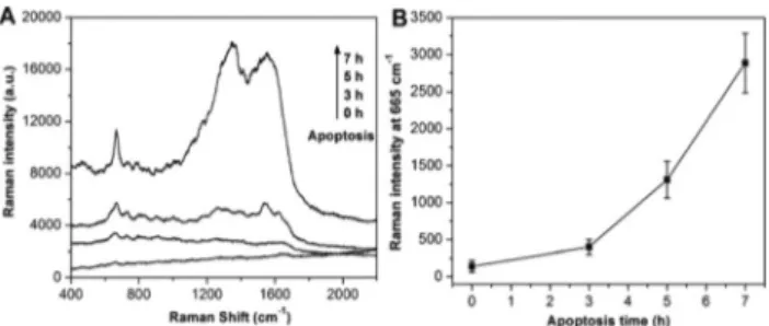
Apoptosis can be seen as a multi-step process, which can be separated into induction to early, intermediate and late-stages.42It is important to develop methods and reagents to distinguish the various apoptotic stages, in particular for the detection of early apoptosis with the intact membranes. In order to further determine whether our SERS method is able to detect in a stage dependent manner the apoptotic process, HL-60 cells at 3 h, 5 h and 7 h after induction of apoptosis were prepared with the in situ coating method described above and SERS was performed. As shown in Fig. 2, we observed that the intensity of the SERS signals significantly increased with
time. A calibration curve derived from multiple independent experiments, i.e., from the analysis of different cultivations, measured from different production batches (each data point representing an average of at least three independent measure-ments), can be found in Fig. 2B. These results indicate that the sensitivity of our method is high as we can differentiate the cell apoptosis with a 2 h interval. Moreover, these results demonstrate that our method is capable of detecting early cell apoptosis, which cannot yet be recognized based on changes in morphology.
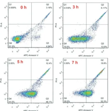
The early stage of apoptosis is characterized by the loss of mitochondrial membrane potential and the release of cytosolic Cyt c accumulations from the mitochondria.43 Fujita et al. developed a label-free method to observe Cyt c dynamics during apoptosis by SERS, based on its unique structure.44 Another key apoptotic process is the translocation of PS, a phospholipid that transmits from the cytosolic (inner) side of the cell membrane to the extracellular (outer) surface in the early/middle apoptosis. The externalization of PS increases with the apoptotic process. Many methods have been develo-ped for the analysis of early apoptosis based on these mole-cular events. Due to the specific binding ability to PS, annexin-V staining, paired with fluorochrome (e.g. propidium iodide, PI), is widely used to identify early cell apoptosis by flow cyto-metry.45To compare our SERS approach with flow cytometry, samples at the same time stage of apoptosis of HL-60 cells after treatment with staurosporine were analyzed by flow cyto-metry with the annexin-V/PI protocol. As shown in Fig. 3, the percentages of apoptotic HL-60 cells at 0 h, 3 h, 5 h and 7 h are 4.9%, 13.9%, 46.1% and 52.3%, respectively. These results indicate that our approach has the similarly sensitivity as flow cytometry, which is commonly used to detect time stages of apoptosis. Notably, the SERS technique is much faster, simpler and lower-cost than flow cytometry. Thus, it has a potential to replace the time-consuming, laborious, sophisti-cated and expensive flow cytometry technique in the future.
For a more precise explanation of the distinct SERS signal source for normal and apoptosis cells by the in situ coating method, bright-field microscopy was performed to investigate the morphology of the cells and the distribution of AgNPs. Fig. 4A and B depict the bright-field images of normal cells Fig. 1 SERS spectra of normal and apoptosis cells obtained by the
in situ coating method. The inset picture shows the corresponding color of the suspension.
Fig. 2 (A) SERS spectra of apoptotic cells with different times (0 h, 3 h, 5 h and 7 h). (B) The corresponding Raman intensity at 665 cm−1vs. time after apoptosis. The error bars represent three independent experiments for each sample.
and apoptotic cells, respectively. In contrast to normal cells that were intact and circled, apoptotic cells showed shrinkage, nuclear fragmentation, and chromatin condensation, which confirm that these cells were in the process of apoptosis. When normal cells were prepared using the in situ coating method (Fig. 4C), the AgNPs (black dots) were randomly dis-tributed in the suspension and did not aggregate on the surface of the normal cells. Some of them were self-aggregated
in the suspension, but they did not adhere to the cellular membrane, which mostly remains uncoated, and these cells remain mostly separated. Consequently, the Raman signals of the cell membrane components cannot be efficiently amplified by electromagnetic field enhancement, resulting in very weak SERS signals. Fig. 4D reveals the intimate contact of AgNPs with the apoptotic cellular membrane. Most of the AgNPs coated the cellular membrane, whereas only a few were found unbound or clustered in the liquid phase. The shape of the AgNPs was irregular. Some of the composites representing large structures (indicated by red arrows in Fig. 4D) were likely to aggregate spontaneously, forming AgNP clusters on the surface of apoptotic cells. Moreover, most of the apoptotic cells were coated by aggregated AgNPs and were assembled in clusters under these conditions, which form highly SERS-active hot spots on the interfaces of apoptotic cells and, conse-quently, strong Raman signals were detected. The growth of AgNPs on the apoptotic cell surface was consistent with the finding of our previously reported paper that AgNPs accumu-late on the bacterial surface.28,29
Rayleigh scattering is a powerful tool to real-time visualize metal nanoparticles in solution.46 Hence, we used dark field microscopy to further investigate the dynamic synthesis of AgNPs on the apoptotic cell membrane by directly monitoring this process. After the preparation using the in situ coating method, 50 μL of samples were dropped on the micro glass slide for real-time monitoring of the synthesis of AgNPs. We observed that AgNPs displayed random Brownian motion and the single AgNP and corresponding aggregates were visualized as blue and orange scattering structures in the blank solution (Fig. 5A). Furthermore, we can clearly see that the AgNPs were synthesized on the apoptotic cell membrane (indicated by red arrows in Fig. 5C), whereas there were almost no AgNPs on the normal cell membrane. To verify that the adhered AgNPs on the apoptotic cell membrane were not from the solution, we moved the objective focal plane from the top of the cell to the bottom. As shown in Fig. 5B and C, AgNPs were always observed (or found), at different focal planes, sitting on the surface (or membrane) of the apoptotic cell but not on the Fig. 3 Detection of apoptosis cells at different time points (0 h, 3 h, 5 h
and 7 h) byflow cytometry. Cells were stained with annexin-V/PI. All the other parameters were consistent with Fig. 2.
Fig. 4 Bright-field images of normal cells (A) and apoptotic cells (B) and the corresponding bright-field images of normal cells (C) and apop-totic cells (D) treated with thein situ coating method. The red arrows in (D) show the aggregated AgNPs on the cellular membrane.
Fig. 5 Rayleigh scattering images of blank (A), normal cells (B) and apoptotic cells (C) treated with thein situ coating method. From left to right, the images were photographed from the same cells from top to bottom. The red arrows show the AgNP coating on the cellular membrane.
normal cell membrane. In addition, we observed that the AgNPs on the membrane of apoptotic cells displayed confined diffusions compared with those in blank solution, which further confirmed that the AgNPs were synthesized in situ on the apoptotic cell membrane using our novel coating method (see ESI Video SI†).
As shown above, we had used annexin-V/PI for the analysis of early cell apoptosis using a Fluorescence Activated Cell Sorter (FACS), annexin-V is a Ca2+-dependent phospholipid-binding protein with high affinity to PS, and binds to exposed PS on the surface of apoptotic cells. Due to the similarity of Ag+and Ca2+, we speculated whether our in situ coating strat-egy shares a mechanism similar to annexin-V based FACS analysis. In order to verify that the main mechanism of our in situ coating strategy is dependent on the binding of Ag+to the negatively charged apoptotic cell membrane, we used a certain concentration of Ca2+(final concentration is 4.8 × 10⁻⁴M) and PS (final concentration is 5 × 10⁻⁵ M). First, we added Ca(NO3)2as the original source of Ca2+to the suspension of
apoptotic cells, followed by the in situ coating method. We observed that the SERS signals dramatically decreased upon adding Ca(NO3)2, to a level almost equal to normal cells
(Fig. 6A). This was due to the first binding of Ca2+with the cel-lular membrane through electrostatic interaction, thus block-ing the adherblock-ing of Ag+to the cellular membrane through the same electrostatic attraction. Furthermore, Ca2+ has stronger electrostatic interaction with the cellular membrane than Ag+ due to its higher positive charge. Thus the AgNPs could not be synthesized on the surface of apoptotic cells. Consequently, very weak SERS signals were observed. Meanwhile, when we added phosphatidylserine (PS) to the cell suspension before the in situ coating sample preparation, no significant SERS signal was detected, indicative of binding competition of Ag+ between the PS in the solution and the PS on the cellular membrane. Inadequate Ag+were synthesized to AgNPs on the PS of apoptotic cells. Consequently, very weak SERS signals were obtained, as shown in Fig. 6B. Moreover, we observed that there was no detection of SERS signals in pure PS with our in situ coating method, further indicating that the SERS signals came from other cell membrane components than PS, such as protein, carbohydrate and cholesterol.
Conclusions
We report a new SERS in vitro sensing strategy for the sensitive detection of early cell apoptosis. This strategy is based on a fast and easy method to in situ synthesize AgNPs on the PS of the apoptotic cell membrane during early apoptosis. The total assay time of the presented method is only 10 min. Remark-ably, the intrinsic mechanism was verified by different approaches. In addition, the detection process is label-free; the cellular membrane intrinsic SERS spectra are directly employed for apoptosis detection, which is free of additional chemical or biological reagents as external signal indicators. Therefore, our SERS approach may serve as a potentially practi-cal tool for the sensitive, precise and real-time detection of apoptotic cells, complementing the state-of-the-art strategies for apoptosis detection. Further investigations will be devoted to further understanding the mechanism of the in situ coating method, and to test it with other agents inducing apoptosis. The current results may provide another choice for early apoptosis real-time detection.
Author contributions
H. B. Z., Q. Q. W. and D. T. Y. contributed equally to this work. H. B. Z., S. Z. C. and Z. J. J. supervised the project and designed the experiments. H. B. Z., Q. Q. W., D. T. Y., J. Y. W., Y. H., H. H. W., J. Y. J., D. T. Y., and S. Z. C. performed the experiments and analysed the data. H. N. and C. H. contributed to the modification of the manuscript. H. B. Z., Z. J. J. and S. Z. C. wrote the manuscript. All authors partici-pated in the discussion of scientific ideas.
Acknowledgements
This work was supported by the Natural Science Foundation of China (21505053 and 21405138), China Postdoctoral Science Foundation (2014M550328), Science and Technology Planning Project of Guangdong Province, China (2015A030401045, 2016A030310089), Guangzhou Key Laboratory of Innovative Chemical Drug Research in Cardiac-cerebral Vascular Disease (151800010), and the Fundamental Research Funds for the Central Universities, Jinan University (21615324 and 21615711). Qiqin Wang gratefully acknowledges funding from the “Excel-lent Doctoral Climb Program” within the Jinan University.
Notes and references
1 S. Elmore, Toxicol. Pathol., 2007,35, 495–516.
2 R. C. Taylor, S. P. Cullen and S. J. Martin, Nat. Rev. Mol. Cell Biol., 2008,9, 231–241.
3 S. Fulda, A. M. Gorman, O. Hori and A. Samali, Int. J. Cell Biol., 2010,2010, 214074.
4 Y. G. Shi, Nat. Struct. Biol., 2001,8, 394–401. Fig. 6 (A) SERS spectra of normal cells, normal cells plus Ca2+,
apopto-tic cells and apoptoapopto-tic cells plus Ca2+. (B) SERS spectra of
phosphatidyl-serine (PS), apoptotic cells and apoptotic cells plus PS.
5 M. Tiwari, S. Prasad, A. Tripathi, A. N. Pandey, I. Ali, A. K. Singh, T. G. Shrivastav and S. K. Chaube, Apoptosis, 2015,20, 1019–1025.
6 L. Portt, G. Norman, C. Clapp, M. Greenwood and M. T. Greenwood, Biochim. Biophys. Acta, 2011, 1813, 238– 259.
7 H. Bayir and V. E. Kagan, Crit. Care, 2008,12, 206.
8 D. R. Hipfner and S. M. Cohen, Nat. Rev. Mol. Cell Biol., 2004,5, 805–815.
9 B. Favaloro, N. Allocati, V. Graziano, C. Di Ilio and V. De Laurenzi, Aging, 2012,4, 330–349.
10 C. M. Rudin and C. B. Thompson, Annu. Rev. Med., 1997, 48, 267–281.
11 D. V. Krysko, T. Vanden Berghe, E. Parthoens, K. D’Herde and P. Vandenabeele, Methods Enzymol., 2008, 442, 307– 341.
12 G. Nestal de Moraes, E. Carvalho, R. C. Maia and C. Sternberg, Anal. Biochem., 2011,415, 203–205.
13 A. E. King, K. A. Southam, J. Dittmann and J. C. Vickers, Acta Neuropathol. Commun., 2013,1, 59.
14 D. Kato, K. M. Boatright, A. B. Berger, T. Nazif, G. Blum, C. Ryan, K. A. Chehade, G. S. Salvesen and M. Bogyo, Nat. Chem. Biol., 2005,1, 33–38.
15 C. J. Vickers, G. E. Gonzalez-Paez, K. M. Litwin, J. C. Umotoy, E. A. Coutsias and D. W. Wolan, ACS Chem. Biol., 2014,9, 2194–2198.
16 T. T. Chen, X. Tian, C. L. Liu, J. Ge, X. Chu and Y. Li, J. Am. Chem. Soc., 2015,137, 982–989.
17 M. O. Li, M. R. Sarkisian, W. Z. Mehal, P. Rakic and R. A. Flavell, Science, 2003,302, 1560–1563.
18 G. Koopman, C. P. Reutelingsperger, G. A. Kuijten, R. M. Keehnen, S. T. Pals and M. H. van Oers, Blood, 1994, 84, 1415–1420.
19 I. Vermes, C. Haanen, H. Steffens-Nakken and C. Reutelingsperger, J. Immunol. Methods, 1995,184, 39–51. 20 M. Fleischm, P. J. Hendra and A. J. McQuilla, Chem. Phys.
Lett., 1974,26, 163–166.
21 D. L. Jeanmaire and R. P. Vanduyne, J. Electroanal. Chem., 1977,84, 1–20.
22 M. G. Albrecht and J. A. Creighton, J. Am. Chem. Soc., 1977, 99, 5215–5217.
23 S. Schlucker, ChemPhysChem, 2009,10, 1344–1354.
24 K. Kneipp, M. Moskovits and H. Kneipp, Surface-enhanced Raman scattering: physics and applications, Springer Science & Business Media, 2006.
25 L. A. Lane, X. Qian and S. Nie, Chem. Rev., 2015, 115, 10489–10529.
26 D. Cialla, S. Pollok, C. Steinbruecker, K. Weber and J. Popp, Nanophotonics, 2014,3, 383–411.
27 R. Zhang, Y. Zhang, Z. C. Dong, S. Jiang, C. Zhang, L. G. Chen, L. Zhang, Y. Liao, J. Aizpurua, Y. Luo, J. L. Yang and J. G. Hou, Nature, 2013,498, 82–86.
28 H. Zhou, D. Yang, N. P. Ivleva, N. E. Mircescu, R. Niessner and C. Haisch, Anal. Chem., 2014,86, 1525–1533.
29 H. Zhou, D. Yang, N. P. Ivleva, N. E. Mircescu, S. Schubert, R. Niessner, A. Wieser and C. Haisch, Anal. Chem., 2015, 87, 6553–6561.
30 H. Zhou, D. Yang, N. E. Mircescu, N. P. Ivleva, K. Schwarzmeier, A. Wieser, S. Schubert, R. Niessner and C. Haisch, Microchim. Acta, 2015,182, 2259–2266.
31 K. A. Willets, Anal. Bioanal. Chem., 2009,394, 85–94. 32 S. McAughtrie, K. Lau, K. Faulds and D. Graham, Chem.
Sci., 2013,4, 3566–3572.
33 L. Sun, K. B. Sung, C. Dentinger, B. Lutz, L. Nguyen, J. W. Zhang, H. Y. Qin, M. Yamakawa, M. Q. Cao, Y. Lu, A. J. Chmura, J. Zhu, X. Su, A. A. Berlin, S. Chan and B. Knudsen, Nano Lett., 2007,7, 351–356.
34 X. M. Qian, X. H. Peng, D. O. Ansari, Q. Yin-Goen, G. Z. Chen, D. M. Shin, L. Yang, A. N. Young, M. D. Wang and S. M. Nie, Nat. Biotechnol., 2008,26, 83–90.
35 Y. Fang, N. H. Seong and D. D. Dlott, Science, 2008,321, 388–392.
36 H. B. Zhou, Z. P. Zhang, C. L. Jiang, G. J. Guan, K. Zhang, Q. S. Mei, R. Y. Liu and S. H. Wang, Anal. Chem., 2011,83, 6913–6917.
37 M. Knauer, N. P. Ivleva, X. J. Liu, R. Niessner and C. Haisch, Anal. Chem., 2010,82, 2766–2772.
38 A. Hashimoto, Y. Yamaguchi, L. D. Chiu, C. Morimoto, K. Fujita, M. Takedachi, S. Kawata, S. Murakami and E. Tamiya, Sci. Rep., 2015,5, 12529.
39 Y. Chen, L. Ding, J. Xu, W. Song, M. Yang, J. Hu and H. Ju, Chem. Sci., 2015,6, 3769–3774.
40 X. X. Jiang, Z. Y. Jiang, T. T. Xu, S. Su, Y. L. Zhong, F. Peng, Y. Y. Su and Y. He, Anal. Chem., 2013,85, 2809–2816. 41 H.-W. Tang, X. B. Yang, J. Kirkham and D. A. Smith, Appl.
Spectrosc., 2008,62, 1060–1069.
42 V. A. Patel, A. Longacre, K. Hsiao, H. Fan, F. Meng, J. E. Mitchell, J. Rauch, D. S. Ucker and J. S. Levine, J. Biol. Chem., 2006,281, 4663–4670.
43 E. Bossy-Wetzel, D. D. Newmeyer and D. R. Green, EMBO J., 1998,17, 37–49.
44 M. Okada, N. I. Smith, A. F. Palonpon, H. Endo, S. Kawata, M. Sodeoka and K. Fujita, Proc. Natl. Acad. Sci. U. S. A., 2012,109, 28–32.
45 I. Vermes, C. Haanen, H. Steffens-Nakken and C. Reutelingsperger, J. Immunol. Methods, 1995,184, 39–51. 46 G. Wang, Y. Zhang, Y. Cui, M. Duan and M. Liu, J. Phys.
Chem. B, 2005,109, 1067–1071.