HAL Id: dumas-00626375
https://dumas.ccsd.cnrs.fr/dumas-00626375
Submitted on 17 May 2012
HAL is a multi-disciplinary open access archive for the deposit and dissemination of sci-entific research documents, whether they are pub-lished or not. The documents may come from teaching and research institutions in France or abroad, or from public or private research centers.
L’archive ouverte pluridisciplinaire HAL, est destinée au dépôt et à la diffusion de documents scientifiques de niveau recherche, publiés ou non, émanant des établissements d’enseignement et de recherche français ou étrangers, des laboratoires publics ou privés.
Traitement endovasculaire du syndrome de la veine cave
supérieure d’étiologie maligne : résultats et facteurs
prédictifs d’efficacité
Dorothée Fagedet
To cite this version:
Dorothée Fagedet. Traitement endovasculaire du syndrome de la veine cave supérieure d’étiologie maligne : résultats et facteurs prédictifs d’efficacité. Médecine humaine et pathologie. 2010. �dumas-00626375�
UNIVERSITE JOSEPH FOURIER FACULTE DE MEDECINE DE GRENOBLE
Année 2010 N°
TRAITEMENT ENDOVASCULAIRE DU SYNDROME DE LA VEINE
CAVE SUPERIEURE D’ETIOLOGIE MALIGNE : RESULTATS ET
FACTEURS PREDICTIFS D’EFFICACITE
THESE PRESENTEE POUR L’OBTENTION DU DOCTORAT EN MEDECINE DIPLOME D’ETAT
Dorothée FAGEDET
Née le 23 Mars 1981 A Saint Martin d’Hères, Isère
THESE SOUTENUE PUBLIQUEMENT A LA FACULTE DE MEDECINE DE GRENOBLE Le 27 Septembre 2010
DEVANT LE JURY COMPOSE DE
Président du jury : Monsieur le Professeur Denis MORO-SIBILOT
Membres du jury : Monsieur le Docteur Frédéric THONY, Directeur de Thèse
Monsieur le Professeur Gilbert FERRETTI Monsieur le Professeur Christian MASSOT Monsieur le Professeur Jean-François TIMSIT
PU-PH - 01/09/2009
NOM PRENOM ADRESSE
ALBALADEJO Pierre
CLINIQUE D'ANESTHESIE
PÔLE 2 ANESTHESIE - REANIMATIONS
ARVIEUX-BARTHELEMY Catherine CLINIQUE DE CHIRURGIE ET DE L'URGENCE POLE 6 DIGIDUNE BACONNIER Pierre BIOSTATISTIQUES ET INFORMATIQUE MEDICALE PAVILLON D
POLE 17 SANTE PUBLIQUE
BAGUET Jean-Philippe
CLINIQUE DE CARDIOLOGIE / HYPERTENSION ARTERIELLE POLE 4 CARDIO VASC. & THORACIQUE
BALOSSO Jacques RADIOTHERAPIE
PÔLE 5 CANCEROLOGIE
BARRET Luc
CLINIQUE MEDECINE LEGALE POLE 8 PLURIDISCIPLINAIRE DE MEDECINE
BAUDAIN Philippe
CLINIQUE RADIOLOGIE ET IMAGERIE MEDICALE POLE 13 IMAGERIE BEANI Jean-Claude CLINIQUE DERMATOLOGIE-VENEREOLOGIE- PHOTOBIOLOGIE ET ALLERGOLOGIE POLE 8 PLURIDISCIPLINAIRE DE MEDECINE
BENHAMOU Pierre Yves
CLINIQUE ENDOCRINO DIABETO NUTRITION EDUCATION
THERAPEUTIQUE/ DIABETOLOGIE - POLE 6 DIGIDUNE
BERGER François ONCOLOGIE MEDICALE POLE 5 CANCEROLOGIE
BLIN Dominique
CLINIQUE CHIRURGIE CARDIAQUE POLE 4 CARDIO VASC. &
THORACIQUE
BOLLA Michel CENTRE COORD. CANCEROLOGIE POLE 5 CANCEROLOGIE BONAZ Bruno CLINIQUE HEPATO-GASTRO- ENTEROLOGIE POLE 6 DIGIDUNE BOSSON Jean-Luc DPT DE METHODOLOGIE DE L'INFORMATION DE SANTE POLE 17 SANTE PUBLIQUE
BOUGEROL Thierry
PSYCHIATRIE D’ADULTES - PAVILLON D. VILLARS
POLE 10 PSYCHIATRIE & NEUROLOGIE
BRAMBILLA Elisabeth
DPT ANATOMIE & CYTOLOGIE PATHOLOGIQUES
BRAMBILLA Christian
CLINIQUE DE PNEUMOLOGIE POLE 7 MEDECINE AIGÜE & COMMUNAUTAIRE
BRICHON Pierre-Yves
CLINIQUE DE CHIRURGIE VASCULAIRE ET THORACIQUE POLE 4 CARDIO VASC. & THORACIQUE
BRIX Muriel
CLINIQUE CHIR. MAXILLO-FACIALE POLE 3 TETE & COU & CHIR.
REPARATRICE CAHN Jean-Yves CANCEROLOGIE
POLE 5 CANCEROLOGIE
CARPENTIER Patrick
CLINIQUE MEDECINE VASCULAIRE POLE 8 PLURIDISCIPLINAIRE DE MEDECINE
CARPENTIER Françoise CLINIQUE URGENCE POLE 1 SAMU SMUR
CESBRON Jean-Yves
IMMUNOLOGIE - BATIMENT J. ROGET FAC MEDECINE
POLE 14 BIOLOGIE CHABARDES Stephan Clinique de Neurochirugie
CHABRE Olivier
CLINIQUE ENDOCRINO DIABETO NUTRITION EDUCATION
THERAPEUTIQUE / ENDOCRINOLOGIE POLE 6 DIGIDUNE
CHAFFANJON Philippe
CLINIQUE CHIRURGIE THORACIQUE, VASCULAIRE ET
ENDOCRINIENNE
CHAVANON Olivier
CLINIQUE DE CHIRURGIE CARDIAQUE POLE 4 CARDIO VASC. &
THORACIQUE
CHIQUET Christophe
CLINIQUE OPHTALMOLOGIQUE POLE 3 TETE & COU & CHIR. REPARATRICE
CHIROSSEL Jean-Paul
ANATOMIE - FACULTE DE MEDECINE POLE 3 TETE & COU & CHIR.
REPARATRICE
CINQUIN Philippe
DPT D'INNOVATIONS
TECHNOLOGIQUES- POLE 17 SANTE PUBLIQUE
COHEN Olivier
DELEGATION - HC FORUM (création entreprise) - rémunération universitaire conservée
COUTURIER Pascal
CLINIQUE MEDECINE GERIATRIQUE POLE 8 PLURIDISCIPLINAIRE DE MEDECINE
CRACOWSKI Jean-Luc Laboratoire de Pharmacologie
DE GAUDEMARIS Régis DPT MEDECINE & SANTE DU TRAVAIL POLE 17 SANTE PUBLIQUE
DEBILLON Thierry
CLINIQUE REA. & MEDECINE NEONATALE
POLE 9 COUPLE/ENFANT
DEMONGEOT Jacques
BIOSTATISTIQUES ET INFORMATIQUE MEDICALE
POLE 17 SANTE PUBLIQUE
DESCOTES Jean-Luc CLINIQUE UROLOGIE POLE 6 DIGIDUNE
DYON J.François
ESTEVE François Dir. Equipe 6 U836 - ID17 /ESRF Grenoble Institut des Neurosciences
FAGRET Daniel CLINIQUE DE MEDECINE NUCLEAIRE POLE 13 IMAGERIE
FAUCHERON Jean-Luc
CLINIQUE DE CHIRURGIE DIGESTIVE ET DE L'URGENCE
POLE 6 DIGIDUNE
FAVROT Marie Christine
DPT DE BIOLOGIE INTEGREE / CANCEROLOGIE
POLE 14 BIOLOGIE
FERRETTI Gilbert
CLINIQUE RADIOLOGIE & IMAGERIE MEDICALE
POLE 13 IMAGERIE
FEUERSTEIN Claude GIN
FONTAINE Eric
CLINIQUE NUTRITION ARTIFICIELLE POLE 7 MED. AIGÜE &
COMMUNAUTAIRE
FRANCO Alain
CLINIQUE VIEILLISSEMENT ET HANDICAP
POLE 7 MED. AIGUE & COMMUNAUTAIRE
FRANCOIS Patrice DPT DE VEILLE SANITAIRE POLE 17 SANTE PUBLIQUE
GARNIER Philippe
GAUDIN Philippe
CLINIQUE DE RHUMATOLOGIE POLE 11 APPAREIL LOCOMOTEUR GERIATRIE CHISSE
GAY Emmanuel
CLINIQUE NEUROCHIRURGIE POLE 3 TETE & COU & CHIR. REPARATRICE
GUIDICELLI Henri HALIMI Serge CLINIQUE ENDOCRINO-DIABETO-NUTRITION POLE 6 DIGIDUNE HOMMEL Marc CLINIQUE DE NEUROLOGIE POLE 10 PSYCHIATRIE & NEUROLOGIE JOUK Pierre-Simon DEPARTEMENT GENETIQUE ET PROCREATION POLE 9 COUPLE/ENFANT JUVIN Robert CLINIQUE DE RHUMATOLOGIE - HOPITAL SUD
POLE 11 APPAREIL LOCOMOTEUR & GERIATRIE CHISSE
KAHANE Philippe
CLINIQUE DE NEUROLOGIE POLE 10 PSYCHIATRIE & NEUROLOGIE
KRACK Paul
CLINIQUE DE NEUROLOGIE POLE 10 PSYCHIATRIE & NEUROLOGIE
KRAINIK Alexandre CLINIQUE NEURORADIOLOGIE & IRM POLE 13 IMAGERIE
LANTUEJOUL Sylvie
DEPARTEMENT D'ANATOMIE ET CYTOLOGIE PATHOLOGIQUES PÔLE 14 BIOLOGIE
LE BAS Jean-François CLINIQUE NEURORADIOLOGIE & IRM POLE 13 IMAGERIE
LEBEAU Jacques
CLINIQUE CHIR. MAXILLO-FACIALE POLE 3 TETE & COU & CHIR.
REPARATRICE LECCIA Marie-Thérèse CLINIQUE DERMATOLOGIE-VENEREOLOGIE- PHOTOBIOLOGIE ET ALLERGOLOGIE POLE 8 PLURIDISCIPLINAIRE DE MEDECINE LEROUX Dominique DEPARTEMENT BIOLOGIE ET PATHOLOGIE DE LA CELLULE POLE 14 BIOLOGIE LEROY Vincent
CLINIQUE D'HEPATO GASTRO ENTEROLOGIE
POLE 6 DIGIDUNE
LETOUBLON Christian
CLINIQUE CHIRURGIE DIGESTIVE & URGENCE
POLE 6 DIGIDUNE
LEVERVE Xavier
LABORATOIRE THERAPEUTIQUE UFR BIOLOGIE BAT 72 UJF BP 53X
LEVY Patrick
PHYSIOLOGIE
POLE 12 REEDUCATION & PHYSIOLOGIE
LUNARDI Joël BIOCHIMIE ADN- POLE 9 COUPLE/ENFANT
MACHECOURT Jacques
CLINIQUE DE CARDIOLOGIE POLE 4 CARDIO VASC. & THORACIQUE
MAGNE Jean-Luc
CLINIQUE CHIRURGIE VASCULAIRE & THORACIQUE
POLE 4 CARDIO VASC. & THORACIQUE
MAITRE Anne
Médecine du travail EPSP/DPT DE BIOLOGIE INTEGREE - POLE 14 BIOLOGIE - J.ROGET 4e ETAGE
MALLION J. Michel
MASSOT Christian
CLINIQUE MEDECINE INTERNE POLE 8 PLURIDISCIPLINAIRE DE MEDECINE
MAURIN Max
DEPARTEMENT DES AGENTS INFECTIEUX / BACTERIOLOGIE POLE 14 BIOLOGIE
MERLOZ Philippe
CLINIQUE CHIR. ORTHOPEDIE TRAUMATOLOGIE
POLE 3 TETE & COU & CHIR. REPARATRICE
MORAND Patrice
DPT DES AGENTS INFECTIEUX / VIROLOGIE
POLE 14 BIOLOGIE
MOREL Françoise
MORO-SIBILOT Denis
MOUSSEAU Mireille ONCOLOGIE MEDICALE POLE 5 CANCEROLOGIE
MOUTET François CHIR. PLASTIQUE ET
RECONSTRUCTRICE ET ESTHETIQUE
PASQUIER Basile
PASSAGIA Jean-Guy
ANATOMIE
POLE 3 TETE & COU & CHIR. REPARATRICE PAYEN DE LA GARANDERIE Jean-François CLINIQUE REANIMATION POLE 2 ANESTHESIE-REANIMATION PELLOUX Hervé
DEPARTEMENT DES AGENTS INFECTIEUX
PARASITOLOGIE ET MYCOLOGIE POLE 14 BIOLOGIE
PEPIN Jean-Louis
CLINIQUE PHYSIOLOGIE SOMMEIL & EXERCICE - POLE 12 REEDUCATION & PHYSIOLOGIE
PERENNOU Dominique
SERVICE DE REEDUCATION POLE 12 REEDUCATION & PHYSIOLOGIE PERNOD Gilles CLINIQUE DE MEDECINE VASCULAIRE- POLE PLURIDISCIPLINAIRE DE MEDECINE - POLE 8
PIOLAT Christian Clinique de chirurgie infantile
PISON Christophe
CLINIQUE PNEUMOLOGIE POLE 7 MEDECINE AIGÜE & COMMUNAUTAIRE
PLANTAZ Dominique CLINIQUE MEDICALE PEDIATRIQUE POLE 9 COUPLE/ENFANT POLACK Benoît DEPARTEMENT DE BIOLOGIE ET PATHOLOGIE DE LA CELLULE POLE 14 BIOLOGIE POLLAK Pierre NEUROLOGIE
POLE 10 PSYCHIATRIE & NEUROLOGIE
PONS Jean-Claude
CLINIQUE UNIVERSITAIRE GYNECOLOGIE OBSTETRIQUE POLE 9 COUPLE/ENFANT
RAMBEAUD J Jacques CLINIQUE UROLOGIE POLE 6 DIGIDUNE
REYT Emile
CLINIQUE O.R.L.
POLE 3 TETE & COU & CHIR. REPARATRICE
ROMANET J. Paul
CLINIQUE OPHTALMOLOGIQUE POLE 3 TETE & COU & CHIR. REPARATRICE
SARAGAGLIA Dominique
CLINIQUE ORTHOPEDIQUE ET TRAUMATOLOGIE
POLE 11 APPAREIL LOCOMOTEUR & GERIATRIE CHISSE HOPITAL SUD SCHAAL Jean-Patrick CLINIQUE UNIVERSITAIRE GYNECOLOGIE OBSTETRIQUE POLE 9 COUPLE/ENFANT SCHMERBER Sébastien CLINIQUE O.R.L.
POLE 3 TETE & COU & CHIR. REPARATRICE
SEIGNEURIN Daniel
DPT ANATOMIE & CYTOLOGIE PATHOLOGIQUES
POLE 14 BIOLOGIE
SEIGNEURIN Jean- Marie DPT AGENTS INFECTIEUX POLE 14 BIOLOGIE
SELE Bernard DPT GENETIQUE & PROCREATION POLE 9 COUPLE/ENFANT
SESSA Carmine
CHIRURGIE THORACIQUE VASCULAIRE
POLE 4 CARDIO VASC. & THORACIQUE
SOTTO Jean-Jacques
STAHL Jean-Paul
CLINIQUE INFECTIOLOGIE POLE 7 MEDECINE AIGÜE & COMMUNAUTAIRE
TIMSIT Jean-François
CLINIQUE REANIMATION MEDICALE POLE 7 MED. AIGUE &
COMMUNAUTAIRE
TONETTI Jérôme
CLINIQUE ORTHOPEDIQUE ET TRAUMATOLOGIE
POLE 11 APPAREIL LOCOMOTEUR & GERIATRIE CHISSE TOUSSAINT Bertrand BIOCHIMIE ET BIOLOGIE MOLECULAIRE POLE 14 BIOLOGIE VANZETTO Gérald CLINIQUE DE CARDIOLOGIE POLE 4 CARDIO VASC. & THORACIQUE
VUILLEZ Jean-Philippe BIOPHYSIQUE ET TRAITEMENT DE L’IMAGE
ZAOUI Philippe CLINIQUE NEPHROLOGIE POLE 6 DIGIDUNE ZARSKI Jean-Pierre CLINIQUE HEPATO-GASTRO-ENTEROLOGIE POLE 6 DIGIDUNE BOLLA Michel Du 13/06/09 au 31/08/2012 DYON J.François (surnombre)
GARNIER Philippe (surnombre) GIRARDET Pierre (surnombre) GUIDICELLI Henri (surnombre) MALLION J. Michel (surnombre)
MOREL Françoise Surnombre depuis le 08/08/2008 --> 31/08/2011
PASQUIER Basile (surnombre)
SEIGNEURIN Jean-Marie Du 11/02/09 au 31/12/2012 SOTTO Jean-Jacques (surnombre)
MCU-PH - 01/09/2009
NOM PRENOM ADRESSE
BOTTARI Serge
Département de Biologie Intégrée Pôle 14: Biologie
BOUTONNAT Jean
Département de Biologie et Pathologie de la Cellule –
Pôle 14: Biologie
BRENIER-PINCHART M.Pierre
Département des agents infectieux Parisitologie Mycologie
Pôle 14: Biologie
BRICAULT Ivan
Clinique de radiologie et imagerie médicale Pôle 13: Imagerie
BRIOT Raphaël
Pôle Urgence SAMU
CALLANAN-WILSON Mary
Génétique IAB
CARAVEL Jean-Pierre
Clinique de médecine Nucléaire Pôle 13: Imagerie
CRACOWSKI Jean Luc
Laboratoire de Pharmacologie
CROIZE Jacques
Département des agents infectieux Microbiovigilance
Pôle 14: Biologie
DEMATTEIS Maurice
Clinique de physiologie sommeil et exercice
Pôle 12: Rééducation et physiologie
DERANSART Colin
GIN - BATIMENT E. SAFRA Equipe 9
DETANTE Olivier
Clinique de Neurologie
DROUET Christian
Département de Biologie et Pathologie de la Cellule
Centre angiodème - Pôle 14: Biologie
DUMESTRE-PERARD Chantal
EYSSERIC Hélène
Clinique de Médecine Légale
Pôle 8: Pôle Pluridisciplinaire de Médecine
FAURE Anne-Karen
Biologie de la procréation / CECOS Département génétique et procréation Pôle 9: Couple/enfant
FAURE Julien
Département génétique et procréation Pôle 9: Couple/enfant
GARBAN Frédéric
Unité clinique thérapie cellulaire Pôle 5 : Cancerologie
GAVAZZI Gaëtan
Clinique médecine interne gériatrique Pôle 8 : Pôle pluridisciplinaire de Médecine
GRAND Sylvie
Clinique deRadiologie et Imagerie Médicale Pôle 13 : Imagerie
HENNEBICQ Sylviane
Biologie de la procréation / CECOS Département génétique et procréation Pôle 9: Couple/enfant
HOFFMANN Pascale
Clinique Universitaire Gynécologie Obstétrique Pôle 9: Couple/enfant
JACQUOT Claude
Clinique d'Anesthésie
Pôle 2 : Anesthésie - Réanimations
LABARERE José
Département de veille sanitaire Pôle 17 : Santé Publique
LAPORTE François
Département de biologie intégrée Pôle 14: Biologie
LARDY Bernard
Département de biologie et pathologie de la cellule –
Laboratoire d'Enzymologie Pôle 14: Biologie
LARRAT Sylvie
Département des agents infectieux Pôle 14: Biologie
LAUNOIS-ROLLINAT Sandrine
Clinique de Physiologie sommeil et exercice Lab. explor. fonct. cardio-respiratoires Pôle 12 : Rééducation et physiologie
MALLARET Marie-Reine
Unité d'Hygiène Hospitalière Pavillon E
MOREAU-GAUDRY Alexandre
Département d'innovations technologiques Pôle 17 Santé Publique
MOUCHET Patrick
Clinique de Physiologie sommeil et exercice Lab. explor. fonct. cardio-respiratoires Pôle 12 : Rééducation et physiologie
PACLET Marie-Hélène
Département de biologie et pathologie de la cellule –
Laboratoire d'Enzymologie Pôle 14: Biologie
PALOMBI Olivier
Clinique de neurochirurgie
Pôle 3 : Tête et cou et chirugie réparatrice
PASQUIER Dominique
Département d'anatomie et cytologie pathologiques
Pôle 14 : Biologie
PELLETIER Laurent
Centre d'innovation biologique
PAYSANT François
Clinique de Médecine Légale
Pôle 8: Pôle Pluridisciplinaire de Médecine
RAY Pierre
Biologie de la reproduction
Département génétique et procréation Pôle 9: Couple/enfant
RENVERSEZ J.Charles
Département de biologie intégrée Biochimie et Biologie Moléculaire Pôle 14 : Biologie
RIALLE Vincent
Laboratoire TIMC LA TRONCHE
SATRE Véronique
Génétique chromosomique
Département génétique et procréation Pôle 9: Couple/enfant
STANKE-LABESQUE Françoise
Laboratoire de Pharmacologie
STASIA Marie-Josée
Département de biologie et pathologie de la cellule
Pôle 14: Biologie
TAMISIER Renaud
Clinique de Physiologie sommeil et exercice Lab. explor. fonct. cardio-respiratoires Pôle 12 : Rééducation et physiologie
WEIL Georges
Biostatistiques et Informatique Médicale Pôle 17 Santé Publique
REMERCIEMENTS
Je voudrais avant toute chose remercier l’ensemble du jury de m’avoir fait l’honneur de participer à cette thèse.
Monsieur le Professeur Denis Moro-Sibilot, merci d’avoir accepté d’être le Président de ce jury. Vous êtes l’initiateur de ce projet et m’avez ainsi lancée sur la route de la recherche clinique.
Veuillez trouver ici tout mon respect et mes remerciements pour m’avoir fait confiance et m’avoir guidée, par vos conseils avisés, vers l’aboutissement de ce travail.
Monsieur le Docteur Frédéric Thony, je veux vous témoigner ici toute ma gratitude pour avoir bien voulu diriger cette thèse. Votre encadrement patient, votre soutien et votre disponibilité m’ont permis de mener à bien ce travail. Veuillez trouver ici toute ma reconnaissance pour m’avoir amenée à concrétiser ce projet dont vous êtes l’entrepreneur.
Monsieur le Professeur Jean-François Timsit, je vous remercie d’avoir accepté de participer à ce jury. Vous m’avez apporté votre savoir dans le rigoureux domaine de la publication scientifique, contribuant largement à améliorer la qualité de cette thèse.
Veuillez trouver ici toute mon estime pour la compétence et le temps que vous avez consacré à mon travail.
Monsieur le Professeur Christian Massot, merci d’avoir accepté de juger cette thèse, en tant que représentant de la Médecine Interne, discipline chère à mes yeux. Les mois passés à vos côtés dans le service de Médecine Interne ont été des plus
enrichissants et pouvoir continuer dans cette voie sera un plaisir.
Veuillez trouver ici toute ma considération devant vos connaissances, votre sens clinique et votre bienveillance.
Monsieur le Professeur Gilbert Ferretti, je vous remercie pour votre
participation à ce jury. Votre expérience et vos remarques pertinentes m’ont permis, et vont encore me permettre, d’améliorer ce travail avant sa potentielle publication. Merci de l’intérêt que vous avez apporté à ce travail.
Merci à toi, Alexis, d’être là pour moi, d’être mon Mari… Ta présence m’est essentielle.
Merci à mes parents d’avoir fait de moi ce que je suis aujourd’hui. Je leur dois énormément, et ils le savent…
Merci à Lolo et Nico, mes frère et sœur, de m’avoir supportée toutes ces années (et celles à venir, mais Alexis est venu vous aider ;-) !). Je me suis construite avec eux et je sais que je peux compter sur eux… et réciproquement.
Merci à mes beaux frères et belles sœurs, Olivier, Elise, Jérémy et Isabelle, qui sont une vraie famille pour moi. Merci pour les quatre merveilleux neveux et nièces qu’ils m’ont apporté et qui font mon bonheur.
Merci à Daniel et Marie-Noëlle de m’avoir accueillie si chaleureusement dans la famille Arvin-Bérod !
Merci à tous mes amis, qui, ils le savent, sont essentiels pour moi. Vous vous reconnaitrez certainement ;-).
Merci à tous les collègues que j’ai pu croiser au travers de mes nombreux stages, médecins, chercheurs, internes, IDE, AS, secrétaires, et j’en oublie ! Grâce à vous tous, mes différents stages se sont toujours déroulés dans une ambiance chaleureuse, améliorant les moments forts et facilitant les plus difficiles.
Merci à tous ceux qui, depuis quelques années, m’ont écouté parler de ce travail. En voilà aujourd’hui le dénouement !
Merci à Aurélien Vesin d’avoir réalisé l’analyse statistique de ce travail, au Dr Alison Foote d’avoir traduit le manuscrit, au Dr Nagy-Mignotte d’avoir complété efficacement les trous de ma base de données.
Merci à Marie de prendre la suite… et Bonne chance !
TABLES DES MATIERES
RATIONNEL DE L’ETUDE p 17 ABREVIATIONS p 20 TITRE p 21 ABSTRACT p 22ARTICLE
Introduction
p 23
Patients and Methods
Patients
p 24
Revascularisation procedure
p 24
Post-procedural Care and follow-up
p 25
End-points
p 26
Statistical analysis
p 26
Results
Success
p 27
Complications
p 28
Recurrence
p 29
Discussion
p 30
Conclusion
p 33
REFERENCES
p 34
TABLES
p 36
RATIONNEL DE L’ETUDE
Le syndrome de la veine cave supérieure (SVCS), décrit en 1757 par William Hunter, est lié à une compression ou une obstruction de la veine cave supérieure, gênant le retour veineux. D’origine maligne et principalement bronchique dans 85 à 95% des cas, il est présent au diagnostic dans 10% des cancers bronchiques à petites cellules (CPC) et 1,7 à 4% des cancers bronchiques non à petites cellule (CBNPC). Cette obstruction est responsable d’une hyperpression veineuse se manifestant cliniquement par une dilatation des veines collatérales, un œdème du cou et de la face, une dyspnée, une cyanose, une dysphonie…pouvant évoluer jusqu’au coma et, rarement, au décès du patient.
Longtemps, le traitement du syndrome cave a été celui de sa cause.
Dans le cas de lésions tumorales, la radiothérapie permet une amélioration clinique du SVCS de 78% à 94% dans les CPC et de 63% dans les CBNPC. Le délai d’efficacité est long, de 72h à 10J, pour une atteinte qui, si elle n’est pas constamment mortelle, ampute largement la qualité de vie de patients dont la survie reste brève. Un traitement par radiothérapie nécessite, de plus, de disposer d’un diagnostic histologique.
La chimiothérapie présente la même limite, étant également d’efficacité variable selon le diagnostic histologique mais plus rapide. Ainsi, on note une amélioration complète sous traitement de 77 à 80% dans les lymphomes et les CPC contre 40 à 59% dans les CBNPC. Se pose enfin le problème d’une voie d’abord qui peut manquer pour le traitement dans les syndromes cave avec œdème des membres supérieurs. Enfin, la chimiothérapie peut nécessiter une hyperhydratation qui est limitée par le syndrome cave et la dyspnée conséquente.
L’association radio-chimiothérapie permet un taux de succès de 90% avec 10 à 20% de récidives. Ces traitements sont agressifs et ne peuvent être renouvelés en cas de récidive. Si le traitement anticoagulant et la corticothérapie permettent de stabiliser le patient dans l’attente d’un autre traitement, ils n’en demeurent pas, à proprement parlé, un traitement curatif du syndrome cave.
Depuis 1986 et Charnsangavej, la pose d’endoprothèse dans la veine cave supérieure et ses branches (Troncs veineux brachio-céphaliques droit et gauche) associée à une angioplastie, sous contrôle scopique, permet une amélioration quasi immédiate des
symptômes par rapport à la radio-chimiothérapie, permettant une hospitalisation courte chez ces patients fragiles, avec un traitement peu agressif.
Plusieurs auteurs, à travers leurs expériences, ont démontré l’efficacité de ce traitement. Une revue récente de la littérature permet de retenir un taux de succès de 81 à 100% selon l’expérience du radiologue, avec une perméabilité primaire évaluée entre 78 et 92,6% selon les séries, et une perméabilité secondaire de 89 à 98,5%.
Le taux moyen de réocclusion serait de 0 à 33%, avec un taux de mortalité per-procédure (ou dans les suites immédiates) de 2% et un taux de complications sévères (migration du stent, hémorragie, infection) de 4%.
Le suivi est court du fait de la gravité de la pathologie associée, de 2 à 7 mois en moyenne. Le traitement par stent est donc utilisé comme traitement palliatif de symptômes sévères, dans une pathologie grave.
Cette technique présente peu de contre-indications : occlusion ancienne ou très étendue, coagulopathie, insuffisance cardiaque congestive ou ischémique. Elle a fait ses preuves d’efficacité et de sécurité à travers de nombreuses séries publiées mais reste encore trop souvent utilisée lors d’échecs des autres traitements ou, au moment du diagnostic de cancer, quand le type histologique n’est pas encore déterminé.
Le traitement du syndrome cave par stent permet :
- d’améliorer cliniquement rapidement et efficacement le patient pour lui permettre d’être capable de supporter des investigations futures lorsque le diagnostic histologique n’est pas connu
- d’optimiser le traitement en libérant une voie d’abord
- d’accompagner le patient et de le soulager en particulier si le projet de traitement est palliatif
Les études de la littérature restent néanmoins restreintes le plus souvent à une cinquantaine de patients, avec des suivis souvent limités voire non précisés. On ne dispose pas de courbes de récidive.
Le traitement décoagulant, dans les suites de la procédure, reste mal codifié. Un grand nombre d’auteurs s’accordent à dire qu’un traitement de trois mois par anti-agregants plaquettaires permet de diminuer le risque de thromboses intra-stent, le temps de l’endothélialisation de l’endoprothèse. D’autres, néanmoins, proposent des traitements par
héparine, AVK voire abstention de décoagulation, discuté selon la présence ou non d’une thrombose associée.
L’efficacité du traitement par endoprothèse est prouvée mais on ne dispose que de peu de données dans la littérature sur les complications, la mortalité et les récidives en lien ou non avec la pose de stent. Ces éléments sont décrits (cf plus haut) mais concernent des patients souffrant d’une pathologie grave sans qu’un lien avec la procédure ne soit toujours bien établi, dans cette maladie déjà avancée.
Il semblerait ainsi bénéfique de proposer un traitement précoce par stent pour traiter des patients moins fragiles, avec des lésions moins avancées (une obstruction ancienne, par exemple, devient fibreuse avec un risque de rupture de la veine cave ou d’échec).
On ne dispose pas de données sur les facteurs pronostiques de succès (ou d’échec), immédiat et à plus long terme, de complications, de récidive.
L’objectif de ce travail rétrospectif a donc été, à partir des patients traités par endoprothèse pour un syndrome cave d’étiologie maligne au CHU de Grenoble entre Aout 1995 et Décembre 2007, de démontrer l’efficacité du traitement endovasculaire et de mettre en évidence des facteurs prédictifs de succès, complications et récidives.
ABBREVIATIONS
APA Anti-platelet aggregation BCV Brachio-Cephalic Vein EVT Endovascular Treatment HR Hazard Ratio
NSCLC Non Small Cell Lung Carcinoma
OR Odds Ratio
SCLC Small Cell Lung Carcinoma SD Standard Deviation
ENDOVASCULAR TREATMENT OF
MALIGNANT SUPERIOR VENA CAVA
SYNDROME: RESULTS AND FACTORS
PREDICTIVE OF EFFICACY
D Fagedet, F Thony, M Rodiere, V Monnin, GR Ferretti,
A Vesin, JF Timsit, D Moro-Sibilot
ABSTRACT
Background : Superior vena cava syndrome (SVCS) is a common complication of thoracic
malignancies. Revascularisation by endovascular treatment with angioplasty and stent placement (EVT), is safe and effective and has become the first-line therapeutic instead of radiotherapy or chemotherapy.
Objective : To demonstrate the effectiveness of EVT for malignant SVCS and secondary to
analyse predictors of EVT efficacy in this pathology.
Methods : Retrospective review of the 164 patients with malignant SVCS treated with EVT
in our hospital from August 1992 to December 2007 and followed until February 2009.
Results : EVT was successful in 95.7% of cases. Thrombosis of the superior vena cava was
an independent factor for EVT failure.
Twenty-one complications (12.8%) occurred during the follow-up with 13 bleeding complications. Dilation or stenting over 16 mm was a predicting factor of complication (OR=3.19 [1.38-7.39] p=0.008). 4 patients died related to the EVT in the first 8 days. Relapse occured in 36 patients (21.9%) and was accessible to an effective re-treatment in 27. Recurrence-free rate at one year was 67.1% [57.9% ; 77.7%] Recurrence-free survival was significantly decreased in case of occlusion (HR=2.50 [1.20-4.90], p=0.01), initial associated thrombosis (HR2.60 [1.30-5.20].p=0.006). or steel stents use (HR=3.70 [1.50-9.10].p=0.004). Long term anticoagulant did not influence the risk of recurrence nor the risk of complications.
Conclusion : In malignancies, EVT is effective and safe in SVCS therapy. These results
should prompt us to indicate this treatment early in the clinical course of the patient and to avoid dilation over 16 mm.
INTRODUCTION
Superior vena cava syndrome (SVCS), first described by Hunter in 1757,1 is due to impaired venous return by compression or obstruction of the superior vena cava, and affects 15,000 people per year in the USA.2 Orthopnoea and oedema of the upper body are the main symptoms but it can also evolve up to respiratory distress and convulsions or coma due to cerebral oedema (Figure 1-2).3 The etiologies are tumour invasion in 60-97% of cases.4-7 Broncho-pulmonary cancers are most common (60-90% of cases), followed by lymphomas (6-25%) and then metastases. The tumour may be treated with radiation or by chemotherapy, with efficacy rates of up to 94% and 77% respectively,8 but this varies according to the histological type. Corticosteroid treatment has little effect.
Revascularisation by endovascular treatment (EVT), is safe and effective and has become the first-line therapeutic strategy in recent years.9-11
However, in the literature there are currently only few reports of series of over 50
patients.5,9,10,12-17 The predictors of treatment efficacy, relapses and complications have never been fully investigated.
Our purpose is to demonstrate the effectiveness of EVT from a large monocentric series of 164 consecutive patients with malignant SVCS, and secondary to analyse predictors of EVT’s failure, complications and SVCS recurrence.
PATIENTS AND METHODS
Patients :
We collected data retrospectively for patients referred to our hospital from August 1992 to December 2007 for EVT for clinical SVCS with a malignant etiology (extrinsic compression, vascular invasion or neoplastic thrombosis) confirmed by angiography. Patients with begnin etiologies, included isolated catheter thrombosis, were excluded. The follow-up period was considered as the day of the procedure to that of latest information, death or otherwise, with the end of data collection fixed at February 28, 2009. All patients gave their consent after being informed of the benefits and risks of the procedure. We used the Kishi score to grade the severity of SVCS (Table 1).18
Revascularisation procedure:
SVCS was treated in first line by EVT as soon as technically possible. Time between first symptoms and EVT and previous treatment varied according to the time that practionners addressed us the patient.
The intervention was performed under local anaesthesia with cardiopulmonary monitoring in an angiography suite. Femoral venous access alone was used when possible, femoral and brachial or jugular approaches when technique required a double venous access. The lesion was pre-dilated by balloon after venography tracking and one or more stents were positioned, followed by another angioplasty for full stent deployment and control angiography. If the obstruction concerned only the SVC with a landing zone greater than or equal to 10 mm, a stent was put in place in the SVC. When the obstruction extended to the venous confluence or to a brachio-cephalic vein (BCV) the stent was deployed from the ipsilateral BCV into the SVC. If the blockage concerned both BCV, angioplasty of one, like proposed by Dinkel13 or the two BCV and the SVC was performed. Boston Scientific Wallstent® (Galway, Ireland), steel stents, were used for complex lesions with high extrinsic compression because of their
high radial expansion force. Bard Memotherm® (Salt Lake City, USA), nitinol stents were used to treat great diameter vena cava, and Cordis Smart® (Miami Lakes, USA) were used in cases of mild stenoses with limited extent because of their weak radial expansion force but with an easy deployement.
Superimposed cruoric thrombi were treated by thrombo-aspiration, fibrinolysis,
fragmentation or crushing simply with the balloon, depending of the lesion and the operator. Fibrinolysis was performed in the absence of contraindications, by injection of 100 000-300 000 IU of Urokinase ® in situ and, if necessary, by prolonged fibrinolysis with a dose of 1000-2000 IU/kg/hour for 12 to 24 hours. A bolus of 3000 IU of heparin was administered during the procedure.
Post-procedural Care and Follow-up
After procedure, anti-platelet aggregation treatment (APA; aspirin 75 to 325 mg/day) was prescribed for 3 to 6 months. This treatment was contraindicated in cases of hemoptysis, neoplastic pericarditis, neoplastic or other lesions with hemorrhagic risk.
In cases of associated thrombosis, heparin therapy was sometimes prolonged according to practionners decision . In case of paraneoplastic hypercoagulability, other indication or recurrence of SVCS despite satisfactory revascularisation, long term treatment with coumarin derivatives was introduced. Association of these treatments was also possible according to practicionners decision.
Anti-tumoral treatment was then proposed according to international consensus.
All patients received six monthly follow-up by computed tomography (CT) with contrast medium enhancement, during the first year and then annually, as part of the monitoring of their neoplasia. In cases of recurrence, a new EVT was proposed when possible.
End-points:
Primary endpoint of our study was to evaluate total (Kishi score<2 48 hours after procedure) and partial (Kishi score<4 48 hours after procedure) clinical success of EVT in malignant SVCS.
We have also evaluated recurrence-free survival and risks factors for failure, recurrence and complications of EVT
Statistical analysis
The Kaplan-Meier estimators were used to represent the curve for recurrence-free survival. The risk factors for total or partial failure of the procedure and the risk factors for
complications were evaluated by Fisher's test (qualitative variables) and the Wilcoxon test (quantitative variables).
The risk factors for recurrence were tested by a univariate proportional hazard model. A multivariate proportional hazard model was constructed by stepwise selection of variables with p value < 5% in the univariate analysis and not colinear.
Statistical analysis was performed using SAS software 9.1 (Cary, NC) and graphics with R 2.8.1 (R Foundation, Vienna, Austria)
RESULTS
One hundred and sixty four patients have been treated, whose clinical and vascular characteristics are summarized in table 2.
Fifty-four patients presented with vena cava total oclusion and 110 with stenosis; 51 patients presented with superimposed cruoric thrombi treated by thrombo-aspiration (n=17),
fibrinolysis (n=13) and fragmentation or crushing with the stent (n=29). In 58 cases the blockage concerned both BCVs ; in 16 cases (27.6%), we performed revascularisation of the two BCV and the SVC, in 41 cases (70.7%) of one BCV and the SVC and in one case (1.7%), no revasculariasation of the BCV was achieved. Example of procedure is shown in figure 3. The median follow-up of the 164 patients was 107 days. Only 8 patients remained alive at the end of follow-up (lasting at least 321 days) including only 3 patients with a lung neoplasm. Two patients was lost of follow-up after 222 and 363 days, in palliative care.
Post procedural treatment consists in APA (n=109), coumarin derivatives (n=13) or long course heparinotherapy (n=8). For 16 and 4 patients, APA was associated respectively with coumarin derivatives or heparin.
Success
Total or partial clinical success was reached in 95.7% of cases. There were 7 clinical failures: death during the procedure (n = 2), stent migration (n = 1), cardiac dysrhythmia requiring the interruption of the intervention (n = 1), fibrino-cruoric thrombosis resistant to fibrinolysis (n = 1) and technical success in revascularisation of the vena cava but without associated clinical improvement (n = 2).
An analysis of factors predictive of partial (Kishi score post procedure>2) or complete (Kishi score>4) clinical failure of EVT is presented in Table 3.
We only found a trend for thrombosis and occlusion (opposite to stenosis) as failure risk factors.
Complications:
At 8 days, 15 patients (9%) had died. Of these, 4 deaths were related to the procedure: rupture of the SVC and cardiac tamponade (n = 3) and hemopericardium on fibrinolysis in a patient with prior metastatic pericarditis (n = 1). Six deaths were related to the cancer itself and five had uncertain etiology.
Moreover, 21 patients had complications related to EVT or to the post-procedure
anticoagulant therapy. The complications linked to the EVT were acute pulmonary edema on reopening the SVC (n = 4), stent migration (Wallstent®) (n = 2), axial stent plication
(Memotherm®) (n = 1) and a febrile episode in the aftermath of the intervention for which no implication concerning the procedure was established (n = 1); that is, 4.9% complications directly related to the intervention.
13 patients (7.9%) had bleeding complications that may have been favored by anticoagulation. Among those two patients died from their hemorrhage. These complications were
haemoptysis (n = 4 including one fatal but also associated with thrombopenia), hematoma (n = 3), epistaxis (n = 2), fatal intra-cerebral hemorrhage (n = 1), rectal bleeding (n = 1), other (n = 2).
We did not find statistically more bleeding complications in patients treated with long term heparin or not (12.5% versus 7.9%, p=0.4), coumarine derivatives or not (17.6% versus 7.5%, p=0.2), APA or not (7.8% versus 11.1%, p=0.5), nor in patients with anticoagulant treatement versus no treatment ( 8.7% vs 6.7%, p=1).
The only statistically significant risk factor, for all complications, was the use of stents of diameter greater than 16mm (Table 4).
Recurrence
During follow-up, 36 stent re-obstructions were detected, with revascularisation during a second procedure attempted in 32 cases and successfull in 27.
The median period for recurrence was 30 days. Figure 4 represents recurrence-free curve. The risk factors for recurrence are shown in Table 5, with more recurrence for total occlusion than for stenosis, even in multivariate analysis (HR=2.50 [1.20-4.90], p=0.01).
Initial thrombosis was another risk factor for recurrence, with a risk twice as high (HR=2.60 [1.30-5.20] p=0.006). However, treatment by fibrinolysis, in contrast to thrombo-aspiration, removed this association, with a rate of recurrence after fibrinolysis returning to that found in the absence of thrombosis.
The use of the stent Wallstent® emerged as a risk factor for recurrence even in multivariate analysis (HR=3.70 [1.50-9.10] p=0.004).
Interestingly, the risk of recurrence was not influenced by the anticoagulant treatment used (p=0,76), even in the subgroup with thrombosis (p=0,54 with heparinotherapy and 0.96 with APA).
DISCUSSION
EVT in malignant SVCS presents a high level of clinical success with relatively low rate of morbi-mortality in the context of neoplasm and the possibility of re-intervention in case of recurrence. Those results encourage to place EVT as first-line treatment of malignant related SVCS. Our results highlight the factors that predict success of this procedure in this patient population.
The success rate we observed was consistent with previous studies6,8,10,12,15,17,19,20 We observed more clinical failure in case of thrombosis (p=0.06). Indeed, in absence of thrombosis, lesions are less complex and so, easier to treat.
This finding has not been evaluated in previous study but is consistent with the better success rate observed in case of extrinsic compression as compared to lesion totally enveloped by tumour.21 In the same line, Tanigawa et al observed a 93% success rate in case of external stenosis versus 50% when intraluminal tumors.22 These criteria were not directly evaluated in our study but we didn’t find any relation with TNM stage.
The median and mean survival of our study are consistent with results from other series in respect of neoplastic etiologies, tumour progression often being the cause of death.5,6,9-15,17-20 We observed a procedure-related mortality of 2.4% in our series, consistent with
others.9,17,19,23 This rate is acceptable in this context, as stents provide significant symptomatic improvement for patients with a short life expectancy and to do without would seriously affect their end-life quality .
The rate of complications in our study was similar to previous series,9,13,15,17,19,20 both in proportion as well as in type. The only significant factor for all complications was the size of the stent. Consequently, in our hospital, the current rule is to never try to restore a diameter greater than 16 mm.
The rate of recurrence we observed was consistent with those found in the other
series.6,8,10,12,15,17,19,20 Straightforward reinterventions encourage us to treat patients with EVT, in addition to the etiological treatment of the tumour .
In the literature, the risk factors for recurrence are essentially inadequate coverage of the lesion by the stent or insufficient stent deployment responsible for stent thrombosis.24 In our series, we did not encounter the problem of inadequate coverage as our stenting protocol required ample coverage of the lesion. Furthermore, if the tumour exerted excessive
compression or was very extensive we used self-expanding steel stents (Wallstent®) with the greatest radial expansion force of currently available stents, and therefore also good efficiency in long and narrow stenoses.20 Despite this, these stents can remained compressed by the tumour, which may explain their poorer performance compared to nitinol stents that we used only when compression by the tumour was low. The use of Wallstent® appeared thus as a risk factor for recurrence, which was rarer with Memotherm® . This result was never reported and might thus be explained by the Wallstent® use in complex and more thrombogenic situations.
Initial occlusion (versus stenosis), as well as thrombosis, were also risk factors for recurrence (p=0.007 and p=0.006 respectively). These results might encourage to treat patients early, before excessive tumour progression, even though the clinical signs may still be only moderate.
Lanciego found thrombosis as factor influencing survival.10 This could possibly be a reflection of advanced malignant disease rather than being related to recurrence. We noted that, the anticoagulant treatment neither influenced recurrence rates nor
complications . We have, in most cases, treated our patients with 3 months of APA (69%) to cover the period of re-endothelialization during which the stent is thrombogenic. This treatment is not supported by evidence based medicine.
Lanciego6 has shown that the choice of this therapy does not influence survival. Thus, all different possibilities of treatment are proposed.2,5,12,14,19,23,25,26 Our results confirm that no more recurrences occur with APA or nothing than with discoumarinics.15,27
In this neoplastic context, the bleeding risk is high (7.9% in our series). Nevertheless, despite a tendency, we didn’t observe a statistically significant excess risk of bleeding with heparin or discoumarinics compared to APA or with anticoagulant treatement.
In light of these data, it seems reasonable to be cautious with anticoagulant therapy and we propose halting the treatment after the first month, as half of the recurrences occurs during this first month. This management strategy should obviously be discussed on a case by case basis. Only a highly powered comparative study, with primary objective of assessing anticoagulant treatment as a function of clinical situation, will enable a final decision to be made on the issue.
CONCLUSION
With more than 95% of success and of secondary patency, the efficacy of endovascular treatment of SVCS of malignant etiology is shown, and this with an acceptable rate of mortality (2.4%) and morbidity (12.8%). The occurrence of thrombosis or complete occlusion of the SVC are predictors of failure or recurrence, and argue in favour of early intervention, especially in patients with NSCLC. Furthermore, stents of the smallest possible diameter according to the diameter of the SVC should be used to avoid an excess risk of complications. Long term anticoagulant therapy does not appear to influence recurrence and the lightest possible regimen should be chosen. For these often fragile patients with malignant disease, as first option treatment, EVT can effectively provide improvement with a low complication rate and its use should no longer be neglected.
REFERENCES
1 Hunter W. The history of an aneurysm of the aorta, with some remarks on aneurysms in general. Med Observ Inq (London). 1757;1:323-57
2 Wilson LD, Detterbeck FC, Yahalom J. Clinical practice. Superior vena cava syndrome with malignant causes. N Engl J Med. 2007;356(18):1862-1869
3 Watkinson AF, Yeow TN, Fraser C. Endovascular stenting to treat obstruction of the superior vena cava. Bmj. 2008;336(7658):1434-1437
4 Ostler PJ, Clarke DP, Watkinson AF, Gaze MN. Superior vena cava obstruction: a modern management strategy. Clin Oncol (R Coll Radiol). 1997;9(2):83-89
5 Kee ST, Kinoshita L, Razavi MK, Nyman UR, Semba CP, Dake MD. Superior vena cava syndrome: treatment with catheter-directed thrombolysis and endovascular stent placement. Radiology. 1998;206(1):187-193
6 Uberoi R. Quality assurance guidelines for superior vena cava stenting in malignant disease. Cardiovasc Intervent Radiol. 2006;29(3):319-322
7 Rizvi AZ, Kalra M, Bjarnason H, Bower TC, Schleck C, Gloviczki P. Benign superior vena cava syndrome: stenting is now the first line of treatment. J Vasc Surg.
2008;47(2):372-380
8 Rowell NP, Gleeson FV. Steroids, radiotherapy, chemotherapy and stents for superior vena caval obstruction in carcinoma of the bronchus: a systematic review. Clin Oncol (R Coll Radiol). 2002;14(5):338-351
9 Urruticoechea A, Mesia R, Dominguez J, et al. Treatment of malignant superior vena cava syndrome by endovascular stent insertion. Experience on 52 patients with lung cancer. Lung Cancer 2004;43(2):209-214
10 Lanciego C, Pangua C, Chacon JI, et al. Endovascular stenting as the first step in the overall management of malignant superior vena cava syndrome. AJR Am J Roentgenol 2009;193(2):549-558
11 Berna P, Bagan P, Renard C, Auquier M, Remond A, Riquet M. Pulmonary malignant superior vena cava obstruction: endovascular stent therapy. Rev Pneumol Clin. 2008;64(3):129-132
12 de Gregorio Ariza MA, Gamboa P, Gimeno MJ, et al. Percutaneous treatment of superior vena cava syndrome using metallic stents. Eur Radiol. 2003;13(4):853-862 13 Dinkel HP, Mettke B, Schmid F, Baumgartner I, Triller J, Do DD. Endovascular
treatment of malignant superior vena cava syndrome: is bilateral wallstent placement superior to unilateral placement? J Endovasc Ther. 2003;10(4):788-797
14 Shah R, Sabanathan S, Lowe RA, Mearns AJ. Stenting in malignant obstruction of superior vena cava. J Thorac Cardiovasc Surg. 1996;112(2):335-340
15 Lanciego C, Chacon JL, Julian A, et al. Stenting as first option for endovascular treatment of malignant superior vena cava syndrome. AJR Am J Roentgenol. 2001;177(3):585-593
16 Marcy PY, Ianessi A, Poudenx M. Percutaneous management of SVC syndrome: update. J Radiol. 2009;90(3Pt1):335-338
17 Nagata T, Makutani S, Uchida H, et al. Follow-up results of 71 patients undergoing metallic stent placement for the treatment of a malignant obstruction of the superior vena cava. Cardiovasc Intervent Radiol. 2007;30(5):959-967
18 Kishi K, Sonomura T, Mitsuzane K, et al. Self-expandable metallic stent therapy for superior vena cava syndrome: clinical observations. Radiology. 1993;189(2):531-535 19 Nguyen NP, Borok TL, Welsh J, Vinh-Hung V. Safety and effectiveness of vascular
endoprosthesis for malignant superior vena cava syndrome. Thorax. 2009;64(2):174-178
20 Ganeshan A, Hon LQ, Warakaulle DR, Morgan R, Uberoi R. Superior vena caval stenting for SVC obstruction: current status. Eur J Radiol. 2009;71(2):343-349
21 Furui S, Sawada S, Kuramoto K, et al. Gianturco stent placement in malignant caval obstruction: analysis of factors for predicting the outcome. Radiology. 1995;195(1):147-152
22 Tanigawa N, Sawada S, Mishima K, et al. Clinical outcome of stenting in superior vena cava syndrome associated with malignant tumors. Comparison with conventional treatment. Acta Radiol. 1998;39(6):669-674
23 Smayra T, Otal P, Chabbert V, et al. Long-term results of endovascular stent placement in the superior caval venous system. Cardiovasc Intervent Radiol. 2001;24(6):388-394
24 Thony F, Moro D, Witmeyer P, et al. Endovascular treatment of superior vena cava obstruction in patients with malignancies. Eur Radiol. 1999;9(5):965-971
25 Wilson E, Lyn E, Lynn A, Khan S. Radiological stenting provides effective palliation in malignant central venous obstruction. Clin Oncol (R Coll Radiol). 2002;14(3):228-232
26 Oudkerk M, Heystraten FM, Stoter G. Stenting in malignant vena caval bstruction. Cancer. 1993;71(1):142-146
27 Gross CM, Kramer J, Waigand J, et al. Stent implantation in patients with superior vena cava syndrome. AJR Am J Roentgenol. 1997;169(2):429-432
TABLES
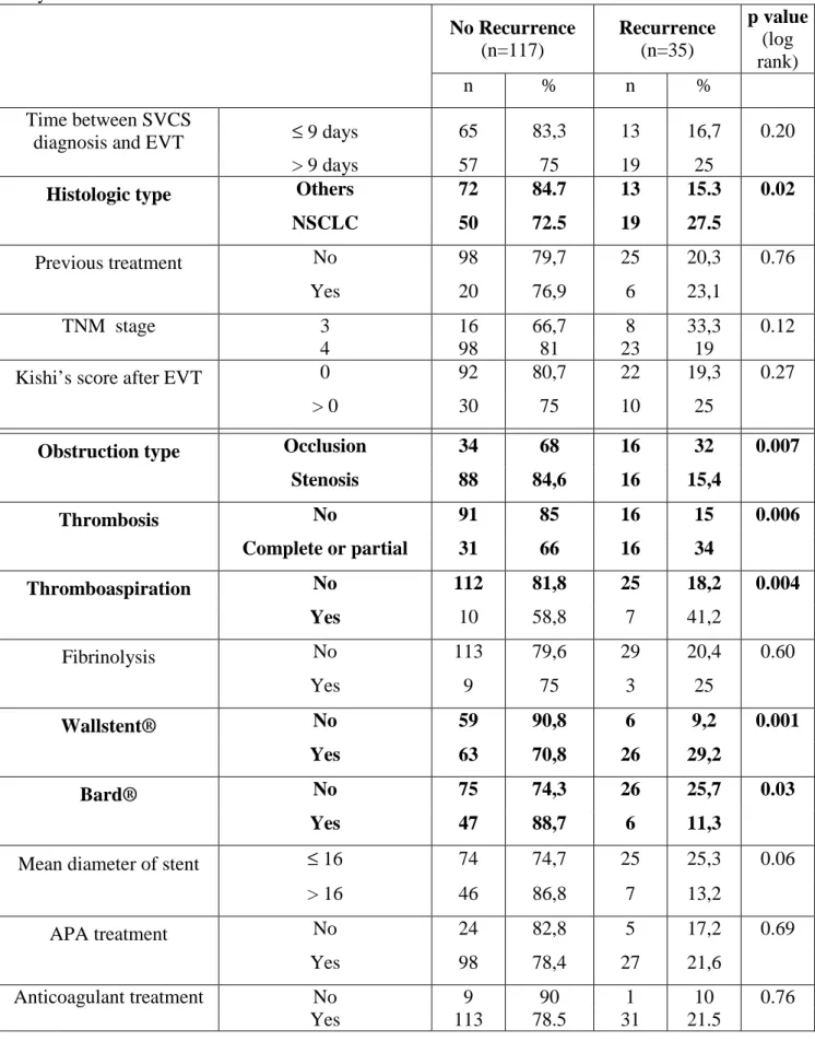
Table 1 : Kishi’s scoring System for Signs and Symptoms of SVC Syndrome18
Signs and Symptoms Grade
Neurologic symptoms
Stupor, coma, or blackout
Blurry vision, headache, dizziness, or amnesia Changes in mentation Uneasiness 4 3 2 1 Laryngopharyngeal or thoracic symptoms
Orthopnea or laryngeal edema
Stridor, hoarseness, dysphagia, glossal edema, or shortness of breath
Cough or pleural effusions
3
2 1
Nasal and facial signs or symptoms
Lip edema, nasal stiffness, epistaxis, or rhinorrhea Facial swelling
2 1
Venous dilatation Neck vein or arm vein distention, upper extremity
swelling, or upper body plethora 1
The total score for signs and symptoms was calculated as the sum of the highest grade in each class.
Table 2 : Characteristics of patients and stents Characteristics n % Gender Male Female 123 patients 41 patients 75% 25% Age 59.9 years(SD 11,7)
Cancer diagnosed before SVCS Yes No 120 patients 44 patients 73.2% 26.8% Tumour type Lung cancer
NSCLC SCLC
Other (metastasis, lymphoma)
132 patients 82 patients 50 patients 32patients 80.5% 50% 30.5% 19.5% Tumour stage Locally advanced (stage 3)
Metastatic (stage 4) 25patients 139 patients 15.2% 84.8% Time between SVCS
diagnosis and EVT 39.5 days (SD 73.5)
Previous treatment None RT/CT Medical Surgery 137 patients 9 patients 17 patients 1 patients 83.5% 5.5% 10.4% 0.6% Mean Kishi’s score before
EVT 5.3 (SD1.4)
Number of stents 186
Number of stents/patients 0 stent 1stent 2stents 3stents 7 patients 132 patients 21 patients 4 patients 4.3% 80.5% 12.8% 2.4% Type of stent Boston Scientific Wallstent® (Galway, Ireland)
Bard Memotherm® (Salt Lake City, USA) Cordis Smart® (Miami Lakes, USA) Other 104 patients 60 patients 20 patients 2 patients 55.9% 32.3% 10.7% 1.1% Mean dimensions of stent Diameter
Length
15.7mm (8-22) 63.7mm (23-90)
NSCLC, Non Small Cell Lung Cancer; SCLC, Small Cell Lung Cancer; RT, Radiotherapy; CT, Chemotherapy; SD, Standard Deviation
Table 3 : Total or partial failure risk factors (not including the three per-procedure death). Complete success (n=136) Partial or total failure (n=25) p value (Log Rank) n % n % Time between SVCS
diagnosis and EVT Median [IQR] 12 [27] 8 [9] 0.23
NSCLC 60 83,3 12 16,7 0.97 SCLC 41 83,7 8 16,3 Metastases 17 85 3 15 Histologic type Other histology 18 90 2 10 Previous treatment. No 107 82,9 22 17,1 0.57 Yes 24 88,9 3 11,1 TNM stage 3 21 84 4 16 1 4 106 83,5 21 16,5
Kishi’s score before EVT Mean ± SD 5.2 ± 1.4 5.5 ± 1.0 0.20
Obstruction type Occlusion 40 76,9 12 23,1 0.10 Stenosis 96 88,1 13 11,9
Number of engaged
vessels 1 65 86,7 10 13,3 0.38
2 22 75,9 7 24,1
3 49 86 8 14
Associated thrombosis No thrombosis 98 88,3 13 11,7 0.06 Partial or complete 38 76 12 24 Thromboaspiration No 123 85,4 21 14,6 0.31 Yes 13 76,5 4 23,5 Fibrinolysis No 126 85,1 22 14,9 0.43 Yes 10 76,9 3 23,1 Wallstent® No 60 83,3 12 16,7 0.83 Yes 76 85,4 13 14,6 Bard® No 88 83 18 17 0.65 Yes 48 87,3 7 12,7
Mean dimensions of stent Diameter (Mean±SD) 15.6 ± 2.5 15.9 ± 2.4 1 Length (Mean±SD) 63.1 ± 19.7 69.4 ± 16.8 0.24 EVT, Endovascular treatment; NSCLC, Non Small Cell Lung Cancer; SCLC, Small Cell Lung Cancer ; SD, Standard Deviation ; SVCS, Superior Vena Cava Syndrome
Table 4 : Complications risk factors Absence of complications (n=132) Complications (n=32) p value (Log Rank) n % n % Time between SVCS
diagnosis and EVT ≤ 9 days 63 75,9 20 24,1 0.17
> 9 days 69 85,2 12 14,8
Previous treatment No 102 78,5 28 21,5 0.29
Yes 25 89,3 3 10,7
TNM stage 3 21 84 4 16 0.60
4 101 78,3 28 21,7
Obstruction type Occlusion 42 77,8 12 22,2 0.54
Stenosis 90 81,8 20 18,2 Thrombosis No 90 79,6 23 20,4 0.83 Complete or partial 42 82,4 9 17,6 Thromboaspiration No 118 80,3 29 19,7 1 Yes 14 82,4 3 17,6 Fibrinolysis No 121 80,1 30 19,9 1 Yes 11 84,6 2 15,4 Wallstent® No 55 76,4 17 23,6 0.32 Yes 77 83,7 15 16,3 Bard® No 89 81,7 20 18,3 0.68 Yes 43 78,2 12 21,8
Mean diameter of stent ≤≤≤≤ 16 91 88,3 12 11,7 0.008 > 16 38 70,4 16 29,6
Heparinotherapy No 114 81,4 26 18,6 0.58
Yes 18 75 6 25
APA treatment No 26 72,2 10 27,8 0.16
Yes 106 82,8 22 17,2
Kishi’s score after EVT 0 95 83,3 19 16,7 0.26
> 0 33 75 11 25
APA, Antiplatelet Aggregation Agent; EVT, Endovascular treatment; SVCS, Superior Vena Cava Syndrome
Table 5 : Recurrence risk factors (not including failures and lost of follow-up); univariate analysis. No Recurrence (n=117) Recurrence (n=35) p value (log rank) n % n % Time between SVCS
diagnosis and EVT ≤ 9 days 65 83,3 13 16,7 0.20
> 9 days 57 75 19 25
Histologic type Others 72 84.7 13 15.3 0.02 NSCLC 50 72.5 19 27.5
Previous treatment No 98 79,7 25 20,3 0.76
Yes 20 76,9 6 23,1
TNM stage 3 16 66,7 8 33,3 0.12
4 98 81 23 19
Kishi’s score after EVT 0 92 80,7 22 19,3 0.27
> 0 30 75 10 25
Obstruction type Occlusion 34 68 16 32 0.007 Stenosis 88 84,6 16 15,4 Thrombosis No 91 85 16 15 0.006 Complete or partial 31 66 16 34 Thromboaspiration No 112 81,8 25 18,2 0.004 Yes 10 58,8 7 41,2 Fibrinolysis No 113 79,6 29 20,4 0.60 Yes 9 75 3 25 Wallstent® No 59 90,8 6 9,2 0.001 Yes 63 70,8 26 29,2 Bard® No 75 74,3 26 25,7 0.03 Yes 47 88,7 6 11,3
Mean diameter of stent ≤ 16 74 74,7 25 25,3 0.06
> 16 46 86,8 7 13,2
APA treatment No 24 82,8 5 17,2 0.69
Yes 98 78,4 27 21,6
Anticoagulant treatment No 9 90 1 10 0.76
Yes 113 78.5 31 21.5
APA, Antiplatelet Aggregation Agent; EVT, Endovascular treatment; NSCLC, Non Small Cell Lung Cancer; SVCS, Superior Vena Cava Syndrome
FIGURES AND PHOTOGRAPHS
Figure 1 : Anatomy of Superior Vena Cava Syndrome
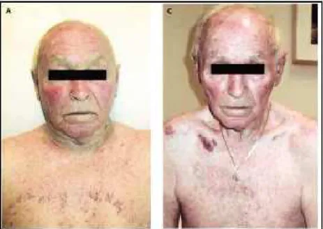
Figure 2 : Clinic aspect of a patient with Superior Vena Cava Syndrome before and after
Figure 3: Example of an EVT:
3a – Angiography before EVT, by injection into the right innominate vein. This opacification shows the proximal edge of the occlusion.
3b – Angiography before EVT, by injection into the right innominate vein which gives
the precise extent of the occlusion from the right innominate vein to the superior vena cava with occlusion of the left innominate vein.
3c - Balloon angioplasty after stent deployment.
3d - Angiography after EVT, by injection into the right innominate vein which
demonstrates the full deployment of the stent and a good flow into the superior vena cava.
d
a
b
Figure 4 : Kaplan-Meier Recurrence-free curve with 95% confidence intervals 0 10 20 30 40 50 0 .5 0 .6 0 .7 0 .8 0 .9 1 .0 Follow up (Months)
En présence des Maîtres de cette Faculté, de mes chers condisciples et devant l’effigie d’HIPPOCRATE,
Je promets et je jure d’être fidèle aux lois de l’honneur et de la probité dans l’exercice de la Médecine.
Je donnerais mes soins gratuitement à l’indigent et n’exigerai jamais un salaire au dessus de mon travail. Je ne participerai à aucun partage clandestin d’honoraires. Admis dans l’intimité des maisons, mes yeux n’y verront pas ce qui s’y passe ; ma langue taira les secrets qui me seront confiés et mon état ne servira pas à corrompre les mœurs, ni à favoriser le crime.
Je ne permettrai pas que des considérations de religion, de nation, de race, de parti ou de classe sociale viennent s’interposer entre mon devoir et mon patient.
Je garderai le respect absolu de la vie humaine.
Même sous la menace, je n’admettrai pas de faire usage de mes connaissances médicales contre les lois de l’humanité.
Respectueux et reconnaissant envers mes Maîtres, je rendrai à leurs enfants l’instruction que j’ai reçue de leurs pères.
Que les hommes m’accordent leur estime si je suis fidèle à mes promesses. Que je sois couvert d’opprobre et méprisé de mes confrères si j’y manque.
SERMENT D’HIPPOCRATE
SERMENT D’HIPPOCRATE
SERMENT D’HIPPOCRATE
ENDOVASCULAR TREATMENT OF MALIGNANT SUPERIOR VENA CAVA SYNDROME: RESULTS AND FACTORS PREDICTIVE OF EFFICACY
D Fagedet, F Thony, M Rodiere, V Monnin, GR Ferretti, A Vesin, JF Timsit, D Moro-Sibilot
Background : Superior vena cava syndrome (SVCS) is a common complication of thoracic
malignancies. Revascularisation by endovascular treatment with angioplasty and stent placement (EVT), is safe and effective and has become the first-line therapeutic instead of radiotherapy or chemotherapy.
Objective : To demonstrate the effectiveness of EVT for malignant SVCS and secondary to
analyse predictors of EVT efficacy in this pathology.
Methods : Retrospective review of the 164 patients with malignant SVCS treated with EVT
in our hospital from August 1992 to December 2007 and followed until February 2009.
Results : EVT was successful in 95.7% of cases. Thrombosis of the superior vena cava was
an independent factor for EVT failure.
Twenty-one complications (12.8%) occurred during the follow-up with 13 bleeding complications. Dilation or stenting over 16 mm was a predicting factor of complication (OR=3.19 [1.38-7.39] p=0.008). 4 patients died related to the EVT in the first 8 days. Relapse occured in 36 patients (21.9%) and was accessible to an effective re-treatment in 27. Recurrence-free rate at one year was 67.1% [57.9% ; 77.7%] Recurrence-free survival was significantly decreased in case of occlusion (HR=2.50 [1.20-4.90], p=0.01), initial associated thrombosis (HR2.60 [1.30-5.20].p=0.006). or steel stents use (HR=3.70 [1.50-9.10].p=0.004). Long term anticoagulant did not influence the risk of recurrence nor the risk of complications.
Conclusion : In malignancies, EVT is effective and safe in SVCS therapy. These results
should prompt us to indicate this treatment early in the clinical course of the patient and to avoid dilation over 16 mm.