HAL Id: hal-03124337
https://hal-univ-lemans.archives-ouvertes.fr/hal-03124337
Submitted on 28 Jan 2021
HAL is a multi-disciplinary open access
archive for the deposit and dissemination of
sci-entific research documents, whether they are
pub-lished or not. The documents may come from
teaching and research institutions in France or
abroad, or from public or private research centers.
L’archive ouverte pluridisciplinaire HAL, est
destinée au dépôt et à la diffusion de documents
scientifiques de niveau recherche, publiés ou non,
émanant des établissements d’enseignement et de
recherche français ou étrangers, des laboratoires
publics ou privés.
Distributed under a Creative Commons Attribution| 4.0 International License
against Metabolic Disorders Associated with Metabolic
Syndrome and Obesity
Claire Mayer, Léo Richard, Martine Côme, Lionel Ulmann, Hassan Nazih,
Benoît Chénais, Khadija Ouguerram, Virginie Mimouni
To cite this version:
Claire Mayer, Léo Richard, Martine Côme, Lionel Ulmann, Hassan Nazih, et al.. The Marine
Mi-croalga, Tisochrysis lutea, Protects against Metabolic Disorders Associated with Metabolic Syndrome
and Obesity. Nutrients, MDPI, 2021, 13 (2), pp.430. �10.3390/nu13020430�. �hal-03124337�
Nutrients 2021, 13, 430. https://doi.org/10.3390/nu13020430 www.mdpi.com/journal/nutrients
Article
The Marine Microalga, Tisochrysis lutea, Protects against
Metabolic Disorders Associated with Metabolic Syndrome
and Obesity
Claire Mayer 1, Léo Richard 2, Martine Côme 1, Lionel Ulmann 1, Hassan Nazih 3, Benoît Chénais 4,
Khadija Ouguerram 5 and Virginie Mimouni 1,*
1 EA 2160 MMS, Mer Molécules Santé, IUML FR 3473 CNRS, Institut Universitaire Technologique,
Département Génie Biologique, Le Mans Université, CEDEX 9, 53020 Laval, France; claire.mayer@univ-lemans.fr (C.M.); martine.come@univ-lemans.fr (M.C.); lionel.ulmann@univ-lemans.fr (L.U.)
2 GP Solutions, 98000 Monaco, Monaco; leo@gpsolutions.fr
3 EA 2160 MMS, Mer Molécules Santé, IUML FR 3473 CNRS, UFR Pharmacie, Université de Nantes,
CEDEX 1, 44035 Nantes, France; el-hassane.nazih@univ-nantes.fr
4 EA 2160 MMS, Mer Molécules Santé, IUML FR 3473 CNRS, UFR Sciences et Techniques, Le Mans
Université, CEDEX 9, 72085 Le Mans, France; benoit.chenais@univ-lemans.fr (B.C.)
5 UMR1280 PhAN, Physiopathology of Nutritional Adaptations, INRAe, University of Nantes, CHU Hôtel
Dieu, IMAD, CRNH Ouest, 44000 Nantes, France; khadija.ouguerram@univ-nantes.fr * Correspondence: virginie.mimouni@univ-lemans.fr; Tel.: +33-243-594-953
Abstract: Long-chain polyunsaturated fatty acids n-3 series and especially docosahexaenoic acid are known to exert preventive effects on metabolic disturbances associated with obesity and de-crease cardiovascular disease risk. n-3 LC-PUFAs are mainly consumed in the form of fish oil, while other sources, such as certain microalgae, may contain a high content of these fatty acids. The aim of this study was to evaluate the effects of Tisochrysis lutea (Tiso), a microalga rich in DHA, on met-abolic disorders associated with obesity. Three male Wistar rat groups were submitted for eight weeks to a standard diet or high-fat and high fructose diet (HF), supplemented or not with 12% of T. lutea (HF-Tiso). The supplementation did not affect plasma alanine aminotransferase (ALAT). Bodyweight, glycemia and insulinemia decreased in HF-Tiso rats (ANOVA, p < 0.001), while total plasma cholesterol, high-density lipoprotein-cholesterol (HDL-C) increased (ANOVA, p < 0.001) without change of low-density lipoprotein-cholesterol (LDL-C) and triacylglycerol (TAG) levels. Tiso supplementation decreased fat mass and leptinemia as well as liver TAG, cholesterol and plasma tumor necrosis factor-alpha levels (ANOVA, p < 0.001) while it did not affect interleukin 6 (IL-6), IL-4 and lipopolysaccharides levels. HF-Tiso rats showed an increase of IL-10 level in ab-dominal adipose tissue (ANOVA, p < 0.001). In conclusion, these results indicated that DHA-rich T. lutea might be beneficial for the prevention of obesity and improvement of lipid and glucose metab-olism.
Keywords: Tisochrysis lutea; DHA; obesity; metabolic syndrome; fat mass; glycemia; dyslipidemia; nonalcoholic fatty liver disease
1. Introduction
The evolution of our society’s lifestyle, characterized by an increase in energy intake and a decrease in physical activity, is the main cause of the dramatic increase in the prev-alence of overweight and obesity. Globally, the number of overweight and obese people tripled in 40 years, reaching more than 1.9 billion adults in 2016 [1]. Abdominal obesity is often associated with metabolic syndrome, a pathophysiological state defined by at least three of the following criteria: abdominal obesity, hyperglycemia, hypertriglyceridemia,
Citation: Mayer, C.; Richard, L.; Côme, M.; Ulmann, L.; Nazih, H.; Chénais, B.; Ouguerram, K.; Mimouni, V. The Marine Microalga,
Tisochrysis lutea, Protects against
Metabolic Disorders Associated to Metabolic Syndrome and Obesity.
Nutrients 2021, 13, 430.
https://doi.org/10.3390/nu13020430 Academic Editor: Lindsay Brown Received: 19 December 2020. Accepted: 27 January 2021 Published: 28 January 2021 Publisher’s Note: MDPI stays neutral with regard to jurisdictional claims in published maps and institutional affiliations.
Copyright: © 2021 by the authors. Licensee MDPI, Basel, Switzerland. This article is an open access article distributed under the terms and conditions of the Creative Commons Attribution (CC BY) license (http://creativecommons.org/licenses /by/4.0/).
and a decrease of plasma high-density lipoprotein-cholesterol (HDL-C) levels [2]. Inflam-mation, insulin resistance and an increase of plasma small, dense low-density lipoprotein-cholesterol (LDL-C) levels are often associated with metabolic syndrome and obesity [3]. These physiological and metabolic disturbances can lead to the development of nonalco-holic fatty liver disease (NAFLD), characterized by an excessive hepatic accumulation of triglycerides, more than 5.5% of liver weight [4,5].
Long-chain polyunsaturated fatty acids n-3 series (n-3 LC-PUFAs), particularly eicosapentaenoic acid (EPA) and docosahexaenoic acid (DHA) are known for their cardi-oprotective effects, as evidenced in numerous studies, notably for their antiatheroscle-rotic, antithrombotic and anti-arrhythmic effects [6–8]. Moreover, n-3 LC-PUFAs improve endothelial function and vasodilatation, decrease platelet aggregation, heart rate, blood pressure and the risk of ischemia and coronary artery disease [9]. The cardioprotective effects of n-3 LC-PUFAs are partly due to their preventive effects against dyslipidemia. Thus, n-3 LC-PUFAs exert hypotriglyceridemic effects that contribute to the decrease of plasma LDL-C and to the increase of plasma HDL-C levels [8]. n-3 LC-PUFAs can also inhibit crystalline cholesterol, thanks to their antioxidant capacities against reactive oxy-gen species (ROS) associated with cell membranes and lipoproteins [8].
n-3 LC-PUFAs exert anti-inflammatory effects by their ability to partially inhibit leu-kocyte chemotaxis, the expression of adhesion molecules and interactions between leuko-cytes and endothelial cells. n-3 LC-PUFAs decrease the production of pro-inflammatory eicosanoids (prostaglandins, leukotrienes) from arachidonic acid and decrease the pro-duction of pro-inflammatory cytokines through the inhibition of nuclear factor-kappa B (NF-κB). EPA and DHA can produce anti-inflammatory mediators such as resolvins, pro-tectins and maresins [10]. n-3 LC-PUFAs also have the ability to interact with lipid rafts as mediators with anti-inflammatory properties [8].
n-3 LC-PUFAs have beneficial effects against NAFLD by decreasing lipid accumula-tion in the liver. In fact, n-3 LC-PUFAs inhibit hepatic lipogenesis by changing the expres-sion and nuclear localization of sterol regulatory element-binding protein-1c (SREBP-1c), a transcription factor that controls several genes involved in lipogenesis, including acetyl-CoA carboxylase (ACC), fatty acid synthase (FAS) [8,11,12].
Moreover, in animal studies, n-3 LC-PUFAs were reported to improve insulin sensi-tivity despite the difficulty of demonstrating direct effects [8]. The increase of adiponecti-nemia induced by n-3 LC-PUFAs is correlated with an improvement of insulin sensitivity [8].
n-3 LC-PUFAs are highly present in fish such as salmon, herring or cod and are mainly commercialized as oils [13,14]. However, the decrease of fisheries resources related to marine pollution and overabundant fishing requires the search for new alternative sources rich in n-3 LC-PUFAs [13]. Thanks to their first place in the food chain, microalgae could be an interesting alternative to replace fish oils. In addition, they are less sensitive to heavy metal contamination [15]. Moreover, microalgae are a potential source of other highly bioactive molecules such as pigments, dietary fiber (soluble and insoluble), phy-tosterols or proteins [16]. These bioactive molecules are known to exert preventive effects against metabolic disturbances associated with fatness and would be an interesting alter-native in the prevention of metabolic syndrome and obesity [17]. Nowadays, in the Euro-pean Union (EU), the most popular microalgae widely commercialized as food ingredi-ents are Chlorella, Odontella aurita and the procaryotic cyanobacterium Arthrospira platensis. Although they can be of interest to human health, other microalgae used in aquacultures, such as Diacronema lutheri, Phaeodactylum tricornutum and Tisochrysis lutea, are not yet con-sidered as a food ingredient by the Novel Food Regulation of the EU [18].
T. lutea could be an interesting alternative to fish oil because of its many advantages:
it has a rapid growth rate, a wide range of physicochemical tolerance and contains high levels of n-3 LC-PUFAs, especially DHA, as well as other bioactive molecules such as pig-ments (fucoxanthin, chlorophylls) phytosterols (brassicasterol, stigmasterol, fucosterol)
and soluble fiber [19–23]. To our knowledge, no study has analyzed the effect of T. lutea as a dietary supplement against metabolic disorders associated with obesity.
The aim of this study was to evaluate the effects of the marine Haptophyte T. lutea, used as a food supplement, on metabolic syndrome associated metabolic disturbances. Thus, our work was to compare the effect of a high-fat (HF) diet associated with 10% of fructose in drinking tap water, known to induce obesity and metabolic syndrome [24,25], to that of the same HF diet supplemented with 12% of freeze-dried T. lutea. The animal model was a young male Wistar rat, which is commonly used to study metabolic syn-drome and obesity. When this model is submitted to an HF diet for eight weeks, it ade-quately reproduces the different metabolic disturbances encountered in human disease [24,26–28].Thus, in animal experimentations, over-consumption of fructose is involved in increasing body mass, energy intake, adiposity, glycemia, dyslipidemia and blood pres-sure [29]. The present study showed the preventive effects of T. lutea against metabolic disorders associated with metabolic syndrome and obesity, induced by HF and a high fructose diet.
2. Materials and Methods
2.1. Animal and Diets
To avoid age effects on metabolic disorders associated with obesity and any sexual endocrine fluctuation, eighteen male Wistar rats aged three weeks and weighing 130 ± 10 g were obtained from Janvier Labs (Le Genest Saint Isle, France). They were housed in a room under controlled conditions of temperature (22 ± 2 °C) and humidity (40–60%) and with a 12 h light/dark cycle. The rats were housed two per cage, 1291H Euro standard type III H in polycarbonate 425 × 266 × 185 mm (Tecniplast, Decines Charpieu, France). During one week of acclimatization, all animals were fed ad libitum with the standard diet A04 (SAFE, Augy, France) and with tap water. The nutritional protocol and all the experiments were approved by the Ethical Committee 06 Pays de la Loire and by the French Ministry of National Education, Higher Education and Research (APAFIS 10187, 31 August 2017).
Then, the animals were randomly divided into three groups of six rats and received diets ad libitum for eight weeks as follows: (1) the control (CTRL) group continued to receive the standard diet A04 providing 3.35 kcal/g, 72 kcal%, 19 kcal%, 8 kcal% from carbohydrates, proteins and lipids, respectively; (2) the HF group was fed the 260 HF diet (Safe, Augy, France) with 10% fructose in ad libitum drinking tap water (providing 1.67 kcal/mL) (Distriborg, St. Genis-Laval, France). HF diet provided 22 kcal/g, 61 kcal%, 24 kcal% from fat and carbohydrates, respectively; (3) the HF-Tiso group received an HF diet supplemented with 12% (w/w) of the freeze-dried microalga T. lutea (IBE-CNR, Florence, Italy). The dose of T. lutea supplementation was chosen from our previous studies that showed the beneficial effects of marine microalga O. aurita and P. tricornutum at 12% (w/w) after eight weeks of the diet [30,31]. These microalgae, which are rich in EPA, showed preventive effects against metabolic disturbances associated with obesity, induced by an obesogenic diet in Wistar rats [30,31]. Moreover, the selected dose of fructose supplemen-tation was based on a meta-analysis highlighting that a dose of 10% fructose in drinking water was sufficient to induce the first characteristics of metabolic syndromes, such as an increase in body weight, blood pressure and glucose, insulin and triglyceride plasma lev-els in rats [24]. These data have been confirmed by the study of Toop and Gentili [32]. Freeze-dried T. lutea provided 5.27 kcal/g, 29 kcal%, 63 kcal%, 6 kcal% from crude protein, lipids and carbohydrates, respectively. Microalgal supplementation was incorporated di-rectly in the HF diet to create a homogeneous mixture and provide 5.93 kcal/g in the HF-Tiso diet. DHA content in T. lutea was 1.41% of dry matter, equivalent to an average DHA intake of 20.3 mg/day/rat. HF diet was stored at +4 °C and T. lutea at −20 °C and renewed in the cages every three days for eight weeks.
The daily food and water consumption were evaluated in order to calculate energy intake. Energy intake (kcal/day) was calculated as the product of food consumption and dietary metabolizable energy. Food efficiency was estimated by using the following for-mula: (body weight gain (g)/energy intake (kcal)) × 100, adapted from Novelli et al. [33]. The bodyweight of the rats was monitored three times a week. Daily food and tap water consumption, and energy intake, were reported according to body weight.
The main characteristics and the fatty acid, pigment and sterol compositions of the CTRL diet, HF diet and T. lutea biomass are reported in Tables S1–S3.
The main components of the CTRL diet, HF diet and T. lutea are presented in Table S1. Data from CTRL and HF diets were provided by SAFE (Augy, France). T. lutea biomass was analyzed for proteins, carbohydrates, lipids, dietary fiber, ashes, and moistures. Total protein content was estimated as N × 6.25, where N is the nitrogen content determined through the elemental analysis. Carbohydrates were measured following Dubois et al. [34] and lipids following Marsh and Weinstein [35]. Total dietary fiber (TDF), insoluble and soluble dietary fiber (IDF and SDF) were determined by the AOAC Method 985.29 (AOAC Official Method 985.29) using commercial kits (Megazyme, Bray, Ireland). TDF was ex-perimentally analyzed, not calculated as a sum of SDF and IDF. Moisture and ashes were determined following ISTISAN protocols (ISTISAN Report 1996/34, method B, p. 7; ISTI-SAN Report 1996/34, pp. 77–78, respectively). The fatty acid compositions of the CTRL diet, HF diet and T. lutea biomass are reported in Table S2. The determination of the fatty acid composition of the CTRL diet is described below. For the HF diet, the fatty acid com-position was performed according to Simionato et al. [36]. The fatty acid analysis of T.
lutea was determined according to the ISO 12966-4:2015 + ISO 12966-2:2011 procedures.
Pigment and sterol compositions, in vitro digestibility, and antioxidant activity of T.
lutea are reported in Table S3. Pigment composition was determined by the
SCOR-UNESCO method [37]. Carotenoid content was performed by HPLC analysis according to Van Heukelem and Thomas [38]. In vitro digestibility was evaluated by the method of Boisen and Fernández [39], modified as reported by Batista et al. [40].
Antioxidant activity of extracts in 90% acetone was measured by using the 2,2-diphe-nyl-1-picrylhydrazyle (DPPH) radical scavenger according to the method of Bondet et al. [41] with slight adaptations and reported in Table S3.
2.2. Blood and Organ Sampling
After the nutritional protocol, all rats were fasted for 12 h and anesthetized by intra-peritoneal administration of a diazepam‒ketamine mix (4:3, v/v). Blood was collected from the abdominal aorta and sampled in coated tubes with 10% ethylenediaminetetraacetic acid (EDTA) from Sigma (St. Louis, MO, USA). Total blood was centrifuged at 1000× g for 10 min, the supernatant containing the plasma fraction was aliquoted in polyethylene tubes and stored at ‒20 °C. Liver, epididymal and abdominal adipose tissues were re-moved, rinsed with ice-cold NaCl solution (0.9%), weighed, frozen in liquid nitrogen, and stored at ‒80 °C until analysis.
2.3. Biochemical Analyses
Plasma levels of glucose, triacylglycerol (TAG), total cholesterol (TC), HDL-C, aspar-tate aminotransferase (ASAT) and alanine aminotransferase (ALAT) were measured by enzymatic methods using commercial enzyme kits (BIOLABO, Maizy, France). From ASAT and ALAT measures, ASAT/ALAT ratio was calculated. The atherogenic risk was evaluated by calculating the atherogenic index plasma (AIP) as the log (TAG/HDL-C) [42] and HDL-C/LDL-C ratio. Plasma LDL-C levels were estimated from the difference be-tween TC and HDL-C. The insulin level was evaluated using an ELISA kit from Thermo Scientific (Waltham, MA, USA). The homeostasis model assessment of insulin resistance index (HOMA-IR) was estimated by calculating the fasting plasma glucose concentration (mg/dL) multiplied by fasting insulinemia (µUI/mL), divided by 405 [43]. Plasma pro-inflammatory cytokines, including interleukin-6 (IL-6), tumor necrosis factor-alpha
(TNF-α), plasma and adipose anti-inflammatory cytokines such as interleuk4 (IL-4) and in-terleukin-10 (IL-10), as well as plasma leptin, were quantified using rat enzyme-linked immunosorbent assay kits (ELISA) from Abcam (Cambridge, UK) according to the man-ufacturer’s protocols. The determination of serum endotoxin levels from Gram-negative bacteria was carried out with a commercial kit (Thermo Fisher, Waltham, Massachusetts, USA) using the Limulus amebocyte lysate (LAL) method and detected the serum LPS (lip-opolysaccharide) level. This method is based on the proteolytic activation of proenzymatic factor C (present in circulating Limulus amebocytes) with endotoxins (LPS derived from the outer cell membrane of Gram-negative bacteria such as E. coli). The chromogenic assay of LAL determines endotoxin levels by measuring the activity of this protease in the pres-ence of a synthetic peptide substrate that produces p-nitroaniline (pNA) after proteolysis. A yellow coloration was obtained, and absorbance was measured at 405 nm. Endotoxin levels were quantified from an endotoxin standard provided by the kit and derived from an E. coli O111:B4 strain. Results were expressed in EU (endotoxin unit)/mL.
2.4. Hepatic Lipid Measurements
TC and TAG levels were measured from an aliquot of liver total lipid extract by en-zymatic methods using commercial enzyme kits (BIOLABO, Maizy, France).
2.5. Statistical Analysis
Data from experimental analyses are presented as mean values ± standard deviation (SD) (n = 6). After the analysis of variance by one-way ANOVA, the mean values were compared using Fisher’s least significant difference post hoc test (LSD). All statistical anal-yses were performed with Statgraphics Plus 5.1 (Manugistics Inc., Rockville, MD, USA).
3. Results
3.1. T. lutea Supplementation Decreases Body Weight and Fat Mass in Wistar rats Fed a High-Fat Diet
3.1.1. Nutritional Monitoring: Food and Water Intake
Food, water and energy intake were monitored for eight weeks (Figure 1a–c. The CTRL group displayed the highest food consumption/body weight ratio during the ex-perimental period in comparison with the other groups (Figure 1a, ANOVA, p < 0.001). The HF and HF-Tiso groups presented similar food consumption/body weight ratios ex-cept for the fourth week, where HF rats exhibited a higher ratio than the HF-Tiso group (Figure 1a, ANOVA, p < 0.001). The ratio of water intake/body weight was markedly higher in the HF-Tiso group than other groups (Figure 1b, ANOVA, p < 0.001) except for the period between the third and fifth weeks, where no difference was observed between HF and HF-Tiso groups (Figure 1b). The ratio of water intake/body weight was similar between CTRL and HF rats throughout the protocol (Figure 1b).
Figure 1. Effect of T. lutea supplementation on food intake (a), water intake (b), energy intake (c) and body weight (d) in fed Wistar rats. CTRL ( ), control group; HF ( ), high-fat group; HF-Tiso ( ), the high-fat group supplemented with T. lutea. Values are means (n = 6), with standard deviations represented by vertical bars. Statistical significance was determined using ANOVA with post hoc Fisher’s test, and means associated with letters indicate significative difference at p < 0.05 (week 1, Figure 1c) and p < 0.001 with a > b > c.
3.1.2. Energy Intake and Food Efficiency
Energy intake was calculated from water and food consumption, relative to body weight, and was similar between experimental groups except for the first week, where energy intake/body weight ratio was higher in the HF-Tiso group compared to the CTRL group (Figure 1c, p < 0.001). Food efficiency was calculated from body weight gain relative to energy intake and was similar between experimental groups, with the exception of the seventh week, where food efficiency was lower in CTRL rats compared to other groups (Figure S1, ANOVA, p < 0.01).
3.1.3. Body Weight and Fat Mass
Despite a ratio of energy intake/body weight similar between HF rats and those fed with T. lutea at the end of the nutritional protocol, the bodyweight of these two experi-mental groups was significantly different. Indeed, the final body weight was higher in the HF group compared to the other groups, and that of HF-Tiso rats was lower compared to other groups (Figure 1d, ANOVA, p < 0.001). In parallel, abdominal and epididymal adi-pose tissue weights increased with the HF diet compared with the other groups (Figure 2a,b, ANOVA, p < 0.001). Supplementation with T. lutea in HF rats significantly reduced abdominal as well as epididymal adipose tissue weight/body weight ratios compared to the HF group, while these ratios were similar between the HF-Tiso group and CTRL rats (Figure 2a, ANOVA, p < 0.001).
Figure 2. Effects of T. lutea supplementation on abdominal adipose tissue (a) and epididymal adi-pose tissue (b) weights. AAT, abdominal adiadi-pose tissue; CTRL, control group; EAT, epididymal adipose tissue; HF, high-fat group; HF-Tiso, the high-fat group supplemented with T. lutea. Values are means (n = 6), with standard deviations represented by vertical bars. Statistical significance was determined using ANOVA with post hoc Fisher’s test, and means associated with letters indi-cate significative difference at p < 0.001 with a > b > c.
3.2. Effects of T. lutea Supplementation on Physiological and Metabolic Disorders in Wistar rats Fed a High-Fat Diet
3.2.1. Plasma Biochemical Parameters and HOMA-IR Index
In HF rats, it was observed an increase of ALAT plasma levels, associated with a decrease of plasma ASAT levels and ASAT/ALAT ratio, compared to CTRL rats (Table 1, ANOVA, p < 0.001). Plasma levels of ASAT were decreased in the HF-Tiso group, associ-ated with a low ratio of ASAT/ALAT (Table 1, ANOVA, p < 0.001).
Table 1. Effects of T. lutea on plasma biochemical parameters.
CTRL CTRL HF HF-Tiso
Plasma biochemical parameters
ASAT (UI/L) 58.72± 6.22 51.80 ***± 7.83 45.09 ***† ± 5.39 ALAT (UI/L) 44.46± 4.57 52.10 ***± 4.10 52.00 ***± 6.79 ASAT/ALAT 1.36± 0.11 1.24 ***± 0.17 0.87 ***† ± 0.09 Glucose (mmol/L) 9.17± 1.48 10.01 ***†± 0.58 7.73 ***† ± 0.10 Insulin (µUI/L) 37.68± 8.26 89.17 ***± 12.95 64.41 ***† ± 11.69 Leptin (ng/mL) 2.02± 0.52 3.87 ***± 0.53 2.17 † ± 0.30 HOMA-IR 0.97± 0.34 2.80 ***± 0.57 1.30 ***† ± 0.33 TAG (mmol/L) 3.07± 0.84 6.85 *** ± 1.16 3.53 † ± 1.02 TC (mmol/L) 2.14± 0.36 2.72 ***± 0.45 3.64 ***† ± 0.35 HDL-C (mmol/L) 2.03± 0.27 1.72 ***± 0.20 2.83 ***† ± 0.26 LDL-C (mmol/L) 0.44± 0.12 0.96 ***± 0.21 0.86 ***± 0.19 HDL/LDL 2.65± 0.31 1.62 ***± 0.39 2.73 † ± 0.44 AIP 0.32± 0.08 0.51 ***± 0.13 0.18 ***† ± 0.06
AIP, atherogenic index of plasma; ALAT, alanine amino-transferase; ASAT, aspartate amino-trans-ferase; CTRL, standard diet; HDL-C, high-density lipoprotein cholesterol; HF, high-fat diet; HF-Tiso, high-fat diet supplemented with 12% of T. lutea; HOMA-IR, homeostasis model assessment of insulin resistance; LDL-C, low-density lipoprotein cholesterol; TAG, triacylglycerols; TC, total cholesterol. Values are means (n = 6), and statistical significance was determined using ANOVA with post hoc Fisher’s test. Mean values associated with *** were significantly different from those of the CTRL group (p < 0.001), and mean values with † were significantly different from those of
In the HF group, basal plasma levels of glucose, insulin and leptin were higher com-pared to CTRL and HF-Tiso rats (Table 1, ANOVA, p < 0.001). The supplementation with
T. lutea partially prevented hyperinsulinemia observed in the HF group and restored the
basal leptin level, whereas it was observed the lowest glycemia in the HF-Tiso group (Ta-ble 1, ANOVA, p < 0.001). In accordance with these results, the HOMA-IR index increased with the HF diet compared to CTRL rats, and it was decreased with T. lutea supplemen-tation (Table 1, ANOVA, p < 0.001).
3.2.2. Plasma Lipids, HDL/LDL Ratio and AIP Index
The HF group showed an increase in plasma TG, TC and LDL-C levels and a decrease in plasma HDL-C levels (Table 1, ANOVA, p < 0.001). T. lutea supplementation restored triglyceridemia and increased plasma HDL-C levels as well as plasma TC levels compared to CTRL and HF rats (Table 1, ANOVA, p < 0.001). LDL-C levels have not been restored after T. lutea supplementation (Table 1, ANOVA, p < 0.001). HF fed Wistar rats exhibited a high AIP index, an effective index associated with abdominal obesity, and a decrease in HDL/LDL ratio, an atherogenicity index (Table 1, ANOVA, p < 0.001), while T. lutea sup-plementation decreased the AIP restored HDL-C/LDL-C ratio (Table 1, ANOVA, p < 0.001).
3.3. Effects of T. lutea on Inflammatory Status
3.3.1. Pro-Inflammatory and Anti-Inflammatory Cytokines
As shown in Figure 3, plasma concentrations of pro-inflammatory cytokines, includ-ing TNF-α and IL-6, were significantly increased in the HF group compared to those in the CTRL and HF-Tiso groups (ANOVA, p < 0.001). The results also evidenced that T. lutea supplementation restored TNF-α concentration (ANOVA, p < 0.001), while plasma IL-6 concentration was not significantly decreased compared to the HF group. Anti-inflamma-tory cytokines were investigated in plasma and abdominal adipose tissue. The adipose tissue IL-10 concentrations and plasma levels of IL-4 were decreased with the HF diet compared to the CTRL diet (Figure 3, ANOVA, p < 0.001). Supplementation with T. lutea significantly improved inflammatory status by an increase of IL-10 levels in the adipose tissue, compared to the HF group (Figure 3, ANOVA, p < 0.001). Nevertheless, the plasma concentration of IL-4 in the HF-Tiso group was not restored compared to CTRL rats (Fig-ure 3, ANOVA, p < 0.001).
Figure 3. Effects of T. lutea supplementation on inflammatory and anti-inflammatory biomarkers. The plasma concentrations of (a) TNF-α, (b) IL-6, (c) IL-4 and adipocyte concentration in (d) IL-10.
CTRL, control group; HF, high-fat group; HF-Tiso, the high-fat group supplemented with T. lutea; IL-4, interleukin 4; IL-6, interleukin 6; IL-10, interleukin 10; TNF-α, tumor necrosis factor-α. Values are means (n = 6), with standard deviations represented by vertical bars. Statistical significance was determined using ANOVA with post hoc Fisher’s test, and means associated with letters indi-cate significative difference at p < 0.001 with a > b > c.
3.3.2. Serum LPS Levels
Serum LPS concentrations were measured in the different experimental groups (Fig-ure 4). An increase of the LPS concentration in serum, named endotoxemia, was observed in Wistar rats fed the HF diet compared to those fed the standard diet (Figure 4, ANOVA,
p < 0.001). The HF-Tiso diet improved serum levels of LPS compared to the HF group
(Figure 4, ANOVA, p < 0.001).
Figure 4. Effects of T. lutea supplementation on endotoxemia in HF-fed Wistar rats. CTRL, control group; HF, high-fat group; HF-Tiso, the high-fat group supplemented with T. lutea; LPS, lipopoly-saccharide. Values are means (n = 6), with standard deviations represented by vertical bars. Statis-tical significance was determined using ANOVA with post hoc Fisher’s test, and means associated with letters indicate significative difference at p < 0.001 with a > b > c.
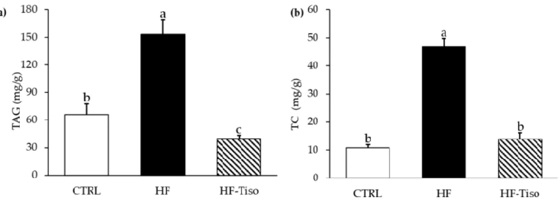
3.4. Effects of T. lutea on Liver Triglyceride and Total Cholesterol Levels
The HF diet induced an increase in hepatic triglyceride and TC contents (Figure 5a,b, ANOVA, p < 0.001). T. lutea supplementation markedly decreased liver levels of triglycer-ides in HF-Tiso rats compared to the other experimental groups (Figure 5a, ANOVA, p < 0.001). In addition, hepatic total cholesterol levels were restored in the HF-Tiso group (Figure 5b).
Figure 5. Effects of T. lutea supplementation on liver triglyceride (a) and cholesterol (b) levels. CTRL, control group; HF, high-fat group; HF-Tiso, the high-fat group supplemented with T. lutea; TAG, triacylglycerols; TC, total cholesterol. Values are means (n = 6), with standard deviations represented by vertical bars. Statistical significance was determined using ANOVA with post hoc Fisher’s test, and means associated with letters indicate significative difference at p < 0.001 with a > b > c.
4. Discussion
The aim of this study was to assess the impact of T. lutea used as a food supplement on metabolic disorders associated with metabolic syndrome and obesity, including over-weight, dyslipidemia, inflammation and NAFLD. The results showed that T. lutea supple-mentation induced body weight and adipose tissue weight reduction. It also contributed to an improvement of plasma lipid parameters, insulinemia, leptinemia and inflammatory status and thus decreased atherogenic risk.
In accordance with the literature [24,26–28], our study highlighted that rats submit-ted to HF diet associasubmit-ted with fructose supplementation in drinking water (10%) pre-sented increased body weight, fat mass and liver weight associated with high plasma lev-els of ALAT, a low ASAT/ALAT ratio and the presence of NAFLD. In addition, dyslipidemia was observed in association with a high AIP index and a low HDL/LDL ratio. Plasma levels of glucose, insulin, leptin, inflammatory cytokines, serum levels of LPS and HOMA-IR index were increased while anti-inflammatory cytokines were de-creased.
4.1. T. lutea Supplementation Reduced Body Weight, Abdominal and Epididymal Adipose Weights in HF-Fed Wistar rats
In the present study, a similar food and energy intake was observed between the HF and HF-Tiso groups, with the exception of the fourth week of the protocol. In parallel, the bodyweight of HF-Tiso rats was significantly reduced compared to the other experimental groups. This suggests that the decreased body weight observed in HF-Tiso rats is not caused by a deficit in energy intake and/or a decrease in food intake but may be due to the nutritional quality provided by T. lutea biomass and the potential synergistic effect of its biomolecules.
The high levels in DHA in T. lutea biomass (1.41% of dry weight, equivalent to 19.1 mg DHA/rat/day for the HF-Tiso group) could explain the reduction of body weight and fat mass observed in the HF-Tiso group. Indeed, previous studies conducted in animals showed the beneficial effects of DHA in reducing hypertrophy and hyperplasia of fat cells by activation of transcription factors involved in pre-adipocyte differentiation such as pe-roxisome proliferator-activated receptor-gamma (PPARγ) and the inhibition of mitogen-activated protein kinases (MAPK), involved in the last phase of adipocyte differentiation [44–46]. The study of Kim et al. [47] showed that DHA induces apoptosis of adipocytes before the differentiation stage and reduces lipid accumulation in adipocytes and the number of lipid droplets, through increased lipolysis associated with activation of PPARα as well as induction of uncoupling protein-2 (UCP2).
Furthermore, high levels of fucoxanthin in T. lutea biomass (64.2% of carotenoids, 9% of total pigments and 0.48% of dry weight, equivalent to 7 mg fucoxanthin/rat/day for the HF-Tiso group) could be another explanation for the decreased body weight and fat mass observed in HF-Tiso rats. In accordance with the present study, similar effects were ob-served in mice supplemented with a lipid fraction rich in fucoxanthin (9.6% of dry weight) from the macroalga Undaria pinnatifida [48]. Another study demonstrated that fucoxanthin could stimulate energy expenditures and β-oxidation, leading to a loss of body weight and fat mass [49]. Thus, it was shown that supplementation with synthetic fucoxanthin or
Undaria pinnatifida-derived fucoxanthin (0.98 mg/g dry weight) at 400 mg/kg body weight
in HF-fed rats, increased energy expenditures and β-oxidation, associated with decreasing gene expression involved in adipogenesis such as PPAR-α, peroxisome proliferator-acti-vated receptor-gamma coactivator-1 alpha (PGC-1α), PPAR-γ and UCP1 [49].
A previous animal study demonstrated that chlorophyll supplementation (0.18 mg/10 g body weight/day) could also exert beneficial effects against obesity by the de-crease of body weight gain, the improvement of glucose tolerance and the reduction of low-grade inflammation in HFD-fed C57BL/6 J male mice [50]. These effects could be due to the preventive action of chlorophyll supplementation on gut dysbiosis, characterized
by the decreased Firmicutes/Bacteroidetes ratios, the increased abundance of Blautia bac-teria, and the significant decrease of Lactococcus and Lactobacillus bacteria [50].
According to the study of Jung et al. [51], it was demonstrated that fucosterol, a phy-tosterol from the macroalga Ecklonia stolonifera, decreased the accumulation of 3T3-L1 pre-adipocytes via the inhibition of transcription factors PPARγ and CCAAT/enhancer-bind-ing protein alpha (C/EBPα) [51]. Thus, fucosterol, abundantly found in T. lutea biomass (16.5% of total sterols, 0.23% of dry weight), could exert anti-obesogenic effects in rats supplemented with T. lutea.
Dietary fiber has positive effects for reducing body weight because it has decreased gastric emptying, slow energy and nutrient absorption, and may influence oxidation and storage of fat [52]. Their anti-obesogenic potential cannot be excluded and would be due to increasing intraluminal viscosity and fermentation of short-chain fatty acids [52]. These physiological changes would promote satiation and/or satiety and decrease food intake, as observed in rats supplemented by T. lutea in the fourth week of the protocol.
Furthermore, we can suggest that food intake/body weight ratio and indirectly the body weight and fat mass could be influenced by bioactive molecules like microalgal pol-ysaccharides. Indeed, the effects of microalgal polysaccharides on intestinal microbiota are different depending on their nature and influence the regulation of appetite and weight gain. For example, it has been previously suggested that polysaccharides from the microalga Isochrysis galbana exert prebiotic effects through the beneficial increase of
Lacto-bacillus lactis bacteria activity and reducing the growth and activity of enterobacteria and
pathogens in diabetic rats supplemented with Isochrysis galbana, at a dose of 50 mg/day [53].
4.2. Effects of T. lutea, as a Dietary supplement, in the Prevention of Dyslipidemia and Athero-sclerosis
In the present study, T. lutea showed anti-dyslipidemic effects when used as a dietary supplement and could be explained by its high levels in bioactive molecules such as n-3 LC-PUFA, dietary fiber, phytosterols or fucoxanthin, which have beneficial effects in the regulation of lipid metabolism [54–56]. Fucosterol is also abundantly found in T. lutea bi-omass and showed the ability to increase plasma HDL-C levels [57].
Our results suggest the beneficial effects of soluble fiber (equivalent to 43 mg/rat/day for HF-Tiso rats) and DHA from T. lutea in reducing dyslipidemia. Indeed, dietary fiber is known for its beneficial effects on intestinal transit, thus playing a preventive role against cardiovascular diseases [58,59]. Moreover, hypotriglyceridemic properties of DHA have been shown in the literature and could be explained by the inhibition of triglyceride pro-duction and hepatic lipogenesis [8,60].
Dyslipidemia is a major risk factor in the development of atherosclerosis, and high AIP index and HDL/LDL ratio are, respectively, efficient markers of atherosclerosis and cardiovascular diseases [61–63]. In our study, rats supplemented with T. lutea showed a low AIP index compared to the other groups. In parallel, the HDL/LDL ratio was restored in HF-Tiso rats. These findings could be explained by the various molecules present in T.
lutea, such as n-3 LC-PUFA, phytosterols and fiber, known to exert cardioprotective effects
[64–67].
4.3. Effects of T. lutea Supplementation on Inflammatory Status in HF-Fed Wistar rats
Decreased plasma levels of TNF-α pro-inflammatory cytokines have been observed in HF-Tiso rats. In parallel, although plasma levels of 6 and 4 were not restored, IL-10 level in abdominal adipose tissue was increased in the HF-Tiso group, reflecting the partial restoration of basal inflammatory status in rats supplemented with T. lutea. Our data may suggest that DHA from T. lutea, known for its anti-inflammatory effects, re-duced production of pro-inflammatory cytokines and increased anti-inflammatory cyto-kine production [68]. Furthermore, similar effects were observed in a previous animal
study that used DHA-rich oil from the microalga Schizochytrium sp. as a dietary supple-ment in C57BL/6 mice submitted to HF diet [69].
Fucosterol is known to have anti-inflammatory properties, mainly related to the in-hibition of the nuclear factor-kappa B (NF- B) and p38 mitogen-activated protein kinases (p38 MAPK) pro-inflammatory pathways [70]. In addition, T. lutea is a significant source of anti-inflammatory compounds, including carotenoids [71]. As shown previously, fuco-xanthin, at a dose of 0.6%, decreased the production of pro-inflammatory markers such as TNF-α and cyclooxygenase-2 (COX-2) in mice submitted to HF diet for four weeks [72]. In the present study, decreased inflammation in the HF-Tiso group could be explained by the synergistic effects of various bioactive molecules, including n-3 LC-PUFA and pig-ments.
In order to better understand the anti-inflammatory effects of T. lutea supplementa-tion, serum LPS level was quantified in HF-Tiso rats. In agreement with the literature, the HF diet induced an increase of serum LPS level, defined as metabolic endotoxemia, by its capacity to modulate the intestinal microbiota. This change leads to an increase of intesti-nal permeability and then to the passage of LPS into the bloodstream [73]. Subsequently, the binding of LPS to adipocyte Toll-like receptor 4 (TLR4) receptors and pattern recogni-tion receptors (PRRs) activates inflammatory pathways such as Nf-κB, inducing pro-inflammatory cytokine production from adipose tissue [74]. By contrast, serum LPS level decreased in the HF-Tiso group, suggesting a preventive effect of T. lutea against meta-bolic endotoxemia. These observations could be attributed to n-3 LC-PUFA from T. lutea supplementation. Indeed, n-3 LC-PUFA demonstrated preventive effects against meta-bolic endotoxemia by their protective role against intestinal dysbiosis and permeability [73].
In accordance with our results, inflammation leads to hyperleptinemia, a marker of pro-inflammatory state and positively correlated with fat mass [74,75]. T. lutea supple-mentation, rich in DHA, restored plasma leptin level in HF-fed Wistar rats. The study conducted by Yook et al. [76] showed a leptinemia decrease after eight weeks of treatment in HF-fed C57BL/6 J mice supplemented with Aurantiochytrium microalga oil rich in DHA. Fucoxanthin could be involved in leptinemia regulation in HF-Tiso rats. Indeed, a previous study showed the decrease of leptinemia in C57BL/6 J mice submitted for eight weeks to HF diet combined with P. tricornutum extract rich in fucoxanthin (corresponding to 0.2% fucoxanthin) [77].
4.4. Hypoglycemic and Hypoinsulinemic Effects of T. lutea Supplementation in HF-Fed Wistar rats
Glycemia was significantly lower in HF-Tiso rats compared to the other groups. In parallel, plasma insulin level and HOMA-IR index were improved in the HF-Tiso group, suggesting a protective effect of T. lutea against insulin resistance. As well as other micro-alga species, T. lutea could have an anti-diabetic activity due to its high levels of bioactive molecules such as DHA and fucoxanthin [17,53,78–80]. Thus, the study of Yook et al. [76] showed preventive effects of Aurantiochytrium sp. microalga oil (rich in n-3 PUFA, DHA) against hyperinsulinemia in C57BL/6 J mice submitted to a hyperlipidic diet, suggesting the beneficial effects of n-3 LC-PUFA, and particularly DHA, in the improvement of insu-lin sensitivity. Indeed, a recent study showed that DHA significantly inhibits protein ex-pression of the mechanistic target of rapamycin complex 1 (mTORC1) signaling pathway and increases phosphorylated-AKT protein (p-AKT) expression to reduce insulin re-sistance [81].
In another study, a lipid extract rich in fucoxanthin from the macroalga Undaria
pin-natifida showed beneficial effects in the reduction of glycemia insulinemia in obese and
diabetic mice [82]. This study suggests that fucoxanthin could improve insulin sensitivity and carbohydrate homeostasis through the regulation of glucose transporter-4 (GLUT-4),
the reduction of hyperinsulinemia and neoglucogenesis, and the modification of the en-zymatic activity of liver glucose regulatory enzymes such as glucose-6-phosphatase and phosphoenolpyruvate carboxykinase [83].
4.5. Effects of T. lutea on Non-Alcoholic Fatty Liver Disease
An excess of liver TAG and cholesterol levels can be hepatotoxic [84]. Thus, the in-tegrity and metabolic functions of the liver were studied. In HF rats, the plasma ALAT level was increased and associated with a decrease of the ASAT/ALAT ratio, an indicator of hepatotoxicity in NAFLD [85]. Although ASAT/ALAT ratio was significantly lower in the HF-Tiso group due to low plasma ASAT levels, T. lutea supplementation did not im-pact plasma ALAT level and hepatomegaly induced by the HF diet, suggesting an absence of hepatotoxicity.
In parallel, T. lutea supplementation improved liver total cholesterol levels and sig-nificantly decreased TAG levels, suggesting NAFLD preventive effects of T. lutea. Similar effects were observed in Wistar rats supplemented for 66 days with freeze-dried
Diacro-nema vlkianum, a marine microalga (equivalent to 101 mg/kg EPA and DHA in the diet),
suggesting the preventive effect of n-3 LC-PUFA in the development of NAFLD [86]. DHA has been shown to inhibit lipid accumulation, particularly triglycerides, through the acti-vation of free fatty acid 4 (FFA4) membrane receptor. Its actiacti-vation prevents hepatic stea-tosis by inhibiting gene and protein expression of SREBP-1c through signaling cascade activation that involves Gq/11, calcium/calmodulin-dependent protein kinase kinase 2 (CaMKK) and AMP-activated protein kinase (AMPK) [87].
In addition, a protective effect of fucoxanthin from dried U. pinnatifida brown algae (0.2% fucoxanthin in diet) against liver lipid accumulation was demonstrated in C57BL/6 mice submitted to an HF diet through the reduction of activity of lipogenic hepatic en-zymes such as glucose-6-phosphate dehydrogenase (G6PD), FAS and phosphatidate phosphatase (PAP), as well as the stimulation of enzymes involved in β-oxidation such as carnitine palmitoyltransferase 1a (CPT1a) [88]. Fucoxanthin also regulates the expression of genes associated with lipid metabolism, such as ACC, FAS and acyl-CoA cholesterol acyltransferase (ACAT), an enzyme that converts free cholesterol into cholesterol ester [89–92]. In parallel, fucoxanthin increases lipolysis through increasing expression of adi-pose triglyceride lipase (AGTL) and phosphorylation of hormone-sensitive lipase (HSL) [93,94]. The study of Chang et al. [94] demonstrated that increasing lipolysis associated with decreasing lipogenesis could be induced by activation of sirtuin 1 (Sirt1)/AMPK pathway [94].
5. Conclusions
This study highlighted the beneficial effect of the marine microalga T. lutea, as a die-tary supplement, in the prevention of metabolic syndrome and obesity in Wistar rats. Met-abolic disturbances associated with obesity were induced by a hyperlipidic diet and fruc-tose-rich drinking water. Our results showed that T. lutea has an effective potential to re-duce hyperglycemia, hypertriglyceridemia by restoring HDL level, hyperleptinemia and an excess of liver lipid levels, body weight and fat mass. Thus, the observed effects could be assigned to the bioactive molecules such as n-3 PUFAs, fucoxanthin, phytosterols, fiber, etc.) and their potential synergy within the biomass of T. lutea. However, this study did not determine the specific effect of each biomolecule from T. lutea biomass on physiolog-ical parameters involved in obesity and metabolic syndrome. The bioactivity of T. lutea purified extracts on inflammation, insulin resistance, and lipotoxicity should be further explored.
Supplementary Materials: The following are available online at www.mdpi.com/2072-6643/13/2/430/s1, Figure S1: comparison of food efficiency between the different experimental groups, Table S1: biochemical composition of standard, high-fat diets, and freeze-dried T. lutea bio-mass, Table S2: fatty acid composition of diets, Table S3: pigment and sterol composition, antioxi-dant activity and in vitro digestibility of freeze-dried T. lutea biomass.
Author Contributions: Conceptualization, C.M., H.N., K.O. and V.M.; methodology, C.M., L.R., M.C., L.U. and V.M.; validation, H.N., K.O. and V.M.; investigations, C.M and V.M; data curation, C.M. and V.M.; writing—original draft preparation, C.M. and V.M; writing—review and visualiza-tion, L.U., B.C., H.N., K.O. and V.M.; editing, CM and V.M.; supervision, B.C., K.O. and V.M.; project administration, V.M.; funding acquisition, V.M. All authors have read and agreed to the published version of the manuscript.
Funding: This research was funded by the financial support of the regional program “Food for To-morrow-Cap Aliment” Research, Formation and Innovation in Pays de la Loire, including a co-fi-nancing at 50% of a Ph.D. grant to CM. The thesis was also co-funded at 50% by Le Mans University with joint financial support from the Conseil Général de la Mayenne, Laval Agglomération and CCI de la Mayenne.
Institutional Review Board Statement: The study was conducted according to the guidelines of the Declaration of Helsinki and approved by the Ethical Committee 06 Pays de la Loire and by the French Ministry of National Education, Higher Education and Research (APAFIS 10187, 31 August 2017).
Informed Consent Statement: Not applicable. Data Availability Statement: Not applicable.
Acknowledgments: The authors thank Frédérique Guéno and Gilles Braud for their technical assis-tance and important advice during animal experimentation. They also thank Graziella Chini Zittelli and Cécilia Faraloni for providing us with freeze-dried T. lutea and carrying out the biomass anal-yses.
Conflicts of Interest: The authors declare no conflicts of interest. References
1. World Health Organization (WHO). Global Health Estimates 2016: Deaths by Cause, Age, Sex, by Country and by Region, 2000–2016; World Health Organization: Genève, Switzerland, 2018.
2. Alberti, K.G.M.M.; Eckel, R.H.; Grundy, S.M.; Zimmet, P.Z.; Cleeman, J.I.; Donato, K.A.; Fruchart, J.C.; James, W.P.; Loria, C.M.; Smith, S.C., Jr. Harmonizing the metabolic syndrome: A joint interim statement of the International Diabetes Federation Task Force on Epidemiology and Prevention; National Heart, Lung, and Blood Institute; American Heart Association; World Heart Federation; International Atherosclerosis Society; and International Association for the Study of Obesity. Circulation 2009, 120, 1640–1645.
3. Després, J.-P.; Lemieux, I. Abdominal obesity and metabolic syndrome. Nat. Cell Biol. 2006, 444, 881–887, doi:10.1038/na-ture05488.
4. Browning, J.D.; Szczepaniak, L.S.; Dobbins, R.; Nuremberg, P.; Horton, J.D.; Cohen, J.C.; Grundy, S.M.; Hobbs, H.H. Prevalence of hepatic steatosis in an urban population in the United States: Impact of ethnicity. Hepatology 2004, 40, 1387–1395, doi:10.1002/hep.20466.
5. Luyendyk, J.P.; Guo, G.L. Steatosis DeLIVERs High-Sensitivity C-Reactive Protein. Arter. Thromb. Vasc. Biol. 2011, 31, 1714–1715, doi:10.1161/atvbaha.111.230722.
6. Marhuenda, J.; Villaño, D.; Cerdá, B.; Zafrilla, P. Cardiovascular disease and nutrition. In Nutrition in Health and Disease; Mogzik, G., Fliger, M., Eds.; IntechOpen: London, UK, 2019.
7. Manson, J.E.; Cook, N.R.; Lee, I.-M.; Christen, W.; Bassuk, S.S.; Mora, S.; Gibson, H.; Albert, C.M.; Gordon, D.; Copeland, T.; et al. Marine n−3 Fa y Acids and Prevention of Cardiovascular Disease and Cancer. N. Engl. J. Med. 2019, 380, 23–32, doi:10.1056/nejmoa1811403.
8. Albracht-Schulte, K.; Kalupahana, N.S.; Ramalingam, L.; Wang, S.; Rahman, S.M.; Robert-McComb, J.; Moustaid-Moussa, N. Omega-3 fatty acids in obesity and metabolic syndrome: A mechanistic update. J. Nutr. Biochem. 2018, 58, 1–16, doi:10.1016/j.jnutbio.2018.02.012.
9. Yashodhara, B.M.; Umakanth, S.; Pappachan, J.M.; Bhat, S.K.; Kamath, R.; Choo, B.H. Omega-3 fatty acids: A comprehensive review of their role in health and disease. Postgrad. Med. J. 2009, 85, 84–90, doi:10.1136/pgmj.2008.073338.
10. Calder, P.C. Marine omega-3 fatty acids and inflammatory processes: Effects, mechanisms and clinical relevance. Biochim. Bio-phys. Acta (BBA) Mol. Cell Biol. Lipids 2015, 1851, 469–484, doi:10.1016/j.bbalip.2014.08.010.
11. Kim, H.-J.; Takahashi, M.; Ezaki, O. Fish oil feeding decreases mature sterol regulatory element-binding protein 1 (SREBP-1) by down-regulation of SREBP-1c mRNA in mouse liver: A possible mechanism for down regulation of lipogenic enzyme mRNAs. J. Biol. Chem. 1999, 274, 25892–25898.
12. Chadli, F.K.; Andre, A.; Prieur, X.; Loirand, G.; Meynier, A.; Krempf, M.; Nguyen, P.; Ouguerram, K. n-3 PUFA prevent meta-bolic disturbances associated with obesity and improve endothelial function in golden Syrian hamsters fed with a high-fat diet. Br. J. Nutr. 2011, 107, 1305–1315, doi:10.1017/s0007114511004387.
13. Martins, D.A.; Custódio, L.; Barreira, L.; Pereira, H.; BeHamadou, R.; Varela, J.C.; Abu-Salah, K.M. Alternative Sources of n-3 Long-Chain Polyunsaturated Fatty Acids in Marine Microalgae. Mar. Drugs 201n-3, 11, 2259–2281, doi:10.n-3n-390/md11072259. 14. Abedi, E.; Sahari, M.A. Long-chain polyunsaturated fatty acid sources and evaluation of their nutritional and functional
prop-erties. Food Sci. Nutr. 2014, 2, 443–463, doi:10.1002/fsn3.121.
15. Lakra, N.; Mahmood, S.; Marwal, A.; Sudheep, N.M.; Anwar, K. Bioengineered plants can be an alternative source of omega-3 fatty acids for human health. In Plant and Human Health; Ozturk, M., Hakeem, K.R., Eds.; Springer International Publishing: Cham, Switzerland, 2019; Volume 2, pp. 361–382.
16. Hamed, I. The Evolution and Versatility of Microalgal Biotechnology: A Review. Compr. Rev. Food Sci. Food Saf. 2016, 15, 1104– 1123, doi:10.1111/1541-4337.12227.
17. Zhao, C.; Wu, Y.; Yang, C.; Liu, B.; Huang, Y. Hypotensive, hypoglycaemic and hypolipidaemic effects of bioactive compounds from microalgae and marine micro-organisms. Int. J. Food Sci. Technol. 2015, 50, 1705–1717, doi:10.1111/ijfs.12860.
18. García, J.L.; de Vicente, M.; Galán, B. Microalgae, old sustainable food and fashion nutraceuticals. Microb. Biotechnol. 2017, 10, 1017–1024.
19. Liu, C.-P.; Lin, L.-P. Ultrastructural study and lipid formation of Isochrysis sp. CCMP1324. Bot. Bull. Acad. Sin. 2001, 42, 207– 214.
20. O’Shea, S.K.; Holland, F.; Bilodeau, A. Modeling the Effects of Salinity and pH on the Cadmium Bioabsorptive Properties of the MicroalgaeIsochrysis galbana(T-Iso) in Coastal Waters. J. Coast. Res. 2010, 261, 59–66, doi:10.2112/08-1073.1.
21. de Jesus Raposo, M.F.; de Morais, R.M.S.C.; de Morais, A.M. Health applications of bioactive compounds from marine micro-algae. Life Sci. 2013, 93, 479–486.
22. Alkhamis, Y.; Qin, J.G. Comparison of pigment and proximate compositions of Tisochrysis lutea in phototrophic and mixo-trophic cultures. J. Appl. Phycol. 2016, 28, 35–42.
23. Mimouni, V.; Couzinet-Mossion, A.; Ulmann, L.; Wielgosz-Collin, G. Lipids from microalgae. In Microalgae in Health and Disease Prevention; Levine, I., Fleurence, J., Eds.; Academic Press: Cambridge, MA, USA, 2018; pp. 109–131.
24. Wong, S.K.; Chin, K.-Y.; Suhaimi, F.H.; Fairus, A.; Soelaiman, I.-N. Animal models of metabolic syndrome: A review. Nutr. Metab. 2016, 13, 1–12, doi:10.1186/s12986-016-0123-9.
25. Lima, M.L.R.P.; Leite, L.H.R.; Gioda, C.R.; Leme, F.O.P.; Couto, C.A.; Coimbra, C.C.; Leite, V.H.R.; Ferrari, T.C.A. A Novel Wistar Rat Model of Obesity-Related Nonalcoholic Fatty Liver Disease Induced by Sucrose-Rich Diet. J. Diabetes Res. 2015, 2016, 1–10, doi:10.1155/2016/9127076.
26. Buettner, R.; Schölmerich, J.; Bollheimer, L.C. High-fat Diets: Modeling the Metabolic Disorders of Human Obesity in Rodents. Obesity 2007, 15, 798–808, doi:10.1038/oby.2007.608.
27. Kakimoto, P.A.; Kowaltowski, A.J. Effects of high fat diets on rodent liver bioenergetics and oxidative imbalance. Redox Biol. 2016, 8, 216–225, doi:10.1016/j.redox.2016.01.009.
28. Marques, C.; Meireles, M.; Norberto, S.; Leite, J.; Freitas, J.; Pestana, D.; De Faria, A.M.C.; Calhau, C. High-fat diet-induced obesity Rat model: A comparison between Wistar and Sprague-Dawley Rat. Adipocyte 2015, 5, 11–21, doi:10.1080/21623945.2015.1061723.
29. Panchal, S.K.; Poudyal, H.; Iyer, A.; Nazer, R.; Alam, A.; Diwan, V.; Kauter, K.; Sernia, C.; Campbell, F.; Ward, L.; et al. High-carbohydrate, High-fat Diet–induced Metabolic Syndrome and Cardiovascular Remodeling in Rats. J. Cardiovasc. Pharmacol. 2011, 57, 611–624, doi:10.1097/fjc.0b013e3181feb90a.
30. Haimeur, A.; Mimouni, V.; Ulmann, L.; Martineau, A.-S.; Messaouri, H.; Pineau-Vincent, F.; Tremblin, G.; Meskini, N. Fish Oil and Microalga Omega-3 as Dietary Supplements: A Comparative Study on Cardiovascular Risk Factors in High-Fat Fed Rats. Lipids 2016, 51, 1037–1049, doi:10.1007/s11745-016-4177-2.
31. Mayer, C.; Côme, M.; Ulmann, L.; Zittelli, G.C.; Faraloni, C.; Nazih, H.; Ouguerram, K.; Chénais, B.; Mimouni, V. Preventive Effects of the Marine Microalga Phaeodactylum tricornutum, Used as a Food Supplement, on Risk Factors Associated with Metabolic Syndrome in Wistar Rats. Nutrients 2019, 11, 1069, doi:10.3390/nu11051069.
32. Toop, C.R.; Gentili, S. Fructose Beverage Consumption Induces a Metabolic Syndrome Phenotype in the Rat: A Systematic Review and Meta-Analysis. Nutrients 2016, 8, 577, doi:10.3390/nu8090577.
33. Novelli, E.; Diniz, Y.S.; Galhardi, C.M.; Ebaid, G.M.X.; Rodrigues, H.G.; Mani, F.; Fernandes, A.A.H.; Cicogna, A.C.; Filho, J.L.V.B.N. Anthropometrical parameters and markers of obesity in rats. Lab. Anim. 2007, 41, 111–119, doi:10.1258/002367707779399518.
34. Dubois, M.; Gilles, K.A.; Hamilton, J.K.; Rebers, P.A.; Smith, F. Colorimetric Method for Determination of Sugars and Related Substances. Anal. Chem. 1956, 28, 350–356, doi:10.1021/ac60111a017.
35. Marsh, J.B.; Weinstein, D.B. Simple charring method for determination of lipids. J. Lipid Res. 1966, 7, 574–576, doi:10.1016/s0022-2275(20)39274-9.
36. Simionato, J.I.; Garcia, J.C.; Santos, G.T.; Oliveira, C.C.; Visentainer, J.V.; Souza, N.E. Validation of the determination of fatty acids in milk by gas chromatography. J. Braz. Chem. Soc. 2010, 21, 520–524.
37. Vohra, D.F. Determination of photosynthetic pigments in sea-water. In Monographs Onocéanographie Methodology; UNESCO, Eds.; UNESCO: Paris, France, 1966; p. 66.
38. Van Heukelem, L.; Thomas, C.S. Computer-assisted high-performance liquid chromatography method development with ap-plications to the isolation and analysis of phytoplankton pigments. J. Chromatogr. A 2001, 910, 31–49, doi:10.1016/s0378-4347(00)00603-4.
39. Boisen, S.; Fernández, J. Prediction of the total tract digestibility of energy in feedstuffs and pig diets by in vitro analyses. Anim. Feed. Sci. Technol. 1997, 68, 277–286, doi:10.1016/s0377-8401(97)00058-8.
40. Batista, A.P.; Niccolai, A.; Fradinho, P.; Fragoso, S.; Bursic, I.; Rodolfi, L.; Biondi, N.; Tredici, M.R.; De Sousa, I.M.N.; Raymundo, A. Microalgae biomass as an alternative ingredient in cookies: Sensory, physical and chemical properties, antioxidant activity and in vitro digestibility. Algal Res. 2017, 26, 161–171, doi:10.1016/j.algal.2017.07.017.
41. Bondet, V.; Brand-Williams, W.; Berset, C. Kinetics and Mechanisms of Antioxidant Activity using the DPPH. Free Radical Method. LWT 1997, 30, 609–615, doi:10.1006/fstl.1997.0240.
42. Frohlich, J.; Dobiásová, M. Fractional Esterification Rate of Cholesterol and Ratio of Triglycerides to HDL-Cholesterol Are
Pow-erful Predictors of Positive Findings on Coronary Angiography. Clin. Chem. 2003, 49, 1873–1880,
doi:10.1373/clinchem.2003.022558.
43. Matthews, D.R.; Hosker, J.P.; Rudenski, A.S.; Naylor, B.A.; Treacher, D.F.; Turner, R.C. Homeostasis model assessment: Insulin resistance and beta-cell function from fasting plasma glucose and insulin concentrations in man. Diabetologia 1985, 28, 412–419. 44. Parrish, C.; Pathy, D.; Angel, A. Dietary fish oils limit adipose tissue hypertrophy in rats. Metabolism 1990, 39, 217–219,
doi:10.1016/0026-0495(90)90038-e.
45. Ruzickova, J.; Rossmeisl, M.; Prazak, T.; Flachs, P.; Sponarova, J.; Vecka, M.; Tvrzicka, E.; Bryhn, M.; Kopecký, J. Omega-3 PUFA of marine origin limit diet-induced obesity in mice by reducing cellularity of adipose tissue. Lipids 2004, 39, 1177–1185, doi:10.1007/s11745-004-1345-9.
46. Murali, G.; DeSouza, C.V.; Clevenger, M.E.; Ramalingam, R.; Saraswathi, V. Differential effects of eicosapentaenoic acid and docosahexaenoic acid in promoting the differentiation of 3T3-L1 preadipocytes. Prostaglandins Leukot. Essent. Fat. Acids 2014, 90, 13–21, doi:10.1016/j.plefa.2013.10.002.
47. Kim, H.-K.; Della-Fera, M.A.; Lin, J.; Baile, C.A. Docosahexaenoic Acid Inhibits Adipocyte Differentiation and Induces Apopto-sis in 3T3-L1 Preadipocytes. J. Nutr. 2006, 136, 2965–2969, doi:10.1093/jn/136.12.2965.
48. Maeda, H.; Hosokawa, M.; Sashima, T.; Funayama, K.; Miyashita, K. Fucoxanthin from edible seaweed, Undaria pinnatifida, shows antiobesity effect through UCP1 expression in white adipose tissues. Biochem. Biophys. Res. Commun. 2005, 332, 392–397, doi:10.1016/j.bbrc.2005.05.002.
49. Grasa-López, A.; Miliar-García, Á.; Quevedo-Corona, L.; Paniagua-Castro, N.; Escalona-Cardoso, G.; Reyes-Maldonado, E.; Jaramillo-Flores, M.-E. Undaria pinnatifida and Fucoxanthin Ameliorate Lipogenesis and Markers of Both Inflammation and Cardiovascular Dysfunction in an Animal Model of Diet-Induced Obesity. Mar. Drugs 2016, 14, 148, doi:10.3390/md14080148. 50. Li, Y.; Cui, Y.; Hu, X.; Liao, X.; Zhang, Y. Chlorophyll supplementation in early life prevents diet-induced obesity and modulates
gut microbiota in mice. Mol. Nutr. Food Res. 2019, 63, 1801219.
51. Jung, H.A.; Jung, H.J.; Jeong, H.Y.; Kwon, H.J.; Kim, M.S.; Choi, J.S. Anti-adipogenic activity of the edible brown alga Ecklonia stolonifera and its constituent fucosterol in 3T3-L1 adipocytes. Arch Pharm. Res. 2014, 37, 713–720.
52. Slavin, J.L. Dietary fibers and body weight. Nutrition 2005, 21, 411–418.
53. Nuño, K.; Villarruel-López, A.; Pueblaperez, A.M.; Romerovelarde, E.; Puebla-Mora, A.; Ascencio, F. Effects of the marine mi-croalgae Isochrysis galbana and Nannochloropsis oculata in diabetic rats. J. Funct. Foods 2013, 5, 106–115, doi:10.1016/j.jff.2012.08.011.
54. Anderson, J.W.; Jones, A.E.; Riddell-Mason, S. Ten Different Dietary Fibers Have Significantly Different Effects on Serum and Liver Lipids of Cholesterol-Fed Rats. J. Nutr. 1994, 124, 78–83, doi:10.1093/jn/124.1.78.
55. Ostlund, R.E. Phytosterols in human nutrition. Annu. Rev. Nutr. 2002, 22, 533–549.
56. Egert, S.; Kannenberg, F.; Somoza, V.; Erbersdobler, H.F.; Wahrburg, U. Dietary α-Linolenic Acid, EPA, and DHA Have Differ-ential Effects on LDL Fatty Acid Composition but Similar Effects on Serum Lipid Profiles in Normolipidemic Humans. J. Nutr. 2009, 139, 861–868, doi:10.3945/jn.108.103861.
57. Hoang, M.-H.; Jia, Y.; Jun, H.-J.; Lee, J.H.; Lee, B.Y.; Lee, S.-J. Fucosterol Is a Selective Liver X Receptor Modulator That Regulates the Expression of Key Genes in Cholesterol Homeostasis in Macrophages, Hepatocytes, and Intestinal Cells. J. Agric. Food Chem. 2012, 60, 11567–11575, doi:10.1021/jf3019084.
58. Threapleton, D.E.; Greenwood, D.C.; Evans, C.E.; Cleghorn, C.L.; Nykjaer, C.; Woodhead, C.; Cade, J.E.; Gale, C.P.; Burley, V.J. Dietary fiber intake and risk of cardiovascular disease: Systematic review and meta-analysis. BMJ 2013, 347, f6879–f6879. 59. McRorie, J.; McKeown, N.M. Understanding the Physics of Functional Fibers in the Gastrointestinal Tract: An Evidence-Based
Approach to Resolving Enduring Misconceptions about Insoluble and Soluble Fiber. J. Acad. Nutr. Diet. 2017, 117, 251–264, doi:10.1016/j.jand.2016.09.021.
60. Tanaka, N.; Sano, K.; Horiuchi, A.; Tanaka, E.; Kiyosawa, K.; Aoyama, T. Highly Purified Eicosapentaenoic Acid Treatment Improves Nonalcoholic Steatohepatitis. J. Clin. Gastroenterol. 2008, 42, 413–418, doi:10.1097/mcg.0b013e31815591aa.
61. Eliasson, B.; Gudbjörnsdottir, S.; Zethelius, B.; Eeg-Olofsson, K.; Cederholm, J. LDL-cholesterol versus non-HDL-to-HDL-cho-lesterol ratio and risk for coronary heart disease in type 2 diabetes. Eur. J. Prev. Cardiol. 2014, 21, 1420–1428, doi:10.1177/2047487313494292.
62. Adhyaru, B.B.; Jacobson, T.A. New Cholesterol Guidelines for the Management of Atherosclerotic Cardiovascular Disease Risk. Endocrinol. Metab. Clin. N. Am. 2016, 45, 17–37, doi:10.1016/j.ecl.2015.09.002.
63. Shen, S.-W.; Lu, Y.; Li, F.; Yang, C.-J.; Feng, Y.-B.; Li, H.-W.; Yao, W.-F.; Shen, Z.-H. Atherogenic index of plasma is an effective index for estimating abdominal obesity. Lipids Heal. Dis. 2018, 17, 1–6, doi:10.1186/s12944-018-0656-1.
64. de Jesus Raposo, M.; de Morais, A.; De Morais, R.M. Marine polysaccharides from algae with potential biomedical applications. Mar. Drugs 2015, 13, 2967–3028.
65. Luo, X.; Su, P.; Zhang, W. Advances in Microalgae-Derived Phytosterols for Functional Food and Pharmaceutical Applications. Mar. Drugs 2015, 13, 4231–4254, doi:10.3390/md13074231.
66. Lovegrove, A.; Edwards, C.H.; De Noni, I.; Patel, H.; El, S.N.; Grassby, T.; Zielke, C.; Ulmius, M.; Nilsson, L.; Butterworth, P.J.; et al. Role of polysaccharides in food, digestion, and health. Crit. Rev. Food Sci. Nutr. 2017, 57, 237–253, doi:10.1080/10408398.2014.939263.
67. Yanai, H.; Masui, Y.; Katsuyama, H.; Adachi, H.; Kawaguchi, A.; Hakoshima, M.; Waragai, Y.; Harigae, T.; Sako, A. An Im-provement of Cardiovascular Risk Factors by Omega-3 Polyunsaturated Fatty Acids. J. Clin. Med. Res. 2018, 10, 281–289, doi:10.14740/jocmr3362w.
68. Calder, P.C. Dietary modification of inflammation with lipids. Proc. Nutr. Soc. 2002, 61, 345–358, doi:10.1079/pns2002166. 69. Yu, J.; Ma, Y.; Sun, J.; Ran, L.; Li, Y.; Wang, N.; Yu, T.; Gao, W.; Jia, W.; Jiang, R.; et al. Microalgal Oil fromSchizochytriumsp.
Prevents HFD-Induced Abdominal Fat Accumulation in Mice. J. Am. Coll. Nutr. 2017, 36, 347–356, doi:10.1080/07315724.2017.1302366.
70. Abdul, Q.A.; Choi, R.J.; Jung, H.A.; Choi, J.S. Health benefit of fucosterol from marine algae: A review: Health benefit of fucos-terol. J. Sci. Food Agric. 2016, 96, 1856–1866.
71. Fernando, I.S.; Nah, J.-W.; Jeon, Y.-J. Potential anti-inflammatory natural products from marine algae. Environ. Toxicol. Pharmacol. 2016, 48, 22–30, doi:10.1016/j.etap.2016.09.023.
72. Tan, C.-P.; Hou, Y.-H. First Evidence for the Anti-inflammatory Activity of Fucoxanthin in High-Fat-Diet-Induced Obesity in Mice and the Antioxidant Functions in PC12 Cells. Inflammation 2013, 37, 443–450, doi:10.1007/s10753-013-9757-1.
73. Bellenger, J.; Bellenger, S.; Escoula, Q.; Bidu, C.; Narce, M. N-3 polyunsaturated fatty acids: An innovative strategy against obesity and related metabolic disorders, intestinal alteration and gut microbiota dysbiosis. Biochimie 2019, 159, 66–71, doi:10.1016/j.biochi.2019.01.017.
74. Reilly, S.M.; Saltiel, A.R. Adapting to obesity with adipose tissue inflammation. Nat. Rev. Endocrinol. 2017, 13, 633–643, doi:10.1038/nrendo.2017.90.
75. Moraes, A.D.S.; Pisani, L.; Corgosinho, F.; Carvalho, L.T.; Masquio, D.; Jamar, G.; Sanches, R.B.; Oyama, L.; Dâmaso, A.; Belote, C.; et al. The Role of Leptinemia State as a Mediator of Inflammation in Obese Adults. Horm. Metab. Res. 2013, 45, 605–610, doi:10.1055/s-0033-1343450.
76. Yook, J.-S.; Kim, K.-A.; Park, J.E.; Lee, S.-H.; Cha, Y.-S. Microalgal Oil Supplementation Has an Anti-Obesity Effect in C57BL/6J Mice Fed a High Fat Diet. Prev. Nutr. Food Sci. 2015, 20, 230–237, doi:10.3746/pnf.2015.20.4.230.
77. Kang, M.-J.; Kim, S.M.; Jeong, S.-M.; Choi, H.-N.; Jang, Y.-H.; Kim, J.-I. Antioxidant effect of Phaeodactylum tricornutum in mice fed high-fat diet. Food Sci. Biotechnol. 2013, 22, 107–113, doi:10.1007/s10068-013-0015-y.
78. Noguchi, N.; Konishi, F.; Kumamoto, S.; Maruyama, I.; Ando, Y.; Yanagita, T. Beneficial effects of Chlorella on glucose and lipid metabolism in obese rodents on a high-fat diet. Obes. Res. Clin. Pr. 2013, 7, e95–e105, doi:10.1016/j.orcp.2013.01.002.
79. Vecina, J.F.; Oliveira, A.G.; Araujo, T.G.; Baggio, S.R.; Torello, C.O.; Saad, M.J.A.; Queiroz, M.L.D.S. Chlorella modulates insulin signaling pathway and prevents high-fat diet-induced insulin resistance in mice. Life Sci. 2014, 95, 45–52, doi:10.1016/j.lfs.2013.11.020.
80. Nasirian, F.; Sarir, H.; Moradi-Kor, N. Antihyperglycemic and antihyperlipidemic activities of Nannochloropsis oculata micro-algae in Streptozotocin-induced diabetic rats. Biomol. Concepts 2019, 10, 37–43, doi:10.1515/bmc-2019-0004.
81. Liu, R.; Chen, L.; Wang, Z.; Zheng, X.; Hou, Z.; Zhao, D.; Long, J.; Liu, J. Omega-3 polyunsaturated fatty acids prevent obesity by improving tricarboxylic acid cycle homeostasis. J. Nutr. Biochem. 2021, 88, 108503, doi:10.1016/j.jnutbio.2020.108503. 82. Maeda, H.; Hosokawa, M.; Sashima, T.; Miyashita, K. Dietary Combination of Fucoxanthin and Fish Oil Attenuates the Weight
Gain of White Adipose Tissue and Decreases Blood Glucose in Obese/Diabetic KK-AyMice. J. Agric. Food Chem. 2007, 55, 7701– 7706, doi:10.1021/jf071569n.
83. Peng, J.; Yuan, J.-P.; Wu, C.-F.; Wang, J.-H. Fucoxanthin, a Marine Carotenoid Present in Brown Seaweeds and Diatoms: Me-tabolism and Bioactivities Relevant to Human Health. Mar. Drugs 2011, 9, 1806–1828, doi:10.3390/md9101806.
84. Dupas, J.; Goanvec, C.; Feray, A.; Guernec, A.; Alain, C.; Guerrero, F.; Mansourati, J. Progressive Induction of Type 2 Diabetes: Effects of a Reality–Like Fructose Enriched Diet in Young Wistar Rats. PLoS ONE 2016, 11, e0146821, doi:10.1371/jour-nal.pone.0146821.
85. Cleveland, E.; Bandy, A.; VanWagner, L.B. Diagnostic challenges of nonalcoholic fatty liver disease/nonalcoholic steatohepatitis. Clin. Liver Dis. 2018, 11, 98–104, doi:10.1002/cld.716.