HAL Id: inserm-00342056
https://www.hal.inserm.fr/inserm-00342056
Submitted on 26 Nov 2008
HAL is a multi-disciplinary open access
archive for the deposit and dissemination of
sci-entific research documents, whether they are
pub-lished or not. The documents may come from
teaching and research institutions in France or
abroad, or from public or private research centers.
L’archive ouverte pluridisciplinaire HAL, est
destinée au dépôt et à la diffusion de documents
scientifiques de niveau recherche, publiés ou non,
émanant des établissements d’enseignement et de
recherche français ou étrangers, des laboratoires
publics ou privés.
Pathological lesions in colonic biopsies during
Parkinson’s disease.
Thibaud Lebouvier, Tanguy Chaumette, Philippe Damier, Emmanuel Coron,
Yann Touchefeu, Sylvie Vrignaud, Philippe Naveilhan, Jean-Paul Galmiche,
Stanislas Bruley Des Varannes, Pascal Derkinderen, et al.
To cite this version:
Thibaud Lebouvier, Tanguy Chaumette, Philippe Damier, Emmanuel Coron, Yann Touchefeu, et al..
Pathological lesions in colonic biopsies during Parkinson’s disease.. Gut, BMJ Publishing Group,
2008, 57 (12), pp.1741-3. �10.1136/gut.2008.162503�. �inserm-00342056�
doi:10.1136/gut.2008.162503
2008;57;1741-1743
Gut
Naveilhan, J-P Galmiche, S Bruley des Varannes, P Derkinderen and M Neunlist
T Lebouvier, T Chaumette, P Damier, E Coron, Y Touchefeu, S Vrignaud, P
Parkinson’s disease
Pathological lesions in colonic biopsies during
http://gut.bmj.com/cgi/content/full/57/12/1741
Updated information and services can be found at:
These include:
References
http://gut.bmj.com/cgi/content/full/57/12/1741#BIBL
This article cites 6 articles, 1 of which can be accessed free at:
service
Email alerting
the top right corner of the article
Receive free email alerts when new articles cite this article - sign up in the box at
Notes
http://journals.bmj.com/cgi/reprintform
To order reprints of this article go to:
http://journals.bmj.com/subscriptions/
go to:
Gut
(Sanofi-Winthrop, Riells y Viabrea, Spain) for 30 days. Criteria for eligibility, randomisation of patients and blinding were those used in our previous randomised study.1Following laparo-scopic cholecystectomy, GB specimens were collected for routine histology and immuno-histochemisty, the latter performed by the streptavidin–biotin method. Mouse monoclo-nal antibodies against COX-2 protein (clone CX-294; Dako, Glostrup, Denmark) and mouse monoclonal antibodies against CD68 (clone M7103; Dako) were used. Part of the GB tissue was collected for muscle cell con-traction to cholecystokinin 8 (CCK-8) studies as well as for measurement of prostaglandin (PG) E2 levels as described elsewhere.1
Following randomisation, eight patients received UDCA and 11 received placebo, for 30 days. Staining with haematoxylin & eosin revealed chronic cholecystitis, with mild inflammatory infiltrates, in all GBs with gallstones, treated with UDCA or placebo. No histopathological lesions were
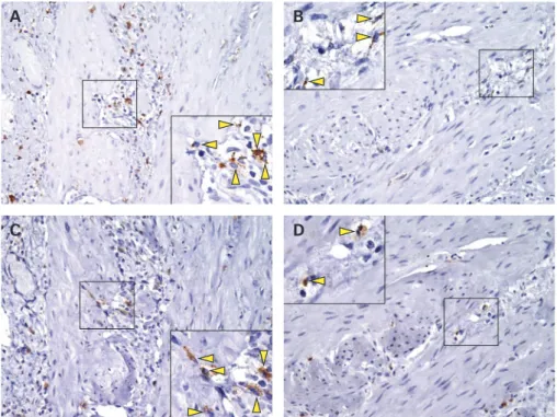
observed in GBs from a control group, represented by 10 alithiasic GBs removed from patients with neoplastic diseases not involving the GB. The number of CD68 positive macrophages in the muscle layer of GBs from gallstone patients was signifi-cantly higher compared to that in control patients. In UDCA-treated patients, the number of CD68 positive macrophages, in GB muscle, was significantly lower when compared to that in placebo-treated patients. Positive COX-2 expression was almost exclusively present in macrophages within the muscle layer (fig 1). The number of COX-2 positive cells was higher in muscle from symptomatic gallstone patients compared to controls and, likewise macrophages, significantly lower following UDCA treatment (table 1, fig 1). A direct and significant correlation was observed between positivity for CD68 and COX-2 (Spearman’s r = 0.7, p,0.01). As in our previous study, the production of PGE2 was significantly lower, following UDCA than after placebo. Furthermore, muscle contraction, induced by increasing concen-trations of CCK-8 (assessed in four patients in each group) was significantly higher in the UDCA, compared to placebo, group (maximal contraction to CCK-8 1028mol/l
was 25.1 (SD 3) vs 12 (SD 4)%, respec-tively; p,0.001). Our more recent data show that an inflammatory macrophage infiltrate is present in the GB muscle layer
of cholesterol gallstone patients. UDCA decreases the presence of macrophages in the muscle layer and confirms improve-ment in GB muscle cell contraction. These results suggest that activated macrophages play a role in muscle cell dysfunction and add insight into the anti-inflammatory action of UDCA, which may explain some of the therapeutic effects of this bile acid in liver diseases as well as other gastrointest-inal inflammatory conditions.
M P L Guarino,1S Carotti,2S Morini,2G Perrone,3
J Behar,4A Altomare,1R Alloni,1R Caviglia,1
S Emerenziani,1C Rabitti,3M Cicala1
1Department of Digestive Diseases, Campus Bio-Medico
University, Rome, Italy;2Department of Biomedical
Research, Campus Bio-Medico University, Rome, Italy;
3Surgical Pathology, Campus Bio-Medico University, Rome,
Italy;4Rhode Island Hospital and Brown University Medical
School, Providence, Rhode Island, USA
Correspondence to: Dr M P L Guarino, Dipartimento di Malattie dell’Apparato Digerente, Universita` Campus Bio-Medico, Via Alvaro del Portillo, 21–00128 Rome, Italy; [email protected]
Competing interests: None.
Ethics approval: This study was approved by the Ethics Committee of Campus Bio-Medico University on 21 June 2001.
Gut 2008;57:1740–1741. doi:10.1136/gut.2008.160333
REFERENCES
1. Guarino MP, Cong P, Cicala M, et al. Ursodeoxycholic acid improves muscle contractility and inflammation in symptomatic gallbladders with cholesterol gallstones. Gut 2007;56:815–20.
2. Ljubuncic P, Fuhrman B, Oiknine J, et al. Effect of deoxycholic acid and ursodeoxycholic acid on lipid peroxidation in cultured macrophages. Gut 1996;39:475–8.
3. Iwaki T, Ishizaki K, Kinoshita S, et al. Protective effects of ursodeoxycholic acid on chenodeoxycholic acid-induced liver injury in hamsters.
World J Gastroenterol 2007;13:5003–8.
4. Wehner S, Behrendt FF, Lyutenski BN, et al. Inhibition of macrophage function prevents intestinal inflammation and postoperative ileus in rodents. Gut 2007;56:176–85.
5. The FO, Boeckxstaens GE, Snoek SA, et al. Activation of the cholinergic anti-inflammatory pathway ameliorates postoperative ileus in mice. Gastroenterology 2007;133:1219–28.
6. Eskandari MK, Kalff JC, Billiar TR, et al. LPS-induced muscularis macrophage nitric oxide suppresses rat jejunal circular muscle activity. Am J Physiol 1999;277:G478–86.
Pathological lesions in colonic
biopsies during Parkinson’s
disease
Parkinson’s disease (PD) is a neurodegenera-tive condition that affects 1% of the population over 65 years of age. The two pathological hallmarks of PD are a loss of dopaminergic neurons in the substantia nigra (SN) and the presence of cytoplasmic eosinophilic inclusions termed Lewy bodies (LBs), whose main component is phosphory-lated a-synuclein.1
This degeneration of SN neurons leads to a dopamine deficiency
Figure 1 Immunohistochemistry for CD68 and cyclooxygenase-2 (COX-2) in muscularis macrophages. CD68 positive cells, were quite evenly distributed in muscle layer of the gallbladder in placebo-treated gallstones patients (A) and were scarcely present in gallbladder of ursodeoxycholic acid (UDCA)-treated patients (B). A similar pattern was exhibited by COX-2 positive cells, in muscle layer of gallbladder in placebo-treated patients (C) and UDCA-treated patients (D). A/C, B/D refer to the same patient. The figure shows corresponding fields for each patient. Original magnification, 6200. High power field, 6400.
Table 1 Mean number (with SD in parentheses) of positively stained cells in 10 consecutive microscopic fields
Controls Placebo UDCA
CD68 11.9 (8.1) 36.2 (11)* 19.6 (6)** COX-2 10.5 (6.8) 30.6 (12)* 15.2 (5.5)**
*p,0.001 vs controls; **p,0.01 vs placebo. COX-2, cyclooxygenase-2; UDCA, ursodeoxycholic acid.
PostScript
responsible for the major motor symptoms. Nevertheless, it has become increasingly evident that PD is a multicentric neurode-generative process that also affects neuronal structures outside the SN.2In this context, various reports performed on surgical or autopsy specimens have shown that the enteric nervous system (ENS) is affected during PD.3 4
However, it is still a matter of debate whether these alterations occur early in the course of the disease. This is mainly due to a lack of accessibility to the ENS in the living patients. Therefore, demonstrating (1) the ability to study the ENS using routine colonic biopsies and (2) the presence of lesions characteristics of PD could be relevant for an early diagnosis of the disease and to better understand its pathophysiology.
We therefore performed routine colonic biopsies in five PD patients complaining of functional constipation (63 (SD 7) years, three men; all had disease duration .5 years). Five healthy age-matched patients (61 (SD 6.5) years, one man) requiring a total colono-scopy for colorectal cancer screening were included as controls. They had no known neurological disease. None suffered from functional digestive symptoms. In order to avoid any specific role for chronic constipa-tion, we included three additional non-age-matched patients (52 (SD 5) years, no men) who underwent total colonoscopy for the assessment of a chronic intractable
constipation as additional controls. Written consent was obtained according to the principles of the Declaration of Helsinki.
Four biopsies were taken from the ascend-ing colon durascend-ing colonoscopy. Biopsies were performed using standard biopsy forceps without needles (FB210K; Olympus, Tokyo, Japan). Samples were immediately immersed in 4uC saline solution and microdissected in order to separate the submucosa (containing the internal submucosal plexus) from the mucosa. The submucosa were then fixed in 4% para-formaldehyde. Imunohistochemical studies were then performed on these tissues using a combination of antibodies against rabbit anti-tyrosine hydroxylase (TH) (1:500, Pel-Freez, Rogers, Arkansas, USA), rabbit anti-dopamine-b-hydroxylase (DBH) (1:250, Millipore, Saint-Quentin-en-Yvelines, France), mouse anti-Hu C/D (1:200, Invitrogen, Cergy-Pontoise, Fance), rabbit anti-phosphorylated a-synuclein (1:5000, WAKO, Osaka, Japan) or rabbit anti-neurofilament 200 kDa (1:250; Millipore) as previously described.5
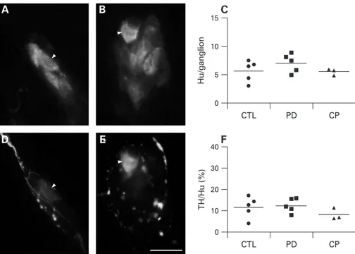
In control patients, individual biopsies contained 11.2 (SD 7.9) ganglia and each ganglia contained 5.6 (SD 1.9) Hu-immu-noreactive (IR) neurons. In PD patients, the number of ganglia per biopsies was similar to controls (13.6 (SD 5.3); p = 0.22). In addition, the number of Hu-IR neurons per ganglion in PD was unchanged as compared to controls (7.0 (SD 1.6); p = 0.25) (fig
1A,C). Constipated controls did not differ from PD patients in the number of ganglia per biopsy (11.3 (SD 1.5); p = 0.57) or in the number of neurons per ganglion (5.5 (SD 0.6); p = 0.19) (fig 1C).
In healthy controls, 11.6 (SD 5.0)% of Hu-IR neurons were TH-IR. In PD patients, the proportion of TH-IR neurons was unchanged as compared to controls (12.3 (SD 3.3)%; p = 0.80) (fig 1D–F). In constipated patients, the proportion of TH-IR neurons was similar to the one of PD patients (8. (SD 2.7)%; p = 0.12) (fig 1F). In all groups no neuronal body was DBH positive, suggesting that all TH-IR neurons in the submucosal plexus were dopaminergic. These results are consistent with a previous report by Singaram et al6 showing the absence of loss of TH-IR neurons in the submucosal and myenteric plexuses of PD patients, suggesting that it is not a marker of choice for detecting PD lesions in the ENS.
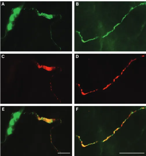
However, immunohistochemical staining with an antibody against phosphorylated a-synuclein, revealed that 4 out of 5 PD patients had phospho-a-synuclein-IR neur-ites (identified with neurofilament (NF) in the submucosa (fig 2A,F). These phospho-a-synuclein-IR neurites were absent in both control and constipated patients. In some cases, large aggregates were observed in dystrophic NF-IR neurites (fig 2E), a pattern reminiscent of Lewy neurites.
Taken together, our pilot study showed that routine colonic biopsies can be used to study the submucosal plexus of the ENS. In addition, we identified for the first time in the gut of living PD patients lesions similar to the ones observed in the brain. This technique could be a reliable tool to detect early lesions in the gut during the course of PD in order to better understand the pathogenesis of the disease and/or to iden-tify novel biomarkers.
T Lebouvier,1,2,3,4T Chaumette,1,2,3P Damier,2,4,5
E Coron,1,2,3,5Y Touchefeu,1,2,3S Vrignaud,6
P Naveilhan,2,7J-P Galmiche,1,2,3,5
S Bruley des Varannes,1,2,3,5P Derkinderen,1,2,3,4,5
M Neunlist1,2,3,4
1Inserm, U913, Nantes, France;2University Nantes, Nantes,
France;3CHU Nantes, Institut des Maladies de l’Appareil
Digestif, Nantes, France;4CHU Nantes, Department of
Neurology, Nantes, France;5Inserm, CIC-04, Nantes,
France;6CHU Nantes, Department of Anesthesia, Nantes,
France;7Inserm, U643, Nantes, France
Correspondence to: Dr M Neunlist, Inserm U913, 1 place Alexis Ricordeau, 44093 Nantes, France; michel.neunlist@ univ-nantes.fr or Dr P Derkinderen, Department of Neurology, CHU Nantes, 44093 Nantes, France; pascal.derkinderen@ chu-nantes.fr
Acknowledgements: The authors wish to thank M Roy and F Vavasseur for their help in the assessment of patients and controls.
Funding: This work was supported by a grant from France Parkinson, ADPLA (association des parkinsoniens de Loire Atlantique), Groupement de Parkinsoniens de Vende´e and Inserm/DHOS (to PDe and MN). PDe and MN are recipients of a Contrat d’Interface Inserm.
Competing interests: None.
Figure 1 Submucosal neuron counts and dopaminergic phenotype are unchanged in patients with Parkinson’s disease (PD). Hu-immunoreactive (IR) submucosal neurons were identified in the colon of controls (n = 5) (A), PD patients (n = 5) (B) and constipated patients (n = 3). There was no change in the number of Hu-IR submucosal neurons per ganglion in the three conditions (C). Double labelling with antibodies against Hu (A,B) and tyrosine hydroxylase (TH) (D,E) showed that occasional submucosal neurons were TH-IR (arrow heads). No significant decrease in the proportion of TH-IR submucosal neurons occurred in PD and in constipated patients (F). Each circle, square and triangle represents one control, PD or constipated patient, respectively. Horizontal bars represent the mean. Scale bar: 20mm. CTL, control; CP, constipated patient.
Ethics approval: The study protocol was approved by the local Committee on Ethics and Human Research on 27 February 2007.
TC and TL as well as PD and MN contributed equally to this work.
Gut 2008;57:1741–1743. doi:10.1136/gut.2008.162503
REFERENCES
1. Fujiwara H, Hasegawa M, Dohmae N, et al. a-synuclein is phosphorylated in a-synucleinopathy lesions. Nat Cell Biol 2002;4:160–4.
2. Braak H, Del Tredici K. Nervous system pathology in sporadic Parkinson disease. Neurology
2008;13;70:1916–25.
3. Braak H, de Vos RA, Bohl J, et al. Gastric alpha-synuclein immunoreactive inclusions in Meissner’s and Auerbach’s plexuses in cases staged for Parkinson’s disease-related brain pathology. Neurosci Lett 2006;396:67–72.
4. Wakabayashi K, Takahashi H, Takeda S, et al. Parkinson’s disease: the presence of Lewy bodies in Auerbach’s and Meissner’s plexuses. Acta Neuropathol 1988;76:217–21.
5. Neunlist M, Aubert P, Toquet C, et al. Changes in chemical coding of myenteric neurones in ulcerative colitis. Gut 2003;52:84–90.
6. Singaram C, Ashraf W, Gaumnitz EA, et al. Dopaminergic defect of enteric nervous system in Parkinson’s disease patients with chronic constipation. Lancet 1995;346:861–4.
CORRECTION
doi:10.1136/gut.2007.119446corr1
R Spiller, Q Aziz, F Creed, et al. Guidelines on the irritable bowel syndrome: mechan-isms and practical management (Gut 2007;56:1770–98). In paragraph 4.4.1 the sentence ‘‘This in turn acts on the adrenal medulla, resulting in cortisol secretion into the circulation’’ should read ‘‘This in turn acts on the adrenal cortex, resulting in cortisol secretion into the circulation’’.
Figure 2 Phospho-a-synuclein-positive submucosal neurites differentiate Parkinson’s disease patients from controls. Double labelling with antibodies against neurofilament (NF) (A,B) and phosphorylateda-synuclein (C,D) revealed that some NF-immunoreactive (IR) neuritic structures were also phospho-a-synuclein-IR (merged image in E,F) in the majority of Parkinson’s disease patients, but in none of the controls. Occasionally the inclusion-bearing neurites displayed dystrophic alterations (A,C,E). Scale bar: 30mm.
ANSWER
From the question on page 1673
The patient had a large inflammatory abdominal aortic aneurysm. The abdominal CT scan shows a large infrarenal aortic aneurysm with a maximum diameter of 7.5 cm extending into the iliac vessels. There is an enhancing soft-tissue cuff surrounding the anterolateral margin of the aneurysm. The aneurysm appears to compress the third part of the duodenum (fig 1 below), which, however, was not detected at endoscopy. These CT findings were suggestive of an inflammatory aneurysm. Inflammatory abdominal aortic aneurysms represent 3–10% of all abdominal aortic aneurysms and occur predomi-nantly in men.1They differ from atherosclerotic aneurysms in
that patients often present with abdominal symptoms or anorexia, weight loss, and raised inflammatory markers. CT has a specificity of 99.7% for diagnosis of inflammatory
aneurysms,2 usually showing periaortic fibrosis as a cuff of
enhancing soft tissue surrounding the anterolateral margin of the aneurysm. If periaortic fibrosis is extensive, adjacent abdominal structures may be compressed and adherent, most commonly the third part of the duodenum.1
Although rare, inflammatory abdominal aortic aneurysms should be kept in mind as a cause of abdominal pain and/or anorexia, weight loss, and raised inflammatory markers. The natural history of inflammatory abdominal aortic aneurysms remains unknown, with 3.3–14% patients presenting with acute or chronic rupture.1
As regards to management, the literature supports an operative approach with a 30 day operative mortality rate of up to 9%.1
Complete regression of fibrosis and inflammatory process occurs in up to one-half of patients at long-term follow-up post-operatively. Clinical symptoms (such as weight loss and gastro-intestinal symptoms) reverse in 93% of the patients after an operation.3 Endovascular therapy is also a potential treatment