HAL Id: hal-01860675
https://hal-univ-rennes1.archives-ouvertes.fr/hal-01860675
Submitted on 23 Aug 2018HAL is a multi-disciplinary open access
archive for the deposit and dissemination of sci-entific research documents, whether they are pub-lished or not. The documents may come from teaching and research institutions in France or abroad, or from public or private research centers.
L’archive ouverte pluridisciplinaire HAL, est destinée au dépôt et à la diffusion de documents scientifiques de niveau recherche, publiés ou non, émanant des établissements d’enseignement et de recherche français ou étrangers, des laboratoires publics ou privés.
Bacteria electrical detection using 3D silicon nanowires
based resistor
Brice Le Borgne, Laurent Pichon, Anne-Claire Salaün, Baptiste Le Bihan,
Anne Jolivet-Gougeon, Sophie Martin, Regis Rogel, Olivier de Sagazan
To cite this version:
Brice Le Borgne, Laurent Pichon, Anne-Claire Salaün, Baptiste Le Bihan, Anne Jolivet-Gougeon, et al.. Bacteria electrical detection using 3D silicon nanowires based resistor. Sensors and Actuators B: Chemical, Elsevier, 2018, 273, pp.1794-1799. �10.1016/j.snb.2018.07.101�. �hal-01860675�
Bacteria electrical detection using 3D silicon nanowires based resistor
Brice Le Borgne1, Laurent Pichon1*, Anne Claire Salaun1, Baptiste Le Bihan1, Anne Jolivet-Gougeon2, Sophie Martin3, Regis Rogel1 and Olivier de Sagazan1
1
Institut d’Electronique et de Télécommunications de Rennes, IETR UMR CNRS 6164,
Université de Rennes 1, Rennes, France,
2
Nutrition Métabolisme et Cancer, NuMeCan UMR INSERM 1241, Université de Rennes
1, Rennes, France
3
Société ARCLYNN, Missillac, France
*corresponding author: [email protected], , tel: +332 23 23 56 65, fax:+332 23 23 56 57
Resistors based on silicon nanowires (SiNWs) are developed for the direct detection of
Escherichia coli bacteria. The devices are manufactured using conventional silicon technology. The SiNWs are synthesized by the vapor liquid solid process (VLS) using gold as
the catalyst. The electrodes of the device consist of highly in situ doped polycrystalline
silicon. The results show that the corresponding resistances can be potentially used for the
detection of bacteria by electrical measurements, using SiNWs as sensitive units. The bacteria
are preferentially hooked in the SiNWs network, which causes a drastic reduction in the
electrical resistance of the component. Such resistor manufactured in a simple and low-cost
CMOS (Complementary Metal Oxide Semiconductor) technology acts as proof of concept.
platform for alternative direct electrical detection of bacteria to monitor the contamination in
environment for hygiene applications.
I. Introduction
One of the main challenges in terms of a public health is the rapid detection of bacteria in
food, water and air, particularly because of the emergence of multi-resistant bacteria.
Although standard microbiological cell culture for identifying bacterial strains are well
controlled, the detection process can often take several days. In addition, complex
instrumentation [1] is required in most conventional methods, and cannot be used on site.
Classical methods for the detection of pathogens are polymerase chain reaction (PCR) [2],
colony culture and enumeration methods [3], immunological methods [4] and more recently
detection of pathogens bacteria using mass spectrometry [5]. The main drawbacks of these
methods are significant analysis time, high cost equipment, and complexity of detection
process.
The use of biosensors [6] is an alternative method for detecting bacteria combining a
biological recognition mechanism with a physical transduction technique. In this case
transduction may be micromechanical, electrochemical, piezoelectric, thermometric, magnetic
or optical. In particular, the electrochemical transduction methods such as amperometry [7],
impedance spectroscopy [8-10], potentiometry [11], are much faster and more sensitive than
other techniques.
Bacteria detection systems are based on multidisciplinary fields requiring knowledge of
physics, chemistry, biology, instrumentation, microelectronics and fabrication technology. In
the objective of a lab on chip system for rapid detection of bacteria which may revolutionize
sensors that enable direct bacteria detection at low level. But none of the detection methods
previously reported fulfils all these criteria.
However, electrical detection of charged (bio)chemical species or polar molecules by
measurement of the conductance change could be an alternative approach. Indeed,
conductance variation, due to the charge effect facilitated by positioning species between two
interconnected electrodes, preserves high sensitivity with less detection time [12]. In addition,
based on this detection method, many groups have reported on field effect transistors (FET)
used as biochemical sensors. In particular, chemical or biochemical (DNA hybridization) [13,
14], gas [15] or mechanical [16] sensors, have been demonstrated using silicon nanowires
(SiNWs) based FETs. In this case nanowires act as sensing elements. However, few works
report on SiNWs use as great promising method for label free, ultra-sensitive and real-time
detection of bacteria [17, 18].
The recent progress in micro- and nano-technologies for fabrication of nano–objects
compatible with silicon technology offer great opportunities for the development of
innovative biosensors integrated on CMOS (Complementary Metal Oxide Semiconductor)
platforms. These sensors could be interestingly directly coupled with any existing circuitry or
signal conditioning systems to transmit their output data. Furthermore, due to their specific
properties at nanoscale (high surface/volume ratio, carrier confinement), SiNWs are
promising candidates as sensing elements for high sensitive bacteria detection at low level.
The main advantages of the use of such nano-objects are: the possibility of the mass
production, the use of the volume of materials at low level, the micro-fluidic facilities for
transport of reagents, the reduction of analysis time, and thus the reduced costs of fabrication
and analysis.
However, the development of a small sized, reliable and versatile low-cost sensor based on
structures and functionalization are reported into literature, but the proposed solutions do not
fulfill the requirements for integration into a sensor. The major problem is the realization of
such high-performance sensor with a low-cost technology, while the technological means
used for the realization of nanowires require high cost and complex technologies based on
ebeam lithography [14] or transfer techniques [19]. Nevertheless, alternative self-assembly
methods [20] are promising for synthesis of nanowires and do not require expensive
lithographic tools.
Moreover, many chemical or biological silicon nanowires based sensors are issued from field
effect transistor technology, but the manufacturing process includes many fabrication steps (at
least four masks for lithography), with possible transfer techniques and sophisticated chemical
steps for functionalization. On the other hand, the fabrication of sensors based on basic
electronic devices (for instance resistors, capacitors), with direct integration of the nanowires
as sensing elements, refers as simple and low-cost technology and constitutes a technological
progress.
In this way, in this study the feasibility of SiNWs based resistor for bacteria detection with
SiNWs used as sensing elements synthesized by vapour liquid solid (VLS) mechanism [20]
(self-assembly method) is investigated. Such device acts as proof of concept and fulfills the
following requirements: i) low cost technology, ii) high sensitivity, iii) real-time analysis, iv)
electrical detection of bacteria, and v) portability and on site measurements.
II. Material and method
Devices
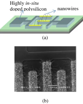
The resistors used for bacteria detection applications (fig. 1) are fabricated in a classical
the devices are made of a highly phosphorous in-situ doped amorphous silicon layer deposited
by Low Pressure Chemical Vapour Deposition (LPCVD) technique at 550°C, and solid phase
crystallized by thermal annealing at 600°C under vacuum to get a highly in-situ doped
polycrystalline silicon (polySi). Silicon nanowires (sensitive units) are locally grown on the
teeth of the polySi inter-digitated electrodes by the well-known VLS mechanism at 460°C
using gold as catalyst. In this way no patterning technique was used for elaboration of
nanowires. The diameter and length of the SiNWs are 100nm and more than 20µm
respectively. Nanowires bridge between two adjacent teeth and thus ensure electrical
connection between the two highly doped polySi electrodes (fig. 1. (b)). The potential use as
high sensitive gas (ammonia) sensors of such SiNWs based resistors was previously reported
[22].
(a)
(b)
Figure 1: VLS SiNWs based resistor: schematic view (a). SEM top view observation (b).
Method
nanowires Highly in-situ
Bacteria/SiNWs interactions were studied with two types of bacterial reference strains
Escherichia coli ATCC 35218 (NCTC 11954; CIP 102181) and Escherichia coli K.12 deposited on the SiNWs based resistors.
Escherichia coli ATCC 35218 bacteria deposition was carried out from serial dilutions in sterile distilled water of an overnight bacterial culture in trypticase soy broth (BBL Trypticase
Soy Broth, Becton Dickinson) incubated at 37°C. This culture was adjusted to 8×10⁸ CFU/mL by measurement of OD with spectrophotometer at 600 nm (OD600 = 0.6). Then, a
serie of dilutions of bacteria in sterile distilled water was performed from 8×10⁸ to 8×10³ CFU/mL. Bacterial counts were verified by plating each dilution (100µ L) on trypticase agar
plates (Oxoid), and enumerating the colonies after 24h of incubation at 37°C.
Escherichia coli K.12 bacteria deposition was carried out from serial dilutions in sterile distilled water of an overnight bacterial culture in LB broth Lennox (Difco) incubated at
37°C. This culture was adjusted to 6×10⁸ CFU/mL by measurement of OD with spectrophotometer at 600 nm, OD = 0,1. Then, a serie of dilutions of bacteria was performed
from 6×10⁸ to 6×10⁴ CFU/mL.
After bacteria solution deposition (30µ L), devices were dried at room temperature and tested
24h later to avoid any moisture effect on the electrical behaviour of the resistor. Note that,
considering the size of the device, a 2µL droplet could be used to limit the waste of material and to drastically decrease the drying time. It would consequently significantly reduce the
response time of the sensor from 24h to less than one hour. In addition, some devices
submitted to a mixture of sterile distilled water and culture solution without any bacteria were
used as references.
Bacteria detection was checked by measuring static I-V curves of the SiNWs based resistors
at room temperature on devices based on n = 20, 40, and 100 teeth of the comb shape
electrode, with each tooth spaced from W = 5µm.
III. Results and discussion
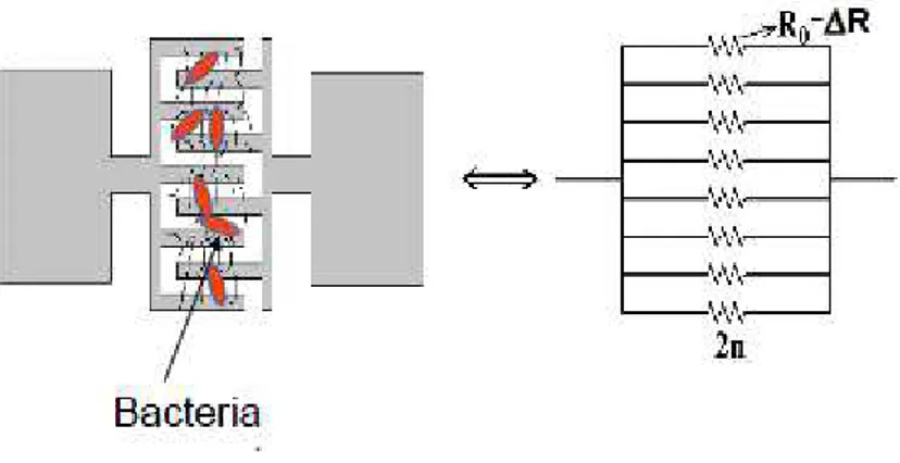
Because of the SiNWs VLS process growth, for a fixed value of W, the number of
interconnected nanowires (conducting paths) between two adjacent N-type polySi teeth can
change. In addition, the sizes of the nanowires are distributed over an average value, which
means that the corresponding elementary resistance value can fluctuate. Thus, it might play a
role in the electrical resistance measurements between two adjacent N+ polySi electrodes. However, in order to simplify and because of the high number of the nanowires we assume
that electrical and geometrical characteristics are identical for all interconnected nanowires. In
this case, these elementary resistances, R0, are assumed to be identical. Thus the equivalent
electrical circuit of the whole resistor represented in the figure 2 results to 2n elementary
resistances connected in parallel. Therefore, the total resistance of the resistor can be
expressed as the following formula:
n R I V R 2 0 ≈ = (1)
In these conditions, macroscopic theoretical model for calculation of R0 has been previously
reported by our group to estimate the average electrical resistivity of a single undoped SiNW
(106-107 Ω cm) [23]. In this model R0 results from interconnections of nanowires and is
related to the average electrical resistivity of the nanowire, ρNW, following:
K
with K the geometrical factor depending of the total conducting paths. The high value
reported of the average electrical resistivity of a single nanowire for undoped SiNWs is
related to carriers trapping effect at defects located at interconnections between nanowires and
induced by gold impurities at the surface of the nanowires.
W
Figure 2: Equivalent circuit diagram of SiNWs based resistors
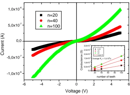
Electrical characterization for three types of resistors with n = 20, 40, 100 are reported in the
figure 3. Plots show that the current level through the global electrical resistance increases as
the number of teeth of the interdigitated electrodes increases. These results are related to the
linear variations of the conductance (G=I/V≈2n/R0) with n reported in the inset of the figure 3
(for an average value R0 ≈1.9×10⁹ Ω), and validate the equivalent electrical model given by
(1).
Interaction of bacteria with the surface of materials is mainly controlled by the charge effects
and hydrophobic properties between bacteria and materials [24]. In particular, the adhesion
can occur when the van der Waals forces of attraction between the membrane of the bacteria
-6 -4 -2 0 2 4 6 -1,0x10⁻⁶ -5,0x10⁻⁷ 0,0 5,0x10⁻⁷ 1,0x10⁻⁶ C u rr e n t (A ) Voltage (V) n=20 n=40 n=100 20 40 60 80 100 0,0 5,0x10⁻⁸ 1,0x10⁻⁷ 1,5x10⁻⁷ 2,0x10⁻⁷ 2,5x10⁻⁷ 3,0x10⁻⁷ C o n d u c ta n c e ( S ) number of teeth 0.2V 2V 4V linear fit Average R 0 = 1,9,10 9Ω
Figure 3: I-V characteristics of the SiNWs based resistors made of interdigitated comb
electrodes with 20, 40 and 100 teeth. Inset: electrical conductance variations with the number
of teeth at 0.2V, 2V and 4V.
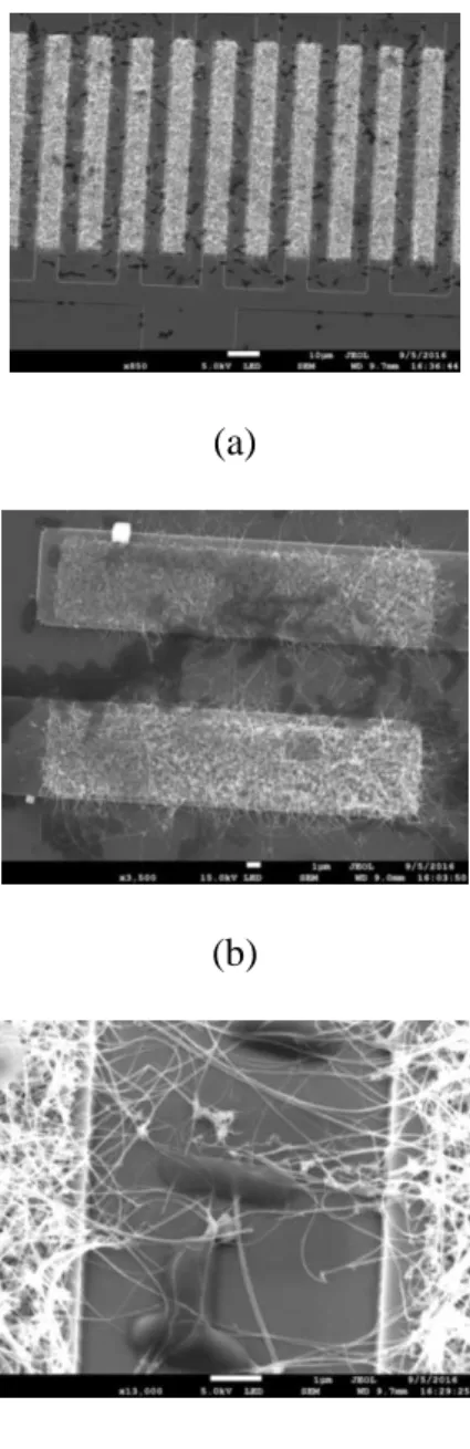
After bacteria immobilization on the device, bacteria interaction with SiNWs is firstly
analysed by SEM observations (fig. 4). The bacteria optical count has not been performed in
this study. Interestingly, in our case, pictures clearly show that bacteria are mainly attached
into the silicon nanowires mesh rather than in other parts of the device. This observation
shows the interest to use a 3D SiNWs tangled mesh to promote bacteria immobilization and
thus detection. Functionalization of active parts for detection, usually based on sophisticated
chemical steps, is a common method to allow immobilization and identification of chemical
(a)
(b)
(c)
Figure 4: SEM pictures of bacteria (dark areas) hanged into SiNWs network of the resistor
shown at three different scales
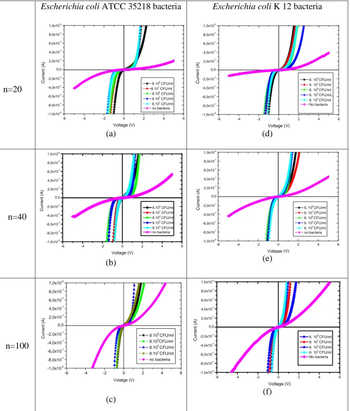
Detection was studied for the two bacteria Escherichia coli, ATCC 35218 and K12 with
different ranging concentrations (8×10⁸ to 8×10³ CFU/mL and 6×10⁸ to 6×10⁴ CFU/mL respectively). Electrical effect of bacteria immobilization is then checked by electrical
n=20
Escherichia coli ATCC 35218 bacteria
-6 -4 -2 0 2 4 6 -1,0x10⁻⁶ -8,0x10⁻⁷ -6,0x10⁻⁷ -4,0x10⁻⁷ -2,0x10⁻⁷ 0,0 2,0x10⁻⁷ 4,0x10⁻⁷ 6,0x10⁻⁷ 8,0x10⁻⁷ 1,0x10⁻⁶ 8.108 CFU/ml 8.107 CFU/ml 8.106 CFU/ml 8.105 CFU/ml 8.104 CFU/ml no bacteria C u rr e n t (A ) Voltage (V) (a)
Escherichia coli K 12 bacteria
-6 -4 -2 0 2 4 6 -1,0x10⁻⁶ -8,0x10⁻⁷ -6,0x10⁻⁷ -4,0x10⁻⁷ -2,0x10⁻⁷ 0,0 2,0x10⁻⁷ 4,0x10⁻⁷ 6,0x10⁻⁷ 8,0x10⁻⁷ 1,0x10⁻⁶ 6. 108 CFU/ml 6. 107 CFU/ml 6. 106 CFU/ml 6. 105 CFU/mL 6. 104 CFU/ml No bacteria C u rr e n t (A ) Voltage (V) (d) n=40 -6 -4 -2 0 2 4 6 -1,0x10⁻⁶ -8,0x10⁻⁷ -6,0x10⁻⁷ -4,0x10⁻⁷ -2,0x10⁻⁷ 0,0 2,0x10⁻⁷ 4,0x10⁻⁷ 6,0x10⁻⁷ 8,0x10⁻⁷ 1,0x10⁻⁶ 8.108 CFU/ml 8.107 CFU/ml 8.106 CFU/ml 8.105 CFU/ml 8.104 CFU/ml no bacteria C u rr e n t (A ) Voltage (V) (b) -6 -4 -2 0 2 4 6 -1,0x10⁻⁶ -8,0x10⁻⁷ -6,0x10⁻⁷ -4,0x10⁻⁷ -2,0x10⁻⁷ 0,0 2,0x10⁻⁷ 4,0x10⁻⁷ 6,0x10⁻⁷ 8,0x10⁻⁷ 1,0x10⁻⁶ C u rr e n t (A ) Voltage (V) 6. 108 CFU/ml 6. 107 CFU/ml 6. 106 CFU/ml 6. 105 CFU/ml 6. 104 CFU/ml no bacteria (e) n=100 -6 -4 -2 0 2 4 6 -1,0x10⁻⁶ -8,0x10⁻⁷ -6,0x10⁻⁷ -4,0x10⁻⁷ -2,0x10⁻⁷ 0,0 2,0x10⁻⁷ 4,0x10⁻⁷ 6,0x10⁻⁷ 8,0x10⁻⁷ 1,0x10⁻⁶ C u rr e n t (A ) Volatge (V) 8.108 CFU/ml 8.106 CFU/ml 8.105 CFU/ml 8.103 CFU/ml no bacteria (c) -6 -4 -2 0 2 4 6 -1,0x10⁻⁶ -8,0x10⁻⁷ -6,0x10⁻⁷ -4,0x10⁻⁷ -2,0x10⁻⁷ 0,0 2,0x10⁻⁷ 4,0x10⁻⁷ 6,0x10⁻⁷ 8,0x10⁻⁷ 1,0x10⁻⁶ 6. 108 CFU/ml 6. 107 CFU/ml 6. 105 CFU/ml 6. 104 CFU/ml No bacteria C u rr e n t (A ) Voltage (V) (f)
Figure 5: I-V curves of the SiNWs based resistors with n = 20, 40, 100 teeth and 5 µm interspacing, after Escherichia coli ATCC 35218 immobilization (a), (b), (c), and
Escherichia coli K12 bacteria immobilization (d), (e), (f). Curves for devices submitted to a mixture of distilled water and culture solution without bacteria are given as reference.
measurements performed on SiNWs based resistors. As shown in the figure 5, for the two
types of bacteria I-V curves exhibit a drastic increase of the current corresponding to a
lowering of the R value of the resistor due to the presence of bacteria. This phenomenon is
probably due to the negative charge of their peptidoglycan (PG) based membrane [27, 28].
Indeed, two main types of bacteria are listed related to their membrane: Gram positive (+) and
Gram negative (–) bacteria. For the Gram-positive bacteria, membrane contains a thick (20–
50 nm) PG layer attached to teichoic acids [29]. In the case of Gram-negative bacteria as E.
Coli, the membrane is made of a thin PG layer covered by an extern membrane containing lipopolysaccharides components, responsible of a higher negative charge of the bacteria [30].
To explain the decrease of the global electrical resistance due to the capture of bacteria three
effects can be invoked. One first explanation could be a transfer of charges (electrons) of the
bacteria into the nanowires which fill the traps previously mentioned, and thus could and
contribute to the decrease of the electrical resistivity of the nanowires [23]. Another possible
reason is that the electrically charged bacteria may behave as biological gate resulting as
well-known field effect [13-15, 17] on the SiNWs. At last, other explanation is that bacteria may
act as additional conducting paths randomly distributed between teeth thanks to hooking
effect of bacteria into the SiNWs network. This phenomenon would contribute to the
variations of R0 values, ∆R, strongly dependent on the number of bacteria hanged between
two adjacent teeth (see fig. 6). Based on these three considerations, further investigations for a
well understanding of the carrier transport in these devices in relation with bacteria and
Figure 6: Illustration of the electrical resistance change with bacteria interaction
Furthermore, as shown in the figures S1 reported in the supplementary material, no significant
change of the global electrical resistance (of the resistor) was observed at low bias voltages
(-1V < V < +1V) with the bacteria concentration, and thus the domain of interest for bacteria
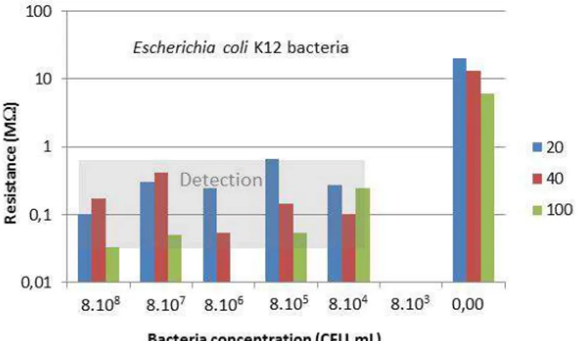
detection corresponds to the highest bias voltages. In this way, we reported in the figure 7 the
values of the global resistance measured at ± 4V for the two types of detected bacteria. No
tendency can be clearly observed in function of the number of teeth and neither in function of
the studied bacteria concentrations. Such unexplicated results must be related to the intricate
model of the global electrical resistance of the device with the presence of bacteria.
(b)
Figure 7: Histograms of the global electrical resistance of the sensor made of 20, 40, 100 teeth
interdigitated electrode measured at ± 4V for (a) Escherichia coli ATCC 35218, (b)
Escherichia coli K.12. Grey area: detection range value
Because the performances of a bacteria sensor are strongly related to its abilities for
quantitative recognition of target analytes, further studies in function of the size, the quantity,
and the type of targeted bacteria have to be carried out for applications of our SiNWs based
resistors as selective bacteria sensors. Nevertheless, sensing properties of our SiNWs based
resistors have s low detection limit, working range and time analysis to many others bacteria
detection techniques (Enzyme Linked ImmunoSorbent Assay-ELISA, PCR, QCM-Quartz
Crystal Microbalance immunosensor, impedimetric, amperometric…) [31, 32].
IV. Conclusion
In this work, investigations on a SiNWs based resistor were carried out to study the potential
development of a sensitive real-time detection bacteria sensor, fabricated by a simple low-cost
elements. Such a technology takes advantages of both benefits at nanoscale and
microtechnology for miniaturization and integration on microsystems, and of the use of
SiNWs network to favour bacteria hanging to increase the sensitivity for detection of bacteria.
The detection is based on the electrical measurement. The resistor showed its ability for
bacteria detection (Escherichia coli ATCC 35218 and K12) at concentration as low as 6x10³ CFU/mL. Since the change into electrical resistances values of the device were not correlated
with bacteria concentrations, it could be used only for yes/no diagnosis. Further studies are
needed to use the SiNWs based resistor as quantitative detection system and at lower
detection limit. However, because such device is easy to use, reagent-less, and portable, it has
the potential to be used as further detection tool for biosafety.
References
[1] S. P. Diggle, A.S. Griffin, G. S. Campbell, S. A. West Cooperation and conflict in
quorum-sensing bacterial populations. Nature, 450(7168), (2007) 411-414.
[2] A. K. Bej, M. H. Mahbubani, J. L. Dicesare, R. M. Atlas, Polymerase chain reaction-gene
probe detection of microorganisms by using filter-concentrated samples, Appl. Environ.
Microbiol. 57 (12), (1991) 3529–3534
[3] E. Leoni, P. P. Legnani, Comparison of selective procedures for isolation and enumeration
of Legionella species from hot water systems, J. Appl. Microbiol. 90, (2001) 27–33
[4] H. Gu, K. Xu, C. Xu, B. Xu, Biofunctional magnetic nanoparticles for protein separation
and pathogen detection, Chem. Commun. 9, (2006) 941–949
[5] W. S. Shell, M. L. Sayed, F. M. G. Allah, F. E. M. Gamal, A. A. Khder, A. A. Samy, A.
H. M. Ali Matrix-assisted laser desorption-ionization-time-of-flight mass spectrometry as a
Vet World. 2017 Sep;10(9):1083-1093. doi: 10.14202/vetworld.2017.1083-1093. Epub 2017
Sep 19. PubMed PMID: 29062198; PubMed Central PMCID: PMC5639107.
[6] A.K. Deisingh, M. Thompson Can. Biosensors for the detection of bacteria, J. Microbiol. 50, (2004) 69–77
[7] H. Tang, W. Zhang, P. Geng, Q. Wang, L. Jin, Z. Wu, M. Lou, A new amperometric
method for rapid detection of Escherichia coli density using a self-assembled
monolayer-based bienzyme biosensor,. Anal. Chim. Acta 562, (2006) 190–196
[8] S. M. Radke, E. C. Alocilja, A high density microelectrode array biosensor for detection
of E. coli O157:H7, Biosens. Bioelectron. 20, (2005)1662–1667
[9] S. M. Radke, E. C. Alocilja, A microfabricated biosensor for detecting foodborne
bioterrorism agents, IEEE Sens. J. 5 (4), (2005) 744–750
[10] M. Varshney, Y. Li, Interdigitated array microelectrode based impedance biosensor
coupled with magnetic nanoparticle-antibody conjugates detection for Escherichia coli
O157:H7 in food sample, Biosens. Bioelectron. 22, (2007) 2408–2414
[11] P. Bergveld, Thirty years of ISFETOLOGY: What happened in the past 30 years and
what may happen in the next 30 years, Sens. Actuators B 88, (2003) 1–20
[12] Z. Mahummad-Tahir, E.C. Alocilja A conductometric biosensor for biosecurity, Bios.
Bioelectron. 18, (2003) 813-819
[13] Y. Cui et al., Nanowire nanosensors for highly sensitive and selective detection of
biological and chemical species. Science, 293(5533) (2001) 1289-1292.
[14] Z. Li, Y. Chen, X. Li, T. I. Kamins, K. Nauka, R.S Williams Sequence-Specific
Label-Free DNA Sensors Based on Silicon Nanowires, Nano Letters 4 (2), (2004) 245–247
[15] X. Chen, C. K. Y. Wong, C. A. Yuan, G. Zhang, Nanowires based gas sensor, Sensors and Actuat. B, 17, (2013) 178-195
[16] R. He, P. Yang, Giant piezoresistance effect in silicon nanowires, Nature Nanotech. 1, (2006) 42-46
[17] N.N. Mishra, W. C. Maki, E. Cameron, R. Nelson, P. Winterrowd, S. K. Rastogi, B.
Filanoski, G. K. Maki Ultrasensitive detection of bacterial toxin with silicon nanowire
transistor Lab. Chip 8, (2008) 868-871
[18] B. Le Borgne, A. C. Salaun, L. Pichon « Silicon nanowires based resistors for bacteria
detection », Eurosensors (2017) 1, 496
[19] M. Lee, Y. Jeon, T. Moon, S. Kim Top-Down Fabrication of Fully CMOS-Compatible
Silicon Nanowire Arrays and Their Integration into CMOS Inverters on Plastic, Nano Lett. 2,
5(4), (2011) 26-29
[20] A. I. Hochbaum, R. Fan, R. He, P. Yang Controlled Growth of Si Nanowire Arrays for
Device Integration, Nano Lett. , 5 (3), (2005) 457–460
[21] R. S. Wagner, W. C. Ellis Vapor-liquid-solid mechanism of single crystal growth, Appl.
Phys. Lett., 4(5), (1964) 89-91
[22] F. Demami, L. Ni, R. Rogel, A.C. Salaun, L. Pichon Silicon nanowires based resistors as
gas sensors Sensors and Actuators B: Chemical vol 170, (2012) 158-162
[23] L. Pichon, R. Rogel, E. Jacques Electrical properties of phosphorus in-situ doped
Au-catalyst vapor liquid solid silicon nanowires Journ. Appl. Phys. 118, (2015) 185701
[24] T.R. Neu, K. C. Marshall. Bacterial polymers: physicochemical aspects of their
interactions at interfaces. J. Biomater. Appl. 5 (1990) 107–133.
[25] K. C. Marshall Adsorption and adhesion process in microbial growth at interfaces. Adv.
Colloid Interface Sci. 25(1986) 59–86
[26] H. H. M. Rijnaarts, W. Norde, E. J. Bouwer, J. Lyklema, A. J. B. Zehnder. Reversibility
and mechanism of bacterial adhesion. Colloids Surf. B: Biointerface 4 (1995) 5–22
[27] Y. C. Lu, Y. S. Chuang, Y. Y. Chen, A. C. Shu, H. Y. Hsu, H. Y. Chang, T. R. Yew
[28] B. Gottenbos, D. W. Grijpma, H. C. van der Mei, J. Feijen, H. J. Busscher,
Antimicrobial effects of positively charged surfaces on adhering positive and
Gram-negative bacteria. Journal of antimicrobial chemotherapy, 48(1), (2001) 7-13
[29] J. R. Scott, T. C. Barnett Surface proteins of gram-positive bacteria and how they get
there. Annu. Rev. Microbiol. 60 (2006) 397–423
[30] I. S. Roberts The biochemistry and genetics of capsular polysaccharide production in
bacteria. Annu. Rev. Microbiol. 50 (1996) 285–315
[31] O. Lazcka, F. J. Del Campo, F. X Minoz, Pathogen detection : A perspective of traditional methods and biosensors, Biosen. And Biolect. 22 (2007) 1205-1217
[32] A. Ahmed, J. V. Rushworth, N. A. Hirst, P. A. Millner, Biosensors for whole-cell bacteria detection, Clinic. Microbiol. Rev. 27-3 (2014) 631-646