RT-PCR-ELOSA tests on pooled sample units for the
detection of virus Y in potato tubers
A. Chandelier
a,*, N. Dubois
a, F. Baelen
a, F. De Leener
b, S. Warnon
c,
J. Remacle
c, P. Lepoivre
aaFaculte´ Uni6ersitaire des Sciences Agronomiques de Gembloux, Unite´ de Phytopathologie, Passage des De´porte´s, 2B-5030Gembloux, Belgium
bLambdatech s.a., Parc Crealys, rue Phocas Lejeune30/8B-5032Les Isnes, Belgium
cFaculte´s Uni6ersitaires Notre-Dame de la Paix, Laboratoire de Biochimie et Biologie Cellulaire rue de Bruxelles, 61B-5000Namur, Belgium
Received 2 May 2000; received in revised form 26 July 2000; accepted 26 July 2000
Abstract
A highly sensitive RT-PCR protocol able to detect potato virus Y (PVY) in pooled sample units (tubers) was developed. PVY-specific primers selected in the coat protein gene were found to amplify a 359 bp fragment from diluted crude extract of infected tubers. For the detection of the amplification products, a colorimetric detection procedure in microtiter plates was established. The amplicons are hybridized between a covalently linked capture probe and a specific biotinylated detection probe ELOSA tests. This detection method detects at least 50 pg of virus per reaction for the four cultivars tested. The RT-PCR-ELOSA assay was adapted to pooled units in order to increase the sample size while reducing the number of tests. © 2001 Elsevier Science B.V. All rights reserved.
Keywords:Diagnosis; Plant; Virus; Enzyme-linked immunosorbent assay (ELISA); Potato virus Y (PVY) 1. Introduction
Potato virus Y (PVY), the type member of the genus Poty6irus in the family Poty6iridae is a widespread virus leading to severe damage in
Solanaceae, including potato, tomato, pepper and
tobacco (De Bokx and Huttinga, 1981), with losses ranging from 15 to 70% of tuber yield (Van der Zaag, 1987). This virus is transmitted by
aphids in the non-persistent manner. The different strains infecting naturally potato have been classified into three different groups according to symptoms on selected potato and tobacco culti-vars: the PVYN group (tobacco veinal necrosis strains), the PVYO group (common or ordinary strains), and the PVYC group (stipple-streak strains) (De Bokx and Huttinga, 1981). Encapsi-dated into long flexuous rods of 730 nm in length, the virus is made up of a single strand positive sense RNA approximately 10 kb long which en-codes a polyprotein later processed to generate functional viral proteins (Robaglia et al., 1989).
* Corresponding author. Tel.: + 81622431; fax: + 32-81610126.
0166-0934/01/$ - see front matter © 2001 Elsevier Science B.V. All rights reserved. PII: S 0 1 6 6 - 0 9 3 4 ( 0 0 ) 0 0 2 3 8 - X
In addition to visual inspections undertaken in the field, the detection of PVY in the frame of certification programs usually rests on the use of serological tests, and notably on an enzyme-linked immunosorbent assay (ELISA) (Vetten et al., 1983; Deltour et al., 1987). These tests are carried out on a batch of individual samples (tubers or leaves) collected from the field in order to give an accurate estimation of the field plot infection level. This estimation, compared to a predeter-mined tolerance level, will lead to the acceptance or rejection of the field plot in the frame of the seed potato certification process. Geng et al. (1983) have demonstrated using cumulative prob-ability curves for the Poisson distribution that the confidence in the evaluation of a seed lot infection level increases with the number of sample units collected in the field and submitted to the analy-sis. On the other hand, both the cost of the certification protocol as well as ‘physical con-straints’ linked to the laboratory equipment act as limitations upon an increase of the number of tests undertaken currently. Although ELISA has been adopted for routine testing, it is not suffi-ciently sensitive to be applied to dormant tubers due to the low virus concentration after harvest-ing (De Bokx and Cuperus, 1987). More sensitive techniques thus represent a possible alternative. In this respect, RT-PCR tests combining a cDNA synthesis and a PCR amplification have been described for the detection of potato viruses like PVYO (Singh and Singh, 1996), PVYNTN (Weide-mann and Maiss, 1996), PLRV (Schoen et al., 1996; Singh et al., 1995) or PVA (Cerovska et al., 1998; Singh and Singh, 1998).
While the ELISA can be carried on diluted crude sap and leads to a colorimetric detection of the antigen:antibody complex, the RT-PCR tests developed so far for the detection of PVY are not adapted to routine testing. Indeed, potato tuber extracts contain polyphenolics or polysaccharides which are known to inhibit enzyme activity in (RT)-PCR (Staub et al., 1995; Singh, 1998). Dif-ferent protocols have been tested in order to eliminate these inhibitors (Singh et al., 1998; Singh and Singh, 1996). For all of these protocols, the sample preparation step prior to the RT-PCR usually rests on nucleic acids extraction methods
which are too expensive and time-consuming for high throughput analyses.
A RT-PCR for the detection of PVY in potato tubers is described. In this test, crude saps from dormant tubers are used directly in the RT-PCR reaction. Moreover, an ELOSA (enzyme-linked-oligosorbent-assay) test (Lambdatech – Namur, Belgium) already described for the detection of HIV-1 amplicons (Zammatteo et al., 1995) has been optimized for the detection of PVY amplifi-cation products. In that system, the amplicons are detected in microtiter plates after sandwich hy-bridization between a capture probe linked cova-lently to the well and a specific biotinylated detection probe. The RT-PCR ELOSA test devel-oped can be partly automated and is sufficiently sensitive to be applied to pooled units.
2. Materials and methods
2.1. Virus source and plant material
The different virus isolates used in this study are described in Table 1. They were inoculated on host plants according to protocols described pre-viously (Le Romancer et al., 1994). For each virus isolate, the inoculation efficiency was tested by assessing the reactivity of inoculated plants against potato viruses polyclonal antibodies (Pabs against PVA, PVX, PVS, and PVY) provided by Loewe Biochemica GmbH using the DAS-ELISA technique (Clark and Adams, 1977) 2 weeks after the inoculation.
For the optimization of the sample preparation step, crude saps from PVY-naturally infected tu-ber (cv Nicola and Bintje) were diluted in DEPC-treated water and 1 ml of each dilution was used for the RT-PCR.
In order to determine the detection limit of the RT-PCR-ELOSA test, virus-free dormant tubers (cv. Nicola, Charlotte, Bintje and Climax) were ground in plastic bags (Extraction bag, Bioreba). The crude extracts, put on ice, were then diluted 100 times in DEPC-treated water. The diluted saps were infected artificially by adding known amounts of partially purified virus (PVYNTN-Gx, Table 1). The RT-PCR-ELOSA tests were carried out with 1 ml of the diluted infected sap.
For the analysis of pooled sample units, virus-free plantlets (cv. Nicola and Charlotte) were cultivated in a regulated insect-proof greenhouse with an ambient temperature range of 20 – 22°C and a photoperiod of 16 h. Three weeks after planting, some of the plantlets were inoculated mechanically with a PVYNTNisolate (PVYNTN-Gx, Table 1). About 2 months (for the cv. Charlotte) and 3 months (for the cv. Nicola) after the inocu-lation, new tubers were harvested from both in-fected and healthy plants. They were ground in plastic bags and their saps were mixed in different proportions. One volume of infected sap was mixed with N − 1 volumes of healthy one (N ranging from 10 to 200). The different mixtures were then diluted either 100 times in DEPC-treated water using 1 ml of the resulting diluted extract for the RT-PCR-ELOSA test or 20 times in the extraction buffer proposed for the use of the polyclonal serum from Loewe Biochemica GmbH using 200 ml for the DAS-ELISA.
2.2. Total RNA extraction
Total RNA was extracted from infected plants using the Tripure Isolation Reagent from Roche Diagnostic, after grinding the tissues (leaves or
tubers) in liquid nitrogen. The RNA concentra-tion was calculated by measuring the absorbance at 260 nm.
2.3. Partial purification of PVY particles
Tobacco leaves infected with PVYNTN
(PVYNTN-Gx, Table 1) were harvested (about 100 g) and a partial purification was carried out ac-cording to the method of Hammond and Lawson (1988) with some modifications. After the first ultracentrifugation step on a 30% sucrose cushion (87 000 × g during 2 h in a Sorvall fixed angle rotor A-641), the pellet containing partially purified virus particles was resuspended in 8 ml BK buffer (0.1 M boric acid, 0.1 M KCl adjusted to pH 8.0 with sodium hydroxide) and centrifuged in Corex tubes for 10 min at 2000 × g (Sorvall, fixed angle rotor SS34). The supernatant was sub-mitted to a second ultracentrifugation step on a 30% sucrose cushion (146 550 × g during 1 h in a Sorvall fixed angle rotor T-1270). The pellet was resuspended in 0.5X BK buffer and stored at − 70°C. The virus concentration was estimated on the basis:
E2600.1%, 1 cm= 2.9 (Huttinga and Maat, 1987).
Table 1
Virus isolates used in the study Host plant
Virus isolate Origin
PVYO-0343 Nicotiana DSMZ collection 1997, H.–L. Weidemann, BBA isolated from S. tuberosum cv. Christa tabacum L.
PVYC-0346 Nicotiana DSMZ collection 1997, van den Heuvel isolated from S. tuberosum cv. Lichte Rode Star tabacum L.
PVYN-0321 Nicotiana DSMZ collection 1997, H.–L. Weidemann, BBA isolated from S. tuberosum cv. Amigo tabacum L.
Nicotiana
PVYNTN-Gx Kindly provided by S. Steyer, De´partement de lutte biologique et ressources ge´ne´tiques, tabacum L. CRA Gembloux (Belgium)
PVYNWilga Nicotiana Kindly provided by Dr Chrzanowska Mlochow Research Center – Rozalin (Poland) tabacum L.
Nicotiana
PVX-0015 DSMZ collection 1997, E Ko¨hler isolated from S. tuberosum cv. Erstling tabacum L.
PVS-0274 Chenopodium DSMZ collection 1997, H.–L. Weidemann, BBA isolated from S. tuberosum cv. Ponto quinoa Willd.
PVA-0320 Nicotiana DSMZ collection 1997, H.–L. Weidemann BBA isolated from S. tuberosum cv. Datura tabacum L.
2.4. Sapex use
For tubers, the use of a tuber sap extractor (Sapex device, Bioreba) (Gugerli, 1979) has been optimized. Two samples of 20ml were taken at the level of buds, automatically diluted with 180 ml DEPC-treated water and mixed together on ice. The resulting extract (400 ml) was diluted 10-fold in DEPC-treated water prior to the RT-PCR reaction using 1 ml of the final diluted sap. The borer and the capillary tube were rinsed successively in distilled water, 0.2 N sodium
hy-droxide and again distilled water between
each sample in order to avoid any false positive results.
2.5. RT-PCR amplification
RT-PCR amplifications were carried out either on total RNA or on diluted crude sap from
tobacco leaves or potato tubers using the
TITANTM One Tube RT-PCR System from
Roche Diagnostic. The forward primer
5%-ACGTCCAAAATGAGAATGCC-3% named
PVYF, located in the coat protein gene position 8717 – 8736 according to the accession number U09509, was described previously (Singh et al., 1996). A reverse primer 5%-TACGCTTCTGCAA-CATCTGAG-3% named PVYR4, located down-stream at position 9055 – 9075, was selected by computer analysis with the PILEUP, FASTA and PRIME programs (Wisconsin Package Version
10.0, Genetics Computer Group (GCG),
Madison, WI) in the EMBL and Genbank data-bases in order to meet the technical requirements of both the PCR amplification and the sandwich hybridization in microtiter plates (for the colori-metric detection of the amplicons). A 25 ml RT-PCR reaction mixture containing 0.2 mM of each dNTPs, 0.4mM of both primers and the reagents
from the TITANTM One Tube RT-PCR System
from Roche Diagnostic (enzymes, DTT and buffer) was submitted to a cDNA synthesis (30 min, 55°C) and to a PCR amplification with a denaturation of 3 min at 94°C; 40 cycles at 94°C, 30 s; 58°C, 1 min; 72°C, 1 min; and a final extension of 10 min at 72°C.
2.6. Post-RT-PCR detection
The PCR products (359 bp) were detected ei-ther after electrophoresis of an aliquot (10 ml) in 1.5% agarose gels stained with ethidium bromide (10 mg ml− 1) or after a sandwich hybridization in a microtitration plate between a covalently linked capture probe produced by PCR and a 3% biotiny-lated detection probe purchased from Eurogentec. Both the capture and the detection probes de-signed according to a patent scheme (Remacle, 1997) are part of a detection kit in development (Lambdatech s.a., Belgium). The capture probe was produced by PCR using: (1) the
PVYF-PVYR4 amplicon obtained from strain PVYO
(PVYO-0343, Table 1) and cloned in plasmid vector (TA cloning kit, Invitrogen) as the target; (2) a forward primer internal to the PVYF-PVYR4 amplicon and (3) the PVYR4 primer. The capture probe was denatured and immobilized on the plastic of the plate (Covalink, Nunc) using the protocol described previously (Rasmussen et al., 1991). The single strand detection probe (5% GAATGCAAAACCAACACTTAGGCAAAT-CATGGCACA 3%) biotinylated at its 3% end was selected in a region of the PVYF-PVYR4 ampli-con non complementary to the capture probe and used at 25 ng per well. Prior to the hybridization, the PVYF-PVYR4 amplicons were diluted ten times in water and denatured by 10 min at 100°C. A hybridization at 65°C for 2 h was then carried out between the capture probe, the denatured amplification product and the detection probe. The nucleic acid hybrids were detected using a streptavidine-peroxidase conjugate, revealed by the addition of (3, 3%, 5, 5% tetramethylbenzidine) and incubating the plate in the dark for 10 min at room temperature. The enzymatic reaction was stopped by addition of stopping solution (sulfuric acid). The hybridization buffer, the washing buffer and the concentration of the different reagents are those recommended by the manufac-turer (Lambdatech s.a.). The optical density
va-lues were measured at 450 nm on a
spectrophotometer Titertek Multiscan Plus. The results were considered as positive when their optical density values were greater or equal to two times the OD value of the negative control. The
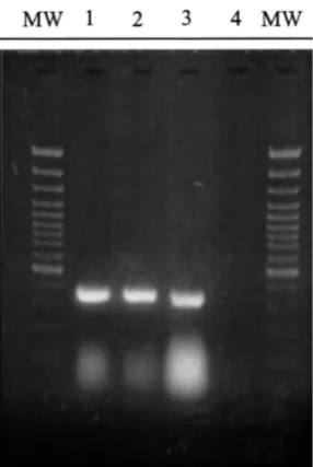
Fig. 1. PVY RT-PCR using the primers PVYF and PVYR4 on total RNA from tobacco leaves infected with PVYN-0321 (1) PVYO-0343 (2) and PVYc -0346 (3). The RT-PCR negative control is presented in channel 4. MW corresponds to the molecular weight markers (GeneRulerTM100 bp DNA Ladder Plus, Fermentas). Agarose gel (1.5%).
Different dilutions were carried out, from ten times to 20 000 times in sterile water and 1 ml of each dilution was used as template for the RT-PCR amplification. As shown in on Fig. 2A for tuber extracts from cv. Nicola, the specific 359 bp signal was observed for each dilution except for the sap diluted 20 000 times. For the sap diluted ten times, the signal was rather weak thus suggest-ing the activity of potential inhibitors of
Fig. 2. PVY RT-PCR using the primers PVYF and PVYR4 on crude sap from PVY infected tubers diluted between 10 and 20,000 times. The RT-PCR positive control (total RNA from PVY infected tobacco leaf) and the RT-PCR negative control (water) are presented in channels ( + ) and ( − ) respectively. A : Cultivar Nicola ; B : Cultivar Bintje. MW corresponds to the molecular weight markers (GeneRulerTM 100 bp DNA Ladder Plus, Fermentas). Agarose gel (1.5%).
total procedure (for a 96 wells plate) can be conducted in less than 4 h.
3. Results
3.1. Optimization of the RT-PCR-ELOSA test The primers PVYF and PVYR4 were used in a one step RT-PCR reaction on total RNA prepara-tions from leaves of tobacco plants infected with the different reference strains of PVY including PVYN, PVYO and PVYC. A 359 bp band corre-sponding to the expected size of the sequence included between both primers was observed for the different samples examined (Fig. 1).
In order to develop a sample preparation proto-col adapted to routine applications, RT-PCR am-plifications were carried out from diluted crude sap of PVY infected tubers (cv. Nicola and Bintje).
the RT-PCR reaction when the sap is not diluted sufficiently. The higher intensity of the signal was observed for sap diluted 100 times. For tuber extracts from the cultivar Bintje (Fig. 2B), similar intensities were observed for tuber sap diluted between ten times and 10 000 times. Therefore, the 100 times dilution was chosen as it represented the best compromise between the level of in-hibitory components and the amount of target sequences. A sapex device currently used in certifi-cation laboratories for the sample preparation in serological assays was tested and found to give the expected results provided that the device was well decontaminated between each sample (data not shown).
The PVY amplification products were then de-tected in microtitration plates after sandwich hy-bridization between a well linked capture probe and a biotinylated specific detection probe (ELOSA tests). The sensitivity of this detection method has been determined. Different concentra-tions of PVYF-PVYR4 amplicons were sandwich-hybridized between the capture probe and the detection probe. Each concentration was tested in triplicate. In such conditions, up to 1 ng of ampli-cons per well was detected (Fig. 3B) while the detection threshold of the same samples was 10 ng after electrophoresis in agarose gel stained with ethidium bromide (Fig. 3A).
3.2. Specificity, poly6alence and detection limit of
the PVY-RT-PCR-ELOSA test
In order to check both the specificity and the polyvalence of the RT-PCR-ELOSA test, crude saps from host plants infected with PVX, PVS, PVA and PVY isolates were submitted to RT-PCR-ELOSA. As observed in Fig. 4, a high opti-cal density value was observed for the PVY isolates thus demonstrating the polyvalence of the test between the different strains of the pathogen at the level of the primers, the capture probe and the detection probe. In contrast, for the other samples, the optical density values were close to that of the negative control.
The amount of virus detected per reaction was determined. Partially purified virus particles were added to the sap of healthy tuber and a
RT-PCR-Fig. 3. Comparison between electrophoresis analysis (agarose gel 1.5%) (A) and ELOSA test (B) for the detection of different amounts (expressed in ng per test) of PVYF-PVYR4 RT-PCR amplification products. For the ELOSA test, the mean OD values of triplicate assay measured at 450 nm (A450) are also provided.
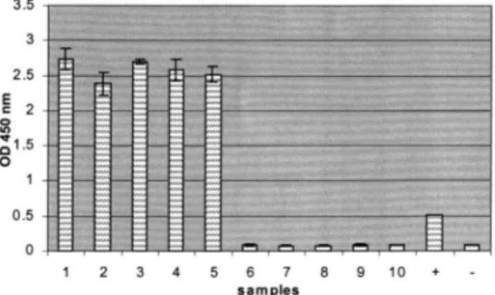
ELOSA was carried out on this artificially con-taminated sap. As observed on Fig. 5, with the cv. Nicola, the RT-PCR-ELOSA test detected up to 0.5 pg of virus per reaction. For the cv. Charlotte and Climax, the detection limit was 5 pg while for the cultivar Bintje, detection of less than 50 pg was not achieved.
3.3. Analysis on pooled sample units
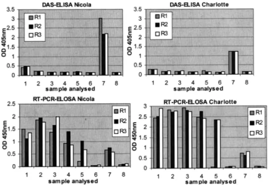
The DAS-ELISA was compared with the RT-PCR-ELOSA on batches of tubers from cv. Nicola and Charlotte. New tubers were harvested from both infected and healthy plants and their saps were mixed in such a way that one infected sap was mixed with N − 1 healthy one (N ranging from 10 to 200).
Using the DAS-ELISA for both cultivars, the optical density value measured for the infected tuber examined individually was slightly higher
Fig. 4. Detection by a sandwich hybridization in microtiter plate of the PVYF-PVYR4 RT-PCR products amplified from diluted crude sap of N. tabacum leaves infected with PVYN -0321 (1) PVYN- wilga(2) PVYNTN(3) PVYO(4) PVYC(5) PVA (6) PVX (7), of C. quinoa leaves infected with PVS (8) and of healthy potato tubers cv Bintje (9). The channel 10 corre-sponds to the RT-PCR negative control. + = ELOSA posi-tive control (purified PVYF-PVYR4 amplicon, 5 ng); − = ELOSA negative control (unrelated DNA).
tive samples were high compared to those of the negative control, thus leading to an unambiguous test interpretation (Fig. 6). When the infected sap was 200 times diluted with healthy sap, the RT-PCR-ELOSA was not reproducible. Indeed, out of the three tests repeated, only two gave a posi-tive signal.
4. Discussion
In a context of routine applications, a detection procedure must be rapid, sensitive, specific, reli-able, easy-to-use and cost-effective. In the terms of a detection kit, it is thus important to integrate the different steps of the procedure in a ‘global approach’ taking into account the different con-straints linked to high throughput analyses.
The sample units taken from the field can be tested in two different ways, regardless of the diagnostic method. The analysis can be performed on individual units (tubers or leaves) which com-pose the sample (individual analysis) or on batches (analysis on pooled units). According to the chosen approach, the number of tests will correspond to the number of individual units or the number of batches. As the number of sample units currently examined in certification laborato-ries by ELISA (individual analysis) can not be easily increased for economical reasons, a test carried out on batches could improve the statisti-cal reliability of the analysis without increasing the number of tests. That approach requires the use of a detection method sufficiently sensitive to detect one infected unit among pooled healthy units.
Considering the great sensitivity of the RT-PCR technology, we aimed at developing a PVY RT-PCR detection kit that could be used on pooled sample units. Furthermore, in order to be applied in certification laboratories, the different steps of the procedure were optimized including (1) the sample preparation prior to the RT-PCR; (2) the amplification step by RT-PCR and; (3) the detection of the amplification products in a con-text of routine applications.
The main drawback linked to the use of the PCR technology concerns both its lack of robust-than that of the negative control making the test
interpretation uncertain. When the infected sap was mixed with healthy sap, the optical density values were close to those of the negative control whatever the proportion would be. In contrast, using the RT-PCR-ELOSA, the infected tuber was detected even when its sap was diluted 100 times with healthy sap. Moreover, contrary to the observation with the DAS-ELISA, the optical density values measured at 450 nm for the
posi-Fig. 5. RT-PCR-ELOSA with the primers PVYF and PVYR4 on diluted crude sap from dormant tubers where virus parti-cles were added at different concentrations. 1 : 5 ng; 2 : 500 pg; 3 : 50 pg; 4 : 5 pg; 5 : 0.5 pg; 6 : 0.05 pg; 7 : RT-PCR negative control (water); 8 : ELOSA positive control (purified PVYF-PVYR4 amplicon, 5 ng); 9 = ELOSA negative control (unre-lated DNA).
Fig. 6. Comparison between DAS-ELISA and RT-PCR-ELOSA on batch of saps from dormant tubers where one infected sap was mixed with N − 1 healthy ones, N ranging from 10 to 200. 1 : undiluted infected sap (i.e. not in batch); 2 : N = 10; 3 : N = 50; 4 : N = 100; 5 : N = 200; 6 : healthy sap; 7 : positive control (DAS-ELISA: crude sap from PVY-infected tobacco leaf; RT-PCR-ELOSA: purified PVYF-PVYR4 amplicon, 5 ng); 8: negative control (DAS-ELISA: sample buffer; RT-PCR-RT-PCR-ELOSA: unrelated DNA). R1-R3: the 3 test repetitions.
ness in terms of sample preparation and its cost. Generally, either immunocapture of virus particles (Nolasco et al., 1993; Weidemann and Maiss, 1996) or extraction of total nucleic acids (Singh and Singh, 1996; Nakahara et al., 1999) precedes the (RT)-PCR amplification. These procedures have proved to be reliable but are either expensive (when an immunocapture of the virus particles is performed) or time-consuming and increase the risk of samples contamination (when a nucleic acid extraction procedure is used). The use of diluted crude sap to carry out the RT-PCR am-plification as undertaken by ELISA represents a significant progress. Marinho et al. (1998) have shown that it was possible to use clarified diluted crude sap of leaves or bark tissues for the detec-tion of the Apple Stem Grooving Virus, at least in their RT-PCR conditions. By designing primers meeting all the technical requirements of the PCR (based on computer analysis), we were able to carry out the RT-PCR reaction using diluted crude sap from dormant tubers of different potato
cultivars used as the target. The decrease in tem-plate concentration due to the extract dilution has been compensated by an efficient amplification step. This simplification of the sample preparation for the RT-PCR reaction is essential to be appli-cable in certification laboratories involved in the detection of potato viruses, where the number of samples processed daily can be important (up to 3000 ELISA tests per day in Belgium, (JL Rolot, personal communication)). The optimization of the use of the Sapex device constitutes a further improvement of the procedure, at least when tu-bers are submitted to the detection test.
The use of a ‘one step RT-PCR protocol’ combining a mix of two enzymes offers several advantages compared to the currently used two steps protocols. Firstly, both the cDNA synthesis and the cDNA amplification are carried out in the same reaction mixture, in one step, thus avoiding the risk of samples contamination which could occur between the reverse transcription and the PCR amplification. Secondly, the protocol used in
this study increases the specificity of the test as a specific primer is used for the cDNA synthe-sis. Thirdly, using the ‘TITAN one tube system’ (from Roche Diagnostic), a high temperature is used during the reverse transcription, thus re-ducing the risk of RNA secondary structures which could inhibit the enzymatic reaction. And finally, it has been demonstrated that a continu-ous RT-PCR in which RT and PCR occur in an uninterrupted reaction with a mix of two en-zymes is more sensitive than a two steps proto-col (Sellner and Turbett, 1998).
Concerning the detection of the amplification products, electrophoresis in agarose gels stained with ethidium bromide is used frequently. How-ever, this detection procedure suffers from sev-eral problems. Firstly, the technique cannot be automated. Secondly, both specific and non spe-cific amplification products are detected, thus in-creasing the risk of false positive results, at least if the non specific signal has the same or ap-proximately the same size as that of the targeted amplicon. We have thus developed a colorimet-ric detection test in microtiter plates using a kit in development (Lambdatech s.a.). This detec-tion method is ten times more sensitive than de-tection carried out in ethidium bromide agarose gel. A PCR-microplate hybridization method for the detection of PVY RT-PCR products has al-ready been described (Hataya et al., 1994). In this method, the amplification products are ad-sorbed directly in the well of the plate and the detection is carried out using a DIG-labeled probe produced by PCR and corresponding to the complete targeted sequence. These character-istics suggest that non specific products could easily be detected. Moreover, the detection pro-cedure needs at least 16 h to be complete. In the ELOSA test described in this study, the de-tection takes about 4 h. The hybridization is carried out at 65°C with a short biotinylated detection probe located in a specific region of the amplicon. A third level of discrimination against false positive results (after the cDNA synthesis and the PCR reaction) is thus ensured by the ELOSA step. Other gel-free methods have been proposed for the detection of PCR amplification products, and notably those based
on the fluorogenic 5% nuclease assay (Schoen et al., 1996). In these methods, the amplification products are detected in real time during the PCR, thus speeding up the detection procedure. Moreover, as the PCR tubes are not open be-tween the amplification and the detection steps, carry-over contaminations are eliminated. How-ever, such devices combining a PCR machine and a fluorometer are expensive. Furthermore, the presence of plant extract in the sample could affect the fluorescence measure, thus making necessary the use of purified nucleic acids as the template for the PCR.
By comparing DAS-ELISA and RT-PCR-ELOSA on batches of pooled tubers of different sizes where one infected sap was mixed with
N − 1 healthy ones, the RT-PCR-ELOSA
method was found to be more sensitive than DAS-ELISA in detecting the infected sap even when it was 100 times diluted with healthy sap. It is important to note that the infected plants used in the comparison between the DAS-ELISA and the RT-PCR-ELOSA techniques have been mechanically inoculated and belong to cultivars that are susceptible to PVY infec-tions. Moreover, the dormant tubers have been collected at least 2 months after the inoculation. These characteristics suggest that a natural in-fection by aphid vectors could lead to virus con-centrations in the tubers lower than those described here. It is thus important, at this stage of the work, to perform a large scale analysis in reference laboratories in order to validate the routine RT-PCR-ELOSA for high throughput analyses on infected naturally tubers.
Acknowledgements
We thank the Ministry of the Walloon Re-gion (Belgium), which supported the present re-search through the ‘Convention 961/3334’. We are also grateful to JL Rolot from the ‘De´parte-ment Production Animales et Syste`mes Agricoles (Libramont, Belgium) du Centre de Recherches Agronomiques de Gembloux’ for letting use the sapex device for our tests.
References
Clark, M.F., Adams, A.N., 1977. Characteristics of the mi-croplate method of enzyme-linked immunosorbent assay for the detection of plant viruses. J. Gen. Virol. 34, 475 – 483.
Cerovska, N., Petrzik, K., Moravec, T., Mraz, I., 1998. Potato virus A detection by reverse transcription-polymerase chain reaction. Acta Virol. 42, 83 – 85.
De Bokx, J.A., Cuperus, C., 1987. Detection of potato virus Y in early-harvested potato tubers by cDNA hybridisation and three modifications of ELISA. OEPP EPPO Bull. 17, 73 – 79.
De Bokx, J.A., Huttinga, H., 1981. Potato virus Y. CMI/AAB Descriptions of plant viruses 242, 6.
Deltour, A., Guillou, M., Kerlan, C., 1987. Trends in France for the use of the ELISA method in routine seed-potato testing and problems of cross reactions between potato viruses A and Y. OEPP EPPO Bull. 17, 69 – 72.
Geng, S., Campbell, R.N., Carter, M., Hills, F.J., 1983. Qual-ity-control programs for seedborne pathogens. Plant Dis. 67, 236 – 242.
Gugerli, P., 1979. Le test immuno-enzymatique (ELISA) et son application pour le diagnostic rapide des viroses de la pomme de terre. Revue Suisse Agric. 11, 253 – 260. Hammond, J., Lawson, R.H., 1988. An improved purification
procedure for preparing potyviruses and cytoplasmic inclu-sions from the same tissue. J. Virol. Methods 20, 203 – 217. Hataya, T., Inoue, A.K., Shikata, E., 1994. A PCR-microplate hybridization method for plant virus detection. J. Virol. Methods 46, 223 – 236.
Huttinga, H., Maat, D.Z., 1987. Characterization and identifi-cation of potato viruses and viroids. Physical and chemical properties. In: De Bokx, J.A., van der Want, J.P.H. (Eds.), Viruses of potatoes and seed-potato production, Second edition. Pudoc, Wageningen, pp. 33 – 44.
Le Romancer, M., Kerlan, C., Nedellec, M., 1994. Biological characterisation of various geographical isolates of potato virus Y inducing superficial necrosis in potato tubers. Plant Pathol. 43, 138 – 144.
Marinho, V.L.A., Kummert, J., Rufflard, G., Colinet, D., Lepoivre, P., 1998. Detection of apple stem grooving virus in dormant apple trees with crude extracts as templates for the one-step RT-PCR. Plant Dis. 82, 785 – 790.
Nakahara, K., Hataya, T., Uyeda, I., 1999. A simple, rapid method of nucleic acid extraction without tissue homogeni-sation for detecting viroids by hybridihomogeni-sation and RT-PCR. J. Virol. Methods 77, 47 – 58.
Nolasco, G., de Blas, C., Torres, V., Ponz, F., 1993. A method combining immunocapture and PCR amplification in a microtiter plate for the detection of plant viruses and subviral pathogens. J. Virol. Methods 45, 201 – 218. Rasmussen, S.R., Larsen, M.R., Rasmussen, S.E., 1991.
Cova-lent immobilization of DNA onto polystyrene microwells: the molecules are only bound at the 5% end. Anal. Biochem. 198, 138 – 142.
Remacle, J., 1997. Method and kit for diagnosing and/or quantifying by sandwich hybridisation of nucleic acid se-quences on solid support. Patent W098/11253.
Robaglia, C., Durand-Tardif, M., Tronchet, M., Boudazin, G., Astier-Manifacier, S., Casse-Delbart, F., 1989. Nucle-otide sequence of potato virus Y (N strain) genomic RNA. J. Gen. Virol. 70, 935 – 947.
Schoen, C.D., Knorr, D., Leone, G., 1996. Detection of potato leafroll virus in dormant potato tubers by immuno-capture and a fluorogenic 5% nuclease RT-PCR assay. Phytopathol 86, 993 – 999.
Sellner, L.N., Turbett, G.R., 1998. Comparison of three RT-PCR methods. Biotechniques 25, 230 – 234.
Singh, R.P., 1998. Reverse-transcription polymerase chain re-action for the detection of viruses from plants and aphids. J. Virol. Methods 74, 125 – 138.
Singh, R.P., Kurz, J., Boiteau, G., 1996. Detection of stylet-borne and circulative potato viruses in aphids by duplex reverse transcription polymerase chain reaction. J. Virol. Methods 59, 189 – 196.
Singh, R.P., Kurz, J., Boiteau, G., Bernard, G., 1995. Detec-tion of potato leafroll virus in single aphids by the reverse transcription polymerase chain reaction and its potential epidemiological application. J. Virol. Methods 55, 133 – 143.
Singh, M., Singh, R.P., 1996. Factors affecting detection of PVY in dormant tubers by reverse transcription poly-merase chain reaction and nucleic acid spot hybridization. J. Virol. Methods 60, 47 – 57.
Singh, R.P., Singh, M., 1998. Specific detection of potato virus A in dormant tubers by reverse transcription polymerase chain reaction. Plant Dis. 82, 230 – 234.
Singh, R.P., Singh, M., King, R.R., 1998. Use of citric acid for neutralizing polymerase chain reaction inhibition by chlorogenic acid in potato extracts. J. Virol. Methods 74, 231 – 235.
Staub, U., Polivka, H., Gross, H.J., 1995. Two rapid mi-croscale procedures for isolation of total RNA from leaves rich in polyphenols and polysaccharides: application for sensitive detection of grapevine viroids. J. Virol. Methods 52, 209 – 218.
Van der Zaag, D.E., 1987. Yield reduction in relation to virus infection. In: De Bokx, J.A., van der Want, J.P.H. (Eds.), Viruses of potatoes and seed-potato production, Second edition. Pudoc, Wageningen, pp. 146 – 150.
Vetten, H.J., Ehlers, U., Paul, H.L., 1983. Detection of potato viruses Y and A in tubers by enzyme-linked immunosor-bent assay after natural and artificial break of dormancy. Phytopath. Z. 108, 41 – 53.
Weidemann, H.L., Maiss, E., 1996. Detection of the potato tuber necrotic ringspot strain of potato virus Y (PVYNTN) by reverse transcription and immunocapture polymerase chain reaction. J. Plant Dis. Protect. 103, 337 – 345.
Zammatteo, N., Moris, P., Alexandre, I., Vaira, D., Piette, J., Remacle, J., 1995. DNA probe hybridisation in microwells using a new bioluminescent system for the detection of PCR-amplified HIV-1 proviral DNA. J. Virol. Methods 55, 185 – 197.