HAL Id: tel-02399228
https://tel.archives-ouvertes.fr/tel-02399228
Submitted on 9 Dec 2019Identifications de nouvelles voies de régulation
impliquées dans la résistance non enzymatique aux
aminosides chez Pseudomonas aeruginosa
Arnaud Bolard
To cite this version:
Arnaud Bolard. Identifications de nouvelles voies de régulation impliquées dans la résistance non enzymatique aux aminosides chez Pseudomonas aeruginosa. Bactériologie. Université Bourgogne Franche-Comté, 2019. Français. �NNT : 2019UBFCE006�. �tel-02399228�
Doctoral thesis from UNIVERSITÉ BOURGOGNE FRANCHE-COMTÉ, prepared at UFR SCIENCES MÉDICALES ET PHARMACEUTIQUES
École doctorale Environnements-Santé n°554
Doctorate in Biochemistry and Molecular Biology
by
Arnaud BOLARD
Identification of novel regulatory pathways
involved in non-enzymatic resistance to
aminoglycosides in
Pseudomonas aeruginosa
Thesis presented and defended at Besançon, France, on July 5
th, 2019
Thesis committee:
Pr. Christophe BORDI President
Dr. Ina ATTRÉE Reviewer
Dr. Thierry NAAS Reviewer
Dr. Thilo KӦHLER Moderator
Dr. Katy JEANNOT Moderator
UBFC
UNIVERSIT É
BOURGOGNE FRANCHE-COMTÉ
Thèse de doctorat de l'UNIVERSITÉ BOURGOGNE FRANCHE-COMTÉ, préparée à l'UFR SCIENCES MÉDICALES ET
PHARMACEUTIQUES
École doctorale Environnements-Santé n
°554
Doctorat en Biochimie et Biologie moléculaire
par
Arnaud HOLARD
Identification de nouvelles voies de régulation
impliquées dans la résistance non enzymatique
aux aminosides chez
Pseudomonas aeruginosa
Thèse présentée et soutenue à Besançon, France,
le 5 juillet
2019
Composition du jury: Pr. Christophe BORD! Dr. Ina A TTRÉE Dr. Thierry NAAS Dr. Thilo KÔHLER Président Rapporteur Rapporteur Examinateur
Ce manuscrit de thèse est l’aboutissement de plusieurs années de travail qui ont pu se concrétiser grâce à l’expertise et au soutien sans faille de nombreuses personnes auxquelles j’adresse mes plus vifs remerciements.
Je tiens tout d’abord à remercier très sincèrement le Docteur Ina Attrée et le Docteur Thierry Naas pour m’avoir fait l’honneur d’accepter d’être les rapporteurs de ce manuscrit ainsi que pour leur participation au jury de soutenance de thèse.
Mes remerciements s’adressent également au Docteur Thilo Köhler qui a accepté de partager son expertise scientifique en examinant mon travail de thèse.
Je remercie avec beaucoup de respect le Professeur Christophe Bordi qui a contribué à la réalisation de cette thèse par ses précieux conseils et par les échanges que nous avons pu avoir au cours des comités annuels de suivi de thèse. Un grand merci pour avoir accepté de faire partie de mon jury de thèse.
Je remercie très chaleureusement le Docteur Delphine Destoumieux-Garzón pour son implication et sa contribution dans la maturation de mon doctorat lors des réunions annuelles de suivi de thèse. Je la remercie, ainsi que toute son équipe, et notamment Daniel Oyanedel pour m’avoir accueilli lors de mon séjour à Montpellier. J’ai été ravi de pouvoir passer ces quelques jours au sein de son équipe soudée et dynamique.
J’adresse mes remerciements à mon directeur de thèse, le Professeur Patrick Plésiat pour m’avoir permis d’effectuer mon doctorat au sein de son équipe. Je le remercie tout particulièrement pour son expertise précieuse et son implication apportées au cours de la rédaction de ce manuscrit.
Je remercie ma tutrice, le Docteur Katy Jeannot avec qui j’ai étroitement travaillé au cours de mon doctorat. Je la remercie tout particulièrement pour avoir initié une collaboration réussie avec le Docteur Yanyan Li.
J’adresse tout mon respect et mes remerciements éternels au Docteur Catherine Llanes. J’espère pouvoir suivre un jour ses pas.
Je remercie très amicalement le Docteur Benoît Valot pour m’avoir débloqué lorsque je pensais être dans une impasse.
Je dois avouer que ma thèse n’aurait pas été la même sans des collègues d’exception: Anaïs, Paulo, Loïs, Hélène, Mélanie, Alexandre, Eleni, Isabelle, Damien, Sophie, Jean-Baptiste, Camille, Gwendoline, Elisa, Antoine, Thomas, Maxime, Pauline, Amélie, Emilie, Steffi, Adeline, Audrey, Alice, Coralie, Chloé, Clothilde, Malik, Sandra, Marie, Didier, Charlotte, Jenny ; ce fut un réel plaisir de travailler avec vous et je vous remercie sincèrement pour votre soutien.
Pour finir, je remercie sans aucune commune mesure ma famille pour son soutien indéfectible depuis toujours et pour la liberté si précieuse qu’elle me procure. Vous êtes ma plus grande force.
Table of contents
I. INTRODUCTION ... 1
II. BIBLIOGRAPHIC REVIEW ... 7
1. Aminoglycosides: overview ... 9
1.1. Structure... 9
1.2. Aminoglycosides uptake ... 9
1.2.1. Cell envelope of Gram-negative bacteria ... 11
1.2.2. Initial phase: ionic binding ... 13
1.2.3. Energy Dependent Phase I (EDPI) ... 13
1.2.4. Energy Dependent Phase II (EDPII) ... 13
1.3. Mode of action of aminoglycosides ... 15
1.3.1. The ribosome, target of aminoglycosides ... 15
1.3.2. Elongation factor G (EF-G) ... 19
1.4. Epidemiology of resistance ... 23
2. Non-enzymatic resistance mechanisms to aminoglycosides ... 25
2.1. Aminoglycosides resistance in biofilms ... 25
2.2. Alteration of the cellular targets of aminoglycosides ... 27
2.3. Membrane modifications ... 29
2.4. Resistance mediated by the efflux pump MexXY(OprM) ... 31
2.4.1. The RND efflux pump structure and assembly ... 31
2.4.2. Intrinsic resistance ... 33
2.4.3. Adaptive resistance ... 35
2.4.4. Acquired resistance ... 35
3. Two-component signal transduction ... 41
3.1. Molecular mechanisms of signal transduction ... 41
3.1.1. The HK protein: signal detection and kinase activation ... 43
3.1.1.1. The periplasmic sensing domains ... 43
3.1.1.2. The transmembrane helical segments ... 45
3.1.1.3. The signal transduction domains: HAMP and STAC ... 45
3.1.1.4. The cytoplasmic sensor domains: PAS and GAF ... 47
3.1.1.5. The kinase core ... 47
3.1.1.6. The helical linker ... 49
3.1.2. The response regulator (RR): phosphotransfer and response ... 49
3.1.2.1. The REC domain ... 49
3.1.2.2. The effector domain ... 51
3.2. TCS and antibiotic resistance in P. aeruginosa ... 51
3.2.1. TCS and resistance to colistin ... 53
3.2.2. Aminoglycoside resistance mediated by TCS ... 53
3.2.3. TCS-dependent susceptibility to carbapenems, fluoroquinolones and β-lactams ... 57
3.3. PmrAB two-component system ... 57
3.3.1. Structure of the TCS PmrAB... 57
3.3.2. The regulon of PmrAB ... 59
3.3.3. Constitutive activation of PmrAB ... 61
4. Nonribosomal peptides ... 67
4.1. NRPs biosynthesis ... 67
4.1.1. The building block assembly ... 67
4.1.2. The assembly of the polypeptide by NRPS ... 69
4.2. Biological activities of NRPs ... 71
4.3. NRPs in P. aeruginosa ... 73
4.3.1. Pyoverdines ... 73
4.3.2. Pyochelin ... 77
4.3.3. L-2-Amino-4-methoxy-trans-3-butenoic acid (AMB) ... 79
4.3.4. Pyoluteorin ... 81
4.3.5. Uncharacterized NRPS clusters ... 83
4.3.5.1. PA7-cluster ... 83
4.3.5.2. PA1221, PA3327 and PA4078 clusters ... 83
III. RESULTS ... 87
Chapter 1. Mutations in fusA1 confer aminoglycoside resistance in P. aeruginosa ... 89
1. Context ... 91
2. Objective ... 91
3. Results ... 91
3.1. Mutations in Gene fusA1 as a Novel Mechanism of Aminoglycoside Resistance in Clinical Strains of Pseudomonas aeruginosa ... 91
3.2. Supplementary results ... 105
3.2.1. Genetic polymorphism of fusA1 among environmental strains of P. aeruginosa ... 105
3.2.2. Prevalence of fusA1 mutations in a collection of isolates exhibiting a non-enzymatic resistance to aminoglycosides ... 105
3.2.3. fusA1 mutations do not contribute to high aminoglycoside resistance in a collection of agrZ and agrW1 mutants isolated from patients ... 107
3.2.4. Susceptibility to β-lactams, carbapenems, fluoroquinolones and tetracycline is unchanged in fusA1 mutants ... 107
3.2.5. fusA1 mutations decrease gentamicin-induced expression of mexY and armZ ... 109
4. Conclusion ... 109
Chapter 2. Mutations in pmrAB mediate aminoglycoside resistance in P. aeruginosa ... 111
1. Context ... 113
2. Objective ... 113
3. Results ... 113
3.1. Article in preparation ... 113
3.2. Supplementary results ... 161
3.2.1. Characterization of a collection of P. aeruginosa clinical strains resistant to colistin….…. ... 161
3.2.2. Transcriptional analysis of mutant AB16.2 compared to PAO1 reveals a high number of PmrAB-regulated genes ... 163
3.2.3. Role of PA4773, PA4774 and PA4775 genes in AB16.2: growth curves, colony morphology and surface attachement ... 165
3.2.4. Polyamines and cell surface modifications ... 167
3.2.5. Contribution of additional genetic loci to antibiotic resistance of AB16.2 ... 171
4. Conclusion ... 171
Chapter 3. The efflux pump MexXY(OprM) contributes to acquired resistance to colistin in P. aeruginosa ... 173
1. Context ... 185
2. Objective ... 185
3. Results ... 185
3.1. Azetidine-containing alkaloids produced by a quorum-sensing regulated non-ribosomal peptide synthetase pathway in Pseudomonas aeruginosa ... 185
3.2. Supplementary results ... 237
3.2.1. Characterization of nonribosomal peptide synthetase biosynthetic gene cluster PA3326-PA3335 ... 237
3.2.2. Nonribosomal peptides derived from the PA3327 biosynthesis cluster do not impact antibiotic resistance, plastic surface attachement or growth at low cell densities in AB16.2…….. ... 239
3.2.3. pmrAB mutations identified from in vitro mutants and clinical strains activate the expression of PA3327 gene ... 241
4. Conclusion ... 241
IV. DISCUSSION AND PERSPECTIVES ... 243
V. MATERIALS AND METHODS ... 267
1. Microbiology ... 269
1.1. Bacterial strains ... 269
1.2. Plasmids ... 274
1.3. Culture media ... 278
1.4. Selection of antibiotic-resistant mutants ... 279
1.5. Drug susceptibility testing ... 279
1.5.1. Antibiograms ... 279
1.5.2. Minimum Inhibitory Concentration (MIC) ... 279
1.6. Growth curves ... 279
1.7. Cytochrome c binding assay ... 280
2. Molecular biology ... 280
2.1. Primers ... 280
2.2. Nucleic acid purification ... 290
2.2.1. Genomic DNA extraction ... 290
2.2.2. Plasmid DNA preparation ... 290
2.3. DNA amplification via Polymerase Chain Reaction (PCR) ... 290
2.4. DNA cloning ... 290
2.4.1. Enzymatic restriction of DNA ... 290
2.4.2. Separation of DNA fragments by agarose gel electrophoresis ... 291
2.4.3. Extraction of DNA fragments from agarose gels ... 291
2.4.4. Ligation of DNA fragments ... 291
2.5. Bacterial DNA transfer ... 291
2.5.1. Heat-shock method ... 291
2.5.2. Electro-competent method ... 292
2.5.3. Conjugation method ... 292
2.6. Gene inactivation ... 292
2.7. Gene complementation and mutagenesis ... 293
2.7.1. Chromosomal complementation ... 293
2.7.2. Plasmid-based complementation ... 294
2.7.3. Mutagenesis of fusA1 gene by homologous recombination ... 294
2.7.4. Site-directed mutagenesis ... 294
2.8. Quantification of mRNA transcripts by RT-qPCR ... 295
2.9. Quantification of mRNA transcripts by RNA seq ... 296
2.9.2. Sample analysis and mRNA transcripts quantification ... 296
3. DNA sequencing ... 297
4. Purification of His6-tagged protein... 297
5. Surface polyamine analysis ... 298
5.1. Surface polyamine sample preparation ... 298
5.2. Surface polyamine sample analysis ... 299
5.2.1. Principle ... 299
5.2.2. Polyamine standards analysis ... 299
5.2.3. Surface-washed polyamines analysis ... 300
5.3. Surface polyamine sample quantification ... 300
VI. APPENDIX ... 303
Figure 1: Structures of representative aminoglycosides from the four subclasses.. ... 8
Figure 2: Schematic representation of the bacterial cell envelope of Gram-negative bacteria... ... 10
Figure 3: Lipopolysaccharide (LPS) structure of P. aeruginosa. ... 10
Figure 4: Schematic representation of aminoglycoside (streptomycin) accumulation in P. aeruginosa. ... 12
Figure 5: Representation of the 70S ribosome of E. coli. ... 14
Figure 6: Functional organization of mRNA translation by the ribosome. ... 16
Figure 7: Interactions of aminoglycoside molecule neomycin with the decoding center of the ribosome. ... 16
Figure 8: The structures of the elongation factor G in complex with the ribosome in pre- and post-translocational states. ... 18
Figure 9: Epidemiology of P. aeruginosa strains resistant to aminoglycosides in Europe. ... 22
Figure 10: Schematic representation of global non-enzymatic resistance mechanisms to aminoglycosides developed by planktonic P. aeruginosa cells. ... 24
Figure 11: Schematic representation of the formation and dispersion of a P. aeruginosa biofilm.... ... 26
Figure 12: Structure of the RND efflux pump MexXY(OprM) based on the MexAB-OprM model from P. aeruginosa. ... 30
Figure 13: Schematic representation of the regulation of mexXY operon expression in P. aeruginosa. ... 34
Figure 14: Schematic representation of a typical TCS. ... 40
Figure 15: Schematic representation of His-Asp phosphotransfer between the sensor kinase and its cognate response regulator in classical (A), unorthodox (B) and hybrid (C) TCSs. ... 40
Figure 16: Structures of extra-cytoplasmic (A), HAMP signal transduction (B) and cytoplasmic (C) sensing domains ... 42
Figure 17: Structure of the kinase core in complex with RR during phosphotransfer. ... 48
Figure 18: Structure of DNA-binding domain subfamilies of RRs. ... 50
Figure 19: TCS contributing to antibiotic resistance in P. aeruginosa. ... 50
Figure 20: LPS modification by addition of 4-amino-4-desoxy-L-arabinose (L-Ara-4N) molecule on a phosphate group of the lipid A... 52
Figure 21: Schematic representation of functional domains of the PmrB sensor protein from P. aeruginosa. ... 58
Figure 23: Nonribosomal peptides representing the diversity of monomers which can be
incorporated by nonribosomal peptide synthetases. ... 66
Figure 24: NRP synthesis pathway. ... 68
Figure 25: Nonribosomal code. ... 68
Figure 26: Examples of reactions catalyzed by NRPS domains. ... 70
Figure 27: Pie chart repartition of the 1,190 NRPs listed in the Norine database according to their reported biological activities and therapeutic applications. ... 70
Figure 28: NRPs synthesized by P. aeruginosa. ... 72
Figure 29: Genetic cluster responsible of type I pyoverdine biosynthesis and export in P. aeruginosa strain PAO1... 72
Figure 30: Schematic representation of pyoverdine synthesis, secretion and uptake in P. aeruginosa. ... 74
Figure 31: Genetic cluster for synthesis of pyochelin in P. aeruginosa strain PAO1. ... 76
Figure 32: Schematic representation of pyochelin synthesis, secretion and uptake in P. aeruginosa. ... 76
Figure 33: Genetic cluster for the biosynthesis of L-2-amino-4-methoxy-trans-3-butenoic acid in P. aeruginosa strain PAO1. ... 78
Figure 34: Schematic representation of L-2-amino-4-methoxy-trans-3-butenoic acid synthesis and export in P. aeruginosa. ... 78
Figure 35: Genetic cluster for synthesis and export of pyoluteorin in P. aeruginosa strain M18…. ... 80
Figure 36: Schematic representation of pyoluteorin synthesis and export in P. aeruginosa...… ... 80
Figure 37: Genetic clusters of uncharacterized NRPS in P. aeruginosa. ... 82
Figure 38: Antibiograms of PAO1 and PAOR13 derivative mutant. ... 90
Figure 39: Effects of fusA1 mutations on gene mexY and gene armZ expression in strain PAO1 submitted to gentamicin exposure. ... 108
Figure 40: Antibiograms of PAO1 and AB16.2 derivative mutant. ... 112
Figure 41: Timeline of isolation of strains 3095 to 3089, and periods of antibiotic treatement... ... 160
using cationic probe cytochrome c. ... 166 Figure 47: SDS-PAGE analysis of samples used for characterization of surface-bound polyamines. ... 168 Figure 48: Structures of several polyamines. ... 168 Figure 49: Comparison of LPS profiles by SDS-PAGE. ... 168 Figure 50: MALDI-TOF mass spectra of lipid A from strains PAO1, AB16.2 and AB16.2ΔmexXY. ... 178 Figure 51: Antibiograms of clinical isolates 2243 and 3795, and their respective mexXY-inactivated mutants. ... 180 Figure 52: Schematic representation of quorum sensing-dependent signaling in P. aeruginosa... ... 184 Figure 53: Agarose gel electrophoresis showing the inactivation of genes PA3326, PA3327 and pqsA in mutant AB16.2, by deletion of internal DNA fragments. ... 236 Figure 54: PA3327 gene expression in the pmrB mutant AB16.2 and various derivatives, as assessed by RT-qPCR. ... 236 Figure 55: Gene expression of PA3335 in AB16.2ΔPA3326,35-complementated strain. ... 238 Figure 56: Growth curves of strains PAO1, AB16.2 and AB16.2ΔPA3327 cultivated in Mueller-Hinton broth at 37°C with shaking (250 rpm). ... 238 Figure 57: Characterization of the role of the NRPS genetic cluster PA3327 in the capacity of a pmrB mutant to adhere to a plastic surface. ... 240 Figure 58: Schematic representation of P. aeruginosa adaptation mediated by mutations in genes fusA1 and pmrB. ... 244 Figure 59: Proposed model for the synthesis of spermidine and norspermidine in P. aeruginosa….. ... 256 Figure 60: Gene inactivation by homologous recombination between a recombinant derivative of plasmid pKNG101 and the P. aeruginosa chromosome ... 293
List of tables
Table 1: Antimicrobial categories and agents used to define multidrug resistant (MDR),
extensively drug-resistant (XDR) and pandrug-resistant (PDR) strains of P. aeruginosa. ... 4
Table 2: List of EF-G1A/B alterations in in vitro-selected and clinical strains of P. aeruginosa…. ... 20
Table 3: Genetic loci confirmed to be involved in the resistance of P. aeruginosa biofilms to aminoglycosides. ... 26
Table 4: List of PmrAB substitutions identified in vitro and in clinical strains of P. aeruginosa…. ... 60
Table 5: Determination of polymorphic mutations in EF-G1A by DNA sequencing of environmental strains. ... 104
Table 6: Characterization of a collection of non-CF clinical strains from the University Hospital of Besançon. ... 104
Table 7: Sequence analysis of fusA1 gene in a clinical collection of previously characterized agrZ and agrW1 mutants. ... 106
Table 8: Phenotypic characterization of engineered EF-G1A mutants. ... 106
Table 9: Characterization of a collection of colistin-resistant clinical strains. ... 160
Table 10: PmrB mutants susceptibility to β-lactams. ... 166
Table 11: Effect of pmrB mutations on the susceptibility to β-lactams... 166
Table 12: Antibiotic susceptibility of AB16.2 derivatives. ... 170
Table 13: Contribution of several TCSs and RND efflux pumps in acquired resistance to colistin. ... 176
Table 14: Analysis of the synergy between lipid A modification and efflux. ... 178
Table 15: Contribution of the NRPS genetic cluster PA3327 in the susceptibility of P. aeuginosa to antibiotics. ... 238
Table 16: Effects of PmrAB substitutions on expression of gene PA3327 and two PmrAB regulated genes (PA4774 and arnA). ... 240
Table 17: Bacterial strains used to characterize the role of elongation factor EF-G1A in aminoglycoside resistance of P. aeruginosa clinical strains. ... 269
Table 22: Plasmids used in all projects. ... 274
Table 23: Plasmids used to study the impact of EF-G1A amino acid substitutions in vitro…….... ... 274
Table 24: Plasmids used to study the impact of PmrAB amino acid substitutions... 275
Table 25: Plasmids used to characterize the colistin resistome in P. aeruginosa... 276
Table 26: Plasmids used to characterize the biosynthesis of new azetidine-containing alkaloids… ... 277
Table 27: Composition of culture media. ... 278
Table 28: Antibiotic concentrations used to maintain plasmids in cultures of E. coli and P. aeruginosa strains. ... 278
Table 29: Primers used in the study of elongation factor EF-G1A. ... 280
Table 30: Primers used in the project related to PmrAB substitutions and increased resistance to aminoglycosides. ... 281
Table 31: Primers used for investigating the role of pump MexXY(OprM) as determinant of acquired resistance to colistin. ... 286
Table 32: Primers used for the characterization of the PA3327 cluster. ... 287
Table 33: Primers used in several projects. ... 289
Abbreviations
A530: absorbance at 530 nm A600: absorbance at 600 nm
(p)ppGpp: guanosine pentaphosphate or tetraphosphate
∆ψ: membrane electrical potential 3OC12-HSL: N-3-oxododecanoyl-HSL 5FA: penta-acyl molecular species 6FA: hexa-acyl molecular species A: adenylation aa: aminoacyl ACN: acetonitrile AMB: L-2-amino-4-methoxy-trans-3-butenoic acid AMK: amikacin
AMP: adenosine monophosphate AMPs: antimicrobial peptides Ampr: ampicillin resistance
ANL: acyl-CoA synthetases, NRPS
adenylation domains, and Luciferase enzymes APH(3’): aminoglycoside O-3’
phosphoryltransferase AraN: aminoarabinose
arn: arnBCADTEF-ugd
A-site: aminoacyl-site ATM: aztreonam
ATP: adenosine triphosphate C: condensation
C4-HSL: N-butanoyl-HSL
CA: C-terminal catalytic and ATP binding CAMPs: cationic antimicrobial peptides CAZ: ceftazidime
c-di-GMP: bis-(3’-5’)-cyclic dimeric GMP cDNA: complementary DNA
CF: cystic fibrosis
CFU: colony forming unit CHL: chloramphenicol CIP: ciprofloxacin
CLSI: clinical and laboratory standards institute Cm: carbamoyl CST: colistin Ct: cycle threshold DABA DC: L-2,4-diaminobutyrate decarboxylase
DHp: dimerization and histidine phosphotransfer
DMSO: dimethyl sulfoxide DNA: deoxyribonucleic acid DSF: cis-2-unsaturated fatty acids E: epimerization
EARS-Net: european antimicrobial resistance surveillance network
ECDC: european centre for disease prevention and control
EDP: energy dependent phase EEA: european economic area EFF: effector
EPS: exopolysaccharides E-site: exit-site
Etn: ethanolamine EU: european union FA: fatty acid FEP: cefepime FOF: fosfomycin
FucNAc: 2-acetamido-2-deoxy-D-fucose GAF: c-GMP and c-GMP-stimulated phosphodiesterases, Anabaena adenylate cyclases and E. coli FhlA
GalN: 2-amino-2-deoxy-galactose gDNA: genomic DNA
GEN: gentamicin GI: genomic island Glc: glucose
GlcN: 3-(acetylamino)-3-deoxy-D-glucose Gmr: gentamicin resistance
GTP: guanosine triphosphate H: histidine
HAMP: linker domain
HATPase: histidine kinase-like ATPases Hep: L-glycero-D-manno-heptose HEPES: 4-(2-hydroxyethyl)-1-piperazineethanesulfonic acid HHQ: 4-hydroxy-2-heptylquinoline HisKA: dimerization and phosphoacceptor
Kan: kanamycin resistance
Kdo: 3-deoxy-D-manno-oct-2-ulosonic acid L-Ara-4N: 4-amino-4-desoxy-L-arabinose L-ASA: L-aspartate β-semialdehyde L-AZC: L-azetidine 2-carboxylic acid LB: lysogeny broth
LC-ESI-MS: liquid chromatography-electrospray ionization-mass spectrometry LC-HRMS: liquid chromatography coupled to high resolution mass spectrometry
L-Dab: L-2,4-diaminobutyrate L-fOHOrn: L-N5-formyl-N5-hydroxyornithine L-OHOrn: L-N5-hydroxyornithine L-Orn: L-ornithine LPS: lipopolysaccharide m/z: mass/charge ratio
MALDI-TOF: matrix-assisted laser desorption/ionization with time-of-flight ManNAc3NAcA: 2,3-diacetamido-2,3-dideoxy-D-mannuronic acid ManNAc3NAmA: 2-acetamido-3-acetamidino-2,3-dideoxy-D-mannuronic acid McF: McFarland MDR: multidrug-resistant MEM: meropenem
MHA: Mueller-Hinton agar
MHBc: Mueller-Hinton broth (cation-adjusted) MIC: minimum inhibitory concentration MOPS: 3-(N-morpholino)propanesulfonic acid mRNA: messenger RNA
NA: non applicable
NADH: nicotinamide adenine dinucleotide NADPH: nicotinamide adenine dinucleotide phosphate (reduced) nd: not determined N-Mt: N-methylation NOV: novobiocin NRP: nonribosomal peptide NRPS: NRP synthetase OM: outer membrane OXA: oxacillinase P: phosphate
PAS-like: Per (period circadian protein)-ARNT (vertebrate aryl hydrocarbon receptor nuclear translocator)-Sim (single-minded) PATRIC: pathosystems resource integration center
PCP: peptidyl carrier protein PCR: polymerase chain reaction PDB: protein data bank
PDR: pandrug-resistant
PIA: Pseudomonas isolation agar
PKS: polyketide synthase PLP: pyridoxal phosphate Pmx: polymyxins PPi: pyrophosphate PQS: 2-heptyl-3-hydroxy-4-quinolone P-site: peptidyl-site PVD: pyoverdine qPCR: quantitative PCR QS: quorum sensing REC: N-terminal receiver Rec: recycled
Rha: rhamnose
RNA seq: high-throughput RNA sequencing RNA: ribonucleic acid
RND: resistance nodulation cell division rpm: revolutions per minute
RR: response regulator rRNA: ribosomal RNA RT: reverse transcription SAM: S-adenosylmethionine
SDS-PAGE: sodium dodecyl sulphate-polyacrylamide gel electrophoresis
SMART: simple modular architecture research tool
STAC: solute carrier and two-component signal transduction associated component STR: streptomycin
Strr: streptomycin resistance
T: thiolation
TAE: tris-acetate 40 mM, EDTA 1 mM TCS: two-component system
TE: thioesterase TET: tetracycline
Tetr: tetracycline resistance
TFA: trifluoroacetic acid TIC: ticarcillin
TIM: ticarcillin-clavulanic acid TM: transmembrane TMAO: trimethylamine-N-oxide TMP: trimethoprim TMPP: N-succinimidyloxycarbonylmethyl)-tris-(2,4,6-trimethoxyphenyl)-phosphonium bromide TOB: tobramycin
Trans_reg_C: transcriptional regulatory protein, C-terminal
tRNA: transfer RNA
TZP: piperacillin-tazobactam U: enzyme unit
UV: ultraviolet VAN: vancomycin
XDR: extensively drug-resistant Zeor: zeocin resistance
to grow in a wide variety of environmental conditions such as soil, water as well as in plant-, animal- and human-host-associated environments (Kazmierczak et al., 2015; Sitaraman, 2015). This great adaptability may result in part from its relatively large genome (≈ 6.3 Mb) carrying a substantial number of genes enabling the utilization of various carbon sources, efficient energy metabolism, and encoding many regulatory systems (Stover et al., 2000).
In the clinical context, this non-fermentative bacillus is a major human opportunistic pathogen infecting immunocompromised patients, especially in intensive care units, as well as those suffering from chronic respiratory diseases such as cystic fibrosis (CF). In CF patients, the bronchial colonization by various microorganisms leads to significant alteration of respiratory function. Unfortunately, P. aeruginosa is difficult to treat because of its intrinsic resistance to several anti-Gram-negative antibiotic classes including β-lactams and aminoglycosides. This resistance is mainly due to the low permeability of the outer membrane, the action of enzymes which degrade or modify antibiotics (AmpC, OXA-50 and APH(3’)-IIb) and finally the complementary action of two multi-drug efflux systems [MexAB-OprM and MexXY(OprM)] able to export a huge panel of antibacterials outside the cell (Lister et al., 2009).
In addition, resistance-associated genes may be acquired by P. aeruginosa from mobile genetic elements such as plasmids and transposons that can reduce ultimately the efficacy of all of the available antibiotic families (aminoglycosides, β-lactams, fluoroquinolones and polymyxins). Finally, the basal resistance levels of P. aeruginosa may also be increased through mutational processes altering the expression and/or function of chromosomally-encoded resistance-associated proteins. For example, mutational inactivation of MexR, a transcriptional repressor of the mexAB-oprM operon, is associated with a 4- to 8-fold increase in resistance to β-lactams (Cao et al., 2004).
As a result, the accumulation of several of these genetic events is the driving force of emergence of clinical isolates highly resistant to multiple antibiotic families. To characterize the different patterns of resistance, a standardized international terminology defined (i) multidrug-resistant (MDR) strains as presenting an acquired resistance to at least one agent in three or more antimicrobial categories, (ii) extensively drug-resistant
Table 1: Antimicrobial categories and agents used to define multidrug resistant (MDR), extensively drug-resistant (XDR) and pandrug-resistant (PDR) strains of P. aeruginosa.
Antimicrobial categories Antimicrobial agents
Aminoglycosides gentamicin, tobramycin, amikacin, netilmicin Antipseudomonal carbapenems imipenem, meropenem Antipseudomonal cephalosporins ceftazidime, cefepime Antipseudomonal fluoroquinolones ciprofloxacin, levofloxacin
Antipseudomonal penicillins/ β-lactamase inhibitors
ticarcillin/clavulanic acid, piperacillin/tazobactam
Monobactams aztreonam
Phosphonic acids fosfomycin
Polymyxins colistin, polymyxin B
categories and (iii) pandrug-resistant (PDR), the ones resistant to all agents in all antimicrobial categories (Table 1) (Magiorakos et al., 2012).
Among the eight categories of antipseudomonal agents, aminoglycosides are widely used to treat acute and chronic infections (Krause et al., 2016). In CF patients, molecules such as tobramycin are administered by aerosols over long periods of time to eradicate or control lung colonization by P. aeruginosa. Identification of aminoglycoside resistance mechanisms is thus considered as a prerequisite for the development of novel and innovative inhibitory molecules. In general, therapeutic treatments often associate an aminoglycoside with an antibiotic from another family (β-lactams or fluoroquinolones) (Tamma et al., 2012). In addition to antimicrobial resistance mechanisms, P. aeruginosa counts on an arsenal of virulence factors to generate infections. Drug-resistant strains should be tackled in a global and integrated way to control and combat them.
While the main objective of this thesis was to identify and characterize novel non-enzymatic mechanisms developed by P. aeruginosa to resist aminoglycosides, we also assessed the impact of these mechanisms on resistance to other antibiotics, especially polymyxins. A specific attention was paid to decipher uncharacterized virulence-associated circuits, via the synthesis of non-ribosomal peptides, in our case, azetidine-containing alkaloids.
The review of the literature in this manuscript describes the mode of action of aminoglycosides and the known mechanisms causing resistance to these major antibiotics. It also makes the point on two-component systems (TCSs) and their implication in resistance to aminoglycosides and other antibiotic families. Furthermore, the virulence traits mediated by non-ribosomal peptide synthesis machineries are presented. The results are divided in four main chapters dealing with: (i) the role of elongation factor EF-G1A in aminoglycoside resistance of clinical strains, (ii) the cross-resistance to aminoglycosides and colistin conferred by PmrAB mutations, (iii) the participation of efflux pump MexXY(OprM) in acquired resistance to colistin and, (iv) the identification of two unusual bicyclic alkaloids produced by P. aeruginosa. A final discussion with a general conclusion ends this manuscript before a last section describing the materials and methods used to achieve these projects.
Figure 1: Structures of representative aminoglycosides from the four subclasses.
The deoxystreptamine (in streptomycin) and the streptidine rings (in apramycin, neomycin B and tobramycin) are in bold.
1. Aminoglycosides: overview
Aminoglycosides are natural antibacterial molecules produced by species from genus
Streptomyces and Micromonospora. The first aminoglycoside to be discovered
(streptomycin) was isolated in 1943 from S. griseus and was used to successfully treat tuberculosis (Becker and Cooper, 2013). After its introduction in antimicrobial chemotherapy in 1944, additional natural aminoglycosides were added to this family as neomycin, kanamycin, gentamicin, tobramycin and sisomycin. Subsequent chemical modifications of some of these natural molecules were then required to resolve the emergence of resistance and to prevent toxic side-effects. This second generation of semisynthetic derivatives includes dibekacin, amikacin, arbekacin, isepamicin and netilmicin. Like the natural molecules, these drugs of second generation target the ribosome and interferes with protein synthesis, leading to cell death.
1.1. Structure
The core structure of aminoglycosides consists of at least one amino sugar connected to a dibasic aminocyclitol via glycosidic linkages. The dibasic aminocyclitol inositol derivative is most often a 2-deoxystreptamine (Mingeot-Leclercq et al., 1999). The whole structure contains several free hydroxyl- and at least two amino-groups, which are important for the interaction with the ribosomal RNA of the 30S subunit (Becker and Cooper, 2013). Aminoglycosides are classified into four subclasses referring to the aminocyclitol moiety (Figure 1): (1) no deoxystreptamine (e.g., streptomycin, which has a streptidine ring); (2) a mono-substituted deoxystreptamine ring (e.g., apramycin); (3) a 4,5-di-substituted deoxystreptamine ring (e.g., neomycin, ribostamycin); or (4) a 4,6-di-substituted deoxystreptamine ring (e.g., gentamicin, amikacin, tobramycin and plazomicin) (Krause et al., 2016).
1.2. Aminoglycosides uptake
Aminoglycosides activity is dependent upon their penetration through bacterial membranes and accumulation into the cytoplasm, where the cellular target (ribosome) is located. The entry of the drug into bacterial cells occurs in three distinct stages, the first of which consists in a self-promoted uptake, whereas the second and the third are
Figure 2: Schematic representation of the bacterial cell envelope of Gram-negative bacteria.
OM: outer membrane, IM: inner membrane, LPS: lipopolysaccharide.
Figure 3: Lipopolysaccharide (LPS) structure of P. aeruginosa.
GlcN: 3-(acetylamino)-3-deoxy-D-glucose, Kdo: 3-deoxy-D-manno-oct-2-ulosonic acid, Hep: L-glycero-D-manno-heptose, Etn: ethanolamine, Cm: carbamoyl, GalN: 2-amino-2-deoxy-galactose, Ala: alanine, Glc: glucose, Rha: rhamnose, FucNAc: 2-acetamido-2-deoxy-D-fucose, ManNAc3NAcA: 2,3-diacetamido-2,3-dideoxy-D-mannuronic acid, ManNAc3NAmA:
2-acetamido-3-acetamidino-2,3-dideoxy-D-mannuronic acid, P: phosphate (King et al., 2009).
energy-dependent, and for this reason are named the Energy Dependent Phases I (EDPI) and II (EDPII) (Krause et al., 2016).
1.2.1. Cell envelope of Gram-negative bacteria
To reach the cytoplasm, aminoglycosides must cross the bacterial cell envelope which is a complex multi-layered structure (Figure 2). The cell wall of P. aeruginosa is composed of (i) an inner membrane (IM) constituted by a phospholipid bilayer surrounding the cytoplasm, (ii) a thin peptidoglycan layer and (iii) an outer membrane (OM) in part constituted of lipopolysaccharide (LPS) molecules (Silhavy et al., 2010). The two concentric membrane layers delimit an aqueous cellular compartment, known as the periplasm.
The OM, absent in Gram-positive bacteria, is an asymmetric bilayer. The inner leaflet mainly contains phosphatidylethanolamine, phosphatidylglycerol and diphosphatidyl-glycerol molecules (Lambert, 1988); while the outer leaflet is made of glycolipids, particularly LPS, which are essential in the barrier function of the OM. LPS consist of three covalently linked regions: the lipid A (a glucosamine disaccharide with six or seven acyl chains), a polysaccharide core, and an extended branched polysaccharide chain also called the O-antigen (Figure 3) (Lambert, 1988). LPS molecules are tightly bound to each other in a packing order via the bridging action of divalent cations, particularly Mg2+, that neutralize the negative charge of LPS phosphate groups.
Lipoproteins and β-barrel proteins are also major constituents of the OM. These latters, including porins, serve as a protection against hostile environments by contributing to the selective permeability of the OM (Silhavy et al., 2010).
The IM is a phospholipid bilayer containing enzymes, permeases and the components of the respiratory chains. The periplasm, delimited by the OM and IM, is rich in proteins whose activity is protected from harmful degradative enzymes such as RNAse or alkaline phosphatase (Silhavy et al., 2010). Periplasm features a peptidoglycan constituted of units of the disaccharide N-acetyl-glucosamine-N-acetyl-muramic acid, cross-linked by pentapeptide side chains (Lambert, 1988).
Figure 4: Schematic representation of aminoglycoside (streptomycin) accumulation in P. aeruginosa.
Strs: streptomycin susceptible strain, Strr: rpsL mutant of Strs.
1.2.2. Initial phase: ionic binding
In Gram-negative organisms, the first stage of aminoglycoside uptake consists of an electrostatic interaction between the polycationic aminoglycosides and the negatively charged components of the membrane (phospholipids, LPS and OM proteins). This electrostatic binding is reversible, concentration-dependent and uninfluenced by inhibitors of energized uptake. Ionic binding leads to a displacement of magnesium ions which are responsible for the cross bridging and stabilization of the membrane lipid components. Therefore, it follows a partial disruption of the OM continuum with local enhanced permeability and self-promoted uptake of aminoglycosides (Hancock et al. 1981; 1991; Hancock 1984; Ramirez and Tolmasky 2010). The two subsequent stages enable aminoglycosides to cross the IM: EDPI and EDPII (Figure 4).
1.2.3. Energy Dependent Phase I (EDPI)
Once aminoglycosides are in the periplasm, they need to cross the IM in order to reach their intracellular target. EDPI is characterized by a slow-rate energy-dependent uptake and is concentration-dependent. This phase precedes the loss of viability and the inhibition of protein synthesis (Becker and Cooper, 2013). It is assumed that during EDPI, aminoglycosides cross the IM in response to the membrane electrical potential (∆ψ). This transport might involve quinone-linked redox energy and/or components of the electron transport chains. That is why this active uptake can be blocked by inhibitors of oxidative phosphorylation (e.g., carbonyl cyanide-m-chlorophenylhydrazone) or electron transport inhibitors (cyanide) (Taber et al., 1987). According to some experimental results, EDPI could benefit from the inhibition of protein synthesis with mistranslated proteins being able to insert into the IM and thus to promote subsequent aminoglycoside entry (Nichols and Young 1985; Davis et al. 1986).
1.2.4. Energy Dependent Phase II (EDPII)
EDPI is followed by EDPII, the third stage of aminoglycoside uptake. It corresponds to a rapid energy-dependent accumulation of aminoglycosides in the cytoplasm increasing inhibition of protein synthesis, mistranslation, and accelerated cell death (Krause et al., 2016). This phase uses the energy from electron transport and possibly from adenosine triphosphate (ATP) hydrolysis, which explains why aminoglycoside uptake during
Figure 5: Representation of the 70S ribosome of E. coli.
The small subunit 30S is composed of a single 16S rRNA (in blue) with 21 ribosomal proteins (S-proteins in orange). The large subunit 50S is formed by two rRNAs 5S and 23S (in red) with 33 ribosomal proteins (L-proteins in green). rRNA: ribosomal ribonucleic acid.
EDPII is blocked by cyanide, sulfhydryl reagents or other uncouplers of the oxidative phosphorylation (Taber et al., 1987). However, the exact mechanism of EDPII remains unclear because some inhibitors of protein synthesis can also reduce or completely abrogate EDPII, suggesting that this step requires protein synthesis. Aminoglycosides are ineffective against bacteria that grow anaerobically, as these microorganisms lack the membrane potential and the electron transport mechanisms necessary for EDPI and EDPII to occur.
1.3. Mode of action of aminoglycosides
Once aminoglycosides have reached the cytoplasm, their major antibacterial activity is due to the inhibition of protein synthesis. Indeed, aminoglycosides introduce errors in the sequence of newly elongated peptides and impact all four principle stages of the translation process (initiation, elongation, termination and recycling) (Prokhorova et al., 2017).
1.3.1. The ribosome, target of aminoglycosides
The ribosome is a ribonucleoprotein complex capable to decode the genetic information from the messenger RNA (mRNA) (Melnikov et al., 2012). Bacterial 70S ribosomes consist of two ribonucleoprotein subunits, a small subunit (30S) and a large one (50S) (Figure 5). In Escherichia coli, the 30S subunit is composed of 21 ribosomal proteins and a single 16S ribosomal RNA (rRNA) of 1,541 nucleotides, whereas the 50S subunit contains 33 ribosomal proteins and two rRNAs: 5S and 23S of 115 and 2,904 nucleotides, respectively (Figure 5) (Arenz and Wilson, 2016).
During the initiation of translation, the small subunit binds to mRNA and decodes the 3-nucleotides codon, to determine the amino acid to incorporate into the nascent polypeptide. The large subunit harbors the peptidyl transferase center, which is the site of peptide-bond formation (Shajani et al., 2011). The ribosome enables the binding of transfer RNAs (tRNAs), adaptor molecules able to recognize a specific codon of the mRNA with their anticodon arm. Next, the amino acid specific for the mRNA codon is covalently linked to the 3’-end of tRNA. The complete 70S ribosome (30S and 50S) contains three tRNA binding sites, termed the A-site, P-site and E-site (Figure 6) (Ling and Ermolenko, 2016).
Figure 6: Functional organization of mRNA translation by the ribosome.
The 70S ribosome composed of the large 50S subunit (light blue) and small 30S subunit (light green) contains three tRNA binding sites, the aminoacyl (A) site, the peptidyl (P) site and the exit (E) site. Respective tRNAs are colored in yellow, orange and red. mRNA is in purple.
Figure 7: Interactions of aminoglycoside molecule neomycin with the decoding center of the ribosome.
(Ling and Ermolenko, 2016)
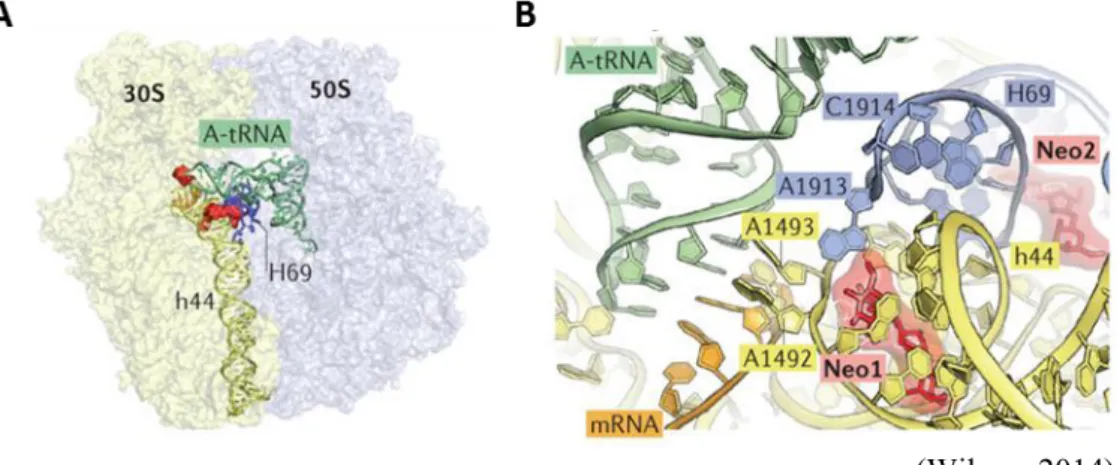
The A-site binds the incoming aminoacylated or charged tRNA while the P-site (peptidyl-tRNA binding site) is occupied by the tRNA carrying the polypeptide chain in elongation (or the initiator tRNA during the initiation step). Finally, the E-site is the exit site which binds only outgoing deacylated or uncharged tRNA. During the stage of translation-elongation, the tRNAs successively pass through the A-, P-, and then E-sites (translocation process), before dissociating from the ribosome (Arenz and Wilson, 2016). Aminoglycosides interact with the ribosome at two distinct sites (Figure 7). The main drug target is at the A-site, on the 16S ribosomal RNA of 30S subunit (Krause et
al., 2016). More precisely, this corresponds to highly conserved nucleotides of 16S
rRNA helix 44 (h44) where monitoring of codon-anticodon interactions takes place (Gutierrez et al., 2013). Members of the aminoglycoside family exhibit affinity with different regions of the A-site (Carter et al., 2000). For example, it was shown that nucleotides C1407/G1494, A1408, A1493 and U1495 are important for paromomycin binding to the A-site (Fourmy et al., 1996). Finally, the majority of aminoglycosides alter the conformation of the ribosome, decreasing translational fidelity by inducing codon misreading on delivery of the aminoacyl transfer RNA (Krause et al., 2016). This results in the formation of erroneous proteins, that once released are assumed to cause damages to the cell membranes. However, some aminoglycosides do not cause misreading, such as apramycin (Matt et al., 2012) or spectinomycin (Vakulenko and Mobashery, 2003). In addition, some aminoglycosides are able to block one of the translation steps. For example, streptomycin blocks the initiation complex and decrease both the rate and the accuracy of translation (Davis, 1987); the 2-deoxystreptamine aminoglycosides are potent inhibitors of translocation (Wilson, 2014).
The second target site of aminoglycosides was identified within another functionally important region of the 50S subunit, the helix 69 (H69) of 23S rRNA (Gutierrez et al., 2013). H69 interacts with helix 44 (h44) of the 16S RNA to form one of the major intersubunit bridges B2a, a site of interaction at the interface between the small and large ribosomal subunits (Wilson, 2014). Following the termination of mRNA translation, the ribosome recycling factor together with elongation factor G (EF-G) disrupt the H69-h44 interaction enabling the two ribosomal subunits to separate. Aminoglycoside binding within H69 stabilizes the interbridge contacts and interfers with the subunit recycling process (Gutierrez et al., 2013).
Figure 8: The structures of the elongation factor G in complex with the ribosome in pre- and post-translocational states.
EF-G conformations during pretranslocation (A and C) and posttranslocation (B and D) complexes are represented. The large ribosomal subunit 50S (gray) and the small subunit 30S (ivory) are shown with the A-site (blue), P-site (pink), and E-site (orange) tRNAs. The five domains of EF-G (I(G), II, II, IV and V) are represented with different colors.
1.3.2. Elongation factor G (EF-G)
The elongation factor G (EF-G) belongs to the GTPase superfamily. This enzyme triggers ribosome-dependent hydrolysis of GTP and thus participates in two different steps during protein synthesis: elongation and ribosome recycling (Palmer et al., 2013). During the elongation step, EF-G enables the translocation of mRNA and tRNA through the ribosome (Nyfeler et al., 2012). EF-G shows two conformations when the mRNA moves from one codon to another: a compact conformation during the pretranslocational state (Figure 8A and C) and an elongated conformation during the postranslocational state (Figure 8B and D) (Lin et al., 2015). Additionally, EF-G collaborates with ribosome recycling factor (RRF) to split the 70S ribosome into its two, 50S and 30S, subunits during a process called ribosome recycling (Borovinskaya et
al., 2007). The ribosome subunits can then be utilized in another round of translation.
The genome of P. aeruginosa possesses two genes coding for two elongation factors G, namely fusA1 (PA4266) and fusA2 (PA2071). The fusA1 gene is predicted to be part of a three-gene operon (PA4265-PA4266-PA4267). PA4265 (tufA) and PA4267 (rpsG) code for the elongation factor Tu and the 30S ribosomal protein S7, respectively. Palmer et al. named the fusA1 encoded protein EF-G1A and the fusA2 protein EF-G1B (Palmer et al., 2013). They consist of 706 and 702 amino acids, respectively. The two protein sequences share 84% identity and 90% similarity. Evidence was provided that EF-G1A plays a role in ribosome recycling while EF-G1B is involved in the translocation process (Palmer et al., 2013). Several mutations in the fusA1 gene were reported to confer an increased resistance to argyrins (increasing the MICs from 16 to >256 µg ml-1 and from 8 to >128 µg ml-1) in mutants selected in vitro from P. aeruginosa PA14 and PAO1, respectively (Table 2) (Bielecki et al., 2012; Nyfeler et al., 2012). These molecules are cyclic octapeptides produced by various Myxobacteria spp and Actinomycetes spp. They inhibit protein synthesis and are active against P. aeruginosa (Selva et al., 1996) through their interaction with EF-G1A (Gao et al.,
2009). This is not the case of fusidic acid, another antibiotic compound targeting EF-G and active against Staphylococcus and Streptococcus species but not against
Pseudomonas species. A putative argyrin-binding site was localized on the domain III
Table 2: List of EF-G1A/B alterations in in vitro-selected and clinical strains of
P. aeruginosa. Reference
strainsa substitutionsEF-G1A b substitutionsEF-G1B b References
in vitro mutants
PAO1 T493I (Klockgether et al., 2010)
PA14 S417L I457T S459F L663M L663Q M685R (Bielecki et al., 2012) PAO1 P414S S417L S459F P486S L663Q T671A Y683C (Nyfeler et al., 2012)
ATCC 27853 N592I (Feng et al., 2016)
PAO1 I61M E100G T671A (López-Causapé et al., 2018)
clinical strains
LESB58 A419T V538A G611V Q678R (Chung et al., 2012)
DK2 G148D V330A E372K D450G R491C R512C Q678R A681T G571D (Marvig et al., 2013) DK1-P33F0 G118S D467G (Markussen et al., 2014) PA14 L40Q G60S G60V V93I V93A T96A E100G R104C G118S P136A G181D Q182R V183I V183A D184N D184G L185P I186V A246V V262A N272S D280N D302E E306D F317L F335L N351S Q365H G387D K389E K430E A439T T456A M461V L464M A481V G484R P486L V488I K504E G516S Y552C N566K S585F D588Gc N592S Q606H P618Q T671A T671I Q678L R680S E28D G74D P80T L161Q A176S D197A Q199K T201A E207D A210S Y238Stop A289V R297K K306N D315N T450S Q456Stop G479S E537D G566S Q601H D632V A690S A695G (Greipel et al., 2016) PAO1 V93A K430E N482S K504E Y552C P554L D588Gc P618L T671I L104P Nt889∆1 N236S P329L S445Stop N561S I640L (López-Causapé et al., 2017) PAO1 V119A E401D K430E Y552C Q161L S176A A197D D373E G479S I570L (Del Barrio-Tofiño
Amino acid substitutions were indirectly identified in EF-G1A by studies investigating the genomic evolution of CF P. aeruginosa strains (Table 2). It was suggested that mutations in gene fusA1 could contribute to the adaptation of P. aeruginosa to the CF lung environment by altering the (p)ppGpp levels and by downregulating the virulence factors of the pathogen (Chung et al., 2012). In another CF context, fusA1 was identified as a pathoadaptive gene from the genomic analysis of clone DK2 prevalent in Denmark (Marvig et al., 2013), suggesting that this gene is important for bacterial persistence in CF patients. This conclusion was inferred from a longitudinal study on a CF patient, during a 32-year period. Two distinct sublineages were identified from the ancestral strain collected from paranasal sinuses; both sublineages were present in the lower airways and harbored alterations in EF-G1A, again highlighting the potential role of fusA1 mutations in the persistence of P. aeruginosa in the CF lung (Markussen et al., 2014).
In Germany, characterization of 361 P. aeruginosa isolates collected from 258 CF patients revealed the presence of 52 and 22 nonsynonymous mutations in fusA1 and
fusA2 genes, respectively (Greipel et al., 2016) (Table 2). Additionally, two stop
mutations (Y238Stop and Q456Stop) were identified in fusA2. Thus, these results strongly
suggested that fusA1/fusA2 genes are under a high evolutionary pressure in the CF context. In another study, Antonio Oliver’s group defined the resistome of isolates belonging to an international CF clone (CC274). Strains with MICs of tobramycin from 6 to >256 µg ml-1 turned out to harbor mutations in fusA1 and fusA2 genes, suggesting a
role of these mutations in high-level aminoglycoside resistance of CF strains (López-Causapé et al., 2017). Finally, substitutions were identified in XDR isolates from Spain (Del Barrio-Tofiño et al., 2017) but their phenotypic impact was not further explored. Not reported in clinical strains, an additional amino acid substitution in EF-G1A (N592I)
was identified in a P. aeruginosa strain subjected in vitro to increasing concentrations of tobramycin. Additional experiments using antibiotics of other families (ciprofloxacin, piperacillin/tazobactam, meropenem or ceftazidime) did not lead to the selection of
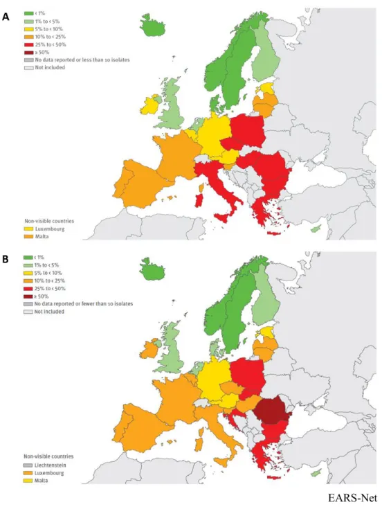
Figure 9: Epidemiology of P. aeruginosa strains resistant to aminoglycosides in Europe.
Data are represented for years 2009 (A) and 2016 (B) (from the annual reports of the European Antimicrobial Resistance Surveillance Network (EARS-Net) 2009 EARS-Net
1.4. Epidemiology of resistance
Antimicrobial resistance of Gram-negative bacteria, including P. aeruginosa is a global issue. Indeed, it was reported in 2011 that 8.9% and 7.1% of healthcare-associated infections were due to P. aeruginosa in Europe and in the United States, respectively (McCarthy, 2015). The development of MDR, XDR and PDR strains is alarming, making this organism a critical priority for the research and development of new antibiotics (Tacconelli et al., 2018).
Resistance to aminoglycoside molecules in invasive strains is monitored in France and in Europe by the European Centre for Disease Prevention and Control (ECDC), which publishes an annual report about the antimicrobial resistance surveillance in Europe [all 28 EU member states and two EEA countries (Iceland and Norway)] (Figure 9). These reports relate that in France 10.7% of the invasive isolates of P. aeruginosa were resistant to aminoglycosides in 2016. This rate is lower than in 2013 and 2009 where 15.5% and 22% were found resistant, respectively. This decreasing trend is also observed in some other European countries such as Italy and the Czech Republic. However, the percentage of aminoglycoside resistant strains remains high in Romania, Ireland and Belgium. Consequently, the ECDC recommends a prudent use of antibacterials and the implementation of infection control measures to prevent a deterioration of the situation.
Figure 10: Schematic representation of global non-enzymatic resistance mechanisms to aminoglycosides developed by planktonic
P. aeruginosa cells.
2. Non-enzymatic resistance mechanisms to aminoglycosides
Several resistance mechanisms to aminoglycosides have been characterized in clinical strains of P. aeruginosa. A high level resistance to these agents can result from drug inactivation or modification by plasmid- or chromosome-encoded enzymes (Poole, 2005). However, this will not be described here as the aim of this research project focused on enzyme-independent resistance.Non-enzymatic resistance to aminoglycosides in P. aeruginosa involve mechanisms such as aminoglycoside-target alterations, membrane modifications, and/or overproduction of the efflux pump MexXY(OprM) (Figure 10). Alternatively, the modification of the bacterial lifestyle via the formation of biofilm may also confer an increased resistance to aminoglycosides (Taylor et al., 2014).
2.1. Aminoglycosides resistance in biofilms
P. aeruginosa often develops in the lung of CF patients as biofilms (Taylor et al., 2014).
Bacterial biofilms are dense microbial communities embedded within an extracellular matrix. The formation and dispersion of a biofilm is summarized in Figure 11. Cells living in biofilms exhibit phenotypic characteristics and a social behaviour quite different from that of planktonic cells. Antibiotic resistance conferred by the biofilm mode of life is a major therapeutic problem. Indeed, compared to planktonic cells, sessile bacteria can be up to 1,000-fold more resistant to antibiotics, including aminoglycosides (Taylor et al., 2014). Genetic determinants participating in this phenotype were investigated in several studies (Table 3). Whiteley et al. showed that 73 genes were differentially expressed in P. aeruginosa biofilms compared to planktonic counter parts. Among the 34 genes found to be overexpressed, tolA is supposed to decrease the affinity of aminoglycosides for the OM by impacting LPS structure. An increase amount of protein TolA might then contribute to the resistance of biofilms to these antibiotics (Whiteley et al., 2001). Among the repressed genes was rpoS, which codes for σ subunit of RNA polymerase. Biofilms formed by rpoS mutant are thicker and more resistant to tobramycin than those formed by wild-type strains (Whiteley et
Figure 11: Schematic representation of the formation and dispersion of a P. aeruginosa biofilm.
The formation of a biofilm starts with a reversible attachment of planktonic bacteria to a cell or abiotic surface (1) followed by an irreversible attachment resulting in a monolayer of cells (2). Then, a microcolony is formed (3) and develops as a macrocolony (4). Finally, cells leave the macrocolony during the dispersion step (5).
Table 3: Genetic loci confirmed to be involved in the resistance of P. aeruginosa biofilms to aminoglycosides.
PA number Gene name Aminoglycosides References
PA0084 tssC1 TOB, GEN (Zhang et al., 2011)
PA0756-PA0757 - TOB, GEN (Zhang et al., 2013)
PA1163 ndvB TOB, GEN (Mah et al., 2003)
PA1875-PA1877 - TOB, GEN (Zhang and Mah, 2008)
PA2070 - TOB, GEN (Zhang et al., 2013)
Another study analyzed a library of about 4,000 random transposon insertion mutants of
P. aeruginosa PA14 for suppression of antibiotic resistance when grown as biofilm (Mah et al., 2003). Amongst this collection, the ndvB mutant was more susceptible to tobramycin. The NdvB protein is involved in the synthesis of periplasmic glucans shown to interact with tobramycin, thus impairing their transport across the cytoplasmic membrane. However, it is worth to mention that the impact of NvdB varies among
P. aeruginosa strains. Indeed, comparison of ndvB mutants highlighted a less important
contribution in resistance of NdvB in the PAO1 and PAK genetic backgrounds than in PA14. For example, only a 2-fold decrease in resistance to tobramycin was observed in PAO1 ndvB knockout mutant compared with an 8-fold decrease in PA14 mutant (Mah
et al., 2003). Later on, additional genetic loci identified from this mutant library were
confirmed to participate in biofilm-associated aminoglycoside resistance, namely: (i) a putative efflux pump encoded by PA1875-PA1877 (Zhang and Mah, 2008), (ii) the
tssc1 encoded protein involved in type VI secretion (Zhang et al., 2011), (iii) a
two-component system encoded by PA0756-PA0757 operon (Zhang et al., 2013), and (iv) two hypothetical proteins encoded by PA5033 and PA2070 (Zhang et al., 2013). Interestingly, the gene upstream of PA2070 (PA2071) encodes the elongation factor EF-G1B.
Finally, biofilm formation is now known to be induced by tobramycin. Gene arr (for aminoglycoside response regulator) was found responsible for biofilm-associated aminoglycoside resistance. Analysis of a transposon mutant library of PAO1 showed, indeed, that the inactivation of arr gene made the strain 100-fold more susceptible to the killing action of tobramycin. The Arr protein, predicted to be inserted in the IM via two transmembrane (TM) domains, contains a periplasmic domain and a EAL domain characteristic of putative phosphodiesterases involved in bis-(3’-5’)-cyclic dimeric GMP (c-di-GMP) degradation (Hoffman et al., 2005).
2.2. Alteration of the cellular targets of aminoglycosides
Methylation of 16S rRNA in bacterial 30S ribosome is another mechanism of resistance to aminoglycosides. In P. aeruginosa, it results from the acquisition of gene rmtA encoding a 16S rRNA methylase (Yokoyama et al., 2003). RmtA shares high similarity with 16S rRNA methylases found in aminoglycoside-producing Actinomycetes spp
(Yokoyama et al., 2003). These bacteria protect themselves from intrinsic aminoglycosides by methylation of their own 16S rRNA. P. aeruginosa strains might have acquired this gene through intergeneric lateral transfer (Yokoyama et al., 2003). Of note, the transfer of a recombinant plasmid carrying gene rmtA in E. coli conferred an increased resistance to 4,6-di-substituted deoxystreptamines. The analysis of 1,113 clinical isolates of P. aeruginosa revealed the presence of gene rmtA in 9 of them (Yokoyama et al., 2003).
The modification of a second aminoglycoside target can lead to a deacrase of susceptibility. A specific resitance to streptomycin was associated with mutations in gene rpsL, encoding ribosomal protein S12 and which is the preferential target of this aminoglycoside (Hancock, 1981).
2.3. Membrane modifications
As aminoglycosides act intracellularly, any modification preventing their uptake and accumulation in the cytosol constitutes potentially a resistance mechanism. The analysis of clinical strains resistant to aminoglycosides revealed that some of them harbor modifications of LPS composition, remaining only with the A-band LPS (Hancock et
al., 1983). Consequently, B-band LPS seem to have an impact on the intracellular
uptake of aminoglycosides (gentamicin) although the exact mechanism remains unclear (Kadurugamuwa et al., 1993). In strains isolated from CF patients, deficiency in LPS O-side-chains is common (Hancock et al., 1983) and could be a widespread mechanism of resistance to aminoglycosides (Bryan et al., 1984).
In our laboratory, the screening of a Tn5-Hg insertional library constructed in reference strain PAO1 showed that the inactivation of the galU gene caused a 2-fold increase in resistance to aminoglycosides (gentamicin, amikacin, tobramycin and netilmicin) (El'Garch et al., 2007). The galU gene codes for an UDP-glucose pyrophosphorylase which catalyzes the conversion of glucose-1-phosphate to UDP-glucose. A previous work showed that the inactivation of galU leads to the synthesis of an incomplete LPS outer core, lacking both A- and B-band polysaccharides (Dean and Goldberg, 2002). The Tn5-Hg insertional library also revealed a role in resistance of a thirteen-gene operon (nuoABDEFGHIJKLMN) which codes for proton-translocating type I NADH
Figure 12: Structure of the RND efflux pump MexXY(OprM) based on the MexAB-OprM model from P. aeruginosa.
The OprM subunit is shown in light blue. The three pairs of MexX dimers are represented in red and deep blue. The MexY protein is represented in pink. OM: outer membrane, IM: inner membrane (Akama et al., 2004).