doi:10.1182/blood-2003-09-3044
Prepublished online May 13, 2004;
2004 104: 1281-1290
Kevin Kipp, Keun Chae, Wilfried Ellmeier, Owen N. Witte and David J. Rawlings
Phyllis W. Yu, Ruby S. Tabuchi, Roberta M. Kato, Alexander Astrakhan, Stephanie Humblet-Baron,
gene transfer
model of X-linked agammaglobulinemia (XLA) using retroviral-mediated
Sustained correction of B-cell development and function in a murine
http://bloodjournal.hematologylibrary.org/content/104/5/1281.full.html
Updated information and services can be found at:
(4699 articles)
Immunobiology
(495 articles)
Gene Therapy
(1333 articles)
Free Research Articles
Articles on similar topics can be found in the following Blood collections
http://bloodjournal.hematologylibrary.org/site/misc/rights.xhtml#repub_requests
Information about reproducing this article in parts or in its entirety may be found online at:
http://bloodjournal.hematologylibrary.org/site/misc/rights.xhtml#reprints
Information about ordering reprints may be found online at:
http://bloodjournal.hematologylibrary.org/site/subscriptions/index.xhtml
Information about subscriptions and ASH membership may be found online at:
Copyright 2011 by The American Society of Hematology; all rights reserved.
Washington DC 20036.
by the American Society of Hematology, 2021 L St, NW, Suite 900,
Blood (print ISSN 0006-4971, online ISSN 1528-0020), is published weekly
GENE THERAPY
Sustained correction of B-cell development and function in a murine model of
X-linked agammaglobulinemia (XLA) using retroviral-mediated gene transfer
Phyllis W. Yu, Ruby S. Tabuchi, Roberta M. Kato, Alexander Astrakhan, Stephanie Humblet-Baron, Kevin Kipp, Keun Chae,Wilfried Ellmeier, Owen N. Witte, and David J. Rawlings
X-linked agammaglobulinemia (XLA) is a human immunodeficiency caused by mu-tations in Bruton tyrosine kinase (Btk) and characterized by an arrest in early B-cell development, near absence of se-rum immunoglobulin, and recurrent bac-terial infections. Using Btk- and Tec-deficient mice (BtkTecⴚ/ⴚ) as a model for XLA, we determined if Btk gene therapy could correct this disorder. Bone marrow (BM) from 5-fluorouracil (5FU)–treated BtkTecⴚ/ⴚ mice was transduced with a retroviral vector expressing human Btk and transplanted into BtkTecⴚ/ⴚ
recipi-ents. Mice engrafted with transduced he-matopoietic cells exhibited rescue of both primary and peripheral B-lineage develop-ment, recovery of peritoneal B1 B cells, and correction of serum immunoglobulin M (IgM) and IgG3 levels. Gene transfer also restored T-independent type II im-mune responses, and B-cell antigen re-ceptor (BCR) proliferative responses. B-cell progenitors derived from Btk-transduced stem cells exhibited higher levels of Btk expression than non-B cells; and marking studies demonstrated a se-lective advantage for Btk-transduced
B-lineage cells. BM derived from primary recipients also rescued Btk-dependent function in secondary hosts that had re-ceived a transplant. Together, these data demonstrate that gene transfer into hema-topoietic stem cells can reconstitute Btk-dependent B-cell development and func-tion in vivo, and strongly support the feasibility of pursuing Btk gene transfer for XLA. (Blood. 2004;104:1281-1290)
©2004 by The American Society of Hematology
Introduction
X-linked agammaglobulinemia (XLA) is the prototypic primary humoral immunodeficiency disorder, first described by Bruton who reported a boy with severe recurrent infections and absence of the gamma-globulin serum fraction.1This disorder has been of major
interest for more than half a century, initiating an ongoing and fruitful search for the genetic basis of this and other primary immunodeficiency diseases.
XLA is characterized by a lack of mature B cells and plasma cells and a profound deficiency of all immunoglobulin types. Most patients develop recurrent infections coincident with the loss of maternal antibodies. Pyogenic infection with encapsulated bacteria is the most common clinical manifestation.2-4 Current therapy
consists of regular immunoglobulin replacement and prompt attention to infection.
Both XLA and a related murine immunodeficiency, X-linked immunodeficiency (Xid), result from deficiencies in Bruton ty-rosine kinase (Btk).5-10 Btk, a member of the Tec family of
nonreceptor kinases,11is expressed throughout B-lineage
develop-ment except in plasma cells.12,13 Btk contains a Src homology 1
(SH1) catalytic domain, SH2 and SH3 protein interaction domains, and a unique amino-terminus with a pleckstrin homology (PH)
domain.14 Mutational studies indicate important functional roles
for each of these domains.15
The major developmental defect in XLA occurs at the pre-B-cell transition, resulting in an increase in pro-B pre-B-cells and a marked reduction in cycling pre-B cells and all subsequent stages.16-18Few
IgM⫹ B cells are detectable in the blood and the hypotrophic lymphoid tissues.19,20 Clonal expansion at the pre-B stage is
regulated by pre-B-cell receptor (pre-BCR) signaling and is essential for generating an adequate pool of immature B cells.21
Pre-BCR engagement leads to the generation of a “signalosome” that includes activated Btk,22an event disrupted in XLA.
Several murine models of Btk deficiency currently exist. Xid mice have a spontaneous missense mutation in the PH domain.23
The mutant protein is inefficiently recruited to the plasma mem-brane and fails to enter the BCR signalosome.24The phenotype of
both Xid mice and Btk null-knockout mice (Btk⫺/⫺) is less severe than that of XLA.25-28These mice produce nearly normal numbers
of peripheral B cells, but splenic B-cell development is signifi-cantly compromised. Btk-deficient transitional 2 (T2) immature B cells fail to generate the BCR-dependent pro-survival, prolifera-tive, and differentiation signals required to produce mature B
From the Molecular Biology Institute, the Howard Hughes Medical Institute, the Department of Microbiology and Molecular Genetics, and the Department of Pediatrics, University of California at Los Angeles; the Departments of Immunology and Pediatrics, University of Washington School of Medicine, Seattle; and the Institute of Immunology, Medical University Vienna, Austria.
Submitted September 4, 2003; accepted April 15, 2004. Prepublished online as
Blood First Edition Paper, May 13, 2004; DOI 10.1182/blood-2003-09-3044.
Supported by a National Institutes of General Medical Sciences National Research Service Award (P.W.Y.), a McDonnell Scholar Award (D.J.R.), a Leukemia and Lymphoma Society Scholar Award (D.J.R.), the Joan J. Drake Grant for Excellence in Cancer Research (D.J.R.), grants from National
Institutes of Health (HD37091, CA81140, AI38348, and AI33617; D.J.R.), and the American Cancer Society (D.J.R.).
An Inside Blood analysis of this article appears in the front of this issue.
Reprints: David J. Rawlings, Children’s Hospital and Regional Medical
Center, 307 Westlake Ave North, Suite 300, Seattle, WA 98109; e-mail: [email protected].
The publication costs of this article were defrayed in part by page charge payment. Therefore, and solely to indicate this fact, this article is hereby marked ‘‘advertisement’’ in accordance with 18 U.S.C. section 1734.
© 2004 by The American Society of Hematology
1281 BLOOD, 1 SEPTEMBER 2004
䡠
VOLUME 104, NUMBER 5cells.29,30 Consistent with these findings, the BCR-dependent
calcium signal is markedly reduced in Btk-deficient B cells.31-33
Thus, Btk plays a crucial role in both primary B lymphopoiesis in the marrow and in splenic peripheral B-lineage development and signaling.
The difference between Btk-deficient humans and mice is partially attributable to the redundant function of Tec. Mice deficient for Btk and Tec (BtkTec⫺/⫺) exhibit an early, severe block in B-lineage development that is nearly identical to XLA.34Tec is
expressed throughout B-lineage development, but at a lower level than Btk (D.J.R., unpublished data, January 2002). While this level of Tec is sufficient to rescue murine pre-B transition, a much higher Btk/Tec dosage is required for mature B-cell signaling in both mice and humans. Overexpression of Tec can rescue BCR-dependent signals in Btk-deficient B cells,33,35but endogenous Tec is insufficient
to restore these events. The inability of residual Tec to rescue human pre-B-cell development is consistent with a more stringent requirement for all components of the pre-BCR complex in humans.36
Several lines of evidence demonstrate a strong selective advan-tage for B-lineage cells expressing wild-type (WT) Btk. Female carriers of XLA exhibit nonrandom X-inactivation of the mutant allele specifically within the B-cell compartment.37Transplantation
of mixtures of CBA/J (WT) and CBA/N (Xid) BM cells into lethally irradiated Xid mice also leads to the selective expansion and survival of WT B cells.38 Immune responses can also be
restored in sublethally irradiated mice with as few as 2.5⫻ 104
total BM cells.39In addition, larger numbers of BM or fetal liver
cells can rescue B-lineage development in the absence of myeloab-lation.40-42These observations suggest that even under suboptimal
conditions, hematopoietic-targeted Btk gene therapy might pro-mote long-term restoration of B-cell function in XLA.
The availability of murine models offers the opportunity to evaluate gene therapy as an alternative treatment for XLA. We tested the capacity of a modified Moloney murine leukemia virus retroviral vector expressing human Btk to implement hematopoietic-based Btk gene therapy in BtkTec⫺/⫺ mice, the model most representative of XLA. Our results demonstrate the progressive outgrowth of expressing B cells and restoration of Btk-dependent B-lineage development and functional responses.
Materials and methods
Retroviral vectors and producer cell lines
The murine stem cell virus (MSCV) backbone was used to enhance sustained expression in hematopoietic cells.43Briefly, the vector MSCV-huBtk-SAR (subsequently referred to as MBS; Figure 1A) was generated as follows: human-interferon scaffold attachment region (SAR),44obtained as an 877-bp fragment after XhoI and ClaI digestion of pMND-IRES-GFP-SAR (kindly provided by Dr Donald Kohn, University of Southern California [USC]), was inserted into the ClaI site of MSCV-ires-GFP45by a blunt-end ligation following end-fill reaction. Full-length human BTK coding sequence (98% identical with murine Btk protein)12 was then inserted into XhoI and HindIII sites.
High-titer 10A.1 pseudotyped retrovirus, generated by cotransfection of 293-T cells with pCL packaging plasmids46by calcium phosphate precipita-tion was used to infect GP⫹E86 producer cell lines to create the MBS producer cell line; titers were estimated by infecting interleukin-7– expanded BM and evaluating transgenic Btk expression by flow cytometry. The control ecotrophic producer cell line, MSCV-ires-GFP (subsequently referred to as MIG), was generated using an identical approach. Producer lines were maintained in Dulbecco modified Eagle medium (DMEM; Mediatech, Herndon, VA) supplemented with 10% fetal bovine serum, 2
mM L-glutamine (Invitrogen, Carlsbad, CA), and 100 U/mL penicillin/ streptomycin (Invitrogen).
Mice
C57BL/6 (Jackson) mice, and BtkTec⫺/⫺mice (generated as previously described34) were bred and maintained in the pathogen-free barrier facilities at the University of California at Los Angeles and the University of Washington.
Transduction and transplantation
BtkTec⫺/⫺donor mice were injected intraperitoneally with 5-fluorouracil (5FU; 150 mg/kg body weight; Sigma, St Louis, MO) 3 days before BM harvest. Following red blood cell lysis, BM cells were cocultured directly on irradiated producer cells in complete media (RPMI; Mediatech), 10% fetal bovine serum, 2 mML-glutamine (Invitrogen), 50M -mercaptoetha-nol (Sigma), 100 U/mL penicillin/streptomycin (Invitrogen) with 4g/mL polybrene (Sigma) and the following recombinant human or murine cytokines (BioSource International, Camarillo, CA): 20 ng/mL interleu-kin-3, 20 ng/mL interleukin-6, 20 ng/mL stem cell factor, 20 ng/mL Flt-3 ligand, and 20 ng/mL thrombopoietin. In experiments 1 to 3, WT C57BL/6 and BtkTec⫺/⫺BM was mock-transduced on GP⫹E86 parental cells. In experiment 4, WT C57BL/6 and BtkTec⫺/⫺BM was prestimulated for 48 hours in media supplemented with the identical murine cytokines (each 10 ng/mL; Peprotech, Rocky Hill, NJ) followed by cocultivation on MBS or MIG producer cells (for BtkTec⫺/⫺BM cells) or GP⫹E86 parental cells (for WT C57BL/6 BM). After 3 days of cocultivation, cells were injected into lethally irradiated (11 Gy, split dose) BtkTec⫺/⫺recipients at 1-5⫻ 106 cells/mouse by retro-orbital injection. Mice were treated with enrofloxacin (0.5 mg/mL in the drinking water) for 2 weeks. For secondary transplants, BM was harvested from primary recipients and 5⫻ 106cells/animal was injected into lethally irradiated BtkTec⫺/⫺recipients.
B-cell purification and semiquantitative real-time polymerase chain reaction (Q-PCR) analysis of Btk retroviral integration
B-cell purification was carried out using magnetically coated beads (Miltenyi Biotec, Auburn, CA) according to manufacturer’s instructions. Splenic B cells were enriched by negative selection using anti-CD43– coated beads. Bone marrow B cells were enriched by positive selection using anti-B220–coated beads. Purity of B-cell populations was more than or equal to 85% based on fluorescence-activated cell sorting (FACS) analysis. Cell populations were pelleted and frozen for Q-PCR analysis. All PCR reagents and instrumentation were obtained from Biorad (Hercules, CA) unless otherwise noted. PCR reactions were carried out using the iCycler real-time PCR detection system with iQ SYBR Green Supermix. Efficiency and threshold cycle (Ct) values were determined with the iCycler
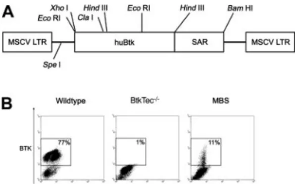
Figure 1. Transduction of 5FU-treated BM with the MBS vector. (A) Structure of
the MSCV-huBtk-SAR (MBS) retroviral vector. The murine stem cell virus (MSCV)
promoter drives expression of human Btk. The human interferon-scaffold
attach-ment region (SAR) was included to facilitate sustained expression of the Btk transgene in differentiated cells. (B) Btk expression in mock-transduced BM from WT
and BtkTec⫺/⫺mice, and in MBS-transduced BM from BtkTec⫺/⫺mice. BM was
analyzed immediately prior to transplantation into BtkTec⫺/⫺ recipients (using
intracellular FACS analysis). Btk expression in BM progenitors was between 6% and 11% in experiments 1 to 3.
IQ optical analysis software and primers designed with Vector NTI software (InforMax, Frederick, MD) and manufactured by MWG Biotech (High Point, NC).
Q-PCR was performed on genomic DNA (using input transduced BM cells, and BM, thymus, or splenic cells derived from animals receiving a transplant), and normalized to a standard DNA derived from an A20 murine B-cell line containing a single copy of the vector MSCV-BTK-IRES-GFP (as determined by Southern blotting). DNA was isolated with QIAamp DNA mini kit (Qiagen, Valencia, CA) according to manufacturer’s instruc-tions. BTK-specific primers were designed to amplify mouse or human exon-spanning products. The mouse-2-microglobulin (2m) gene was selected as a housekeeping control. Primer sequences are available upon request. Products were amplified using a standard PCR protocol (35 cycles of 95°C for 30 seconds, 60°C for 30 seconds, and 72°C for 30 seconds) and primer specificity was determined by melt curve and ethidium bromide gel analysis. BTK or GFP integration was analyzed using the Pfaffl mathemati-cal model for relative quantification.47Initially, BTK, GFP, and2m primer efficiencies were determined by generating a standard curve using dilutions of A20 DNA. Threshold cycles (Cts) were then obtained by running all PCR samples in triplicate. Relative ratios of BTK or GFP expression were calculated using A20 Ct as calibrator values for all samples. All Q-PCR data included at least 3 independent analyses (with 3 replicates per analysis) and statistical significance was determined by a paired, 2-tailed Student t test.
Immunization and ELISA assays
Engrafted mice were immunized intraperitoneally with 10g trinitrophenyl-substituted (TNP)–Ficoll (Bioresearch Technologies, Novato, CA) in 100 L phosphate-buffered saline (PBS). Mice were bled by saphenous vein puncture 7 to 10 days later. Serum was used for enzyme linked immunosor-bent assays (ELISAs) and peripheral blood cells were analyzed by flow cytometry. ELISAs were used to detect TNP-specific IgM and IgG3and total serum IgM and IgG3. Briefly, 96-well Nunc Maxisorp ELISA plates (Fisher, Pittsburgh, PA) were coated with TNP–bovine serum albumin (BSA; Bioresearch Technologies), anti–mouse IgM, or anti–mouse IgG3 (Southern Biotech, Birmingham, AL), incubated for 1 hour in a 37°C humidified chamber, were blocked with PBS/Mg2⫹(Invitrogen) plus 2%
BSA, and incubated with serial dilutions of serum, followed by alkaline phosphatase–conjugated anti–mouse IgM or anti–mouse IgG3. Plates were washed with PBS/Mg2⫹ plus 0.05% Tween-20, incubated with
p-nitrophenyl phosphate substrate (Southern Biotech), and absorbance was measured at 405 nm on a Bio-Rad microplate reader. Ig levels were quantified by comparison with titrated Ig standards.
Flow cytometric analysis
Single-cell suspensions prepared from BM, spleen, peripheral blood, and peritoneal washes were incubated with fluorochrome-conjugated antibodies for 1 hour on ice. Cytofix/Cytoperm (Pharmingen, San Diego, CA) was used for intracellular stains. The following primary antibodies (all from Pharmingen, except where noted) were used: phycoerythrin (PE)– conjugated antimouse CD45R/B220, Cy-Chrome–conjugated antimouse CD45R/B220, fluorescein isothiocyanate (FITC)–conjugated anti–mouse IgM, PE-conjugated anti–mouse IgD (Southern Biotech), PE-conjugated antimouse CD11b/Mac1, conjugated antimouse CD43, and biotin-conjugated antimouse CD5. Spectral red–biotin-conjugated streptavidin (Southern Biotech) was used to visualize biotin-conjugated antibodies. For Btk stains, affinity-purified polyclonal rabbit anti-Btk was used in conjunction with a PE-conjugated donkey anti–rabbit Ig (Jackson ImmunoResearch); all intracellular stains were performed in the presence of blocking total mouse IgG (Accurate Chemical, Westbury, NY). Cell analysis was performed using a FACSCalibur flow cytometer and CellQuest software (Becton Dickinson, San Jose, CA).
B-cell proliferation assay
Total splenocytes were seeded in 96-well flat-bottom plates at 2.5⫻ 105 cells/well in complete media with 10 ng/mL phorbol myristate acetate (PMA; Sigma) plus 1M ionomycin (Calbiochem, San Diego, CA), 10
g/mL lipopolysaccharide (LPS; Sigma), and 20 g/mL or 5 g/mL goat-antimouse IgM F(ab⬘)2fragment (Jackson ImmunoResearch, West-grove, PA).3H-thymidine (1Ci [37 KBq]; DuPont NEN, Boston, MA) was added after 48 hours, and cells were harvested 18 hours later and counted on a scintillation counter.
Colony-forming unit–B-cell (CFU-B) assay
Sheep red blood cells (SRBCs; Mission Labs, Los Angeles, CA) were washed extensively and stored on ice. BM cells and splenocytes were plated in duplicate on 35⫻ 10–mm culture dishes with a 2-mm grid (Nalge Nunc, Rochester, NY) at 5⫻ 104 cells/dish in 1 mL Iscove modification of DMEM (Mediatech) supplemented with 15% FBS, 2 mML-glutamine, 50 M -mercaptoethanol, 25 mM HEPES (Invitrogen), 100 U/mL penicillin/ streptomycin, 10g/mL LPS, and 10% washed SRBCs. A predissolved solution of 3% wt/vol bacto agar (Becton Dickinson) was kept at 55°C and used to add agar to the cells at a final concentration of 0.3% just before plating. Colonies were counted after 7 to 10 days by treating with 1 mL 3% acetic acid.
Statistical analysis
The statistical significance of measurements of B-cell populations and function were determined using a 2-tailed t test of independent sample means. Statistical analyses were performed comparing results obtained from recipient mice of MBS transduced bone marrow versus control mice that received mock-transduced (or MIG-transduced) bone marrow from BtkTec⫺/⫺mice.
Results
Transduction of BtkTecⴚ/ⴚBM progenitors using a retroviral vector expressing human Btk
To transfer Btk into murine hematopoietic stem cells, we generated the MBS vector containing the full-length human Btk cDNA and the human-interferon SAR to promote sustained gene expression (Figure 1A).44Gene transfer studies were carried out using Btk/Tec
doubly deficient mice for several key reasons. First, BtkTec⫺/⫺ mice exhibit a pre-B-cell developmental block essentially identical with human XLA.34Second, because pre-BCR signaling mediates
the major B-lineage clonal expansion in the BM, we predicted that enforced Btk expression at this stage would strongly favor the expansion of transduced cells. Finally, we hypothesized that the stringent requirement for Btk at this checkpoint would promote expansion of cells expressing Btk at a dosage sufficient to also rescue peripheral B-lineage function.
BM from 5FU-treated BtkTec⫺/⫺ mice was transduced by cocultivation on an ecotropic MBS producer cell line in the presence of cytokines for 3 days. In all experiments, lethally irradiated BtkTec⫺/⫺recipients received transplants of 1 to 5⫻ 106
5FU-treated bone marrow cells. Experiments 1 to 3 included cohorts of 2 to 5 animals that underwent transplantation with the following cells: transduced WT (C57/Bl6) BM, mock-transduced BtkTec⫺/⫺BM, a 10% mixture of C57/Bl6 marrow in BtkTec⫺/⫺marrow (designed to simulate MBS transduction), and MBS-transduced BtkTec⫺/⫺BM. In each of these experiments, 5% to 15% of the MBS-transduced, 5FU-treated BM cells expressed exogenous Btk at the time of cell transplantation (as determined by Btk intracellular staining; Figure 1B). The mean fluorescence intensity (MFI) of Btk staining was comparable to that of endoge-nous Btk in WT (C57/Bl6) BM. In experiment 4, BM cells were prestimulated for 2 days prior to cocultivation on the MBS or MSCV-ires-GFP (MIG) producer cell lines and cohorts of 2 to 5 animals underwent transplantation with mock-transduced WT
BTK GENE THERAPY 1283 BLOOD, 1 SEPTEMBER 2004
䡠
VOLUME 104, NUMBER 5(C57/Bl6) BM, MIG-transduced BtkTec⫺/⫺ BM, and MBS-transduced BtkTec⫺/⫺BM.
Btk expression is maintained and positively selected in mice undergoing transplantation with MBS
To monitor transgenic Btk expression and reconstitution, mice that had undergone transplantation were periodically immunized and bled beginning at 5 to 6 weeks after transplantation. Animals in experiments 1 to 3 were killed at 13, 15, and 21 weeks after transplantation, respectively. Animals in experiment 4 were killed at week 14.
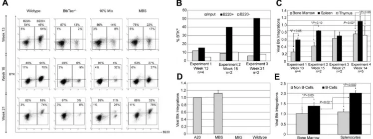
Animals that had received transplants were initially evaluated for cellular reconstitution and transgene expression. Figure 2A demonstrates the Btk expression profile for splenocytes from killed animals. Btk⫹ cells were specifically enriched within the B220⫹ splenic B-cell compartment in MBS-transduced mice. In contrast, Btk expression levels in non-B splenic cells (while clearly higher than BtkTec⫺/⫺ control cells) was significantly less than that observed in B-lineage cells. Similar results were observed for peripheral blood B versus non–B-cell populations (data not shown). The relative level of Btk expression in B-lineage versus non–B-lineage cells from 3 similar experiments is illustrated in Figure 2B. In contrast to Btk expression in B220⫹splenocytes, Btk expression in B220⫺cells remained nearly equivalent to the expression level observed within the input, MBS-transduced stem cell population.
We also used Btk retroviral specific Q-PCR analysis to deter-mine the viral copy number in BM and splenic cell populations isolated from MBS-treated animals (Figure 2C). These data indicated a significant enrichment for Btk marked cells within the spleen. To determine whether there was a specific increase in relative viral copy numbers in B-lineage cells, we isolated B- and non–B-lineage cells. Viral copy number was determined using Q-PCR for input transduced cells; (Figure 2D) and from purified B cells versus non–B cells (Figure 2E) for each of the MBS-treated
animals in experiment 4. These data indicated a significant enrichment for viral marking within B-lineage cells in both BM and spleen. Further, these data demonstrated a significant enrichment for Btk marking of peripheral (splenic) versus primary (BM) B cells. Taken together, these observations indicate that Btk-expressing B cells exhibit a significant selective advantage, leading to their progressive accumulation within the peripheral lymphoid compartment.
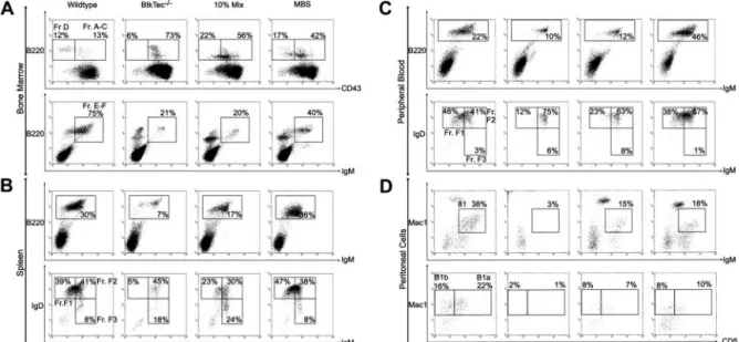
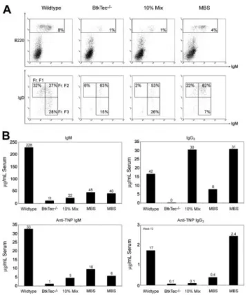
MBS transduction rescues B-cell development in BtkTecⴚ/ⴚmice
We analyzed the relative frequency and absolute numbers of B-cell developmental subsets within the BM, spleen, peripheral blood, and the peritoneal cavity of recipient animals (Figure 3) using the schema developed by Li et al.48 Fractions A to C, identified by
expression of B220 and CD43, represent the earliest stages of B-cell development corresponding to pro-B cells. Fraction D cells, which lose CD43, are primarily pre-B cells. Fractions E to F (immature and mature B cells) are characterized by the acquisition of surface IgM. Fraction F cells emigrate to the spleen, where relative expression levels of IgM and IgD further distinguish immature, transitional, and naive mature B-cell subsets. Differentia-tion proceeds roughly from FracDifferentia-tion F3 (IgMhi/IgDlo, Transitional
1) to F2 (IgMhi/IgDhi, Transitional 2) to F1 (IgMlo/IgDhi). A similar
profile is observed in peripheral blood.
Figure 3A illustrates the pre-B-developmental block in Btk-/ Tec- mice34manifested as an accumulation of Fraction A to C cells
(73% of BM B220⫹cells) and a relative paucity of Fraction D (6%) and Fractions E to F (21%). This contrasts with 13% for Fractions A to C, 12% for Fraction D, and 75% for Fractions E to F, respectively, in a WT recipient. In the representative MBS-treated animal, the corresponding percentages were 17% Fractions A to C, 42% Fraction D, and 40% Fractions E to F, indicating a marked increase in later developmental B-lineage subsets compared with the BtkTec⫺/⫺ control. The block in peripheral BCR-dependent
Figure 2. huBtk expression and viral marking in mice that underwent transplantation with BtkTecⴚ/ⴚ. (A) Splenocytes were harvested from animals in experiments 1, 2, and 3 at weeks 13, 15, and 21, respectively; stained for B220; permeabilized; and stained for transgenic huBtk expression. Representative plots for each time point are shown.
B220⫺non–B-lineage and B220⫹B-lineage regions are indicated, as well as the percentages of each relative to a small lymphocyte gate on total splenocytes. The percentage
of Btk⫹cells within each region is shown inside the region box. (B) The average percentage of Btk⫹cells in B220⫹versus B220⫺splenic populations in MBS-treated recipients
for each experiment was determined and compared to expression at the time of initial BM transduction (as measured by intracellular Btk staining prior to injection). (C) Btk viral copy number was determined using BM and spleen (as well as thymocytes in experiment 4) for all MBS-treated animals at the time of sacrifice (see “Materials and methods”). *Statistically significant differences in splenic versus BM cells. The similar level of marking of BM and spleen in experiment 3 likely reflects increasing numbers of mature recirculating B cells present in the BM by week 21 after transplantation (data not shown). (D) Btk viral copy number in a control B-cell line (A20) containing a single Btk viral integration, and in MBS-, MSCV-ires-GFP (MIG)–, or mock-transduced stem cells at the time of primary transplantation in experiment 4. (E) Btk viral copy number in B versus non-B cells purified from either BM or spleen in experiment 4 (see “Materials and methods”). *Significant differences between relative viral copy number in B versus non-B cells,
development in Btk-deficient mice occurs in the spleen between Fraction F2 and F1 (Figure 3B). In the BtkTec⫺/⫺recipient, F1 cells comprised 5% of the B220⫹cells versus 39% in the WT control. In comparison, 47% of the splenic B cells in the MBS-treated recipient were F1 cells. A similar trend was seen in peripheral blood (Figure 3C).
Another feature of Btk-deficient strains is the complete loss of peritoneal B1 B cells. This population is identified by the coexpres-sion of IgM and Mac1 (CD11b), and can be further separated into CD5⫹B1a, and CD5⫺B1b B cells. To determine if B1 cells were restored, peritoneal cells were isolated and evaluated (Figure 3D). IgM⫹/Mac1⫹B1 cells constitute less than 3% of the total peritoneal population in a BtkTec⫺/⫺recipient as compared with 38% in the WT control. This population was clearly restored in the MBS-treated animal, comprising 18% of the total peritoneal cells in the example shown. Moreover, CD5⫹B1a cells were increased from 1% in the BtkTec⫺/⫺ control to 10% in the MBS animal, as compared with 22% in the WT control. These results demonstrate that peritoneal B1 cells can be reconstituted following MBS transduction.
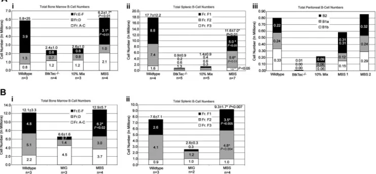
Absolute cell numbers of each B-lineage developmental subset were calculated using the flow cytometry percentages and total cell counts. Figure 4 provides a summary of these data from the 4 experiments. Analysis of BM revealed significant recovery of total B-lineage cellularity in MBS-treated mice as compared with either mock-transduced (Figure 4Ai) or MIG-transduced (Figure 4Bi) BtkTec⫺/⫺control cells. Although the numbers of cells in the pro-B fraction (Fractions A-C) were slightly higher than in WT mice that underwent transplantation, there was a substantial increase in the immature and mature B fractions (Fractions E-F) with restoration to normal cell numbers in the majority of MBS recipients. Striking evidence for MBS-mediated restoration of peripheral B-cell devel-opment was also observed in the spleen (Figure 4Aii, Bii). Fraction
F1, mature splenic B-cell numbers were fully rescued in many animals and were at least 30% to 50% of normal in all MBS recipients. Peritoneal B-cell numbers (Figure 4Aiii) were also restored in experiments where this was evaluated.
Taken together, our data indicate that MBS retroviral gene transfer is capable of restoring both the early and the late developmental blocks associated with the BtkTec⫺/⫺phenotype.
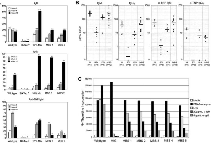
MBS transduction restores immunoglobulin levels and TI-II immune responses in BtkTecⴚ/ⴚmice
Low serum levels of IgM and IgG3 and failure to respond to
T-independent, type 2 (TI-II) antigens are fundamental functional defects present in all Btk-deficient strains.25,27,34Kinetic analysis
from a representative experiment (Figure 5A) demonstrates the progressive increase in serum IgM and IgG3to normal levels within
approximately 8 weeks in MBS-treated animals (versus ⬃ 4-6 weeks in control recipients). Similar results were observed in all experiments (Figure 5B). This kinetic correlated with the progres-sive increase in Btk⫹mature B cells observed over the same period. Notably, recipients of 10% chimeric cell mixtures exhibited serum Ig concentrations similar to the MBS-transduced and WT animals despite a distinctly less robust cellular reconstitution (Figure 4). This latter observation suggests that small numbers of Btk-expressing cells may be sufficient to maintain circulating IgM and IgG3levels and/or that “natural antibody” production is
addition-ally down-regulated in animals with normal numbers of mature B cells.
To ascertain whether MBS transduction could rescue T-independent immune responses, mice were immunized with the TI-II antigen TNP-Ficoll. Strikingly, MBS-treated mice responded with production of normal levels of TNP-specific IgM and IgG3
(Figure 5A-B) antibodies. A slow decline in TNP-specific IgM was
Figure 3. Reconstitution of B-lineage development in MBS-treated BtkTecⴚ/ⴚmice. BM, splenocytes, peripheral blood, and peritoneal cells were harvested from
BtkTec⫺/⫺recipient mice 3 to 5 months after transplantation, evaluated by flow cytometry, and characterized based on “Hardy Fraction” stages of B-cell development. (A) BM
(harvested 21 weeks after transplantation) was stained for B220, CD43, and IgM. Levels of Fractions A to C (pro-B) and Fraction D (pre-B), presented as percentages of the
B220⫹B-cell compartment, are shown in the upper panel (CD43/B220, gated on IgM⫺cells). Fractions E to F (immature B, mature B) are shown in the lower panel (IgM⫹/B220,
gated on live cells as determined by forward and side scatter profiles), and also as percentages of B220⫹cells. (B) Splenocytes (harvested 15 weeks after transplantation) were
stained for B220, IgM, and IgD. B220⫹cells, shown as percentages of total splenocytes, are shown in the upper panel (IgM/B220, gated on live cells). Late-stage splenic B-cell
profiles are shown in the lower panel (IgM/IgD, gated on B220⫹cells); the sequence of maturation is from Fraction FIII (IgMhi/IgDlo) to FII (IgMhi/IgDhi) to FI (IgMlo/IgDhi) as
percentages of B220⫹B cells. (C) Peripheral blood (collected 21 weeks after transplantation) was analyzed by the same parameters as for splenocytes. (D) Representative
plots of peritoneal cells collected from BtkTec⫺/⫺transplant recipients 21 weeks after transplantation and stained for IgM, Mac1, and CD5 are shown. Total B1 cells are shown in
the upper panel (IgM/Mac1, gated on live cells); percentages are relative to total peritoneal cells. Further resolution into CD5⫹B1a and CD5⫺B1b, also presented as a
percentage of the total peritoneal population, is shown in the lower panel (CD5/Mac1, gated on IgM⫺cells). Representative data from 2 of 3 independent experiments are
shown.
BTK GENE THERAPY 1285 BLOOD, 1 SEPTEMBER 2004
䡠
VOLUME 104, NUMBER 5observed in both MBS-transduced and WT control recipients over time (Figure 5A). This correlated with increasing titers of TNP-specific IgG3, consistent with isotype switch to IgG3in response to
repeated immunization.49 Rescue of the T-independent immune
response was observed in all experiments (Figure 5B). These results are in contrast to our previous studies with a Btk transgenic mouse that expressed subendogenous levels of Btk (Btklomice).
Despite restoration of mature follicular B-cell numbers and IgM and IgG3 levels, Btklo mice fail to respond to TI-II antigen
stimulation.50These observations indicate that retroviral-mediated
Btk gene delivery is capable of rescuing a stringent, Btk dose-dependent functional response.
MBS transduction rescues BCR- and mitogen-dependent B-cell proliferative responses in BtkTecⴚ/ⴚmice
To further investigate the degree of functional rescue in the MBS-treated mice, we tested splenocytes from animals that had undergone transplantation for response to B-cell mitogens or BCR engagement (Figure 5C). While untreated BtkTec⫺/⫺–derived cells responded to control stimulation with phorbol ester and ionomycin,
3H-thymidine incorporation was reduced in response to LPS
stimulation and absent with IgM crosslinking. In contrast, both the LPS and anti-IgM dependent responses in splenocytes derived from the treated mice were significantly restored in MBS-treated animals. BCR-dependent proliferative responses were re-stored in all MBS-treated recipients within 13 to 21 weeks in 4 independent experiments (Figure 5C; and data not shown).
Splenocytes and BM from all Btk-deficient strains fail to generate B-lineage colonies when placed in an agar-based, mito-genic colony-forming assay (CFU-B assay).51In all cohorts, cells
from control BtkTec⫺/⫺mice that underwent transplantation pro-duced few or no colonies (Figure 6). In contrast, both BM (Figure 6A) and splenocytes (Figure 6B) from MBS-treated recipients
formed abundant agar colonies. While colony numbers were somewhat variable between individual recipients, the average colony number in MBS-treated mice reached 50% to 100% of the numbers derived from recipients of WT marrow. B-cell colony size was also equivalent in WT and MBS-treated mice (data not shown). Colony numbers in MBS-treated mice were also higher than in the 10% chimeric cell mixture–derived cell populations; or in cells derived from MIG-transduced stem cells.
Taken together, these results demonstrate MBS gene transfer can correct key functional and developmental defects present in BtkTec⫺/⫺mice.
Long-term correcting hematopoietic stem cells are transduced by MBS
Sustained Btk expression in BM, spleen, and peripheral blood was observed for more than 24 weeks in MBS-transduced BM recipi-ents. Production of newly formed IgM⫹immature B cells was also maintained throughout this period. This strongly suggests that the phenotypic rescue resulted from the transduction of early hemato-poietic stem cells capable of self-renewal. To directly evaluate this question, BM from primary transplant recipients was transferred into BtkTec⫺/⫺ secondary recipient hosts. At 6 weeks after transplantation, peripheral blood analysis revealed mature circulat-ing B cells in mice that underwent transplantation with BM from MBS primary recipients. In all 3 experimental MBS-transduced cohorts, the percentage of B cells in the peripheral blood was similar to, or slightly less than, the WT control secondary recipients. In contrast, mature B cells were absent in secondary recipients of BtkTec⫺/⫺ marrow (Figure 7A). In the example shown, the percentage of peripheral blood B cells in the MBS secondary recipients (4%) was lower than the WT control recipient (8%), but significantly higher than the BtkTec⫺/⫺ secondary recipients (1%) and recipients of the 10% chimeric cell mixture
Figure 4. Reconstitution of B-cell numbers in MBS-treated mice. Total numbers for B-cell subsets, based on the developmental schema of Li and colleagues,48were
calculated using flow cytometry percentages and total cell counts. (Ai-ii) Combined results for BM and spleen, respectively, from experiments 1 to 3. (Aiii) Peritoneal B-lineage analysis was performed for 1 experiment, and the results from 1 animal from each control category and 2 MBS-treated animals are shown. MBS-treated mice exhibited
recovery of all peritoneal B-cell subsets. *Statistically significant differences between MBS-treated mice and mock-transduced BtkTec⫺/⫺controls (with P values as noted). (B)
Analysis of BM and spleen B-cell subsets from animals reconstituted with MBS- versus MIG-transduced stem cells. The numbers for each B-cell subset are shown, and mean total B-cell numbers with SEM are included at the top of each bar. *Statistically significant differences between MBS-treated mice and MIG-treated animals (with P values as noted).
(1%). This difference was most apparent with regard to the relative number of mature Fraction FI B cells. In the MBS secondary host, FI cells comprised 22% of circulating B cells compared with 32% in the WT control, 6% in the BtkTec⫺/⫺control, and 2% in chimeric mixture recipient, respectively. We also evaluated B-cell functional activity by analyzing immunoglobulin levels and TI-II responses (Figure 7B). These data demonstrate sustained rescue of these Btk-mediated functional responses. The relatively lower B-cell numbers and less robust developmental rescue observed in both control and MBS-treated secondary recipient mice are consistent with previous studies that report diminished reconstitution capacity with serial transplantation.52Together, these observations
demon-strate that transduction of stem cells leads to sustained rescue of Btk-dependent B-cell development and function.
Discussion
Using the BtkTec⫺/⫺double-knockout mouse as a model of XLA, we have tested whether Btk retroviral gene therapy could correct the BtkTec⫺/⫺phenotype. Analysis of B cells in the BM, spleen, peripheral blood, and peritoneum of engrafted mice demonstrates
Figure 5. Rescue of serum Ig levels, TI-II responses, and BCR-dependent proliferative responses in MBS-treated BtkTecⴚ/ⴚmice. Engrafted recipients were
immunized with TNP-Ficoll, bled, and analyzed for serum IgM, IgG3, and anti-TNP–specific IgM levels. (A) Kinetic data from individual animals in a representative experiment
shows restoration of antibody production and TI-II levels by about 15 weeks after transplantation with MBS-transduced bone marrow cells. (B) IgM, IgG3, and anti-TNP IgM
results from all experiments, analyzed between weeks 7 and 14 after transplantation, show significant improvement in MBS-treated animals versus BtkTec⫺/⫺controls.
*Statistical significance, P⬍.01 for IgM, IgG3, and anti-TNP IgM. Anti-TNP IgG3levels from weeks 13 to 21 are also shown (P⬍.0001). Mean serum antibody levels are
indicated by a horizontal bar. Open versus closed circles denote results for mock- versus MIG-transduced BtkTec⫺/⫺animals, respectively. (C) Total splenocytes were
harvested after transplantation, red blood cells lysed, and cultured with media alone (negative control), phorbol ester and ionomycin (positive control), LPS, or 2 doses of
anti-IgM F(ab)2antibody. Proliferative responses were measured by3H-thymidine uptake. Results from a representative experiment (experiment 4) demonstrate restoration of
LPS and anti-IgM responses in MBS-treated animals (P⬍.001 for MBS- versus MIG-treated animals for all stimuli). Analysis of data from experiments 1 to 3 also demonstrated
statistically significant differences between MBS-treated mice (n⫽7) and BtkTec⫺/⫺control mice (n⫽5; P⬍.01 for 20g and P⬍.05 for 5g anti-IgM stimulation).
Figure 6. Rescue of CFU-B colony formation in MBS-treated BtkTecⴚ/ⴚmice.
BM (A) and splenocytes (B) from individual animals were cultured with LPS and sheep red blood cells (SRBCs) in agar to induce B-cell colony formation. Colonies were enumerated at day 7 and data from all experimental cohorts are represented. The colony count for each animal (average of 2 plates) is shown and the mean for all animals in each experimental category is indicated by a bar. *Statistical difference between MBS-treated mice and mock-transduced (open symbols) or
MIG-transduced (filled symbols) BtkTec⫺/⫺controls (P⬍.000 05).
BTK GENE THERAPY 1287 BLOOD, 1 SEPTEMBER 2004
䡠
VOLUME 104, NUMBER 5that transduction of 5FU-treated BM with the MBS vector can lead to stable expression of exogenous Btk without obvious detrimental effects. Our findings also demonstrate that retroviral-mediated expression of human Btk is sufficient to alleviate both the early and late developmental blocks associated with Btk deficiency in mice. This is evident by the significant increases in both relative population size and absolute cell numbers of increasingly mature B-cell developmental subsets: Hardy Fractions D, E, and F (pre-B and onward) in the BM, and FI (follicular mature IgMlo/IgDhiB
cells) in the spleen and periphery. Both CD5⫹and CD5⫺peritoneal B1 cells were also reestablished, demonstrating transduction of a BM precursor for these unique developmental subsets. Most notably, MBS treatment also led to restoration of normal mature B-cell function, as illustrated by the normalized serum IgM and IgG3, response to immunization with TI-II antigens, and the
proliferative response to BCR engagement. Restoration of these key outcomes represents a highly stringent measure of physiologi-cally relevant levels of Btk enzymatic activity. The progressive accumulation of TNP-specific IgG3also suggests that corrected
cells can undergo isotype switching.
The data presented also provide clear evidence supporting the predicted selective advantage in B-lineage cells linked to the presence of the therapeutic gene. This was demonstrated by the selective accumulation of transgene-expressing B cells, enrichment for higher viral copy number specifically within B-lineage cells, and the progressive increase in immune function over time. Finally,
the results of the secondary transplant studies, in association with the demonstration of sustained Btk expression, strongly support the conclusion that these events were mediated by transduction of early hematopoietic progenitors with self-renewal capacity. Taken to-gether, these data indicate that MBS-mediated stem cell gene transfer supports a broad constellation of complex, Btk-dependent, functional, and developmental B-cell activities.
The results of the current study are in distinct contrast to those observed in Xid or Btk⫺/⫺single knockout murine models (Yu et al53,54; and data not shown). We carried out extensive previous
studies using alternative Btk-ires-GFP retroviral vectors and trans-plantation of transduced Xid or Btk⫺/⫺5FU–treated BM cells into severe combined immunodeficient (SCID) or Btk-deficient recipi-ents. Sustained GFP expression was observed in peripheral B cells for more than 1 year using either an MSCV-based vector or an alternative vector, MND, designed to facilitate viral expression in hematopoietic cells.55However, this approach led to a relatively
limited selective advantage for GFP-expressing B cells and only partial restoration of mature IgMloIgDhiB-cell numbers and serum
IgM and IgG3levels. TI-II and BCR-dependent responses were not
rescued at any time point (4 weeks to 1.5 years after transplanta-tion). Other groups have recently reported similar preliminary observations in Xid mice.36,56
Several important factors may have contributed to the func-tional rescue in the BtkTec⫺/⫺model. First, the crucial role for Btk at the pre-B-cell transition most likely increased the relative survival and clonal expansion of Btk⫹pre-B cells. Xid and Btk⫺/⫺ mice (that express Tec) exhibit nearly normal early B-lineage development and do not experience a similar selective advantage. Second, amplification of Btk⫹ pre-B cells likely increased the number of gene-expressing cells that subsequently completed light chain rearrangement and entered the periphery. This may limit the impact of the approximately 90% cell loss operative at the immature stage. In contrast, in Xid or Btk⫺/⫺mice, this loss and the lack of an early selective advantage may have led to numbers of Btk-expressing immature cells that were below the threshold required for functional reconstitution.39Third, despite high initial
transduction and expression, a combination of factors, including silencing, variegation, or other positional effects associated with exit from cell cycle and/or cell differentiation, led to progressive diminution of retroviral long-terminal repeat–mediated expression in mature B lymphocytes in Xid and Btk⫺/⫺mice.57,58The strong
selective pressure in the BtkTec⫺/⫺model, operative at more than one developmental checkpoint, probably helped to counter these cumulative events. Finally, differences in vector design including use of the SAR element, human Btk, and elimination of cis-linked GFP may also have played a role in mediating a higher sustained level of Btk expression in the current studies.
Our findings in the BtkTec⫺/⫺model constitute strong support for the development of an analogous gene therapy approach in human XLA. Based on these findings, we predict that retroviral-mediated Btk gene transfer could provide a significant advantage over the current supportive therapy for XLA. Recent studies in genetically defined XLA patients indicate that infectious and noninfectious morbidity remains high, and mortality rates can reach 30% over 10 years despite comprehensive care.59,60 The
milder disease phenotype in XLA patients with partial Btk activity suggests that even partial restoration of enzymatic activity may lead to clinical benefit.61,62When compared with other candidate
immunodeficiency disorders where gene therapy is being pursued, XLA exhibits some potential strategic advantages. XLA is the most
Figure 7. Analysis of B-lineage development in recipients of a secondary transplant. Representative data from 1 of 3 experiments showing the following.
(A) Peripheral blood collected from recipients of a secondary transplant 6 weeks after
transplantation and stained for B220, IgM, and IgD. B220⫹cells, presented as
percentages of total peripheral blood mononuclear cells, are shown in the upper panel (IgM/B220, gated on live cells). Fraction F subsets, presented as percentages
of total B220⫹B cells, are shown in the lower panel. (B) Serum IgM, IgG3, and
TNP-specific IgM for TNP-immunized, individual, recipients of a secondary transplant
from the same experiment. TNP-specific IgG3from week 12 is also shown. Combined
data from all 3 experiments demonstrated a statistically significant increase in the
total number of IgM⫹/B220⫹B cells in MBS-treated mice (P⬍.01). These data
suggest that Btk-dependent B-cell development and function are preserved in secondary MBS recipients.
common immunodeficiency with a selective advantage for gene-corrected lymphoid cells, and XLA patients comprise the largest available cohort for evaluation of such a therapy. Because the level of circulating immunoglobulin has no effect on Btk-dependent selection, gene therapy could be administered without discontinu-ing conventional therapy. This distdiscontinu-inguishes XLA, for example, from the confounding effects of polyethylene glycol-modified adenosine deamirase (PEG-ADA) therapy in SCID.63Autologous
stem cell gene therapy for XLA may prove to be more efficient than matched allogeneic stem cell transplantation, which has not provided clinical benefit to date in the absence of BM condition-ing.64Finally, the relatively more stable condition of XLA patients
(in comparison with SCID) will permit extended evaluation as well as additional optimization of this therapeutic approach.
At least 2 key questions with regard to the efficacy of Btk gene therapy in the BtkTec⫺/⫺ model remain to be addressed. First, nontoxic or minimally toxic conditioning is crucial for clinical application. Our preliminary data indicate that relatively small numbers of BM cells can restore immune function in nonmyeloab-lated BtkTec⫺/⫺ mice. Determining whether the selective advan-tage of Btk-corrected cells is sufficient to restore function in a nonmyeloablated setting is a current priority. Second, it remains to be determined whether Btk gene therapy will restore protective response to infectious challenge. Response of MBS-treated mice to S pneumoniae bacteremia is being used to address this question.
This study failed to identify evidence for toxicity related to constitutive Btk expression in marrow-derived cell populations in either primary or secondary recipients. Our results also suggest that
regulated expression of Btk is not essential for therapeutic rescue. Although Btk is a potent mediator of B-cell activation and proliferation, previous studies indicate that even very high levels of transgenic expression (10-fold endogenous) have no obvious deleterious effects.65 In addition, overexpression of activated,
mutant Btk leads only to the reduced survival of immature B cells via enhanced negative selection.66While these findings suggest that
Btk gene transfer is safe, additional experiments with larger numbers of mice and with human systems are required to address this issue.
Our results demonstrate the feasibility of Btk gene therapy and constitute an encouraging first step toward gene therapy for human XLA. The gene transfer and transplantation model we have described provides a valuable approach for the analysis of alterna-tive vectors and for structure/function studies of Btk and other key proteins in the pre-BCR and BCR signaling pathways.
Acknowledgments
We wish to thank the following individuals: Vicky Dang, Maria Garcia-Lloret, Rafael Hernandez, Jimmy Johnson, Socheath Khim, Erick Lansigan, Datian Lin, Enca Montecito-Rodriquez, Karen Sommer, and Matt Wahl for technical assistance; Tony Blau and Ken Dorshkind for critical reading of the manuscript; and members of the Rawlings and Dorshkind laboratories for thoughtful discus-sions, support, and technical advice.
References
1. Bruton O. Agammaglobulinemia. Pediatrics. 1952;9:722-728.
2. Lederman HM, Winkelstein JA. X-linked agam-maglobulinemia: an analysis of 96 patients. Medi-cine (Baltimore). 1985;64:145-156.
3. Hermaszewski RA, Webster AD. Primary hypo-gammaglobulinaemia: a survey of clinical mani-festations and complications. Q J Med. 1993;86: 31-42.
4. Liese JG, Wintergerst U, Tympner KD, Belohrad-sky BH. High- vs low-dose immunoglobulin therapy in the long-term treatment of X-linked agammaglobulinemia. Am J Dis Child. 1992;146: 335-339.
5. Conley ME. X-linked immunodeficiencies. Curr Opin Genet Dev. 1994;4:401-406.
6. Thomas JD, Sideras P, Smith CI, Vorechovsky I, Chapman V, Paul WE. Colocalization of X-linked agammaglobulinemia and X-linked immunodefi-ciency genes. Science. 1993;261:355-358. 7. Vetrie D, Vorechovsky I, Sideras P, et al. The
gene involved in X-linked agammaglobulinaemia is a member of the src family of protein-tyrosine kinases. Nature. 1993;361:226-233. 8. Ochs HD, Smith CI. X-linked
agammaglobuline-mia: a clinical and molecular analysis. Medicine (Baltimore). 1996;75:287-299.
9. Smith CI, Islam TC, Mattsson PT, Mohamed AJ, Nore BF, Vihinen M. The Tec family of cytoplas-mic tyrosine kinases: mammalian Btk, Bmx, Itk, Tec, Txk and homologs in other species. Bioes-says. 2001;23:436-446.
10. Mano H. The Tec family protein-tyrosine kinases: a subset of kinases for a subset of signalings. Int J Hematol. 1999;69:6-12.
11. Rawlings DJ, Witte ON. The Btk subfamily of cy-toplasmic tyrosine kinases: structure, regulation and function. Semin Immunol. 1995;7:237-246. 12. Tsukada S, Saffran DC, Rawlings DJ, et al.
Defi-cient expression of a B cell cytoplasmic tyrosine
kinase in human X-linked agammaglobulinemia. Cell. 1993;72:279-290.
13. de Weers M, Verschuren MC, Kraakman ME, et al. The Bruton’s tyrosine kinase gene is ex-pressed throughout B cell differentiation, from early precursor B cell stages preceding immuno-globulin gene rearrangement up to mature B cell stages. Eur J Immunol. 1993;23:3109-3114. 14. Pawson T. Protein modules and signalling
net-works. Nature. 1995;373:573-580.
15. Vihinen M, Brandau O, Branden LJ, et al. BTK-base, mutation database for X-linked agamma-globulinemia (XLA). Nucleic Acids Res. 1998;26: 242-247.
16. Campana D, Farrant J, Inamdar N, Webster AD, Janossy G. Phenotypic features and proliferative activity of B cell progenitors in X-linked agamma-globulinemia. J Immunol. 1990;145:1675-1680. 17. Nomura K, Kanegane H, Karasuyama H, et al.
Genetic defect in human X-linked agammaglobu-linemia impedes a maturational evolution of pro-B cells into a later stage of pre-B cells in the B-cell differentiation pathway. Blood. 2000;96:610-617. 18. Noordzij JG, de Bruin-Versteeg S, Comans-Bitter WM, et al. Composition of precursor B-cell com-partment in bone marrow from patients with X-linked agammaglobulinemia compared with healthy children. Pediatr Res. 2002;51:159-168. 19. Pearl ER, Vogler LB, Okos AJ, Crist WM, Lawton
AR 3rd, Cooper MD. B lymphocyte precursors in human bone marrow: an analysis of normal indi-viduals and patients with antibody-deficiency states. J Immunol. 1978;120:1169-1175. 20. Good RA. Studies on agammaglobulinemia, II:
failure of plasma cell formation in the bone mar-row and lymph nodes of patients with agamma-globulinemia. J Lab Clin Med. 1955;46:167-181. 21. Karasuyama H, Rolink A, Melchers F. Surrogate light chain in B cell development. Adv Immunol. 1996;63:1-41.
22. Guo B, Kato RM, Garcia-Lloret M, Wahl MI,
Rawl-ings DJ. Engagement of the human pre-B cell receptor generates a lipid raft-dependent calcium signaling complex. Immunity. 2000;13:243-253. 23. Rawlings DJ, Saffran DC, Tsukada S, et al.
Muta-tion of unique region of Bruton’s tyrosine kinase in immunodeficient XID mice. Science. 1993;261: 358-361.
24. Scharenberg AM, El-Hillal O, Fruman DA, et al. Phosphatidylinositol-3,4,5-trisphosphate (PtdIns-3,4,5-P3)/Tec kinase-dependent calcium signal-ing pathway: a target for SHIP-mediated inhibi-tory signals. EMBO J. 1998;17:1961-1972. 25. Wicker LS, Scher I. X-linked immune deficiency
(xid) of CBA/N mice. Curr Top Microbiol Immunol. 1986;124:87-101.
26. Scher I. CBA/N immune defective mice: evidence for the failure of a B cell subpopulation to be ex-pressed. Immunol Rev. 1982;64:117-136. 27. Khan WN, Alt FW, Gerstein RM, et al. Defective B
cell development and function in Btk-deficient mice. Immunity. 1995;3:283-299.
28. Kerner JD, Appleby MW, Mohr RN, et al. Impaired expansion of mouse B cell progenitors lacking Btk. Immunity. 1995;3:301-312.
29. Loder F, Mutschler B, Ray RJ, et al. B cell devel-opment in the spleen takes place in discrete steps and is determined by the quality of B cell receptor-derived signals. J Exp Med. 1999;190: 75-89.
30. Su TT, Rawlings DJ. Transitional B lymphocyte subsets operate as distinct checkpoints in murine splenic B cell development. J Immunol. 2002;168: 2101-2110.
31. Kurosaki T, Tsukada S. BLNK: connecting Syk and Btk to calcium signals. Immunity. 2000;12: 1-5.
32. Rawlings DJ. Bruton’s tyrosine kinase controls a sustained calcium signal essential for B lineage development and function. Clin Immunol. 1999; 91:243-253.
BTK GENE THERAPY 1289 BLOOD, 1 SEPTEMBER 2004
䡠
VOLUME 104, NUMBER 533. Fluckiger AC, Li Z, Kato RM, et al. Btk/Tec ki-nases regulate sustained increases in
intracellu-lar Ca2⫹following B-cell receptor activation.
EMBO J. 1998;17:1973-1985.
34. Ellmeier W, Jung S, Sunshine MJ, et al. Severe B cell deficiency in mice lacking the tec kinase fam-ily members Tec and Btk. J Exp Med. 2000;192: 1611-1624.
35. Tomlinson MG, Kurosaki T, Berson AE, Fujii GH, Johnston JA, Bolen JB. Reconstitution of Btk sig-naling by the atypical tec family tyrosine kinases Bmx and Txk. J Biol Chem. 1999;274:13577-13585.
36. Conley ME, Rohrer J, Rapalus L, Boylin EC, Mi-negishi Y. Defects in early B-cell development: comparing the consequences of abnormalities in pre-BCR signaling in the human and the mouse. Immunol Rev. 2000;178:75-90.
37. Conley ME, Puck JM. Definition of the gene loci in X-linked immunodeficiencies. Immunol Invest. 1988;17:425-463.
38. Sprent J, Bruce J. Physiology of B cells in mice with X-linked immunodeficiency (xid), III: disap-pearance of xid B cells in double bone marrow chimeras. J Exp Med. 1984;160:711-723. 39. Rohrer J, Conley ME. Correction of X-linked
im-munodeficient mice by competitive reconstitution with limiting numbers of normal bone marrow cells. Blood. 1999;94:3358-3365.
40. Porpiglia AS, Rohrer J, Conley ME. Reconstitu-tion of B cell funcReconstitu-tion in murine models of immu-nodeficiency. Clin Immunol. 2003;107:90-97. 41. Quan ZS, Dick RF, Regueiro B, Quintans J. B cell
heterogeneity, II: transplantation resistance in xid mice which affects the ontogeny of B cell sub-populations. Eur J Immunol. 1981;11:643-649. 42. Quintans J. The immune response of CBA/N
mice and their F1 hybrids to 2,4,6-trinitropheny-lated (TNP) antigens, I: analysis of the response to TNP-coupled lipopolysaccharide in vivo and at the clonal level. Eur J Immunol. 1979;9:67-71. 43. Hawley RG, Lieu FH, Fong AZ, Hawley TS.
Ver-satile retroviral vectors for potential use in gene therapy. Gene Ther. 1994;1:136-138.
44. Murray L, Travis M, Luens-Abitorabi K, et al. Addi-tion of the human interferon beta scaffold attach-ment region to retroviral vector backbones in-creases the level of in vivo transgene expression
among progeny of engrafted human hematopoi-etic stem cells. Hum Gene Ther. 2000;11:2039-2050.
45. Kang SW, Wahl MI, Chu J, et al. PKCbeta modu-lates antigen receptor signaling via regulation of Btk membrane localization. EMBO J. 2001;20: 5692-5702.
46. Naviaux RK, Costanzi E, Haas M, Verma IM. The pCL vector system: rapid production of helper-free, high-titer, recombinant retroviruses. J Virol. 1996;70:5701-5705.
47. Pfaffl MW. A new mathematical model for relative quantification in real-time RT-PCR. Nucleic Acids Res. 2001;29:e45.
48. Li YS, Hayakawa K, Hardy RR. The regulated expression of B lineage associated genes during B cell differentiation in bone marrow and fetal liver. J Exp Med. 1993;178:951-960. 49. Le Moal MA, Colle JH, Truffa-Bachi P. Study on
B-memory generation by Tnp-Ficoll: induction but not expression is observed among various inbred mouse strains. Cell Immunol. 1984;87:110-117. 50. Satterthwaite AB, Cheroutre H, Khan WN,
Sideras P, Witte ON. Btk dosage determines sen-sitivity to B cell antigen receptor cross-linking. Proc Natl Acad Sci U S A. 1997;94:13152-13157. 51. Dorshkind K, Phillips RA. Characterization of
early B lymphocyte precursors present in long-term bone marrow cultures. J Immunol. 1983; 131:2240-2245.
52. Mauch P, Hellman S. Loss of hematopoietic stem cell self-renewal after bone marrow transplanta-tion. Blood. 1989;74:872-875.
53. Yu P, Tabuchi R, Kato RM, et al. Correction of X-linked immunodeficiency by retroviral mediated transfer of Bruton’s tyrosine kinase [abstract]. Blood. 2000;100:210a.
54. Yu PW, Tabuchi R, Kato RM, et al. Reconstitution of Bruton’s tyrosine kinase function in murine models of X-linked agammaglobulinemia [ab-stract]. Mol Ther. 2002;5:S22-S23.
55. Robbins PB, Skelton DC, Yu XJ, Halene S, Leo-nard EH, Kohn DB. Consistent, persistent expres-sion from modified retroviral vectors in murine hematopoietic stem cells. Proc Natl Acad Sci U S A. 1998;95:10182-10187.
56. Tanabe H, Miyake K, Shimada T. HIV mediated expression of Bruton’s tyrosine kinase in
hemato-poietic stem cells promotes B cell development but not restore immunoglobulin production in X-linked immunodeficient mice [abstract]. Mol Ther. 2003;7:S410.
57. Hawley RG. Progress toward vector design for hematopoietic stem cell gene therapy. Curr Gene Ther. 2001;1:1-17.
58. Klug CA, Cheshier S, Weissman IL. Inactivation of a GFP retrovirus occurs at multiple levels in long-term repopulating stem cells and their differ-entiated progeny. Blood. 2000;96:894-901. 59. Plebani A, Soresina A, Rondelli R, et al. Clinical,
immunological, and molecular analysis in a large cohort of patients with X-linked agammaglobu-linemia: an Italian multicenter study. Clin Immu-nol. 2002;104:221-230.
60. Van der Hilst JC, Smits BW, van der Meer JW. Hypogammaglobulinaemia: cumulative experi-ence in 49 patients in a tertiary care institution. Neth J Med. 2002;60:140-147.
61. Saffran DC, Parolini O, Fitch-Hilgenberg ME, et al. Brief report: a point mutation in the SH2 do-main of Bruton’s tyrosine kinase in atypical X-linked agammaglobulinemia. N Engl J Med. 1994; 330:1488-1491.
62. Kornfeld SJ, Haire RN, Strong SJ, et al. A novel
mutation (Cys1453Stop) in Bruton’s tyrosine
kinase is associated with newly diagnosed X-linked agammaglobulinemia in a 51-year-old male. Mol Med. 1996;2:619-623.
63. Kohn DB, Hershfield MS, Carbonaro D, et al. T lymphocytes with a normal ADA gene accumulate after transplantation of transduced autologous
umbilical cord blood CD34⫹cells in ADA-deficient
SCID neonates. Nat Med. 1998;4:775-780. 64. Howard V, Myers LA, Williams DA, et al. Stem
cell transplants for patients with X-linked agam-maglobulinemia. Clin Immunol. 2003;107:98-102. 65. Maas A, Dingjan GM, Savelkoul HF, Kinnon C,
Grosveld F, Hendriks RW. The X-linked immuno-deficiency defect in the mouse is corrected by expression of human Bruton’s tyrosine kinase from a yeast artificial chromosome transgene. Eur J Immunol. 1997;27:2180-2187.
66. Dingjan GM, Maas A, Nawijn MC, et al. Severe B cell deficiency and disrupted splenic architecture in transgenic mice expressing the E41K mutated form of Bruton’s tyrosine kinase. EMBO J. 1998; 17:5309-5320.