HAL Id: hal-01230404
https://hal.archives-ouvertes.fr/hal-01230404
Submitted on 17 May 2021HAL is a multi-disciplinary open access archive for the deposit and dissemination of sci-entific research documents, whether they are pub-lished or not. The documents may come from teaching and research institutions in France or abroad, or from public or private research centers.
L’archive ouverte pluridisciplinaire HAL, est destinée au dépôt et à la diffusion de documents scientifiques de niveau recherche, publiés ou non, émanant des établissements d’enseignement et de recherche français ou étrangers, des laboratoires publics ou privés.
Selective estrogen-receptor modulators (SERMs) in the
cyclopentadienylrhenium tricarbonyl series: Synthesis
and biological behaviour
Siden Top, A Vessieres, Pascal Pigeon, Mn Rager, M Huche, E Salomon, C
Cabestaing, J Vaissermann, G. Jaouen
To cite this version:
Siden Top, A Vessieres, Pascal Pigeon, Mn Rager, M Huche, et al.. Selective estrogen-receptor modulators (SERMs) in the cyclopentadienylrhenium tricarbonyl series: Synthesis and biological be-haviour. ChemBioChem, Wiley-VCH Verlag, 2004, 5 (8), pp.1104-1113. �10.1002/cbic.200400067�. �hal-01230404�
Selective Estrogen‐ Receptor Modulators (SERMs) in the
Cyclopentadienylrhenium Tricarbonyl Series: Synthesis and
Biological Behaviour
Siden Top,[a] Anne Vessières,[a] Pascal Pigeon,[a] Marie‐ Noëlle Rager,[a] Michel Huché,[a] Emmanuel Salomon,[a] Claude Cabestaing,[a] Jacqueline Vaissermann,[b] Gérard Jaouen,*[a]
[a]
Laboratoire de Chimie et Biochimie des Complexes Moléculaires, Ecole Nationale
Supérieure de Chimie de Paris, UMR CNRS 7576, 11, rue Pierre et Marie Curie, 75231 Paris Cedex 05 (France), Fax: (+33) 1-43-26-00-61, E-mail: vessieres@ext.jussieu.fr, gerard-jaouen@enscp.jussieu.fr
[b]
Laboratoire de Chimie Inorganique et Matériaux Moléculaires UMR CNRS 7071, Université Pierre et Marie Curie, 75252 Paris Cedex 05 (France)
Abstract
A series of organometallic antiestrogens based on the OH‐ tamoxifen (OH‐ Tam) skeleton and bearing the (η5‐ C5H4)ReI(CO)3 unit has been prepared by using McMurry coupling for the purpose of studying their biological behaviour. The cyclopentadienylrhenium tricarbonyl moiety is indeed stable in biological media, compact, lipophilic and easy to handle. Furthermore, this study allowed us to select the best candidates for subsequent use as radiopharmaceuticals either for imaging or therapy by using appropriate radionucleides, namely 99mTc and 188Re. In these molecules the β‐ phenyl group of OH‐ Tam has been replaced by the (η5‐ C5H4)Re(CO)3 moiety, and the length of the dimethylamino side chain — O(CH2)nN(CH3)2 was varied (n=2, 3, 4, 5 and 8). The compounds 7 a–7 e were obtained as mixtures of their Z and E isomers, which could be separated by semipreparative HPLC. Unlike their ferrocene homologues, the compounds do not isomerise in solution. Structural identification was carried out with NMR spectroscopy by using the HMBC and NOE techniques and was confirmed by the X‐ ray structural determination of (E)‐ 7 a (n=2). These molecules were more lipophilic than OH‐ Tam (log Po/w=4.5–6.3) and they were all
reasonably well recognized by the two forms of the estrogen receptor (ERα and ERβ). For example, (Z)‐ 7 b (n=3) has high relative binding affinity (RBA) values of 31 % for ERα and 16.8 % for ERβ. The antiproliferative effects of two pairs of isomers, (Z)‐ and (E)‐ 7 b (n=3)
and (Z)‐ and (E)‐ 7 d (n=5), were studied at a molarity of 1 μM on two breast‐ cancer cell lines, MCF7 (ERα positive) and MDA‐ MB231 (ERα negative). These molecules had an antiproliferative effect on MCF7 cells slightly higher than that of OH‐ Tam and no effect on MDA‐ MB231 cells. Thus, the antiproliferative effect observed on the MCF7 cells seemed essentially to be linked to an antiestrogenic effect. Molecular modelling studies have allowed us to rationalise these effects and select the best compounds for future development of a radioactive series.
Introduction
Despite the widespread occurrence of breast cancer in the western world, where one woman in eight will be affected during her lifetime, breast cancer therapy still suffers from serious deficiencies in available strategies for treatment, and mortality remains close to 30 %.[1-3]
The personal and social costs involved make it imperative to seek novel approaches to treat the disease. Until recently, two types of breast tumours were generally recognised: those defined as hormone‐ dependent, in which the estrogen receptor is found to be present (ER(+)), and which make up two‐ thirds of tumours; and those considered hormone‐ independent, in which the estrogen receptor is not detected (ER(−)), which account for the remaining third of cases. For ER(+) tumours, adjuvant treatment normally includes administration of an antiestrogen. In most cases, this will be tamoxifen (1 a, Tam), the most widely used of the antiestrogens and the prodrug of hydroxytamoxifen—the active molecule. However, it is known that one‐ third of the tumours will respond to hormone therapy over a long period, another third will only be responsive for a short period (3–6 months), and the final third will not respond at all. It is thus evident that there is a need for new selective estrogen‐ receptor modulators (SERMs) with a different, and possibly broader, therapeutic range.[4,5]
Our understanding of the mechanism of action of estrogens and antiestrogens has recently undergone substantial evolution owing to a number of major advances in endocrinology research, such as i) the discovery of the existence of a second estrogen receptor, so that two differentiated receptors, ERα and ERβ, must now be considered; ii) the X‐ ray structural
determination of the ligand binding domain (LBD) of the two receptors ERα and ERβ with estrogens and antiestrogens docked in the binding site;[6-10] and iii) the discovery of two different mechanisms of gene activation at the DNA level: either via an estrogen response element (ERE), in which the receptor bound to its bioligand dimerises and interacts with DNA, or via an activated protein (AP1), in which the monomeric receptor bound to its bioligand interacts with two proteins (Jun and Fos) to form a complex that binds to DNA.[11,12] In the latter case, the receptor does not bind directly to DNA. Furthermore, the recruitment of coactivators depends on the type of mechanism and the nature of the target cells. This important multifaceted breakthrough adds a degree of complexity to the problem, but conversely also provides the first precise molecular and supramolecular view of the situation, and allows a reasoned chemical approach to be attempted.
It should first be borne in mind that, following the recent breakthroughs, most tumours classed ER(−) as a result of their lack of ERα have now been shown to contain the second form of the receptor, (ERβ), albeit in lower quantities,[13] and it is possible that tamoxifen‐ resistant tumours may respond via ERβ, although a precise role for this receptor has not yet been elucidated despite recent advances.[14-16]
Since breast tumours are usually heterogeneous and tend to consist of cohorts of both hormone‐ dependent and hormone‐ independent cells, finding a single SERM that can resolve the problem is clearly going to be very difficult. Other functionalities need to be added to the SERM in the hope of generating different effects. Accordingly, we decided to
use the “gold‐ standard” SERM, tamoxifen, as the vector which could be targeted to the receptor binding site, and to add a functional or potentially functional group to tamoxifen, in this case an organometallic function, that might lead to an increased range of applications. In this way we were able to show that in the ferrocifen series (Chart 1), complexes such as 2 b and 2 c are both antiestrogenic, owing to their conformational effect on ERα, and cytotoxic, perhaps via ERβ, probably as a result of oxidation of the FeII group of the ferrocene.[17] This new approach produces an antiproliferative effect on breast cancer cell lines, whether classed as hormone‐ dependent (MCF7) or as hormone‐ independent (MDA‐ MB231), although the latter in fact contains ERβ.[18] This increased susceptibility of 2 b and 2 c to oxidation may generate a cytotoxic effect at molarities compatible with therapeutic use[17] and promote apoptosis phenomena.[19] In this context, it was interesting to explore the effect of attaching other organometallic moieties to a nanovector based on a tamoxifen skeleton. The CpRe(CO)3 group appeared particularly appropriate for this approach, since it is lipophilic,
robust, and small enough in size such that its substitution for the β‐ phenyl of OH‐ Tam, the active metabolite of tamoxifen, would not be expected to unduly inhibit the modified SERMs' recognition for their specific receptors. Furthermore, in the group VII series, 99mTc is a γ emitter, while 188Re, a radionucleide of Re, is a 90 % β emitter and a 10 % γ emitter. Therefore, incorporation of these elements should allow the future development of novel organometallic radiopharmaceuticals that can be used in both therapy and imaging.[20-23]
In this work, we present the synthesis and characterisation of organometallic SERMs of CpRe(CO)3 derived from OH‐ Tam. We have also included a set of biological tests designed
to establish the potential interest of these molecules as targets for a future radiopharmaceutical approach, as well as a molecular modelling study to rationalise the observed effects.
Results
Synthesis and characterisation
Two guiding ideas lie behind the preparation of the products shown in Scheme 1. Firstly, the metallic element Re is incorporated into the framework of the OH‐ Tam skeleton (1 b) in the form of (η5‐ C5H5)ReI(CO)3, a robust, compact and nonbulky organometallic moiety which is
not susceptible to oxidation. Previous studies have in fact illustrated the interest of the organometallic approach via the chelate route (e.g. N2S2 and with ReV), leading to charged
and bulky species that are unstable in aqueous solution and mask the recognition factors of OH‐ Tam for the estrogen receptor.[24-26]
Scheme 1 Synthesis of the rhenium derivatives of OH‐ Tam (7 a–e; n=2, 3, 5 and 8) by McMurry cross‐ coupling: i) KH, THF; ii) X(CH2)nX, THF/DMF; iii) TiCl4/Zn, reflux; iv) HNMe2/MeOH, 60 °C,
autoclave. The Z and E isomers are separated by semipreparative HPLC.
In the ferrocifen series, the length of the basic carbon chain proved to be a major factor in the development of antiproliferative activity on breast cancer cell lines.[17] For this reason we varied the length of the carbon chain in our syntheses from n=2 to n=8, so as to cover a wide range of products. We note that a preliminary study with n=4 has already given us a good first approach to the problem.[27]
The McMurry coupling reaction arguably remains the method of choice for preparation of alkene derivatives.[28] There are two ways of accessing the desired compounds: the first strategy is to carry out the coupling reaction between propionylcyclopentadienylrhenium tricarbonyl with dihydroxybenzophenone, and to attach the aminoalkyl chain in the last step; the second approach is to attach the aminoalkyl chain onto the dihydroxybenzophenone before carrying out the coupling reaction. Bearing in mind that rhenium is an expensive metal, and that the reaction to attach the aminoalkyl chain is not a high‐ yielding process as a result of the formation of secondary dialkylated products, it appears preferable to use the second method. However, the coupling reaction with dihydroxybenzophenone bearing the aminated chain does not give good yields because of the insolubility of the ketone, probably as a result of the formation of a zwitterion. To avoid this difficulty, we used a halogenated chain instead of the aminoalkyl chain, and converted the halogenated function to a dimethylamino moiety in the final stage of the synthesis (Scheme 1).
In the first step, 4,4′‐ dihydroxybenzophenone (3) was monoalkylated with the selected halogenoalkyl chain by using the corresponding dihalide and the monopotassium salt of 3 (prepared by the action of potassium hydroxide). McMurry coupling of the corresponding ketone 4 a–e with ketone 5 gave the alkenes 6 a–e in good yield (>60 %). These alkenes were converted to amines 7 a–e in an autoclave by treatment with dimethylamine in methanol at 60 °C. Heating at a higher temperature (120 °C) led to unidentified aminated by‐ products which lowered the yield and were difficult to separate. For 6 a–d, the bromated compounds were synthesised, and heating at 60 °C for 24 h was sufficient to obtain the amines 7 a–d. For 6 e, the selected halogen is chlorine, and the reaction is much slower, requiring 3 days at 60 °C.
The McMurry coupling reaction gave a mixture of Z and E isomers with a slight excess of the
latter. Unlike the ferrocifens, which interconvert rapidly between the
geometric Z and E isomers in solution, particularly in chloroform,[17] none of the rhenium complexes synthesised here showed this tendency to isomerise. This justified the separation of the isomers by HPLC with a reversed‐ phase preparatory column. By using a mixture of phosphate buffer (pH 7) and methanol of varied composition according to the isomer pairs, the less lipophilic Z isomer eluted first from the column. Identification of the Z or E configuration of the isomers was carried out by NMR spectroscopy and confirmed by an X‐ ray structural determination of (E)‐ 7 a. The HMBC technique was used on the (E)‐ 7 b and (E)‐ 7 c isomers to permit definite assignments of the protons and carbons. The NOE technique revealed a through‐ space interaction between the protons of the cyclopentadienyl ring and the aromatic protons of the hydroxyl‐ bearing α′ ring on the one hand, and an interaction between the protons of the ethyl group and those of the aminoalkyl‐ bearing α ring on the other. The existence of these effects is clear proof of the E configuration of these two isomers. This identification is borne out by the X‐ ray structural determination of (E)‐ 7 a.
Structure of (E)‐ 7 a
Crystallization of (E)‐ 7 a from CH2Cl2/hexane produced colourless crystals which were
suitable for an X‐ ray structural determination. (E)‐ 7 a crystallizes in the monoclinic space group P21/a. Crystallographic data are collected in Table 1. A representation of the molecular
Figure 1 View of the molecular structure of (E)‐ 7 a showing the atom numbering. Selected bond lengths [Å]
and angles [°]: C(1)–C(2), 1.33(1); C(1)–C(21), 1.47(1); C(1)–C(31), 1.47(1); C(2)–C(11), 1.48(1); C(11)– C(12), 1.43(1); Re(1)–C(11), 2.33(1); Re(1)–C(16), 1.89(1); C(16)–O(16), 1.12(1); C(2)‐ C(1)‐ C(21), 123.5°(9); C(2)‐ C(1)‐ C(31), 123.1°(8); C(1)‐ C(2)‐ C(11), 124.4°(9); C(1)‐ C(2)‐ C(41), 120.7°(9).
Table 1. Summary of crystallographic data for (E)‐ 7 a.
Parameter Value
Formula C28H28NO5Re
a [Å] 13.900(9) b [Å] 12.039(4) c [Å] 16.030(7) α [°] 90 β [°] 102.00(5) γ [°] 90 V [Å3] 2624(2) Z 4
crystal system monoclinic
space group P21/a
μ [cm−1] 47.3
ρ [g cm−3
] 1.63
diffractometer Enraf–Nonius MACH3
radiation MoKα (λ=0.71069 Å) scan type ω/2θ scan range [°] 0.8 + 0.345 tgθ θ limits [°] 1–26 T room temperature octants collected 0.17; 0.14; −19.19
number of data collected 5636
number of unique data used for refinement 2763 (Fo)2>3σ(Fo)2
R=∑||Fo|−|Fc||/∑|Fo| 0.0420
Rw[a]={∑w(|Fo|−|Fc|) 2/∑
}1/2 0.0498
absorption correction DIFABS (min=0.88, max=1)
extinction parameter none
goodness of fit 1.12
number of variables 317
Δρmin [e Å−3] −0.79
Δρmax [e Å−3] 1.12
[a] w=w′[1−((||Fo|−|Fc||)/6 σ(Fo))2]2 with w′=1/∑rArTr(X) with 3 coefficients 4.89, −2.13 and 3.45 for a Chebyshev Series, for
which X is Fc/Fc(max).
It is interesting to compare the structure of (E)‐ 7 a to that of ferrocifen (2 a).[12]
The C(1)-C(2) bond length is 1.33(1) Å, which is shorter than that of 2 a (1.37(2) Å) but similar to that of tamoxifen (1.34 Å for the Z isomer and 1.33 Å for the E isomer).[29] The C(2)‐ C(1)‐ C(21) and C(2)‐ C(1)‐ C(31) angles are 123.5° and 123.1°, respectively. While 2 a showed a little distortion by widening the C(1)‐ C(2)‐ C(11) angle (129.0°) and by narrowing the C(1)‐ C(2)‐ C(41) angle (115.3°), (E)‐ 7 a exhibits almost normal values of 124.4° and 120.7°, respectively. It is clear that cyclopentadienyltricarbonylrhenium has a smaller steric effect on the α′ ring than ferrocene does in 2 a. The C–C distance values in the double bond are in agreement with the observed order of ease of isomerisation.[30,31]
Biochemical studies
Measurement of the relative binding affinities (RBAs) of the complexes for the estrogen receptors ERα and ERβ: The rhenium complexes 7 a–e were tested as both Z+E mixtures and
purified) and on the β form of the receptor (purified), and the values were compared to those found for OH‐ Tam (1 b). These values are shown in Table 2.
Table 2. Relative binding affinities (RBAs) for ERα (cytosol), ERα (purified), ERβ (purified) and
lipophilicity (logPo/w) of the rhenium derivatives of OH‐ Tam, (Z+E) mixtures, and
separated Z and E isomers.[a]
ERα ERα ERβ log Po/w[b]
(cytosol) (purified) (purified)
1 b (Z+E)‐ 1 b 38.5[c] – 24[c]
(n=2) (Z)‐ 1 b 107[c] – – 3.2[c]
7 a (Z+E)‐ 7 a 6.4±0.6 16.5±1.1[d] 9±0.1[d]
(n=2) (Z)‐ 7 a 11.3±0.9 28±2 12±3 4.5
(E)‐ 7 a 3.5±0.1 4.0±0.5 5.1±0.8 4.7
7 b (Z)‐ 7 b 12.3±0.2 31±3 16.8±0.2 4.3
(n=3) (E)‐ 7 b 10.6±0.7 11.2±0.6 10.4±0.4 4.5
(n=4) (Z)‐ 7 c 7.4±0.2[d] 22.6±0.4 18±1 4.4 (E)‐ 7 c 5.2±0.6 3.6±0.6 5.6±0.1 4.6 7 d (Z+E)‐ 7 d 2±0.2 – – (n=5) (Z)‐ 7 d 1.14±0.05 16.0±2.6[d] 12±0.2 4.7 (E)‐ 7 d 1.13±0.05 7.2±0.1 6.7±0.1 5.0 7 e (Z+E)‐ 7 c 1.0±0.2 – 1.6±0.1 (n=8) (Z)‐ 7 c 1.3±0.1 2.9±0.5 5±0.5 6.0 (E)‐ 7 c 0.7±0.1 0.75±0.05 2.7±0.5 6.3
[a] Measurements performed with stock solutions in DMSO for 3 h at 0 °C. Mean of two experiments (general case) or three (when specified). [b] Measured by reversed‐ phase HPLC. [c] Value from ref. [17]. [d] Mean of three experiments.
The decrease in global recognition of the organometallic complexes of rhenium compared to OH‐ Tam is primarily as a result of the greater steric hindrance of the CpRe(CO)3 moiety
relative to the less bulky phenyl group of OH‐ Tam (see modelling studies below). However, the complexes remain reasonably well recognised by the two forms of the estrogen receptor, particularly in the case of 7 a, 7 b and 7 c (n=2, 3 and 4). As the chain is lengthened, the RBA value declines (in the case of 7 d and 7 e; n=5 and 8). In all cases the RBA values of the Z isomers are higher than those for the E isomers and the Z+E mixture of the two isomers. This is also observed with OH‐ Tam. The difference is more marked when working with the purified receptors. The RBA values obtained for the E isomers are the lowest but remain significant with a maximum of over 10 % for (E)‐ 7 b (n=3).
Determination of logPo/w values: The lipophilicity (logPo/w value) of the complexes was
determined by reversed‐ phase HPLC. All the rhenium complexes have higher logPo/w values
respectively). Substitution of the phenyl group by a CpRe(CO)3 moiety increases the
lipophilicity by 1.3, which is in agreement with the result previously found for the estradiol series.[32] Increasing the length of the side chain results in a progressive increase in the logPo/w value. The values obtained for these rhenium complexes are very close to those found
for the ferrocifens, except in the case of n=2. For any given pair of isomers, the E isomer is always slightly more lipophilic than the Z isomer.
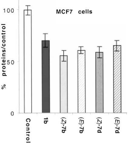
Study of the antiproliferative effects of (Z)‐ and (E)‐ 7 b and (Z)‐ and (E)‐ 7 d on hormone‐ dependent (MCF7) and hormone‐ independent (MDA‐ MB231) breast cancer cell lines: Separation of the Z and E isomers of all the rhenium complexes 7 a–e was carried out
by preparative reversed‐ phase HPLC. Since we have previously shown that these complexes do not isomerise in solution, we have chosen two representative pairs of isomers, (Z)‐ and (E)‐ 7 b and 7 d, to study their proliferative/antiproliferative effect on the MCF7 and MDA‐ MB231 cell lines, which are considered the archetypes of ERα(+) and ERα(−) cell lines, respectively. OH‐ Tam (1 b) and estradiol were added as controls in each series. The results obtained are shown in Figures 2 and 3.
At an incubation molarity of 1 μM, the two isomers (Z)‐ and (E)‐ 7 b and 7 d have an antiproliferative effect on MCF7 cells which is slightly higher (12–15 %) than that of OH‐ Tam. In both series (n=3 and n=5), the Z isomer is more effective than the E isomer. However, the observed difference is quite modest at around 5 % (Figure 2). This antiproliferative effect of the complexes must, like that of OH‐ Tam, be linked to an antiestrogenic effect. However, the significant differences in RBA values of 7 b and 7 d found for cytosolic ER are not reflected in any great variation in antiproliferative effects on these hormone‐ dependent cells.
As expected, on MDA‐ MB231 cells with no ERα, neither estradiol nor OH‐ Tam has any effect (Figure 3). At a concentration of 1 μM, the rhenium complexes have a weak antiproliferative effect of 10 % or slightly above. This antiproliferative effect must be associated with a cytotoxic effect, and the complexes with the longest chain (n=5) are somewhat more effective than those with a 3‐ carbon chain. On these cells, the Z and E isomers have identical behaviour for any given pair.
Figure 2 Antiproliferative effect of 1 μM of OH‐ Tam (1 b), (Z)‐ 7 b, (E)‐ 7 b, (Z)‐ 7 d and (E)‐ 7 d on MCF7 cells (breast cancer cell line ERα‐ positive) after 6 days of culture. In this experiment 10 nM of E2 has a
proliferative effect (198 %; data not shown). Representative data of one experiment performed twice with similar results (8 measurements±limits of confidence; P=0.1, t=1.895).
Overall the results obtained with these stable and lipophilic rhenium complexes are essentially linked to an antiestrogenic effect of the same order as or slightly higher than that of OH‐ Tam. This augurs well for the future introduction of active group‐ VII radionucleides to create another functionality in the binding site.
Figure 3 Effect of 1 μM of OH‐ Tam (1 b), (Z)‐ 7 b, (E)‐ 7 b, (Z)‐ 7 d, (E)‐ 7 d and of 10 nM of E2 on
MDA‐ MB231 cells (breast cancer cell line ERα‐ negative) after 6 days of culture. Representative data of one experiment performed twice with similar results (8 measurements±limits of confidence; P=0.1, t=1.895).
Discussion
The behavioural similarity between the rhenium complexes 7 b and 7 d and OH‐ Tam (1 b) justifies the use of the crystallographic data underpinning the biological behaviour of OH‐ Tam. In particular, the structure of the ligand binding domain (LBD) of human estrogen receptor alpha (hERα) occupied by OH‐ Tam (1 b) has been published.[9]
We digitally removed the bioligand 1 b from the site and replaced it with (Z)‐ 7 b. To obtain Figure 4, we used MacSpartan Pro,[33] Molview,[34] and weblab viewer Accelys35 software successively. This molecular modelling study shows the overall position of (Z)‐ 7 b in the binding site with the phenol of (Z)‐ 7 b bound to the amino acid residues Arg‐ 394 and Glu‐ 353, while Asp‐ 351 reveals the possibility of a hydrogen bond with the nitrogen atom of the basic chain O—(CH2)3NMe2. The CpRe(CO)3 moiety which replaces the phenyl of 1 b is easily inserted
at the level of His‐ 524 causing no notable perturbation. This view shows the steric influence of the basic chain at the level of helix 12 of the protein. The latter can no longer bind to helix 4, and this new conformation prevents fixation of the coactivators present. This new position
of H12, which is stabilised by the bond between Asp‐ 351 and the nitrogen atom of the NMe2 group of the side chain, is responsible, as with OH‐ Tam, for the antagonist effect
observed with the MCF7 cell line.
Figure 4 View of (Z)‐ 7 b docked in the antagonist binding site of hERα. This view is based on the ligand binding site X‐ ray structural determination obtained by Shiau[9] and the use of several computer programs.[33-35] For an explanation see text.
A more detailed molecular modelling study reveals the precise role played by the organometallic moiety, which in this case is based solely on the steric effect, in contrast to the case of the ferrocifens for which the possibility of oxidation of the ferrocene came into play.[17] Here, once again, we used MacSpartan Pro[33] software and the LBD structure published by Shiau.[9] Only the amino acids forming the wall of the cavity were retained,
totalling 757 atoms. OH‐ Tam was removed and replaced with the rhenium complexes 7 b with Z and E geometry, and with a 3‐ carbon side chain. With all the heavy atoms of the cavity immobilised, a position search was carried out to determine the energetically optimal location for the mediator. Next, the side chain of the amino acid Met‐ 343, which represented a steric hindrance for the organometallic moiety, was liberated. This was justified by the fact that this part of the cavity has been shown to be flexible.[36] An energy minimisation was then carried out with all the heavy atoms immobilised, except those of the mediator and of the Met‐ 343 side chain, by using the Merck molecular force field (MMFF). A conformational study was also carried out inside the cavity to determine the best position for the organometallic moiety in relation to the rest of the molecule. This determined the ideal position for the bioligand. Next, the affinity of the bioligand for its cavity was determined by MMFF molecular mechanics, with calculations for the bioligand–cavity combination, and for the cavity and bioligand separately, with the last two retaining the conformations they had in the molecular complex. This gives the ΔE energy change for the reaction bioligand + cavity→molecular complex. This gives a result of ΔE=−42 kcal mol−1 for (Z)‐ 7 b and ΔE=−36 kcal mol−1 for (E)‐ 7 b. The calculation shows that these compounds
have good affinity for the cavity in agreement with the RBA values obtained. Recognition of hERα is better for the Z than the E isomer, despite the fact that these two compounds have identical volumes (483 Å3 for (Z)‐ 7 b and 484 Å3 for (E)‐ 7 b). The difference in affinity lies in the fact that the Z isomer is a better fit for the shape of the cavity than its geometric E isomer (Figure 5).
Figure 5 Space‐ filling models of (Z)‐ 7 b and (E)‐ 7 b in the ligand binding site of the antagonist form of the ERα as a basis for the structure previously described by Shiau.[9]
Calculations of ΔE for a 2‐ carbon chain give ΔE=−38 kcal mol−1 for (Z)‐ 7 a and ΔE=−33
kcal mol−1 for (E)‐ 7 a. Here again, the results are in accord with the RBA values. The reason for this is that the organometallic moiety CpRe(CO)3 fills the volume of the cavity well. The
molecule has little room for free play at this level. Because of these constraints the 2‐ carbon chain at the other end of the molecule does not optimally stabilise the hydrogen bond with Asp‐ 351. The situation is completely different with (Z)‐ OH‐ Tam (1 b). This molecule is less bulky, with a volume of only 395 Å3. Consequently, the phenyl does not by any means occupy the entire cavity. This makes it possible for the molecule to adjust its position at this level, and a 2‐ carbon chain is sufficient to give a good bond with Asp‐ 351 and to stabilise the system. A similar rationale is applicable to the case of the ferrocifens, for which the antiproliferative effects with MCF7 cells are better with a 3‐ carbon than with a 2‐ carbon chain.[17,37] This modelling study is consistent with the published body of work involving hERα.
For hERβ, the available structural data are still insufficient to allow this type of molecular approach.[7] As a first approximation the results obtained with MDA‐ MB231 are weak to
nonexistent, similar to those of OH‐ Tam (1 b). It is possible that the primary target here is estrogen receptor β, for which, to the best of our knowledge, there has been no X‐ ray structural determination of the LBD with OH‐ Tam (1 b) bound to the site. The fact that there is a slight variation in the effect depending on the length of the chain 7 c (n=4) suggests that other targets of tamoxifen, such as the calmodulin receptor, may be partially involved.[38]
Since one of the future aims of this program is to find a route to 99mTc‐ labelled
radiopharmaceuticals analogous to (Z)‐ 7 b, we carried out a ΔE energy variation calculation with this hypothetical technetium molecule and found a variation of ΔE=−36 kcal mol−1. The
result allows us to predict that this radioactive complex should have good affinity for the estrogen receptor, and will be an attractive target molecule to synthesise.
Conclusion
These results reveal a striking analogy between the biochemical behaviour of OH‐ Tam and its organometallic rhenium analogues. The differences between the two series are attributable to the greater volume of CpRe(CO)3 compared to the β‐ phenyl of OH‐ Tam (1 b) which was
substituted. This is very clear in the molecular modelling studies in which moderate lengthening of the basic chain retrieves the initial effect. The (Z)‐ 7 b complex represents the best compromise between recognition and activity.
Based on the results of this study, and assuming that CpRe(CO)3 does not play a functional
role in the site, the effect of the rhenium antiestrogens at the level of estrogen receptor alpha is entirely conformational in nature. The basic carbon chain plays the same role as its equivalent in OH‐ Tam: it changes the conformation of helix 12 of ERα, thus causing an antagonist effect. MDA‐ MB231 cells contain no receptor α but only the β form of the estrogen receptor. Here again, the observed lack of effect is analogous to that of OH‐ Tam, and for the same reasons.[7] The slight chain effect observed may perhaps be compared to the intervention of a partial inhibition mechanism of the calmodulin receptor.[38]
Our results clearly demonstrate the approach to be followed in making use of this type of product in the future: it is a case of functionalising the CpRe(CO)3 moiety of the vectors 7.
There are two objectives: either to prepare a complex of the Cp99mTc(CO)3 type, a γ emitter
for use in medical imaging; or to prepare Cp188Re(CO)3, also on the vectors 7, to give a novel
therapeutic product based on β emission. The chemistry is identical in either case. It is still necessary, however, to find novel reactions that are rapid, high‐ yielding and usable in
water.[24-26] We are actively working on this. In particular, we are exploring two routes, one involving use of Alberto's reagent [(H2O)3Re(CO)3]+ or [(H2O)3Tc(CO)3]+,[39-42] the other a
novel exchange reaction between [Re(CO)6]+ and the ferrocene derivatives developed in our
laboratory.[43] The first results are encouraging, as we have successfully developed a one‐ step synthesis of a 99mTc‐ labelled steroid. This molecule is obtained by exchange of a
ferrocene with a Cp99mTc/Re(CO)3 moiety. The reaction gives high yields of >90 % at 95 °C
in a H2O/dimethyl sulfoxide (DMSO) mixture in 3.5 h (Scheme 2).[44]
Scheme 2 Synthesis of a novel, 99mTc‐ labelled 17α‐ ethynylestradiol derivative: i) [99mTc(H2O)3(CO)3]
+ in
H2O/DMSO 1:1 mixture, 95 °C, 3.5 h at atmospheric pressure. [44]
Experimental Section
General: Starting materials were synthesized by using standard Schlenk techniques under an
argon atmosphere. Anhydrous THF and diethyl ether were distilled from
sodium/benzophenone. Thin‐ layer chromatography was performed on silica gel 60 GF254. FTIR spectra of KBr plates were recorded on a BOMEM Michelson‐ 100 spectrophotometer. 1H and 13C NMR spectra were acquired on Bruker 200 and Bruker 400 spectrometers by using CDCl3 as a solvent. Mass spectrometry was performed on a Nermag R
10–10C spectrometer. Melting points were measured with a Kofler device. Elemental analyses were performed by the Regional Microanalysis Department of the Université Pierre et Marie Curie.
4‐ Halogenoalkoxyphenyl‐ 4′‐ hydroxyphenyl ketones 4 a: Potassium hydride (5.70 g of 35 % dispersion in oil, 50 mmol) was introduced into a Schlenk tube filled with argon. The mixture was washed three times with petroleum ether, and then the rest of the petroleum ether was evaporated under reduced pressure for 10 min. Dry THF (100 mL) was added. A solution
of 4,4′‐ dihydroxybenzophenone (10.7 g, 50 mmol) in N,N‐ dimethylformamide (200 mL) was carefully added with a syringe. The yellow mixture was heated for 10 min and then cooled. Dibromoethane (250 mmol) was rapidly added, and the mixture was then heated again at gentle reflux overnight. The solution was cooled, and the solvent was removed under reduced pressure. The residue was purified by column chromatography with silica gel using dichloromethane/acetone (95:5) as eluent. Recrystallization from dichloromethane furnished pure ketone 4 a (12 % yield). M.p. 141 °C; IR (KBr): =3183 (OH), 1600 cm−1 (C=O); 1H NMR: δ=3.68 (t, J=6.2 Hz, 2 H; CH2), 4.38 (t, J=6.2 Hz, 2 H; CH2), 6.93 (d, J=8.9 Hz, 2 H;
Harom), 6.98 (d, J=8.8 Hz, 2 H; Harom), 7.05 (s, 1 H; OH), 7.74 (d, J=8.8 Hz, 2 H; Harom), 7.79
ppm (d, J=8.9 Hz, 2 H; Harom); 13C NMR: δ=28.5 (CH2), 67.8 (CH2), 114.1 (2 CH), 115.1
(2 CH), 130.0 (C), 131.1 (C), 132.3 (2 CH), 132.6 (2 CH), 160.1 (C), 161.4 (C), 195.2 ppm (CO); MS (IE, 70 eV): m/z: 320 [M]+.
Alkene 6 a: Titanium tetrachloride (2 mL, 18 mmol) was added dropwise to a suspension of zinc powder (2.34 g, 36 mmol) in THF (60 mL) at 0 °C. The mixture obtained was heated at reflux for 2 h and then allowed to cool to room temperature. A second solution was prepared by dissolving 4‐ haloalkoxy‐ 4′‐ hydroxybenzophenone (4 a) (6 mmol) and ketone 5 (2.35 g, 6 mmol) in THF (30 mL). This latter solution was added dropwise to the first solution, and the resulting mixture was then heated at reflux for 2 h. After cooling to room temperature, the mixture was stirred with water and dichloromethane. The mixture was acidified with dilute hydrochloric acid, filtered (Celite), and the organic layer was dried over magnesium sulfate. After concentration under reduced pressure, the crude product was purified by chromatography on silica gel plates with dichloromethane as eluent to give a pure mixture of 6 a (Z+E, 64 % yield) as an oil. IR (KBr): =3422 (OH), 2018 and 1920 cm−1 (C=O); MS (IE, 70 eV): m/z: 680 [M]+, 596 [M−3 CO]+; Major isomer: 1H NMR: δ=1.06 (t, J=7.5 Hz, 3 H; CH3), 2.26 (q, J=7.5 Hz, 2 H; CH2), 3.63 (t, J=6.2 Hz, 2 H; CH2), 4.28 (t, J=6.2 Hz, 2 H;
CH2), 5.01–5.16 (m, 4 H; C5H4), 6.68–7.15 ppm (m, 8 H; Harom); 13C NMR: δ=15.2 (CH3),
29.1 (CH2), 30.8 (CH2), 67.7 (CH2), 82.7 (2 CH, C5H4), 85.6 (2 CH, C5H4), 108.9 (1 C, C5H4),
114.4 (2 CH), 115.5 (2 CH), 130.0 (2 CH), 130.4 (C), 130.6 (2 CH), 135.5 (C), 136.3 (C), 141.9 (C), 154.9 (C), 156.8 (C), 194.6 ppm (3 CO); Minor isomer: 1
H NMR: δ=1.06 (t, J=7.5 Hz, 3 H; CH3), 2.27 (q, J=7.5 Hz, 2 H; CH2), 3.63 (t, J=6.2 Hz, 2 H; CH2), 4.26 (t, J=6.2 Hz,
2 H; CH2), 5.01–5.16 (m, 4 H; C5H4), 6.68–7.15 ppm (m, 8 H; Harom); 13C NMR: δ=15.2
C5H4), 114.7 (2 CH), 115.2 (2 CH), 130.0 (2 CH), 130.4 (C), 130.6 (2 CH), 135.2 (C), 136.6
(C), 141.9 (C), 154.7 (C), 156.9 (C), 194.6 ppm (3 CO).
Amine 7 a: Halide 6 a (3 mmol) was added to a solution of dimethylamine in methanol (2 M, 15 mL, 30 mmol) and placed in an autoclave. The mixture was heated with stirring at 60 °C for 1 day. After cooling, the solution was concentrated under reduced pressure. The residue was purified by chromatography on silica gel plates (chloroform/triethylamine, 80:20 as eluent). After evaporation of the solvent, the residue obtained was dissolved in dichloromethane, and the solution was washed three times with water. The organic layer was dried over magnesium sulfate, and the solution was then concentrated under reduced pressure to furnish a pure mixture of Z and E isomers of 7 a as an oil. The Z and E isomers (ratio 18:82, respectively; 82 % yield) were separated on a reverse‐ phase semipreparative Kromasil C18 column (diameter 2 cm, length 25 cm), eluent methanol/phosphate buffer (KH2PO4, 25
mM, pH 7), flow rate 6 mL min−1. Major (E) isomer: m.p. 141 °C; IR (KBr): =3449 (OH), 2016 and 1913 cm−1 (C=O); 1H NMR: δ=1.04 (t, J=7.4 Hz, 3 H; CH3), 2.29 (q, J=7.4 Hz, 2 H; CH2), 2.43 (s, 6 H; NMe2), 2.83 (t, J=5.3 Hz, 2 H; NCH2), 4.04 (t, J=5.3 Hz, 2 H; OCH2), 5.11 (s, 4 H; C5H4), 6.48 (d, J=8.7 Hz, 2 H; Harom), 6.71 (d, J=8.6 Hz, 2 H; Harom), 6.91 (d, J=8.6 Hz, 2 H; Harom), 6.97 ppm (d, J=8.7 Hz, 2 H; Harom); 13C NMR: δ=15.3 (CH3), 29.1 (CH2), 45.0 (2 CH3), 58.0 (CH2), 64.1 (CH2), 82.6 (2 CH, C5H4), 85.4 (2 CH, C5H4), 109.8 (1 C, C5H4), 113.7 (2 CH), 115.9 (2 CH), 129.5 (C), 130.0 (2 CH), 130.8 (2 CH), 134.6 (C), 135.7
(C), 142.4 (C), 156.2 (C), 157.3 (C), 194.7 ppm (3 CO); MS (IE, 70 eV): m/z: 645 [M]+
, 561 [M−3 CO]+; elemental analysis calcd (%) for C28H28NO5Re: C 52.16, H 4.38, N 2.17; found:
C 51.53, H 4.51, N 2.16; Minor (Z) isomer: m.p. 110 °C; IR (KBr): =3438 (OH), 2016 and 1913 cm−1 (C=O); 1H NMR: δ=1.07 (t, J=7.4 Hz, 3 H; CH3), 2.31 (q, J=7.4 Hz, 2 H; CH2), 2.42 (s, 6 H; NMe2), 2.82 (t, J=5.3 Hz, 2 H; NCH2), 4.04 (t, J=5.3 Hz, 2 H; OCH2), 5.00–5.20 (m, 4 H; C5H4), 6.55 (d, J=8.7 Hz, 2 H; Harom), 6.77 (d, J=8.7 Hz, 2 H; Harom), 6.93 (d, J=8.7 Hz, 2 H; Harom), 7.00 ppm (d, J=8.7 Hz, 2 H; Harom); 13C NMR: δ=15.2 (CH3), 29.0 (CH2), 45.1 (2 CH3), 58.0 (CH2), 64.3 (CH2), 82.5 (2 CH, C5H4), 85.5 (2 CH, C5H4), 109.9 (1 C, C5H4), 114.0 (2 CH), 115.6 (2 CH), 129.7 (C), 130.2 (2 CH), 130.6 (2 CH), 134.2 (C), 136.1
(C), 142.5 (C), 156.0 (C), 157.4 (C), 194.6 ppm (3 CO); MS (IE, 70 eV): m/z: 645 [M]+; elemental analysis calcd (%) for C28H28NO5Re: C 52.16, H 4.38, N 2.17; found: C 52.04, H
X‐ ray crystal structure determination of (E)‐ 7 a: A suitable crystal of (E)‐ 7 a was mounted on an Enraf–Nonius Mach‐ 3 diffractometer. Accurate cell dimensions and orientation matrices were obtained by least‐ squares refinement of 25 well‐ centred reflections. No significant variations were corrected from Lorentz and polarization effects. The structure was solved by direct methods (SHELXS)[45] and refined by using the PC version of CRYSTALS.[46] Scattering factors and corrections for anomalous absorption were taken from ref. [47]. All non‐ hydrogen atoms were anisotropically refined. Hydrogen atoms were introduced in calculated positions in the last refinements and they were allocated one overall isotropic thermal parameter. Main significant crystallographic data and collection details are summarized in Table 1.
CCDC‐ 233202 contains the supplementary crystallographic data for this paper. These data can be obtained free of charge via www.ccdc.cam.ac.uk/conts/retrieving.html (or from the Cambridge Crystallographic Data Centre, 12 Union Road, Cambridge CB2 1EZ, UK; fax: (+44) 1223‐ 336033; or deposit@ccdc.cam.uk).
Biochemical experiments:
Materials: 17β‐ Estradiol and OH‐ Tam (Z+E) were obtained from Sigma–Aldrich (France); Ferrocene [(η5‐ C5H5)2Fe] was from Strem (Newburyport, MA, USA). Stock solutions
(1×10⁻³ M) of the compounds to be tested were prepared in ethanol and were kept at −20 °C in the dark; under these conditions they are stable for at least 2 months. Serial dilutions in ethanol were prepared just prior to use. Dulbecco's modified eagle medium (DMEM) was purchased from Gibco BRL; fetal calf serum was from Dutscher, Brumath, France; glutamine, estradiol, and protamine sulfate were from Sigma. MCF7 and MDA‐ MB231 cells were from the Human Tumor Cell Bank.
Animal tissues: Sheep uteri weighing approximately 7 g were obtained from the slaughterhouse at Mantes‐ la‐ Jolie, France. They were immediately frozen and kept in liquid nitrogen prior to use.
Determination of the relative binding affinity (RBA) of the compounds for ERα and ERβ: RBA values were measured on ERα from lamb uterine cytosol and on ERα and ERβ purchased from Pan Vera (Madison, WI, USA). Sheep uterine cytosol prepared in buffer A (0.05 M Tris‐ HCl, 0.25 M sucrose, 0.1 % β‐ mercaptoethanol, pH 7.4 at 25 °C) as described in ref. [48] was used as a source of ERα. For ERβ, the solution containing 3500
pmol mL−1 (10 μL) was added to buffer B (16 mL, 10 % glycerol, 50 mM Bis‐ Tris‐ Propane, pH 9, 400 mM KCl, 2 mM DTT, 1 mM EDTA, 0.1 % BSA) in a silanised flask. Aliquots (200 μL) of ERα in glass tubes or ERβ in polypropylene tubes were incubated for 3 h at 0 °C with [6,7‐ 3H]‐ estradiol (2×10⁻⁹ M, specific activity 1.62 TBq mmol−1
, NEN Life Science, Boston MA) in the presence of nine concentrations of the hormones to be tested. At the end of the incubation period, the free and bound fractions of the tracer were separated by protamine sulfate precipitation. The percentage reduction in binding of [3H]‐ estradiol (Y) was calculated by using the logit transformation of Y (logitY: ln[y/1−Y] versus the log of the mass of the competing steroid. The concentration of unlabeled steroid required to displace 50 % of the bound [3H]‐ estradiol was calculated for each steroid tested, and the results expressed as RBA. The RBA value of estradiol is by definition equal to 100 %.
Measurement of octanol/water partition coefficient (logPo/w) of the compounds: The logPo/w values of the compounds were determined by reversed‐ phase HPLC on a C‐ 8
column (nucleosil 5 C8, from Macherey Nagel, France) according to the method previously described by Minick[49] and Pomper.[50] Measurement of the chromatographic capacity factors (k′) for each compound was done at various concentrations in the range 85–60 % methanol (containing 0.25 % octanol) and an aqueous phase consisting of 0.15 % n‐ decylamine in 0.02 M MOPS (3‐ morpholinopropanesulfonic acid) pH 7.4 buffer (prepared in 1‐ octanol‐ saturated water). These capacity factors (k′) are extrapolated to 100 % of the aqueous component given the value of k′w. LogPo/w (y) is then obtained from the formula
logPo/w=0.13418 + 0.98452logk′w.
Culture conditions: Cells were maintained in monolayer in DMEM with phenol red (Gibco BRL) supplemented with 8–9 % fetal calf serum (Gibco BRL) and glutamine 2 mM (Sigma) at 37 °C in a 5 % CO2 air‐ humidified incubator. For proliferation assays, cells were plated in
DMEM medium (1 mL) with phenol red, supplemented with 10 % decomplemented and hormone‐ depleted fetal calf serum and 2 mM glutamine and incubated. The following day (D0), 1 mL of the same medium containing the compounds to be tested was added to the plates (final volumes of alcohol: 0.1 %; 4 wells for each condition, one plate per day). After 3 days (D3) the incubation medium was removed and fresh medium containing the compounds was added. After 6 days (D6), the total protein content of the plate was analysed by methylene blue staining as follows: cell monolayers were fixed for 1 h in methanol, stained for 1 h with methylene blue (1 mg mL−1) in PBS, then washed thoroughly with water. HCl (1 mL, 0.1 M)
was then added, and the absorbance of each well was measured at 620 nm with a Biorad spectrophotometer. The results are expressed as the percentage of proteins versus the control.
Acknowledgements
We thank A. Cordaville for technical assistance and B. McGlinchey for her help in translating the manuscript. Financial support from the Ministry of Research and the Centre National de la Recherche Scientifique is gratefully acknowledged.
[1] E. J. Feuer, L. M. Wun, C. C. Boring, W. D. Flanders, M. J. Timmel, T. Tong, J. Natl. Cancer Inst. 1993, 85, 892-897.
[2] V. C. Jordan, J. Med. Chem. 2003, 46, 883-908. [3] V. C. Jordan, J. Med. Chem. 2003, 46, 1081-1111. [4] K. Dhingra, Invest. New Drugs 1999 17, 285-311.
[5] M. J. Meegan, D. G. Lloyd, Curr. Med. Chem. 2003, 10, 181-210.
[6] A. M. Brzozowski, A. C. Pike, Z. Dauter, R. E. Hubbard, T. Bonn, O. Engstrom, L. Ohman, G. L. Greene, J.-A. Gustafsson, M. Carlquist, Nature 1997, 389, 753-758.
[7] A. C. Pike, A. M. Brzozowski, R. E. Hubbard, T. Bonn, A. G. Thorsell, O. Engstrom, J. Ljunggren, J.-A. Gustafsson, M. Carlquist, E MBO J. 1999, 18, 4608-4618.
[8] A. C. Pike, A. M. Brzozowski, J. Walton, R. E. Hubbard, A. G. Thorsell, Y. L. Li, J.-A. Gustafsson, M. Carlquist, Structure (Camb) 2001, 9, 145-153.
[9] A. K. Shiau, D. Barstad, P. M. Loria, L. Cheng, P. J. Kushner, D. A. Agard, G. L. Greene, Cell 1998,
95, 927-937.
[10] D. M. Tanenbaum, Y. Wang, S. P. Williams, P. B. Sigler, Proc. Natl. Acad. Sci. USA 1998, 95, 5998-6003.
[11] J. A. Gustafsson, J. Endocrinol. 1999, 163, 379-383.
[12] K. Paech, P. Webb, G. G. Kuiper, S. Nilsson, J.-A. Gustafsson, P. J. Kushner, T. S. Scanlan, Science 1997, 277.
[13] P. de Cremoux, C. Tran-Perennou, C. Elie, E. Boudou, C. Barbaroux, M. Poupon, Y. De Rycke, B. Asselain, H. Magdelénat, Biochem. Pharmacol. 2002, 64, 507-515.
[14] J. A. Gustafsson, Trends Pharmacol. Sci. 2003, 24, 479-485. [15] V. Speirs, M. J. Kerin, Brit. J. Surgery 2000, 87, 405-409. [16] V. Speirs, J. Pathol. 2002, 197, 143-147.
[17] S. Top, A. Vessières, G. Leclercq, J. Quivy, J. Tang, J. Vaissermann, M. Huché, G. Jaouen, Chem.
Eur. J. 2003, 9, 5223-5236.
[18] E. A. Vladusic, A. E. Hornby, F. K. Guerra-Vladusic, R. Lupu, Cancer Res. 1998, 58, 210-214. [19] C. Ferlini, G. Scambia, M. Marone, M. Distefano, C. Gaggini, G. Ferrandina, A. Fattorossi, G. Isola,
P. Benedetti Panci, S. Mancuso, Brit. J. Cancer 1999, 79, 257-263.
[20] Technetium and Rhenium in Chemistry and Nuclear Medicine n°3, U. Mazzi (Ed.), Cortina
International, Verona, 1990.
[21] Technetium and Rhenium in Chemistry and Nuclear Medicine n°4, U. Mazzi (Ed.), SG Editoriali,
Padova, 1995.
[22] P. Bläuenstein, New J. Chem. 1990, 405-407. [23] R. Alberto, Top. Curr. Chem. 1996, 176, 149-187.
[24] N. Metzler-Nolte, Angew. Chem. 2001, 113, 1072-1076; Angew. Chem. Int. Ed. 2001, 40, 1040-1043. [25] F. Le Bideau, M. Salmain, S. Top, G. Jaouen, Chem. Eur. J. 2001, 7, 2289-2294.
[26] G. Jaouen, S. Top, A. Vessières, R. Alberto, J. Organomet. Chem. 2000, 600, 23-36.
[27] G. Jaouen, S. Top, A. Vessières, P. Pigeon, G. Leclercq, I. Laïos, Chem. Commun. 2001, 383-384. [28] J. E. McMurry, Acc. Chem. Res. 1983, 16, 405.
[29] G. Precigoux, C. Courseille, S. Geoffre, M. Hospital, Acta Crystallogr. 1979, B35, 3070. [30] D. W. Robertson, J. A. Katzenellenbogen, J. Org. Chem. 1982, 47, 2389.
[31] S. Top, B. Dauer, J. Vaissermann, G. Jaouen, J. Organomet. Chem. 1997, 541, 355-361.
[32] S. Top, H. El Hafa, A. Vessières, J. Quivy, J. Vaissermann, D. W. Huges, M. J. McGlinchey, J. P. Mornon, E. Thoreau, G. Jaouen, J. Am. Chem. Soc. 1995, 117, 8372-8380.
[33] Mac Spartam Pro, Wavefunction Co., Irvine CA 92612, USA. [34] J. M. Cense, Phys. Theor. Chem. 1990, 71, 763-766.
[36] R. N. Hanson, E. Napolitano, R. Fiaschi, J. Med. Chem. 1998, 41, 4686.
[37] G. Jaouen, S. Top, A. Vessières, G. Leclercq, J. Quivy, L. Jin, A. Croisy, C.R. Acad. Sci. Paris 2000,
Série IIc, 89-93.
[38] A. A. Colletta, J. R. Benson, M. Baum, Breast Cancer Res. Treat. 1994, 31, 5.
[39] R. Alberto, R. Schibli, A. Egli, P. A. Schubiger, W. A. Hermann, G. Artus, V. Abram, T. A. Kaden, J.
Organomet. Chem. 1995, 492, 217-224.
[40] R. Alberto, R. Schibli, A. Egli, U. Abram, S. Abram, T. A. Kaden, P. A. Schubiger, Polyhedron 1998,
17, 1133-1140.
[41] R. Alberto, R. Schibli, P. A. Schubiger, U. Abram, H. H¸bener, H. Berke, T. A. Kaden, Chem.
Commun. 1996, 1291-1292.
[42] J. Wald, R. Alberto, K. Ortner, L. Candreia, Angew. Chem. 2001, 113, 3152-3156; Angew. Chem. Int.
Ed. 2001, 40, 3062-3066.
[43] S. Top, S. Masi, G. Jaouen, Eur. J. Inorg. Chem. 2002, 1848-1853.
[44] S. Masi, S. Top, L. Boubekeur, G. Jaouen, S. Mundwiler, B. Spingler, R. Alberto, Eur. J. Inorg.
Chem. 2004, 2013-2017.
[45] G. M. Sheldrick, SHELXS86, Program for the solution of crystal structures. Univ. Gottingen, Germany.
[46] D. J. Watkins, C. K. Prout, J. R. Carruthers, P. W. Bettridge, CRYSTALS Issue 10 1996, Chemical Crystallography Laboratory, Oxford, UK.
[47] D. T. Cromer, International Tables for X-ray Crystallography, Kynoch Press, Birmingham, UK, 1974, Vol. IV.
[48] A. Vessières, S. Top, A. A. Ismail, I. S. Butler, M. Loüer, G. Jaouen, Biochemistry 1988, 27, 6659-6666.
[49] D. J. Minick, J. H. Frenz, M. A. Patrick, D. A. Brent, J. Med. Chem. 1988, 31, 1923-1933.
[50] M. G. Pomper, H. VanBrocklin, A. M. Thieme, R. D. Thomas, D. O. Kiese-wetter, K. E. Carlson, C. J. Mathias, M. J. Welch, J. A. Katzenellenbogen, J. Med. Chem. 1990, 33, 3143-3155.
Supporting Information
Experimental SectionGeneral Data. Starting materials were synthesized using standard Schlenk techniques, under an argon atmosphere. Anhydrous THF and diethyl ether were distilled from sodium/benzophenone. Thin layer chromatography was performed on silica gel 60 GF254. FT-IR spectra were recorded on a BOMEM Michelson-100 spectrometer as a KBr plate. 1H and 13C NMR spectra were acquired on Bruker 200 and Bruker 400 spectrometers by using CDCl3 as a solvent. Mass spectrometry was performed on a Nermag R 10-10C spectrometer.
Melting points were measured with a Kofler device. Elemental analyses were performed by the Regional Microanalysis Department of Université Pierre et Marie Curie. 4-Halogenoalkoxyphenyl-4'-hydroxyphenyl ketones 4a-e : In a Schlenk tube filled with argon was introduced potassium hydride (5.70 g of 35% dispersion in oil, 50 mmol). The mixture was washed three times with petroleum ether, then the rest of the petroleum ether was evaporated under reduced pressure for 10 minutes. Dry THF (100 mL) was added. A solution of 4,4'-dihydroxybenzophenone (10.7 g, 50 mmol) in N,N-dimethylformamide (200 ml) was carefully added with a syringe. The yellow mixture was heated for 10 minutes, then was cooled. The corresponding dihaloalkane (250 mmol) was rapidly added, and then the mixture was heated again at gentle reflux overnight. The solution was cooled, the solvent was removed under reduced pressure. The residue was chromatographed with silica gel column by using dichloromethane/acetone 95/5 as eluent. Recrystallization from dichloromethane furnished pure ketone 4a-e.
4a : Yield 12%; mp 141°C. IR: 3183 (OH), 1600 (C=O) cm-1. 1H NMR: 3.68 (t, J = 6.2 Hz, 2H, CH2), 4.38 (t, J = 6.2 Hz, 2H, CH2), 6.93 (d, J = 8.9 Hz, 2H, Harom), 6.98 (d, J = 8.8 Hz,
2H, Harom), 7.05 (s, 1H, OH), 7.74 (d, J = 8.8 Hz, 2H, Harom), 7.79 (d, J = 8.9 Hz, 2H, Harom). 13
C NMR: 28.5 (CH2), 67.8 (CH2), 114.1 (2 CH), 115.1 (2 CH), 130.0 (C), 131.1 (C), 132.3
(2 CH), 132.6 (2 CH), 160.1 (C), 161.4 (C), 195.2 (CO). MS (IE, 70 eV) m/z: 320 [M]+..
4b : Yield 37%; mp 147°C. IR: 3146 (OH), 1600 (C=O) cm-1. 1H NMR: 2.35 (quintuplet, J = 6.1 Hz, 2H, CH2), 3.62 (t, J = 6.1 Hz, 2H, CH2), 4.19 (t, J = 6.1 Hz, 2H, CH2), 6.98 (d, J =
8.8 Hz, 2H, Harom), 6.99 (d, J = 8.7 Hz, 2H, Harom), 7.75 (d, J = 8.7 Hz, 2H, Harom), 7.81 (d, J =
(2 CH), 115.3 (2 CH), 129.5 (C), 130.5 (C), 132.4 (2 CH), 132.8 (2 CH), 161.0 (C), 162.2 (C), 195.9 (CO); MS (IE, 70 eV) m/z: 334 [M]+..
4c : Yield 35%; mp 102°C. IR: 3130 (OH), 1603 (C=O) cm-1. 1H NMR: 1.88-2.25 (m, 4H, CH2-CH2), 3.51 (t, J = 6.0 Hz, 2H, CH2), 4.08 (t, J = 6.0 Hz, 2H, CH2), 6.94 (d, J = 8.4 Hz,
4H, Harom), 7.73 (d, J = 8.4 Hz, 2H, Harom), 7.78 (d, J = 8.4 Hz, 2H, Harom), 8.05 (s, 1H, OH); 13
C NMR: 27.7 (CH2), 29.3 (CH2), 33.2 (CH2), 67.0 (CH2), 113.8 (2 CH), 115.1 (2 CH),
129.8 (C), 130.7 (C), 132.2 (2 CH), 132.5 (2 CH), 160.6 (C), 162.0 (C), 194.9 (CO).
4d : Yield 40%; oil. IR: 3460 (OH), 1602 (C=O) cm-1. 1H NMR: 1.54-2.08 (m, 6H, CH2
-CH2-CH2), 3.46 (t, J = 6.6 Hz, 2H, CH2), 4.06 (t, J = 6.1 Hz, 2H, CH2), 4.75 (s, 1H, OH), 6.92
(d, J = 8.4 Hz, 2H, Harom), 6.95 (d, J = 8.4 Hz, 2H, Harom), 7.73 (d, J = 8.4 Hz, 2H, Harom), 7.78
(d, J = 8.4 Hz, 2H, Harom); 13C NMR: 24.5 (CH2), 28.0 (CH2), 32.1 (CH2), 33.2 (CH2), 67.6
(CH2), 113.7 (2 CH), 115.0 (2 CH), 129.7 (C), 130.1 (C), 132.2 (2 CH), 132.4 (2 CH), 160.2
(C), 162.2 (C), 195.3 (CO); MS (IE, 70 eV) m/z: 362 [M]+..
4e : Yield 37%; mp 97°C. IR: 3374 (OH), 1598 (C=O) cm-1; 1H NMR: 1.291.95 (m, 12H, -(CH2)6-), 3.55 (t, J = 6.6 Hz, 2H, CH2), 4.04 (t, J = 6.4 Hz, 2H, CH2), 6.30 (s, 1H, OH), 6.92
(d, J = 8.9 Hz, 2H, Harom), 6.95 (d, J = 8.9 Hz, 2H, Harom), 7.74 (d, J = 8.9 Hz, 2H, Harom), 7.78
(d, J = 8.9 Hz, 2H, Harom); 13C NMR: 25.8 (CH2), 26.7 (CH2), 28.7 (CH2), 28.9 (CH2), 29.0
(CH2), 32.5 (CH2), 45.0 (CH2), 68.1 (CH2), 113.9 (2 CH), 115.0 (2 CH), 130.2 (C), 130.4 (C),
132.3 (2 CH), 132.5 (2 CH), 159.8 (C), 162.5 (C), 195.1 (CO); MS (IE, 70 eV) m/z: 360 [M]+..
Alkenes 6a-e : Titanium tetrachloride (2 ml, 18 mmol) was added dropwise to a suspension of zinc powder (2.34 g, 36 mmol) in 60 mL of THF at 0°C. The mixture obtained was heated at reflux for 2 h and then allowed to cool to room temperature. A second solution was prepared by dissolving 4-haloalkoxy-4'-hydroxybenzophenone 4a-e (6 mmol) and ketone 5 (2.35 g, 6 mmol) in 30 mL of THF. This latter solution was added dropwise to the first solution and then the resulting mixture was heated at reflux for 2 h. After cooling to room temperature, the mixture was stirred with water and dichloromethane. The mixture was acidified with dilute hydrochloric acid, filtered on celite and the organic layer was dried over magnesium sulfate. After concentration under reduced pressure, the crude product was chromatographed on silica gel plates with dichloromethane as eluent to give pure oily (Z+E) mixture of 6a-e.
6a : Yield 64%. IR: 3422 (OH), 2018 and 1920 (C=O) cm-1. MS (IE, 70 eV) m/z: 680 [M]+ , 596 [M-3CO]+ . Major isomer: 1H NMR: 1.06 (t, J = 7.5 Hz, 3H, CH3), 2.26 (q, J = 7.5 Hz,
2H, CH2), 3.63 (t, J = 6.2 Hz, 2H, CH2), 4.28 (t, J = 6.2 Hz, 2H, CH2), 5.01-5.16 (m, 4H,
C5H4), 6.68-7.15 (m, 8H, Harom). 13C NMR: 15.2 (CH3), 29.1 (CH2), 30.8 (CH2), 67.7 (CH2),
82.7 (2 CH, C5H4), 85.6 (2 CH, C5H4), 108.9 (1C, C5H4), 114.4 (2 CH), 115.5 (2 CH), 130.0
(2 CH), 130.4 (C), 130.6 (2 CH), 135.5 (C), 136.3 (C), 141.9 (C), 154.9 (C), 156.8 (C), 194.6 (3 CO). Minor isomer: 1H NMR: 1.06 (t, J = 7.5 Hz, 3H, CH3), 2.27 (q, J = 7.5 Hz, 2H,
CH2), 3.63 (t, J = 6.2 Hz, 2H, CH2), 4.26 (t, J = 6.2 Hz, 2H, CH2), 5.01-5.16 (m, 4H, C5H4),
6.68-7.15 (m, 8H, Harom). 13C NMR: 15.2 (CH3), 29.1 (CH2), 30.8 (CH2), 67.7 (CH2), 82.7
(2 CH, C5H4), 85.6 (2 CH, C5H4), 108.9 (1C, C5H4), 114.7 (2 CH), 115.2 (2 CH), 130.0 (2
CH), 130.4 (C), 130.6 (2 CH), 135.2 (C), 136.6 (C), 141.9 (C), 154.7 (C), 156.9 (C), 194.6 (3 CO).
6b : Yield 68%. IR: 3431 (OH), 2018 and 1918 (C=O) cm-1. MS (IE, 70 eV) m/e: 694 [M]+., 610 [M-3CO]+. Major isomer: 1H NMR: 1.07 (t, J = 7.4 Hz, 3H, CH3), 2.23-2.40 (m, 4H, 2
CH2), 3.60 (t, J = 6.4 Hz, 2H, CH2), 4.09 (t, J = 5.5 Hz, 2H, CH2), 5.01-5.16 (m, 4H, C5H4),
6.45 (s, 1H, OH), 6.69-6.91 (m, 4H, Harom), 6.91-7.16 (m, 4H, Harom). 13C NMR: 15.2 (CH3),
28.9 (CH2), 30.0 (CH2), 32.3 (CH2), 65.2 (CH2), 82.7 (2 CH, C5H4), 85.6 (2 CH, C5H4), 109.0
(1C, C5H4), 114.2 (2 CH), 115.5 (2 CH), 130.0 (2 CH), 130.2 (C), 130.6 (2 CH), 135.5 (C),
135.8 (C), 142.1 (C), 155.0 (C), 157.4 (C), 194.7 (3 CO). Minor isomer: 1H NMR: 1.07 (t, J = 7.4 Hz, 3H, CH3), 2.23-2.40 (m, 4H, 2 CH2), 3.60 (t, J = 6.4 Hz, 2H, CH2), 4.09 (t, J = 5.5 Hz, 2H, CH2), 5.01-5.16 (m, 4H, C5H4), 6.45 (s, 1H, OH), 6.69-6.91 (m, 4H, Harom), 6.91-7.16 (m, 4H, Harom). 13C NMR: 15.2 (CH3), 28.9 (CH2), 30.0 (CH2), 32.3 (CH2), 65.2 (CH2), 82.7 (2 CH, C5H4), 85.6 (2 CH, C5H4), 109.1 (1C, C5H4), 114.4 (2 CH), 115.2 (2 CH), 130.1 (2 CH), 130.2 (C), 130.5 (2 CH), 135.2 (C), 136.1 (C), 142.1 (C), 154.8 (C), 157.5 (C), 194.7 (3 CO).
6c : Yield 62%; IR: 3438 (OH), 2018 and 1918 (C=O) cm-1. MS (IE, 70 eV) m/z: 708 [M]+., 624 [M-3CO]+. Major isomer: 1H NMR: 1.07 (t, J = 7.6 Hz, 3H, CH3), 1.80-2.18 (m, 4H,
(CH2)2), 2.28 (q, J = 7.6 Hz, 2H, CH2), 3.50 (t, J = 6.4 Hz, 2H, CH2), 3.99 (t, J = 6.4 Hz, 2H,
CH2), 5.01-5.16 (m, 4H, C5H4), 6.67-6.90 (m, 4H, Harom), 6.90-7.14 (m, 4H, Harom). 13C NMR: 15.2 (CH3), 27.8 (CH2), 29.0 (CH2), 29.4 (CH2), 33.4 (CH2), 67.7 (CH2), 82.7 (2 CH, C5H4),
85.6 (2 CH, C5H4), 109.0 (1C, C5H4), 114.2 (2 CH), 115.4 (2 CH), 129.9 (2 CH), 130.3 (C),
isomer: 1H NMR: 1.07 (t, J = 7.6 Hz, 3H, CH3), 1.80-2.18 (m, 4H, (CH2)2), 2.28 (q, J = 7.6 Hz, 2H, CH2), 3.50 (t, J = 6.4 Hz, 2H, CH2), 3.99 (t, J = 6.4 Hz, 2H, CH2), 5.01-5.16 (m, 4H, C5H4), 6.67-6.90 (m, 4H, Harom), 6.90-7.14 (m, 4H, Harom). 13C NMR: 15.2 (CH3), 27.8 (CH2), 29.0 (CH2), 29.4 (CH2), 33.4 (CH2), 67.7 (CH2), 82.7 (2 CH, C5H4), 85.6 (2 CH, C5H4), 109.0 (1C, C5H4), 114.4 (2 CH), 115.2 (2 CH), 130.1 (2 CH), 130.3 (C), 130.5 (2 CH), 135.6 (C), 135.8 (C), 141.9 (C), 154.4 (C), 157.7 (C), 194.6 (3 CO).
6d : Yield 70%. IR: 3422 (OH), 2018 and 1919 (C=O) cm-1. MS (IE, 70 eV) m/z: 722 [M+.], 638 [M+-3CO]. Major isomer: 1H NMR: 1.06 (t, J = 7.5 Hz, 3H, CH3), 1.50-2.07 (m, 6H,
(CH2)3), 2.26 (q, J = 7.5 Hz, 2H, CH2), 3.44 (t, J = 6.8 Hz, 2H, CH2), 3.96 (t, J = 6.2 Hz, 2H, CH2), 5.01-5.16 (m, 4H, C5H4), 6.67-6.90 (m, 4H, Harom), 6.90-7.14 (m, 4H, Harom). 13C NMR: 15.2 (CH3), 24.7 (CH2), 28.3 (CH2), 29.0 (CH2), 32.3 (CH2), 33.5 (CH2), 67.4 (CH2), 82.6 (2 CH, C5H4), 85.6 (2 CH, C5H4), 109.1 (1C, C5H4), 114.1 (2 CH), 115.4 (2 CH), 129.9 (2 CH), 130.2 (C), 130.7 (2 CH), 135.4 (C), 135.7 (C), 142.1 (C), 154.7 (C), 157.7 (C), 194.6 (3 CO). Minor isomer: 1H NMR: 1.06 (t, J = 7.5 Hz, 3H, CH3), 1.50-2.07 (m, 6H, (CH2)3), 2.26 (q, J = 7.5 Hz, 2H, CH2), 3.44 (t, J = 6.8 Hz, 2H, CH2), 3.95 (t, J = 6.2 Hz, 2H, CH2), 5.01-5.16 (m, 4H, C5H4), 6.67-6.90 (m, 4H, Harom), 6.90-7.14 (m, 4H, Harom). 13C NMR: 15.2 (CH3), 24.7 (CH2), 28.3 (CH2), 29.0 (CH2), 32.3 (CH2), 33.5 (CH2), 67.4 (CH2), 82.6 (2 CH, C5H4), 85.5 (2 CH, C5H4), 109.1 (1C, C5H4), 114.4 (2 CH), 115.1 (2 CH), 130.1 (2 CH), 130.2 (C), 130.5 (2 CH), 135.4 (C), 135.7 (C), 142.1 (C), 154.6 (C), 157.8 (C), 194.6 (3 CO).
6e : Yield 68%; IR: 3412 (OH), 2019 and 1913 (C=O) cm-1. MS (IE, 70 eV) m/z: 720 [M+.], 636 [M+-3CO]. Major isomer: 1H NMR: 1.07 (t, J = 7.5 Hz, 3H, CH3), 1.231.90 (m, 12H,
-(CH2)6-), 2.27 (q, J = 7.5 Hz, 2H, CH2), 3.55 (t, J = 6.6 Hz, 2H, CH2), 3.95 (t, J = 6.4 Hz, 2H,
CH2), 5.01-5.16 (m, 4H, C5H4), 6.68-6.90 (m, 4H, Harom), 6.90-7.13 (m, 4H, Harom). 13C NMR: 15.2 (CH3), 25.8 (CH2), 26.7 (CH2), 28.7 (CH2), 29.1 (3 CH2), 32.5 (CH2), 45.0 (CH2), 67.8
(CH2), 82.6 (2 CH, C5H4), 85.5 (2 CH, C5H4), 109.1 (1C, C5H4), 114.1 (2 CH), 115.3 (2 CH),
129.9 (2 CH), 130.2 (C), 130.7 (2 CH), 135.2 (C), 136.0 (C), 142.0 (C), 154.4 (C), 158.0 (C), 194.6 (3 CO). Minor isomer: 1H NMR: 1.07 (t, J = 7.5 Hz, 3H, CH3), 1.231.90 (m, 12H,
-(CH2)6-), 2.27 (q, J = 7.5 Hz, 2H, CH2), 3.55 (t, J = 6.6 Hz, 2H, CH2), 3.93 (t, J = 6.4 Hz, 2H,
CH2), 5.01-5.16 (m, 4H, C5H4), 6.68-6.90 (m, 4H, Harom), 6.90-7.13 (m, 4H, Harom). 13C NMR: 15.2 (CH3), 25.8 (CH2), 26.7 (CH2), 28.7 (CH2), 29.1 (3 CH2), 32.5 (CH2), 45.0 (CH2), 67.8
130.1 (2 CH), 130.2 (C), 130.5 (2 CH), 135.5 (C), 135.7 (C), 142.0 (C), 154.3 (C), 157.9 (C), 194.6 (3 CO).
Amines 7a-e : Halide 6a-e (3 mmol) was added into a solution of dimethylamine in methanol (2M, 15 ml, 30 mmol) placed in an autoclave. The mixture was heated with stirring at 60°C for 1 day (6a-d) or 3 days (6e). After cooling, the solution was concentrated under reduced pressure. The residue was chromatographed on silica gel plates (chloroform:triethylamine 80:20 as eluent). After solvent evaporation, the residue obtained was dissolved in dichloromethane and the solution was washed three times with water. The organic layer was dried over magnesium sulfate then the solution was concentrated under reduced pressure to furnish pure amines 7a-e as oils containing a mixture of Z and E isomers. The Z and E isomers were separated on reverse phase semi-preparative Kromasil C18 column (diameter 2 cm, length 25 cm), eluent methanol:phosphate buffer (KH2PO4, 25 mM, pH 7) ratio : 82:18
for 7a, 83:17 for 7b, 87:13 for 7c, 88:12 for 7d, 92:8 for 7e, flow rate 6 mL/min.
7a Yield 82%. Major isomer (E): mp 141°C. IR: 3449 (OH), 2016 and 1913 (C=O) cm-1; 1H NMR: 1.04 (t, J = 7.4 Hz, 3H, CH3), 2.29 (q, J = 7.4 Hz, 2H, CH2), 2.43 (s, 6H, NMe2), 2.83
(t, J = 5.3 Hz, 2H, NCH2), 4.04 (t, J = 5.3 Hz, 2H, OCH2), 5.11 (s, 4H, C5H4), 6.48 (d, J = 8.7
Hz, 2H, Harom), 6.71 (d, J = 8.6 Hz, 2H, Harom), 6.91 (d, J = 8.6 Hz, 2H, Harom), 6.97 (d, J = 8.7
Hz, 2H, Harom); 13C NMR: 15.3 (CH3), 29.1 (CH2), 45.0 (2 CH3), 58.0 (CH2), 64.1 (CH2),
82.6 (2 CH, C5H4), 85.4 (2 CH, C5H4), 109.8 (1 C, C5H4), 113.7 (2 CH), 115.9 (2 CH), 129.5
(C), 130.0 (2 CH), 130.8 (2 CH), 134.6 (C), 135.7 (C), 142.4 (C), 156.2 (C), 157.3 (C), 194.7 (3 CO). MS (IE, 70 eV) m/z: 645 [M]+., 561 [M-3CO]+. Anal. Calcd for C28H28NO5Re: C,
52.16; H, 4.38; N, 2.17. Found: C, 51.53; H, 4.51; N, 2.16. Minor isomer (Z): mp 110°C. IR: 3438 (OH), 2016 and 1913 (C=O) cm-1. 1H NMR: 1.07 (t, J = 7.4 Hz, 3H, CH3), 2.31 (q, J =
7.4 Hz, 2H, CH2), 2.42 (s, 6H, NMe2), 2.82 (t, J = 5.3 Hz, 2H, NCH2), 4.04 (t, J = 5.3 Hz, 2H,
OCH2), 5.00-5.20 (m, 4H, C5H4), 6.55 (d, J = 8.7 Hz, 2H, Harom), 6.77 (d, J = 8.7 Hz, 2H,
Harom), 6.93 (d, J = 8.7 Hz, 2H, Harom), 7.00 (d, J = 8.7 Hz, 2H, Harom). 13C NMR: 15.2
(CH3), 29.0 (CH2), 45.1 (2 CH3), 58.0 (CH2), 64.3 (CH2), 82.5 (2 CH, C5H4), 85.5 (2 CH,
C5H4), 109.9 (1 C, C5H4), 114.0 (2 CH), 115.6 (2 CH), 129.7 (C), 130.2 (2 CH), 130.6 (2
CH), 134.2 (C), 136.1 (C), 142.5 (C), 156.0 (C), 157.4 (C), 194.6 (3 CO); MS (IE, 70 eV) m/z: 645 [M]+.. Anal. Calcd for C28H28NO5Re: C, 52.16; H, 4.38; N, 2.17. Found: C, 52.04;
7b : Yield 64%. Major isomer (E): mp 161°C. IR: 3431 (OH), 2018 and 1930 (C=O) cm-1. 1H NMR (400 MHz): 1.04 (t, J = 7.4 Hz, 3H, CH3), 1.97 (qint., 2H, CH2CH2CH2), 2.24 (q, J =
7.5 Hz, 2H, CH2CH3), 2.31 (s, 6H, NMe2), 2.55 (t, J = 7.3 Hz, 2H, NCH2), 3.94 (t, J = 6.2 Hz,
2H, OCH2), 5.07 (m, 4H, C5H4), 6.67 (d, J = 8.4 Hz, 2H, Hα’m), 6.76 (d, J = 8.6 Hz, 2H, Hαm),
6.92 (d, J = 8.4 Hz, 2H, Hα’o), 7.02 (d, J = 8.6 Hz, 2H, Hαo). 13C NMR: 15.4 (CH3), 27.0
(CH2CH2CH2), 29.1 (CH2CH3), 45.1 (NMe2), 56.3 (CH2N), 66.0 (OCH2), 82.8 and 85.7 (4C,
C2' and C3', C5H4), 109.4 (C1', C5H4), 114.2 (2 Cαm), 115.8 (2 Cα’m), 130.1 (C2), 130.1 (Cαo),
130.8 (Cα’o), 135.1 (Cα’ip), 135.8 (Cαip), 142.5 (C1), 155.9 (Cαp), 157.8 (Cα’o), 194.9 (3 CO).
MS (IE, 70 eV) m/z: 659 [M]+.. Anal. Calcd for C29H30NO5Re: C, 52.88; H, 4.59; N, 2.13.
Found: C, 52.67; H, 4.78; N, 2.14. Minor isomer (Z): mp: 162°C; IR (KBr): 3432 (OH), 2015 and 1899 (C=O) cm-1; 1H NMR: 1.08 (t, J = 7.4 Hz, 3H, CH3), 1.80-2.10 (m, 2H, CH2), 2.30
(q, J = 7.4 Hz, 2H, CH2), 2.35 (s, 6H, NMe2), 2.59 (t, J = 7.5 Hz, 2H, NCH2), 3.97 (t, J = 6.1
Hz, 2H, OCH2), 5.06 and 5.11 (m, m, 4H, C5H4), 6.73 (d, J = 8.5 Hz, 2H, Harom), 6.75 (d, J =
8.5 Hz, 2H, Harom), 7.00 (d, J = 8.5 Hz, 2H, Harom), 7.01 (d, J = 8.5 Hz, 2H, Harom). 13C NMR: 15.2 (CH3), 26.5 (CH2), 28.9 (CH2), 44.6 (2 CH3), 56.1 (CH2), 65.7 (CH2), 82.6 (2 CH,
C5H4), 85.5 (2 CH, C5H4), 109.2 (1 C, C5H4), 114.1 (2 CH), 115.7 (2 CH), 129.9 (C), 130.0 (2
CH), 130.6 (2 CH), 134.7 (C), 135.7 (C), 142.3 (C), 155.9 (C), 157.5 (C), 194.7 (3 CO). MS (IE, 70 eV) m/z: 659 [M]+.. Anal. Calcd for C29H30NO5Re: C, 52.88; H, 4.59; N, 2.13. Found:
![Figure 1 View of the molecular structure of (E) ‐ 7 a showing the atom numbering. Selected bond lengths [Å]](https://thumb-eu.123doks.com/thumbv2/123doknet/7770171.256739/9.892.105.788.101.732/figure-view-molecular-structure-showing-numbering-selected-lengths.webp)


![Figure 4 View of (Z)‐ 7 b docked in the antagonist binding site of hERα. This view is based on the ligand binding site X‐ ray structural determination obtained by Shiau [9] and the use of several computer programs](https://thumb-eu.123doks.com/thumbv2/123doknet/7770171.256739/17.892.120.701.252.825/figure-antagonist-binding-structural-determination-obtained-computer-programs.webp)

