Domestic fl eas were collected in 12 villages in the western Usambara Mountains in Tanzania. Of these, 7 are considered villages with high plague frequency, where hu-man plague was recorded during at least 6 of the 17 plague seasons between 1986 and 2004. In the remaining 5 vil-lages with low plague frequency, plague was either rare or unrecorded. Pulex irritans, known as the human fl ea, was the predominant fl ea species (72.4%) in houses. The density of P. irritans, but not of other domestic fl eas, was signifi -cantly higher in villages with a higher plague frequency or incidence. Moreover, the P. irritans index was strongly posi-tively correlated with plague frequency and with the logarith-mically transformed plague incidence. These observations suggest that in Lushoto District human fl eas may play a role in plague epidemiology. These fi ndings are of immediate public health relevance because they provide an indicator that can be surveyed to assess the risk for plague.
P
lague, caused by infection with Yersinia pestis, per-sists in many parts of the world; several hundred cases are reported to the World Health Organization each year, mostly from Africa (1,2). In Tanzania, a persistent focus of human plague was discovered in 1980 in the Lushoto District, in the northeastern part of the country. By 2004, 7,603 cases had been reported from this region (3). The distribution of plague cases in Lushoto is limited to an area of ≈1,200 km2, and a strong variation in plague frequency and incidence is seen among the villages in this region (3). Although evidence of infection with Y. pestis has been ob-served in several wild rodent and fl ea species, the actual reservoir in which the infection survives between epidem-ics has not yet been identifi ed, and the ecology of theinfec-tion and the source from which humans acquire infecinfec-tion are poorly understood (4–8). In Lushoto District, frequent plague outbreaks occur in some villages, but the disease is uncommon in other villages in the same vicinity. A study is under way to compare the ecologic conditions in villages having frequent outbreaks with those in villages where plague is relatively rare, with the objectives of understand-ing, predictunderstand-ing, and ultimately controlling human plague. Comparing host and vector communities is an important part of such studies.
In Lushoto District, it has been suggested that the fl eas
Xenopsylla cheopis, X. brasiliensis, and Dinopsyllus lypu-sus are plague vectors among sylvatic rodents, but Pulex irritans, the human fl ea, has received little attention (9). P. irritans has been collected in several plague-affected and
plague-free villages of the Lushoto area during epidemics and interepidemics (10), as well as on Rattus rattus (B.S. Kilonzo and S. Msingwa, unpub. data). Plastering a mud house is recommended in the area as a way of keeping the house free of fl eas (9) and involves mixing soil (with-out manure) with water and rubbing the mixture over the fl oors with a piece of cloth. We report differences between plague-affected and plague-free villages in the numbers of free domestic fl eas present in mud houses and consider whether this variation can be linked to house plastering as an antifl ea measure.
Materials and Methods
Lushoto District is situated in Tanga region, in the West Usambara Mountains, a part of the Eastern Arc Mountains. With an elevation ranging from 900 to 2,250 m above sea level, Lushoto District (04°22′–05°08′S, 038°05′–038°38′E) covers a surface area of 3,500 km2, of which 2,000 km2 are arable land and 340 km2 are forest reserve. Soils are mainly low-pH loams, rich in iron, man-ganese, and magnesium. Agriculture is the major economic activity, on which >90% of the population depends (11,12).
Plague and the Human Flea,
Tanzania
Anne Laudisoit,*† Herwig Leirs,*‡ Rhodes H. Makundi,§ Stefan Van Dongen,* Stephen Davis,* Simon Neerinckx,*¶ Jozef Deckers,¶ and Roland Libois†
*University of Antwerp, Antwerp, Belgium; †University of Liège, Liège (Sart Tilman), Belgium; ‡University of Aarhus, Kongens Lyn-gby, Denmark; §Sokoine University of Agriculture, Morogoro, Tan-zania; and ¶Katholieke Universiteit Leuven, Leuven, Belgium
The temperate climate is characterized by a short rainy sea-son during November–December and a longer one during March–May. A minor and unreliable rain, the Mlwati, oc-casionally occurs in August and September. The region is the most densely populated area in Tanzania, with an annu-al growth rate of 2.8% and 102 inhabitants per square kilo-meter. Inhabitants belong to 1 of 3 major tribes: Wasambaa (80%), Wambugu (10%), and Wapare (5%); the remaining 5% are immigrants from diverse other regions (13) Most Lushoto residents (70%) keep livestock in their houses, but cats and dogs are usually kept outside (9).
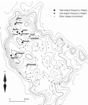
We selected 12 villages from throughout the plague-endemic area, ensuring a variation in both the frequency and incidence of plague, based on the earlier study by Davis et al. (3). Plague frequency is expressed as the percentage of plague seasons from 1986 to 2004 with reported plague cases, while plague incidence is the mean annual number of plague cases per 1,000 inhabitants at village level for the same period. Table 1 lists the 12 villages we surveyed, ranked according to plague frequency, and Figure 1 shows a map of the study area in Lushoto District. The 7 villages where plague on average occurred in >3 years per decade were considered “high plague frequency” villages, and the 5 villages where it occurred on average in <2 years per de-cade were considered “low plague frequency” villages. In all 12 villages, the common housing is mud houses with
dirt fl oors and iron sheet or thatch roofs. Cattle are kept outside the house attached to poles during the day and feed on grass, but they are kept inside overnight.
Collection of domestic fl eas began in May 2005 be-cause earlier literature reported that plague cases in Tanza-nia usually appear 2 times per year, in October/November and May/June (14). A more recent detailed investigation of Lushoto hospital data, however, showed a consistent sea-sonal pattern in which the highest number of plague cases occurs in January (3). Taking into account the practical limitations of extended fi eldwork periods overseas, a sec-ond collection period was started in January 2006. Fleas were thus collected every month from May through August 2005 in 4 core study villages (Gologolo, Emao, Kiranga, and Magamba) and from January through March 2006 in all 12 villages.
Houses surveyed were randomly chosen after the chief of each village granted authorization. Fleas were trapped by using a kerosene hurricane lamp hung above a 3-cm–high tray with a 45-cm diameter, half full of water. The lamp was lit at dusk and switched off at dawn during 3 consecu-tive nights. All traps were checked every morning between 9 AM and 11 AM, and captured fl eas were preserved in 70%
ethanol. The head of each household was interviewed by questionnaire to assess the perception of fl ea nuisance in the house and the frequency of plastering.
Table 1. Data on villages in Lushoto District, Tanzania, surveyed for domestic fleas, ranked by plague frequency*
Village Population† Plague frequency Plague incidence Coordinates, South–East Altitude, m asl Year of most recent plague case Year(s) flea trapping conducted No. forms received‡ Dule 3,036 0.059 0.637 04.58370– 038.31657 1,405 1986 2006 20 Mtae 3,407 0.059 0.121 04.48421– 038.23758 1,632 2000 2006 20 Handei 5,745 0.118 0.137 04.59514– 038.32390 1,376 1990 2006 20 Kiranga 868 0.176 1.056 04.57571– 038.27021 1,821 1996 2005–06 33 Magamba 2,676 0.176 0.836 04.72895– 038.30148 1,743 1998 2005–06 52 Goka 1,116 0.353 1.175 04.56680– 038.25990 1,843 1997 2006 20 Mambo 5,669 0.353 0.722 04.51167– 038.21976 1,828 1997 2006 20 Nkelei 1,305 0.647 3.434 04.56062– 038.24209 1,904 2000 2006 20 Shume-Nywelo 3,757 0.647 10.460 04.70025– 038.19687 1,890 2001 2006 20 Emao 2,054 0.706 5.180 04.56276– 038.25304 1,827 2000 2005–06 41 Gologolo 2,202 0.765 18.544 04.69707– 038.22692 1,950 2002 2005–06 26 Manolo 10,464 0.765 6.320 04.62058– 038.22260 1,809 2003 2006 9
*Data about plague are extracted from (3). Villages with a plague frequency of >0.3 on average, 3 years in a decade, were considered high frequency villages. asl, above sea level.
†From 2002 census.
We calculated the P. irritans index (Pii), per village per month, as the average number of P. irritans collected in a house. Frequency and incidence are village-specifi c char-acteristics based on long-term data, while for Pii we had to rely on relatively sparse and heterogeneous sampling dur-ing a short period. Simple averagdur-ing and testdur-ing for a cor-relation may be misleading if fi ndings vary between days, months, or years. Therefore, we analyzed the relation with a mixed model that included these sources of temporal vari-ation in Pii (fi rst applying a log transformvari-ation to ensure normality). This model included log(Pii) as the dependent variable and frequency or log(incidence) as the (continu-ous) independent factor. Year and month (nested within year) were added as fi xed effects, while village and the year–village interaction were treated as random effects to control for temporal variation in Pii, as well as for the fact that village is the independent unit of observation. Correla-tions in day-to-day estimates of Pii were modeled by using an autoregressive correlation coeffi cient. Model selection was based on backward elimination of nonsignifi cant fi xed effects. All random effects were retained in the model to ensure appropriate weighting and approximation of degrees of freedom by using the Kenward-Roger method.
We also tested for lagged relationships between monthly values of Pii and monthly plague incidence; that is, monthly incidence was related to the mean Pii for the previous month. Per village, the average number of people infected with plague each month was further expressed as a proportion of total number of cases in the district and was termed the monthly incidence. Daily fl ea numbers per house per village were summed to obtain a mean monthly Pii ± SD. The natural logarithm of the monthly incidence +1 was the dependent variable, while the natural logarithm of the mean monthly Pii was the predictor variable. Possible tem-poral dependence of the monthly incidences within villages was modeled by using an exponential decay of the degree of temporal autocorrelation. We also tested for an associa-tion between plastering frequency (independent variable) and the total number of captured fl eas (dependent variable, log transformed) in a mixed analysis of covariance model with village and village-by-frequency as random effects. In all the analyses above, standard error and denominator degrees of freedom were estimated by the Kenward-Roger method. We also tested for an association between altitude and the mean monthly Pii (log transformed to ensure nor-mality) by using a linear regression model. Finally, the as-sociation between fl ea abundance and the time since the previous plastering (the number of days between the last time the householder said the fl oor was plastered and the date of our fi rst visit) was analyzed with a Cox proportional hazards model. If no fl eas were trapped, the observation was considered to be censored. All analyses were performed in SAS version 9 (SAS Institute Inc., Cary, NC, USA).
Results
P. irritans was the predominant species (72.4%)
among domestic fl eas. Other species collected were
Echid-nophaga gallinacea (15.1%), Ctenocephalides felis and C. canis (6.5%), Xenopsylla brasiliensis (3.4%), and Tunga penetrans (2.6%). P. irritans and E. gallinacea were the
only species found in every village. P. irritans accounted for 61.5% and 75.2% of all fl eas collected in low and high plague frequency villages, respectively (Table 2). Twice as many houses were infested by P. irritans in high plague frequency villages than in low plague frequency villages (Table 2).
For all the trapping sessions in 2005 and 2006, the
P. irritans index was 2–9× greater in high plague
fre-quency than in low plague frefre-quency villages (Figure 2). The statistical analysis also showed that Pii was strongly positively correlated with plague frequency (F1,11.3 = 14.08, p = 0.003), with the logarithmically transformed plague incidence (F1,11.5 = 12.62, p = 0.004) and altitude (F1,11H = 8,641, p = 0.015). The abundance of other species was much lower than that of P. irritans, and none of the other Figure 1. Map of the Lushoto District of Tanzania showing locations
of willages with high and low plague frequency. All other villages with known locations are also plotted. The solid lines represent altitude contours (200-m elevation lines). To the west, a steep escarpment demarcates the edge of the district.
fl ea species indexes were correlated with plague frequency or incidence.
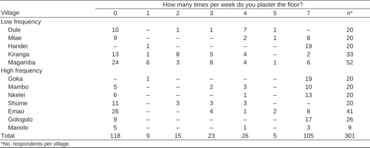
The questionnaires (301 valid responses) showed that in 8 of the 12 studied villages, some persons plaster their houses daily (Table 3). The fi gures suggest a great variabil-ity in the frequency of plastering between and within vil-lages and that frequency of plastering has no relation with the frequency of plague. For example, in Shume-Nywelo (high plague frequency) and Dule (low plague frequency), 55% and 50% of housekeepers, respectively, said they nev-er plastnev-er the house; in Gologolo (high plague frequency), 65.4% plaster their houses 7 times a week, but in Emao (another village with high plague frequency), only 19.5% do so.
The frequency of plastering did not correlate with the natural logarithm of the total number of fl eas caught (t20 = 0.88, p = 0.39), and this lack of association did not vary across villages (no signifi cant random village–frequency interaction, χ2
1 = 0.4, p = 0.47). Frequent plastering did not appear to prolong the time between the last plastering and the occurrence of the fi rst fl eas in the trap (χ2
1 = 0.36, p = 0.55).
Discussion
Our results show that the density of domestic fl eas is higher in villages with a higher plague frequency or inci-dence. Moreover, the human fl ea P. irritans accounts for a larger percentage of the domestic fl eas in these villages. The factors that contribute to the presence of plague in some villages in Lushoto while it is absent from others (3) are so far unknown. It is tempting therefore to attribute an epidemiologic role to P. irritans. This has been suggested recently for another focus of human plague; Arrieta et al. (15), working in the Peruvian Andes, observed that 69.9% of fl eas collected in domestic environments (on domestic animals and inside houses) were P. irritans (or, perhaps,
P. simulans, a sister species) and found the same positive
relation between high plague risk areas and P. irritans densities.
The human fl ea was fi rst mentioned in tropical Africa (Ethiopia) in 1868 (16). In Tanzania, plague was fi rst re-ported in 1886 in the Iringa region, but no information is available about the fl ea species present at that time. The
presence of P. irritans in Tanzania dates at least to 1915, when it was found in Dar-es-Salaam. In northeastern Tan-zania, its presence was reported in 35% of the beds exam-ined by Smith in 1959 (17); in 1977, 82.5% of the fl eas collected in human dwellings belonged to this species (18).
P. irritans is often found in high densities in habitations,
especially those with a dirt fl oor and a thatched roof, and is considered a possible plague vector in Angola, Brazil, Burundi, Democratic Republic of Congo, Iran, Iraq, Nepal, People’s Republic of China, and Tanzania (19–21).
Although a substantial body of literature describes the ecology of plague, the relation between the bacterium Y.
pestis and the human fl ea P. irritans during epizootics and
epidemics is poorly understood. The classic epidemiologic model for plague considers it an enzootic infection of most-ly resistant wild rodents. An outbreak of human plague may begin with an epizootic in peridomestic rats, from which ro-dent fl eas (in tropical regions typically X. cheopis) questing for a host may infect humans (22). In this scenario, human ectoparasites do not play an important role. However, epi-demiologic investigations based on historical accounts of the Black Death in the 14th–16th centuries in Europe show that the epidemics do not conform to this classic model, even leading to suggestions that the Black Death may have had a cause other than Y. pestis plague, an issue that is still hotly debated among historians (23,24). Recently, Dran-court et al. (25) reviewed earlier biologic studies that have presented experimental evidence for or against the role of
P. irritans in the transmission of plague.
Table 2. Distribution of flea species within villages and houses, Lushoto District, Tanzania
Flea species composition, %
Houses with given flea species, %
Domestic flea species Common hosts in Tanzania Low* High† Low* High†
Pulex irritans Humans 61.5 75.2 28.8 65.4
Ctenocephalides felis, C. canis Cats, dogs, other animals 8.8 5.8 6.8 10.7
Echidnophaga gallinacea Domestic fowl, Rattus rattus 19.6 13.7 12.2 15.7
Tunga penetrans Humans, dogs, goats 2.0 2.8 2.0 6.9
Xenopsylla brasiliensis Rattus rattus, Mastomys natalensis 6.8 2.1 4.1 5.0
*Villages designated as low plague frequency. †Villages designated as high plague frequency.
Figure 2. Monthly domestic Pulex irritans index, Pii, averaged for low plague frequency villages (black columns) and high plague frequency villages (white columns). The error bars indicate standard deviation from the mean. No data were available for high plague frequency villages in June 2005 or for low plague frequency villages in July 2005.
P. irritans is frequently infected with Y. pestis
(pestif-erous) but is rarely infective (China, Ecuador, Kazakhstan, Democratic Republic of Congo, Brazil; [21]), mainly be-cause it is not an easily blocked species (21). Blocking of the proventriculus by massive replication of the Y. pestis bacteria is known to enhance fl ea vectorial capacity and occurs in known plague vectors X. cheopis and Nosopsyllus
fasciatus (26). Therefore, the role for Pulex spp. as plagues
vector was classically believed to be no more than me-chanical transmission by way of soiled mouthparts, which is only possible if a high level of bacteremia exists in the pestilent host, if new potential hosts are available within 3 days after the infective blood meal, and if multiple bites occur (21). Such levels of ectoparasitism are realistic in a rural habitat; for example, in 1 night in our study in Golo-golo, a basic light trap caught 26 fl eas in a single room.
The role of unblocked fl eas may, however, be more than just mechanical. Eisen et al. (27), studying alternative fl eaborne transmission mechanisms, recently showed that
Oropsylla montana, which rarely becomes blocked, is
im-mediately infectious, transmits effi ciently for at least 4 days postinfection (early phase), and may remain infectious for up to 8 weeks postinfection because the fl eas do not un-dergo block-induced death. This scenario of effi cient early-phase transmission by unblocked fl eas matches historical observations of rapidly spreading epizootics and epidem-ics and their highly focal nature. During the second plague pandemic, in Europe, P. irritans was a suitable vector be-cause it was abundant on persons and in their homes, as it is today in some remote foci in Central Asia (25,28). In Ec-uador, during a plague outbreak in the Chimborazo region in 1998, P. irritans was abundant in human bedding (29). The fi ndings of the study by Eisen et al. (27) would also be consistent with a role for human fl eas in the epidemiology of plague in Lushoto. In contrast, in the Ituri plague focus
in the Democratic Republic of Congo, Devignat noticed the total absence of domestic P. irritans (16,30), just as in the epidemics in Saigon-Cholon in 1943 (31). P. irritans also appeared later in foci in the Democratic Republic of Con-go, and the primary human fl eas at that time (1946) were X.
cheopis and X. brasiliensis (32).
Among the other domestic species collected, C. felis
strongylus and C. canis are commonly found on cats and
dogs in Lushoto (5). These species are poor plague vec-tors but can be pestiferous, as observed in Democratic Re-public of Congo (30). T. penetrans’ status as plague vector is unknown. The females of this species are embedded in the host epidermis (humans, dog, rat, cat), but males are free hematophagous ectoparasites (33). E. gallinacea is frequent in human homes where hens are kept, but it was never observed on humans in Lushoto. It has been found to be infected with Y. pestis in the fi eld (34,35) but is consid-ered a poor plague vector due to its “stick tight” behavior (36). Finally, X. brasiliensis is the African counterpart to Asian X. cheopis in the sense that it is considered an ex-cellent plague vector (7,30). Notably, the abundance of X.
brasiliensis could not explain the village-level variation in
either incidence or frequency of human plague in the pres-ent study.
During our study, no human plague cases were record-ed in the test region, and the small mammals we trapprecord-ed in the 4 core villages tested negative for Y. pestis (n = 925, tested in a multiplex PCR; data not shown). Thus, the study period could be atypical in the sense that it is a period in which plague was absent. Whatever the explanation for the absence of plague cases, it is nevertheless clear that the abundance of P. irritans differs signifi cantly between vil-lages with different histories of human plague cases.
Because the vectorial status of P. irritans is still under discussion, and because of the correlative nature of our
re-Table 3. Questionnaire responses about plastering frequency, Lushoto District, Tanzania
How many times per week do you plaster the floor?
Village 0 1 2 3 4 5 7 n* Low frequency Dule 10 – 1 1 7 1 – 20 Mtae 9 – – – 2 1 8 20 Handei – 1 – – – – 19 20 Kiranga 13 1 8 5 4 – 2 33 Magamba 24 6 3 8 4 1 6 52 High frequency Goka – 1 – – – – 19 20 Mambo 5 – – 2 3 – 10 20 Nkelei 6 – – – 1 – 13 20 Shume 11 – 3 3 3 – – 20 Emao 26 – – 4 1 2 8 41 Gologolo 9 – – – – – 17 26 Manolo 5 – – – 1 – 3 9 Total 118 9 15 23 26 5 105 301
sults, the observed relations must be interpreted with care. For example, P. irritans may not be a signifi cant plague vector but a biologic indicator of the conditions that are conducive for the occurrence of plague in a village. Flea lar-vae are very sensitive to moisture excess and dehydration, 2 conditions that are caused by abiotic factors, mainly air/soil humidity and temperature, factors likely to vary locally and annually. Climatic conditions are further linked with alti-tude and orientation of slopes in mountainous areas, and those do not change from 1 year to another. Indeed, eleva-tion cannot change the transmission of plague, but it can cre-ate conditions that are more conducive for plague, such as the distribution of particular fl ea species. Altitude effects on the distributions of sylvatic fl ea species are partly explained by host availability and population density but also by local climatic conditions (37). For example, in the Madagascar highlands, at an altitude <800 m, the sylvatic fl ea
Synopsyl-lus fronquerniei is absent, even though its common host, R. rattus, is present (38). Soil texture can also affect both
de-velopment time and survival of preimaginal stages of fl eas through differences in soil moisture (39).
Our data suggest that human fl eas may play an impor-tant role in spreading plague in Lushoto, or that human fl eas at least are correlated with other factors that are important in this respect. These observations are of immediate public health relevance because they provide a clear indicator that can be surveyed to assess plague risk. Also, they suggest a clear target to be included in disease control efforts and in-dicate where to continue looking for factors that are respon-sible for the persistence of plague foci. Earlier studies have so far not been able to pinpoint such factors in the Lushoto plague focus, nor in the similar focus of Okoro County, Nebbi District, Uganda, which has been surveyed for 13 years (4,6,14,40). Plague has always been associated with poor home and environmental sanitation, and plague con-trol in Africa has always focused on rodents and their fl eas. Our results show the importance of including human ecto-parasites in control programs and that plastering of houses, a locally accepted means of fl ea (and plague) control, does not have the expected effect on fl ea densities.
Acknowledgments
We are grateful to J.C. Beaucournu, B.S. Kilonzo, and S. Msingwa for information and advice. For their heavy work in the fi eld, we thank Michael Mkande and Joseph Charles and the vil-lagers who collaborated. Special appreciation goes to Jo Shio for hosting us. For the logistical support and funding, we are very grateful to R. Machangu and the staff at the Sokoine University of Agriculture Pest Management Center, the University of Antwerp, the Fund for Scientifi c Research (Flanders), Pasteur Institute Paris and E. Carniel, M.D. Simonet, and the Lion’s Club Liège.
A.L. holds a PhD grant from Fund for Research in Industry and Agriculture, Belgium.
Ms Laudisoit is a PhD student in biology at the University of Liège and the University of Antwerp in Belgium. Her research interest is in rodents and their fl eas and the role they play in the dispersal of infectious diseases.
References
1. World Health Organization. Human plague in 2002 and 2003. Wkly Epidemiol Rec. 2004;79:301–8.
2. World Health Organization. Outbreak news index 2005. Wkly Epi-demiol Rec. 2005;80:433–40.
3. Davis S, Makundi R, Leirs H. Demographic and spatio-temporal variation in human plague at a persistent focus in Tanzania. Acta Trop. 2006;100:133–41.
4. Kilonzo BS, Mhina JIK. Observations on the current status of plague-endemicity in the western Usambara mountains, north-east Tanzania. Acta Trop. 1983;40:365–73.
5. Kilonzo BS, Mbise TJ, Makundi RH. Plague in Lushoto District, Tanzania, 1980–1988. Trans R Soc Trop Med Hyg. 1992;86:444–5. 6. Kilonzo BS, Makundi RH, Mbise TJ. A decade of plague epidemi-ology and control in the western Usambara mountains, north-east Tanzania. Acta Trop. 1992;50:323–9.
7. Schwan TG. Seasonal abundance of fl eas (Siphonaptera) on grass-land rodents in lake Nakuru National Park, Kenya, and potential for plague transmission. Bull Entomol Res. 1986;76:633–48.
8. Arap Siongok TK, Njagi AM, Masaba S. Another focus of sylvatic plague in Kenya. East Afr Med J. 1977;54:694–700.
9. Kilonzo BS, Mvena ZSK, Machangu RS, Mbise TJ. Preliminary observations on factors responsible for long persistence and contin-ued outbreaks of plague in Lushoto district, Tanzania. Acta Trop. 1997;68:215–27.
10. Kilonzo BS. Observations on the epidemiology of plague in Tanza-nia during the period 1974–1988. East Afr Med J. 1992;69:494–9. 11. Tenge A. Participatory appraisal for farm-level soil and water
con-servation planning in West Usambara Mountains, Tanzania. Doc-toral thesis. Wageningen (the Netherlands): Wageningen University; 2006.
12. Lyamchai CJ, Luimo SD, Ndondi RV, Owenya MZ, Ndakidemi PA, Massawe NF. Participatory rural appraisal for Kwalei catchment, Lushoto District. Report from the African Highlands Ecoregional Program, Selian Agricultural Research Institute, Arusha, Tanzania; 1998.
13. Vainio-Matilla K. Wild vegetables used by the Sambaa in the Usam-bara Mountains, NE Tanzania. Ann Bot Fenn. 2000;37:57–67. 14. Njunwa KJ, Mwaiko GL, Kilonzo BS, Mhina JIK. Seasonal patterns
of rodents, fl eas and plague status in the Western Usambara Moun-tains, Tanzania. Med Vet Entomol. 1989;3:17–22.
15. Arrieta M, Soto R, Gonzales R, Nombera J, Holguin C, Monje J. Caracteristicas de la población de roedores y pulgas en áreas de differente riesgo para peste de tres provincias del departamento de Piura—Peru. Revista Peruana de Medicina Experimental y Salud Pública. 2001;18:90–7.
16. Beaucournu JC, Le Piver M, Guiguen C. The present status of the conquest of tropical Africa by Pulex irritans Linnaeus, 1758 [in French]. Bull Soc Pathol Exot. 1993;86:290–4.
17. Smith A. The susceptibility to dieldrin of Pulex irritans and Pedicu-lus humanus corporis in the Pare area of north-east Tanganyika. Bull World Health Organ. 1959;21:240–1.
18. Kilonzo BS, Mtoi RS. Entomological, bacteriological and serologi-cal observations after the 1977 plague outbreak in Mbulu District, Tanzania. East Afr Med J. 1983;60:91–7.
19. Beaucournu JC, Guiguen C. Presence of Pulex irritans L. (Siphon-aptera) in Burundi, plague risk area [in French]. Bull Soc Pathol Exot Filiales. 1979;72:481–6.
20. Beaucournu JC. Diversity of fl ea vectors as a function of plague foci [in French]. Bull Soc Pathol Exot. 1999;92:419–21.
21. Dennis DT, Gage KL, Gratz N, Poland JD, Tikhonov I. Plague man-ual: epidemiology, distribution, surveillance and control. Geneva: World Health Organization; 1999. WHO/CDS/CSR/EDC/99.2. 22. Perry RD, Fetherston JD. Yersinia pestis—etiologic agent of plague.
Clin Microbiol Rev. 1997;10:35–66.
23. Twigg G. The black death: a biological reappraisal. New York: Schocken Books; 1985.
24. Audouin-Rouzeau F. Les chemins de la peste. Le rat, la puce et l’homme. Rennes, (France): Presses Universitaires de Rennes; 2003.
25. Drancourt M, Houhamdi L, Raoult D. Yersinia pestis as a telluric, human ectoparasite-borne organism. Lancet Infect Dis. 2006;6: 234–41.
26. Bacot AW, Martin C, Martin J. Observations on the mechanism of the transmission of plague by fl eas. J Hyg (Lond). 1914;3(Sup-pl):423–39.
27. Eisen RJ, Bearden SW, Wilder AP, Montenieri JA, Antolin MF, Gage KL. Early-phase transmission of Yersinia pestis by unblocked fl eas as a mechanism explaining rapidly spreading plague epizoot-ics. Proc Natl Acad Sci U S A. 2006;103:15380–5.
28. Gage KL, Kosoy MY. Natural history of plague: perspectives from more than a century of research. Annu Rev Entomol. 2005;50:505– 28.
29. Gabastou JM, Proaño J, Vimos A, Jaramillo G, Hayes E, Gage KL, et al. An outbreak of plague including cases with probable pneumonic infection, Ecuador, 1998. Trans R Soc Trop Med Hyg. 2000;94:387– 91.
30. Devignat R. Epidémiologie de la peste au lac Albert 1944–1945– 1946. Ann Soc Belg Med Trop. 1949;29:277–305.
31. Herivaux G, Toumanoff C. Epidémiologie de la peste à Saïgon-Cho-lon (1943). L’étude de la faune pulicidienne des rats dans ses rap-ports avec la transmission de la peste. Séance du 8 janvier 1947. Bull Soc Pathol Exot. 1948;41:47–59.
32. Misonne X. The rodents of the areas of the Congolese plague [in French]. Ann Soc Belg Med Trop. 1959;39:437–93.
33. Witt LH, Linardi PM, Meckes O. Blood-feeding of Tunga penetrans males. Med Vet Entomol. 2004;18:439–41.
34. Mitchell A. Plague in South Africa: historical summary. Publications of the South African Institute of Medical Research. 1927;20:89. 35. Wheeler C, Douglas J, Evans F. The role of burrowing owl and the
stricktight fl ea in the spread of plague. Science. 1941;94:560–1. 36. Burroughs AL. Sylvatic plague studies. The vector effi ciency of nine
species of fl eas compared with Xenopsylla cheopis. J Hyg (Lond). 1947;45:371–96.
37. Morand S, Poulin R, Krasnov BR. Global changes and the future of micromammal–macroparasite interactions. In: Morand S, Krasnov BR, Poulin R, editors. Micromammals and macroparasites. From evolutionary ecology to management. Tokyo: Springer-Verlag; 2006. p. 617–35.
38. Chanteau S. Atlas de la peste à Madagascar. Institut de recherche pour le développement, Institut Pasteur, Agence universitaire de la francophonie, Paris; 2006.
39. Krasnov BR, Khokhlova IS, Fielden LJ, Burdelova NV. Develop-ment rates of two Xenopsylla fl ea species in relation to air tempera-ture and humidity. Med Vet Entomol. 2001;15:249–58.
40. Orochi Orach S. Plague outbreaks: the gender and age perspective in Okoro County, Nebbi District, Uganda. July 2003. Nebbe, Uganda: Agency for Accelerated Regional Development; 2003.
Address for correspondence: Anne Laudisoit, Boulevard du Rectorat, 27, Batiment B22 – Botanique, B-4000 Liège (Sart Tilman), Belgium; email: [email protected]
The print journal is available at no charge to public health professionals YES, I would like to receive Emerging Infectious Diseases.
Please print your name and business address in the box and return by fax to 404-639-1954 or mail to
EID Editor
CDC/NCID/MS D61 1600 Clifton Road, NE Atlanta, GA 30333
Moving? Please give us your new address (in the box) and print the number of your old mailing label here_______________________________________
Full text free online at www.cdc.gov/eid