Direction des bibliothèques
AVIS
Ce document a été numérisé par la Division de la gestion des documents et des archives de l’Université de Montréal.
L’auteur a autorisé l’Université de Montréal à reproduire et diffuser, en totalité ou en partie, par quelque moyen que ce soit et sur quelque support que ce soit, et exclusivement à des fins non lucratives d’enseignement et de recherche, des copies de ce mémoire ou de cette thèse.
L’auteur et les coauteurs le cas échéant conservent la propriété du droit d’auteur et des droits moraux qui protègent ce document. Ni la thèse ou le mémoire, ni des extraits substantiels de ce document, ne doivent être imprimés ou autrement reproduits sans l’autorisation de l’auteur.
Afin de se conformer à la Loi canadienne sur la protection des renseignements personnels, quelques formulaires secondaires, coordonnées ou signatures intégrées au texte ont pu être enlevés de ce document. Bien que cela ait pu affecter la pagination, il n’y a aucun contenu manquant.
NOTICE
This document was digitized by the Records Management & Archives Division of Université de Montréal.
The author of this thesis or dissertation has granted a nonexclusive license allowing Université de Montréal to reproduce and publish the document, in part or in whole, and in any format, solely for noncommercial educational and research purposes.
The author and co-authors if applicable retain copyright ownership and moral rights in this document. Neither the whole thesis or dissertation, nor substantial extracts from it, may be printed or otherwise reproduced without the author’s permission.
In compliance with the Canadian Privacy Act some supporting forms, contact information or signatures may have been removed from the document. While this may affect the document page count, it does not represent any loss of content from the document.
Polymerie Nanoparticles: From Microporosity and Drug
Release Kinetics to Cellular Interactions
par Shilpa Sant
Faculté de Phannacie
Thèse présentée à la Faculté des études supérieures en vue de l'obtention du grade de Philisophae Doctor
en Sciences Phannaceutiques Option Technologie pharmaceutique
August, 2007
1008 MAlO
1Faculté des études supérieures
Cette thèse intitulée:
Polymerie Nanoparticles: From Microporosity and Drug Release Kinetics to Cellular Interactions
Présentée par:
Shilpa Sant
a été évaluée par un jury composé des personnes suivantes:
Prof. Fahima Nekka, Président-rapporteur Prof. Patrice Hildgen, Directeur de recherche
Prof. Suzanne Giasson, Membre du jury Prof. Abdelwahab Omri, Examinateur externe Prof. Fahima Nekka, représentant du doyen de la FES
Les polymères biodégradables façonnés sous fonne de nanoparticules (NPs) ont été trés étudiés pour leur capacité à libérer une grande variété de médicaments, protéines et peptides de façon contrôlée sur des périodes de plusieurs semaines à plusieurs mois. Plusieurs facteurs, tels que les propriétés physico-chimiques des médicaments et des polymères ainsi que la structure interne de la matrice polymérique de la particule, influencent la libération du médicament.
La taille nanométrique des NPs limite l'étude de leur structure microporeuse. Ainsi l'importance des micropores n'a pas été prise en considération par le passé du fait qu'aucune méthode de mesure n'était disponible. Cependant les pores de tailles nanométriques augmentent de façon significative le ratio surface interne / volume, qui a son tour affecte l 'hydratation des polymères, la pennéabilité et conséquemment la dégradation du polymère qui à l'origine insoluble dans l'eau, est transfonné en fragments solubles. Il est donc d'une grande importance de mieux comprendre la structure interne microporeuse afin de mieux prédire la libération des médicaments des NPs.
Les techniques d'imagerie n'atteignent pas le niveau de résolution requis pour l'analyse de la microporosité. Cependant il existe une méthode par l'adsorption d'azote sur matériau poreux qui pennet l'analyse quantitative de l'aire de surface spécifique, la microporosité et la distribution de taille des pores à l'échelle nanométrique (micro et mésopores).
La première partie de cette thèse consiste en l'application de la technique d'adsorption de gaz à l'étude de la microporosité des NPs polymériques. Les études ont été entreprises afin d'évaluer les effets des facteurs de fonnulation comme la charge en médicament sur la microporosité de NPs fabriquées à partir du même polymère, le poly(ethylene glycol)I%-graft-poly(D,L lactide) ou (PEG1%-g-PLA). Il a été trouvé que bien que la quantité de triethylamine et la charge initiale en médicament augmentent tous les deux la quantité totale de médicament encapsulé, seul la charge initiale en médicament initiale affecte la microporosité des NPs. De façon surprenante ces différences de microporosité bien que marginales peuvent expliquer adéquatement les différences observées dans les cinétiques de libération de la propafenone chlorhydrate (Prop). Il a été
établi qu'une plus grande microporosité entrave les déplacements du médicament dans les pores, diminuant son coefficient de diffusion effectif et ralentissant sa libération.
À partir de ces résultats nous avons été amenés à penser que les changements dans la structure moléculaire devraient conduire à des organisations différentes des chaines de polymère durant la formation des NPs et donc créer des différences dans leur microporosité dépendant des espaces entre chaines et volume libre caractéristiques du type de polymère. Pour tester cette hypothèse, des polymères peggylés avec des structures moléculaires différentes ont été utilisé afin de préparer des NPs. Les études d'adsorption de gaz révèlent des différences dans les isothermes d'adsorption de l'azote, la microporosité et l'aire de surface BET. Ces résultats, ainsi que les analyses de surface par XPS et les propriétés thermiques des NPs montrent que les chaines de polymères se réorganisent de façon distincte et reproductible dans les NPs en fonction de l'architecture moléculaire du polymère. Cette structure microporeuse interne des NPs détermine au final leurs propriétés de libération de médicament, alors que nous avons confirmé qu'une plus grande microporosité ralentissait la libération.
Dans la deuxième partie de ce travail, nous avons entrepris une évaluation détaillée des propriétés de surface des NPs préparées à partir de polymères ayant des architectures différentes. Ces études démontrent que la structure moléculaire du polymère non seulement change la morphologie interne des NPs mais également altère significativement leurs propriétés de surface. En particulier, l'effet des différences chimiques de surface de ces NPs a été étudié en fonction de leur capacité à prévenir l'adsorption de protéines. Également nous avons montré que les taux d'internalisation des NPs sur une lignée de macrophage sont corrélés avec leurs propriétés de surface. Néanmoins toutes les NPs testées montrent une internalisation principalement par le biais d'un processus d'endocytose dépendant de la clathrine.
En bref, ce travail démontre que les NPs polymériques ont une structure interne bien précise et reproductible déterminée par les propriétés des polymères la composant. La structure moléculaire des polymères affecte non seulement l'intérieur de la matrice mais également la morphologie de surface des NPs, qui conséquemment décide de leur performance globale tant au niveau de la libération de médicament que de leurs propriétés biologiques.
Mots clés: Nanoparticules polymériques, adsorption de gaz, structure microporeuse, adsorption de protéines, internalisation intracellulaire,
Abstract
Biodegradable polymers have been investigated extensively for their ability of releasing a wide variety of drug molecules, proteins and peptides in a controlled way over a period of several weeks or months. Factors affecting the drug release rate include physicochemical properties of both, pol ymer & drug, formulation factors and the internaI structure of matrix. However, the nanometric size of nanoparticles (NPs) limits investigation of their internaI microporous structure. Nevertheless, nitrogen adsorption provides a means for quantitative analysis of specific surface area, microporosity and pore size distribution in the nanometer range (micropores and mesopores), where imaging techniques have yet to achieve sufficient resolution in this range. The importance of micropores in release kinetics has not been considered in the past due to the absence of adequate measurement methods. However, small pores in the nanometer size range increase the internaI surface-to-volume ratio significantly, which affects polymer hydration, permeability and consequently, degradation of the originally water-insoluble polymer into water-soluble fragments. Hence, it is of great importance to gain a better understanding of the internaI microporous structure which may permit a more precise prediction of drug release from NPs.
Objective of the present work was to bridge the existing knowledge gap between microporosity of NPs and their drug release kinetics using nitrogen gas adsorption. The first part of the thesis consists of the application of gas adsorption technique to probe the microporosity of polymeric NPs. Studies were undertaken to evaluate the effect of formulation factors like drug loading levels on the microporosity of NPs fabricated from poly(ethylene glycol)l%-graft-poly(D,L lactide) (PEG1%-g-PLA). It was found that both, the amount of triethylamine and initial drug loading level increased the total encapsulated drug in the polymeric matrix. However, only initial drug loading levels affected the miCroporosity of the NPs. Surprisingly, these differences in microporosity, although marginal, could adequately explain the differences in the release kinetics of propafenone hydrochloride (Prop). It was established that higher microporosity can hinder the drug movement through such small pores, decreasing its effective diffusion coefficient and consequently, the drug release rate.
From these results, it was concluded that changes in the molecular structure of the polymer would lead to different chain organization during NP formation. This would create greater differences in their microporosity depending on interchain spacing and free volume, characteristic of polymer type etc. Hence, pegylated polymers of different molecular structure were used for preparation of NPs. Gas adsorption studies revealed differences in their nitrogen adsorption isotherms, microporosity and BET surface areas. These results along with XPS surface analysis and thermal properties of NPs pointed out that polymer chains reorganized distinctively inside the NPs depending on molecular structure of the polymer. This internaI microporous structure of the NPs determined their drug release properties. Once again, it was confirmed that higher microporosity slows down the release.
The second part of this thesis undertook detailed evaluation of surface properties of NPs fabricated from different polymer architectures. These studies demonstrated that molecular structure of polymer did not only change the bulk morphology of NPs, but significantly altered their surface properties. Further, effect of different surface chemistry of NPs on their ability to prevent protein adsorption was studied. AIso, these NPs showed different cellular uptake in macrophage cell line, which was correlated to their surface properties. AlI these NPs showed clathrin-dependent endocytosis as the main mechanism of their cellular uptake.
ln a nutshelI, this thesis demonstrates that polymeric NPs have definite and reproducible internal structure determined by the properties of individual polymer. Molecular structure of polymer affects bulk as well as surface morphology of NPs, which consequently determines their overall performance in terms of drug release and biological properties.
Keywords: Polymeric nanoparticles, Gas adsorption, Microporosity, Protein adsorption, Intracellular uptake
Table of contents
Résumé ... i
Abstract. ... iv
Table of contents ... vi
List of tables ... xi
List of figures ... xii
List of abbreviations ... xvi
L · IS t 0 f equa Ions ... t· XVlll ... Acknowledgements ... xxii
Chapter 1. Review of Literature ... 1
1.1 General overview ... 1
1.2 Characterization ofNPs ... 3
1.2.1 Size and size distribution ... 4
1.2.2 Surface and bulk morphology ... 4
1.2.2.1 Physical Gas Adsorption: Basic Principles [62] ... 5
1.2.2.2 Classification of adsorption isotherms [62-64] ... 6
1.2.2.3 Adsorption in micropores ... 7
1.2.2.4 Specifie surface area ... 9
1.2.2.5 Nitrogen as a standard adsorptive for surface area measurements [62] ... 10
1.2.3 Physical state of drug within NPs ... 10
1.2.4 In vitro drug release kinetics ... Il 1.2.5 Surface charge ... 12
1.2.6 Surface chemistry ... 12
1.3 Optimization of surface chemistry of NPs: Essence for their long-circulating behavior and biological activity ... 13
1.3.1 Approaches used for prolonged circulation ... 14
1.3.2 Physicochemical properties oflong-circulating carriers ... 14
1.3.2.1 Solubility and Hydrophilicity: ... 15
1.3.2.2 Molecular weight (Mw) and Polymer layer thickness: ... 15
1.3.2.3 Grafting density and conformation: ... 16
1.3.2.5 Flexibility ofthe polymer chain: ... 18
1.3.2.6 Presence of side group on the protective polymer: ... 18
1.4 Biological consequences oflong-circulating carriers ... 19
1.5 l'Janoparticle Targeting ... 20
1.5.1 Passive Targeting ... 20
1.5.2 Active targeting ... 21
1.5.3 Intracellular transport mechanisms ... 22
1.6 References ... 23
CHAPTER 2. HYPOTHESES AND OBJECTIVES 2.1 Hypotheses ... 34
2.1.1 Hypothesis 1 ... 34
2.1.1.1 Justification for studying drug release kinetics ... 34
2.1.1.2 Gas adsorption and microstructure of NPs ... 35
2.1.2 Hypothesis 2 ... 36
2.1.2.1 Justification of studying effect of pol ymer architecture on release kinetics. 36 2.1.3 Hypothesis 3 ... 37
2.1.3.1 Justification for studying surface properties ... 37
2.2 Objectives ... 39
2.3 References ... 40
PRESENTATION OF MANUSCRIPTS ... 46
CHAPTER 3. EFFECT OF POROSITY ON THE RELEASE KINETICS OF PROPAFENONE-LOADED PEG-G-PLA NANOPARTICLES 3.1 Abstract ... 46
3.2 Introduction ... 47
3.3 Materials and Methods ... 49
3.3.1. Materials ... 49
3.3.2. Synthesis ofpolymer ... 49
3.3.3. Preparation ofNanoparticles (NPs) ... 50
3.3.4. Characterization ofNPs ... 51
3.3.4.1. Particle size distribution ... 51
3.3.4.2. Morphology ... 51
3.3.4.3. Encapsulation Efficiency (EE) ... 51
3.3.4.5. DifferentiaI Scanning Calorimetry (DSC) ... 52
3.3.4.6. Porosity measurements ... 52
3.3.4.7. In vitro release study ... 53
3.4 Results and Discussion ... 53
3.4.1. Characterization of polymer ... 53
3.4.2. Morphology and partic1e size measurements ... 54
3.4.3. Encapsulation efficiency (EE) ... 55
3.4.4. Residual PV A ... 56
3.4.5. Thermal analysis ... 57
3.4.6. Surface area, pore size distribution and fractal dimension ... 58
3.4.7. In vitro Release ... 61
3.5 Conclusion ... 64
3.6 References ... 65
LINK BETWEEN FIRST AND SECOND ARTICLE References: ... 69
CHAPTER 4. MICROPOROUS STRUCTURE AND DRUG RELEASE KINETICS OF POLYMERIC NANOPARTICLES 4.1 4.2 4.3 4.4 Abstract ... 71 Introduction ... 72
Materials and methods ... 73
4.3.1. Materials ... 73
4.3.2. Synthesis of the polymers ... 73
4.3.3. Preparation ofNanopartic1es (NPs) ... 75
4.3.4. Characterization ofNPs ... 75
4.3.4.1 Partic1e size distribution ... 75
4.3.4.2 Encapsulation efficiency (EE) ... 75
4.3.4.3 DifferentiaI scanning calorimetry (DSC) ... 76
4.3.4.4. Porosity measurements ... 76
4.3.4.5. XPS analysis ... 76
4.3.5 In vitro release study ... 77
Results and Discussion ... 77
4.4.1. Characterization of polymers ... 77
4.5 4.6 4.7 4.8 3.1
4.4.3. Thennal properties ofNPs ... 81
4.4.4. Porosity measurements and XPS analysis ofblank NPs ... 82
4.4.5. Microporosity measurements ofProp-loaded NPs ... 87
4.4.6. In vitro drug release ... 88
Conclusion ... 91
Acknowledgements ... 92
Supporting infonnation available ... 92
References ... 92
Supporting infonnation ... 95
LINK BETWEEN SECOND AND THIRD ARTICLE CHAPTER 5. EFFECT OF POL YMER ARCHITECTURE ON SURF ACE PROPERTIES, PLASMA PROTEIN ADSORPTION AND CELLULAR INTERACTIONS OF PEGYLATED NANOPARTICLES 5.1 Abstract ... 103
5.2 Introduction ... 104
5.3 Materials and methods ... 106
5.3.1 Materials ... 106
5.3.2 Synthesis of polymers ... 106
5.3.3 Preparation of nanopartic1es (NPs) ... 107
5.3.4 Fluorescence labeling of blank NPs ... 108
5.3.5 Characterization ofNPs ... 108
5.3.5.1 Partic1e size distribution ... 108
5.3.5.2 Zeta potential measurement ... 108
5.3.5.3 Surface morphology and phage image analysis ... 108
5.3.5.4 XPS analysis ... 109
5.3.5.5 Plasma protein binding ... 109
5.3.6 Cell culture ... 109
5.3.6.1 Evaluation of cellular toxicity ofblank NPs ... 110
5.3.6.2 Cellular interaction with RA W 264.7 ... 110
5.4 Results and Discussion ... III 5.4.1 Polymer characterization ... 111
5.4.2 Size and charge ofNPs ... : ... 112
5.5 5.6 5.7 5.4.4 5.4.5 5.4.6 XPS analysis ... 114
Plasma protein binding ... 116
Cellular toxicity and uptake studies ... 118
Conclusion ... 121
Acknowledgements ... 122
References ... 122
CHAPTER 6. GENERAL DISCUSSION Introduction ... 127
6.1 Synthesis and characterization of pegylated polymers ... 127
6.1.1 Chemical structure by 'HNMR ... 127
6.1.2 Thermal properties by DSC ... 128
6.2 NP preparation and impact of formulation factors on their microporosity ... 129
6.2.1 Physicochemical properties ofNPs ... 129
6.2.2 6.3 6.4 6.4.1 6.4.2 6.4.3 In vitro release kinetics ... 130
Role of polymer architecture: Study on Blank NPs ... 132
Release kinetics ... 134
Considering conventional factors in the literature ... 134
Significance of microporosity ... 135
Role of polymer chain organization ... 136 .
6.5 Characterization of surface properties and Cellular interaction ofNPs ... 137
6.5.1 Surface structure ... 137
6.5.1.1 'H NMR ofNPs in D20 ... 137
6.5.1.2 AFM image analysis ofNPs ... 139
6.5.1.3 XPS analysis ... 139
6.5.2 Plasma prote in binding ... 139
6.5.3 Cellular uptake studies ... 140
6.6 References ... 141
CHAPTER 7. CONCLUSI01\f ... : ... 145
List of tables
Chapter 3 : Effect of Porosity on the Release Kinetics of Propafenone-Loaded PEG-g-PLA Nanoparticles
Table 3.1. Table 3.2.
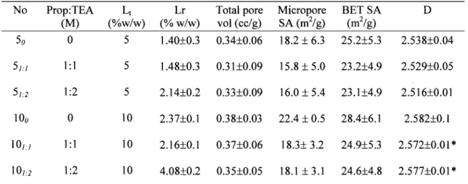
Summary of different formulations and their characterization ... 56 Porosity measurements for different formulations ... 58
Chapter 4: Drug Release Kineties and Microporous Structure of Polymerie
N anoparticles
Table 4.1. Polymer characterization by IH NMR and Size Exclusion Chromatography
Table 4.2. Table 4.3.
and DSC ... 79 Characterization of blank NPs ... 80 Characterization ofProp-loaded NPs ... 87
Chapter 5: Effect of Polymer Architecture on Surface Properties, Plasma Protein Adsorption and Cellular Interactions of Pegylated Nanoparticles
Table 5.1.
Table 5.2. Table 5.3. Table 5.4.
Polymer characterization by gel permeation chromatography, IH NMR and
DSC ... 111 Characterization ofNPs ... 112 Relative peak areas from XPS surface analysis ... 114 Relative percentage of monomer from area under the curves from respective Cls peaks ... 116
List of figures
Chapter 1 : Review of literature
Figure 1.1. IUP AC classification of adsorption isothenns ... 7 Figure 1.2. Mushroom and brush regime []Upper: Mushroom (low density.); Lower:
Figure 1.3.
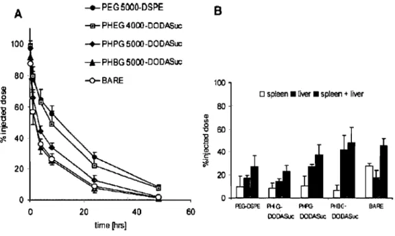
Brush (High density), AL: Area o(particle surface; D: Distance between grafting points; L: Length of the polymer segment (Np); Xp: mole fraction of polymer ... 17 Physicochemical properties and total prote in amounts adsorbed on lattices 1-5 expressed in arbitrary units (cpm). The values are the mean of three experiments; error bars represent the standard deviation [96] ... 18 Figure 1.4. Pharmacokinetics and distribution to the MPS of different poly(hydroxyalkyl L-glutamine)s incorporated with a grafting density of 7.5% in 150 nm DPPC-cholesterolliposomes. (A) %-injected dose in blood-curves of PEG2000-Distearoyl phosphatidylethanolamine (closed circles), PHEG (poly(hydroxyethyl L-glutamine))4000-DODASuc (gray squares), PHPG (poly(hydroxypropyl L-glutamine))5000-DODASuc (gray diamonds), PHBG poly(hydroxybutyl L-glutamine))5000-DODASuc (gray triangles), and bare liposomes without polymer-lipid conjugate (open circles). (B) Distribution to spleen (open bars), liver (gray bars) and total distribution to the MPS (liver and spleen) (black bars). Results are expressed as the mean percentage of the injected dose of four rats
±
SD. (Taken from Ref [90]) ... 19 Figure 1.5. EPR effect, taken from reference [110] ... : ... 21 Figure 1.6. Active targeting ... 21Chapter 3 : Effeet of Porosity on the Release Kineties of Propafenone-Loaded PEG-g-PLA Nanopartieles
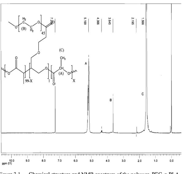
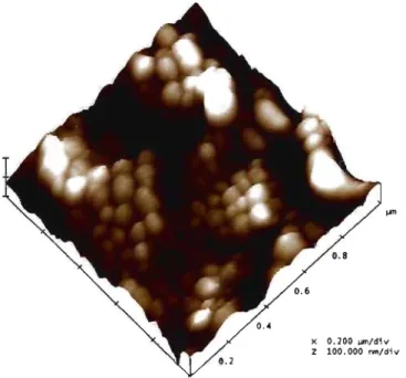
Figure 3.1. Chemical structure and NMR spectrum of the polymer, PEG-g-PLA ... 54 Figure 3.2. Surface morphology ofNPs (fonnulation 10u) by AFM ... 55
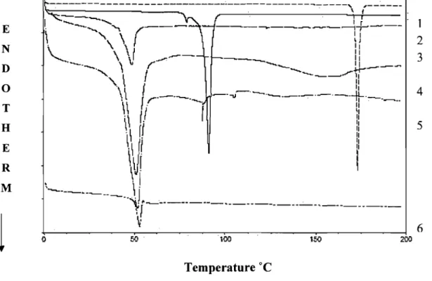
Figure 3.3. DSC curves of (1) Prop.HCI; (2) base (Prop); (3) Polymer; (4) Physical mixture of Prop.HCI: Polymer; (5) Physical mixture of Prop: Polymer and
Figure 3.4.
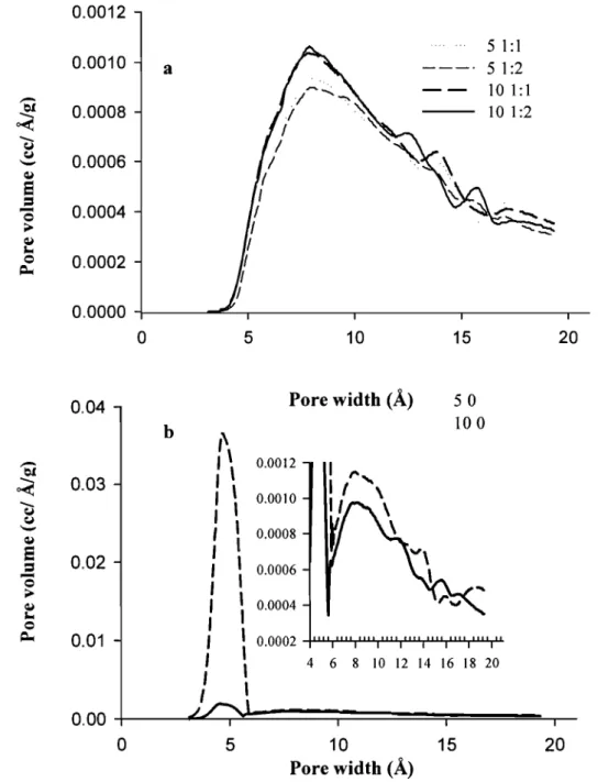
Figure 3.5.
(6) Drug loaded NPs (101:2) ... 57 Pore size distribution for various formulations with (a) or without (b) TEA. AlI values are mean ± S. D. for n= 3, Inset in fig b shows the pore size distribution on the same scale as Fig. 3.4 a ... 59 Effect of TEA (0, 1: 1, 1 :2) on the release profile of NPs at different drug loading levels, a; 10 % and b; 5 %. Error bars indicate mean ± S. D. for n= 3 ... 61 Figure 3.6. Effect of initial drug loading level on the release profile of NPs in the
presence (a, 1:2 M Prop: TEA) or absence (b) of TEA. Error bars indicate mean ± S. D. for n= 3 ... 63
Chapter 4: Drug Release Kinetics and Microporous Structure of Polymerie
N anoparticles
Figure 4.1. 1 H NMR spectra and chemical structures of multiblock copolymer, PEG1 %-g-PLA (m = 1) and PEGs%-%-g-PLA (m = 5) ... 78 Figure 4.2. DSC thermograms of 4 different polymers heated at the rate of 10°C/min
from -50 to 220°C, PLA (a), PEG1%-g-PLA (b), PEGs%-g-PLA (c), and (PLA-PEG-PLA)n (d) ... 80 Figure 4.3. Second run of DSC thermograms of blank NPs heated at the rate of
10°C/min from -50 to 70°C, PLA (a), PEG1%-g-PLA (b), PEG5%-g-PLA (c), and (PLA-PEG-PLA)n (d) ... 81 Figure 4.4. High resolution nitrogen adsorption isotherms of blank NPs of PEG1%-g-PLA (0), PEG1%-g-PLA (6), PEGs%-g-PLA (*) and (PLA-PEG-PLA)n (0). Inset shows magnified region between 0-5 cclg on y-axis to reveal the detailed
Figure 4.5.
adsorption behavior ofnitrogen in relative pressure range 10-6 to 10-1 ... 82 Effect of molecular structure of polymer on microporosity of blank NPs of PEG1%-g-PLA (0), PLA (6), PEGs%- g-PLA (*) and (PLA-PEG-PLA)n (0) ... 84
Figure 4.6.
Figure 4.7.
Schematic representation of arrangement of polymer chains inside NPs and mechanism of drug release; PLA (a), grafted polymers (b) and multiblock copolymer (c ) ... 85 Effect ofpolymer type on in vitro release profile ofProp-loaded NPs; values are represented as mean ± S. D. of 3 independent experiments. PEG1
%-g-PLA (0), (PLA-PEG-PLA)n (x) ... 89
Chapter 5: Effect of Polymer Architecture on Surface Properties, Plasma Protein Adsorption and Cellular Interactions ofPegylated Nanoparticles
Figure 5.1. Chemical structures ofmultiblock copolymer (a) and grafted copolymers (b) where m=l and 5 for PEG1%-g-PLA and PEGs%-g-PLA. ... 111
Figure 5.2. Tapping mode AFM phase images of NPs, aIl images are acquired in air. Scan size: 250 x 250 (nm x nm); PLA (a), PEG1%-g-PLA (b), PEGs%-g-PLA (c), PEGs%-g-PLA-PEG-PEGs%-g-PLA)n (d) ... 113 Figure 5.3. Plasma protein adsorption of different NPs by SDS-PAGE gel electrophoresis ... 117 Figure 5.4. Cytotoxicity ofNPs in RAW 264.7 cells by MTT assay. PLA (+); PEG1
%-g-PLA (.); PEGs%-g-%-g-PLA (Â); (%-g-PLA-PEG-%-g-PLA)n (x) ... 118 Figure 5.5. A. Effect of NP concentration on cellular uptake by RA W 264.7 cells. Cells were incubated with NPs at concentrations 40, 80, 120, 160 and 200 Ilg/mL at 37 oC for 3 h. Mean ± SD, n = 5; PLA (+); PEG1%-g-PLA (0);
PEGs%-g-PLA (Â); (PLA-PEG-PLA)n (x); B. Effect of temperature on cellular uptake ofNPS by RA W 264.7 cells. Cells were incubated with NPs at 37°C (open bar) and 4°C (filled bar) at a dose of20 Ilg/mL ... 121 Figure 5.6. Uptake of fluorescent NPs of different polymers by RA W 264.7 cells at
37°C in the presence of sodium azide (0.1% w/v) , hyperosmolar sucrose (0.45 M), Chlorpromazine (10 Ilg/mL), and Cytochalasin B (10 Ilg/mL) (B), Cells were pretreated with the inhibitors for 1 h followed by NPs (160 Ilg/mL) for another 3 h. Mean ± SD, n 2: 4; *: values significantly different atP>O.Ol ... 122
Chapter 6 : General Discussion
Figure 6.1. Microporosity determines the release; Formulations (51:2, 100) having same
real loading (% w/w), but different microporosity resulted in different release kinetics; higher the microporosity (l00), slower the release is ... 132 Figure 6.2. lH NMR of blank NPs of PLA, PEG1%-g-PLA, PEGs%-g-PLA, and
List of abbreviations
ADR AFM BET DCM DLS DSC EE GPC HKmethod MPS NK Np NPs O/W PCL PCS PE PEG PEG-g-PLA PEG\%-g-PLA PEGs%-g-PLA PLA PLGA PLLA (PLA-PEG-PLA)n Prop PSD PVA SDP AdriamycinAtomic force microscope Brunauer-Emmette-Teller Dichloromethane
Dynamic light scattering
DifferentiaI scanning calorimetry Encapsulation efficiency
Gel permeation chromatography Horvath-Kawazoe method Number average molecular weight
Mononuclear phagocytic system Weight average molecular weight N eimark -Kiselev
Degree of polymerization N anoparticles
oil in water
Poly( E-caprolactone)
Photon correlation spectrosopy Phosphatidylethanolamine
Polyethylene glycol
Poly( ethylene glycol)-graft-poly(D,L lactide) Poly( ethylene glycol)\ %-graft-poly(D,L lactide) Poly( ethylene glycol)s%-graft-poly(D,L lactide) Polylactide
Poly(lactide-co-glycolide) Poly(L-lactide)
(poly(D,L lactide )-block-poly( ethylene glycol)-block-poly(D,L lactide))n
Propafenone hydrochloride Pore size distibution Polyvinyl alcohol
SEC SEM TEA TEM Tg TM-AFM
Size exclusion chromatography Scanning electron microscope Triethylamine
Transmission electron microscopy Glass transition temperature
List of equations
Pln - = - x - - - x
+
Po RT cr4 (l-2do) 3(1-dol 9(1-dol 3(1- dol 9 (dol
... (1) do = ... (2) 2 cr
~ {~r
.do ...• ··· ... (3) 3 Ua + -Xa ... (4) Aa=
-
Ille c2 Ua Xa ... (5) 2 1 C-l P+
-
...
(6) W [P/Po - 1] WmC WmC Po , Q(t) = Ds X Eh X Ca X (2Co - ECa)t ... (7)Q(t)
=
V
DappCa X (2Co - ECa)t ... (8)MIj.
c.ute Uttte da(.l.ghter,. viVliiha for- her love
allW1 affec.twjlt"
MtJ
wollW1erfut hI-LSbaj/l.,d/ vi/l'vafjak for- the
, "
"
sirre~th,c,owf[.de,Vl,pt .tfV1-d emotl,OV1-tl
tsupport;'~~he gave me
A/l'vdmtJ pa re/l'vts' for thetr
btèsst~s,support:
>:
aM vaLues;
ofLifethe/j ta'1?iJht'me
,
,
,
,
,
,
,
,
xx
\\ My
Inspiration"
l
was there standing on the aisle
Looking beyond the horizon
The horizon where the Sun and the Moon meet
The sky and the ocean greet
Suddenly
lfelt weak,
l
saw a cloud that was dark, floating on my moon
It was hazy,
ltried
to
look beyond,
lcouldn't.
Foes disabled me, Friends asked me
to
be brave
l
thought,
Do
lhave courage account in the bank of bravery?
Or am
ltoo lazy
to
draw a chaque?
l
was still on the aisle,
The breeze whipsered,
Look at the cloud once again dear,
It has a silver lining,
It said, the night is adrker just before dawn
The dawn is breaking
l
am looking at the sun
l
hear the sun whispering
to
me
The only thing that makes you strong
Is seeing sombody like you
Achieving something great.
Then you know, how much is possible
&
you reach out further
Than you ever thought you cou Id.
You reach out for the Stars
&Sun;
Look at the sky
It says, don't look back.
l
give you my clouds,
The clouds give you the wings,
AI/ you have todo is
To Fly! Fly 1 Fly 1
... ..
... Author unknown
l
will love the light for it shows me the way
Yet,
lwill endure the darkness for it shows me the stars
... Author unknown
It is the hour of 'Trial'
That makes men Great
Not the hour of 'Triumph'
... Author unknown
Acknowledgements
It gives me great pleasure to present my Ph. D. thesis entitled "Polymerie
Nanoparticles: From Microporosity and Drug Release Kinetics to Cellular Interactions".
The respect and gratitude for my revered teacher and Director of research Dr.
Patrice Hildgen, Professor, Faculty of Pharmacy, University of Montreal cannot be
expressed in words. The complete autonomy given by him to plan the experiments along with his timely guidance and criticism in dijJicult limes has played a big role in shaping my approach towards the research. It is indeed my good fortune to have been able to work under the guidance of the teacher of his caliber. Also, 1 really value his deep involvement and unstintingfatherly support throughout the course ofmy doctoral studies wifhout which if would have been extremely dijJicult to endure the ups and downs in my research work along with my family responsibilities.
1 sincerely thank aU the co-authors of my articles, Matthias Thommes, Suzie
Poulin and Veronique Nadeau. Words fai! to express my deep sense of gratitude towards
Dr. Matthias Thommes, Director, Quantachrome Instruments, Florida, USA for his keen interest, active guidance and precious time in helping me to inter prete the porosimetry results. His priceless suggestions have been an extremely important factor in diffusing our microporosity results to the scientijic community. 1 wholeheartedly acknowledge Suzie Poulin, Research Associate, Ecole polytechnique, Montreal for her unbounded efforts and patience in interpretation of XPS data. Thank you Veronique for helping me in the synthesis of polymers.
My thanks are due to Natural Science and Engineering Research Co un cil
(NSERC) of Canada for the jinancial support through the award of Postgraduate
Scholarship for my Ph. D.
1 am Ihanliful 10 my jury members, Dr. Fahima Nekka, Assistant Professor,
Faculty of Pharmacy, University of Montreal; Dr. Suzanne Giasson, Assistant Professor, Faculty of Pharmacy, University of Montreal; and Dr. Abdel Omri, Associate Professor,
Department of Chemistry and Biochemistry, Laurentian University for accepling 10
1 express my deep sense of gratitude ta Dr. Jean-Cltristoplte Leroux, Professor, Faculty of Pltarmacy, university of Montreal for his valuable suggestions during my predoctoral examination and seminars. 1 also thank him for his wholehearted support including timely recommendations and utilization of facilities in his laboratory like nanosizer, zetasizer and macrophage cellline.
1 am grateful ta Dr. Sophie-Dorothée Clas, R & D department, Merck-Frosst, Canada for granting me permission ta perform difJerential scanning calorimetrie studies at Merck-Frosst. 1 am also thanliful ta Karine Khougaz and Rafik Naccache, Merck-Frosst, Canadafor their help in this work.
My heartfelt thanks are due ta Julie Boivin and Sylvain Essiembre, Research agents, Department of Chemistry, University of Montreal for their willingness ta help me in the interpretation of DSC studies.
1 am ever grateful ta Dr. Christian Pellerin, Professeur Adjoint, Department of Chemistry, University of Montreal for his valuable suggestions in designing DSC experiments on nanoparticles and analysis of data.
1 sincerely appreciate the valuable efforts, timely help and co-operation of Patricia Moraille, Research agent, Department of Chemistry, University of Montreal during AFM studies. 1 was fortunate ta have a friend as nice as her during these studies.
The help extended by Véronique Desjardin, Department of Chemistry, University of Montreal during NMR studies is also appreciated. My thanks are due ta Petra Pohankova, technician, Faculty for Pharmacy, University of Montreal for her help during my studies.
1 find myself lucky ta have a colleague and friend like Jean-Michel. 1 will never forget his ever willingness ta help, overwhelming support and valuable suggestions given ta me during the entire course of my Ph. D. 1 also thank the k.ind co-operation given ta me by my colleagues Névine, Véronique, Nicolas, Taha, Renu and Hamza during my work.
It is my pleasure ta thank Pierre Simard who extended helping hand ta teach me cell culture techniques. My sincere thanks are also due ta Marie-Andrée for ail the minute
details of Gel electrophoresis. 1 also appreciate the help provided by Marie-Christine during my Ph. D.
1 take this opportunity to thank Prashant Satturwar for ail his help and critical suggestions during this work. My heartfelt thanks are due to my close friend, Mohan for being always there in good and bad times lending his constant support and help.
Thankyou very much Dorothée and Sandra for your support and encouragement!
How can Iforget my closefriends, Hetal, Nisha, Sunila, Rupesh, Sonali, Madhuri,
Seema and other marathi community who were always there like my own family to
support me whenever 1 needed a helping hand?
1 express my deepest sense of gratitude towards my parents, my brothers; Vinod
and Sunil; and my sister Swati for their boundless love, affection and continuo us encouragement. 1 always missed them during these years, but at the same time, 1 always found them besides me and encouraging me to fight the tough moments in my life. The position achieved by me today is solely because of their blessings and constant support and
the values oflife they have taught me.
1 wholeheartedly express my gratitude towards my in-laws, Aai-Aba, Appa, Swati
vahini, Jayutai and Vivek bhau for their unstinting support, love and care. 1 consider myself lucky to have in-Iaws like them.
Last but not the least, thank you Vinisha, my Little adorable daughter, whose birth and growth progressed hand-in-hand with my Ph. D.! Her little gestures and big smiles have great power to revive me and it really made me forget ail the bad and tough times during the course ofthis work!
And now, the most important person in my life, my inspiration, my beloved
husband, Vinayak ... ... How do 1 thank you? 1 find ail the words inadequate for his thoughtfulness, constant moral support, helping hand at home, valuable suggestions and brainstorming discussions ... . He is everything to me, a very loving and caring husband, an excellent mentor, an excellent critic, and ab ove ail, a wonderful & perfect friend! 1 will never find enough words to express my feelings towards him, but sujjice to say
that this great moment of triumph would not have arrived without his support and inspiration.
Finally, l am thanliful ta God for giving me lot of patience and perseverance because ofwhich this thesis has become a reality!
Chapter 1.
1.1 Generaloverview
According to the report by the Royal Society of London in July 2004, nanotechnologies are the design, characterization, production and application of structures, devices and systems by controlling shape and size at nanometric scale [1]. Nanotechnologies are not new and have been studied for many decades. However, only in recent years, scientists have gained an in depth understanding of nanostructured substances by using sophisticated tools such as the atomic force microscope. It is reported from different sources that the global market of nanotechnology will expend very fast. This year, it should reach $1 trillion [2]. Its applications in pharmacology are included in this action, and the National Institutes of Health estimated that more than 50% of aIl biomedical advances will be in the nanotechnological sector by the year 2010 [2].
Nobel laureate and immunologist, Paul Ehrlich, proposed the concept of the so-caIled "magic buIlets" for the drugs that might be selectively directed to their site of action [3]. This dream is fast turning into reality, thanks to the recent milestones achieved in the field of 'polymer therapeutics' viz. polymeric drugs, polymer drug-conjugates and polymeric nanoparticles (NPs). This breakthrough was achieved by multidisciplinary approach encompassing physics, chemistry, cellular & molecular biology and biotechnology.
Various steps have been recognized in the development of successful drug delivery device; the first and foremost of which is the availability of well-characterized, biodegradable, biocompatible polymers with potential of controlled release and drug targeting to specific tissue/cells. Biodegradable polymers have had a remarkable impact in the field of controlled drug delivery. Over past two decades, Poly (L-lactide) (PLLA) and other copolymers based on D-lactide or glycolic acid or poly(s-caprolactone) (peL) or polyethylene glycol (PEG) have been extensively studied as controlled drug delivery carriers. Such carriers offer various advantages such as controlled drug release rate, improved therapeutic efficacy, prolonged biological activity and decreased administration frequency [4]. Block and graft copolymers ofpolylactide (PLA) and PEG have opened new avenues in the field of targeted drug delivery by prolonging the circulation time of the polymeric colloidal drug carriers in vivo. However, with in-depth understanding of
pathophysiology and cellular mechanisms of the disease, targeted or cell specific drug delivery is becoming the focus of the CUITent research. Presence of specific ligands on the surface of colloidal carriers is necessary to target specific cell type in the body (as in case of tumour cells). This can reduce systemic si de effects of the drug by improving receptor-mediated uptake by the targeted cells. For instance, targeted doxorubicin delivery could be achieved by folate conjugated mixed micelles of PLGA-b-PEG-folate polymer [5]. Thus, targeted drug delivery has generated a great need for biomaterials with bioadhesive and/or specific recognition properties. The ability to impart bioadhesivity, cell specificity or other specific characteristics to the existing biocompatible polymers represents an important synthetic challenge. Indeed, availability of functional pendant groups is highly desirable for fine-tuning of above mentioned properties. Various efforts are directed towards achieving this goal [6, 7]. However, chemistry involved in the synthesis of functional monomers is complex and/or tedious. For example, Bizzarri et al [6] have synthesized functionalized malolactonate polymers and copolymers, where the synthesis of monomers itself was long with low yields (12-45 %) and polymerization reactions required over 4-30 days. Similarly, Ouchi et al [8] have reported the synthesis of PLA-grafted polysaccharides; however, their method involved protection and deprotection. Thus, there are very few reports on the efficient and easy synthesis of functionalized polyesters. Amongst them, Finne et al [9] . have reported very efficient synthesis method for functionalized peL and PLLA with
controlled molecular weight and low polydispersity.
The next goal is formulation of drug delivery vehic1e like NPs, liposomes, micelles etc. providing protection to the encapsulated therapeutic agent and optimizing its formulation factors to obtain desired release kinetics. The recent advances in nanotechnologies, especially nanopartic1es (NPs) make them very promising in the drug delivery and diagnostics. NPs were first developed around 1970 and are defined as solid colloidal partic1es, less than 1 !lm in size [10]. They can be made from inorganic and polymeric materials, however, polymeric NPs are more desirable because they can be chemically designed to be degradable and biocompatible, the first and foremost requirement of any system for biological application. Despite of their own disadvantages such as low drug-Ioading capacity and wide size distribution [11], NPs offer numerous advantages over conventional dosage forms, including the ability to protect drugs from enzymatic degradation, target the drug to specific tissues, cells & cell compartments and
reduce the side-effects of chemotherapy. Rence, over past few decades, there has been considerable interest in developing biodegradable NPs as effective drug delivery devices [4, 12-16].
Various polymers have been investigated for preparation of NPs including polylactide (PLA) [17-20]; poly(lactide-co-glycolide) (PLGA)[II, 16, 21-27]; poly(c-caprolactone) (peL) [28,29] and pegylated polymers [29-40]. NPs have also been prepared using polyalkylcyanoacrylate polymers [41-45]. The drug of interest is dissolved, entrapped, adsorbed or attached to the nanoparticle matrix. Depending on the method of preparation, nanospheres or nanocapsules can be obtained with different properties and release characteristics. Nanocapsules are vesicular systems in which the drug is confined to a cavity surrounded by a unique polymeric membrane, whereas nanospheres are matrix systems in which the drug is physically and uniformly dispersed [33, 46-49]. A drug can be loaded into NPs by oil-in-water emulsion method if the drug is water insoluble [25, 50] or water-in-oil-in-water double emulsion [11, 19, 30, 50, 51] if the drug is water soluble. Other methods reported for preparation of NPs include nanoprecipitation [17, 52] and electrospraying [20]. Drug loading efficiency of NPs depends on the properties of the drug and polymer as weIl as formulation parameters.
1.2 Characterization of NPs
The therapeutic success of any smart drug delivery system requires it to i) be non-toxic, non-immunogenic and stable in the biological fluids, especially during circulation ii) protect the encapsulated moiety from premature degradation, iii) overcome biological barriers that it will encounter once administered in vivo, iv) be recognized efficiently and selectively by the target cells and tissues, v) be able to penetrate the target cell membranes and gain access to the intracellular structure and of course, vi) release the encapsulated therapeutic moiety in a controlled and/or sustained manner depending on the need of the application. In short, innovative and successful developments in the se drug delivery systems will require a multidisciplinary approach encompassing engineering, physical chemistry and biological sciences. Fine-tuning of various physicochemical properties is equally necessary to obtain biologically successful therapeutic system. Behavior of NPs within the biological microenvironment, stability, extracellular and cellular distribution
varies with their chemical makeup, morphology, and size. The following section gives the brief summary of these properties.
1.2.1 Size and size distribution
Most widely studied parameter in case of particulate carriers is the size. The size and size distribution are important parameters in determining their release kinetics as weIl as the interaction of NPs with the cells and penetration through biological barri ers such as gastrointestinal barrier for oral chemotherapy and blood brain barrier for the treatment of brain cancers or Alzheimer' s disease. It has been confirmed with repetitive studies that generally larger particles are rapidly cleared by mononuclear phagocytic system (MPS) than smaller particles [13, 53, 54]. Complement activation is also suggested to be dependent on the size [53, 54]. Even in case oflong-circulating colloidai particles, optimum size is proposed to be 150-200 nm [14, 55, 56].
1.2.2
Surface and bulk morphology
The surface and bulk morphology are important in determining the drug release kinetics from NPs. NPs are too small to be visualized under the light microscope. As a result of the rapid progress in high-resolution microscopies such as scanning electron microscopy (SEM), atomic force microscopy (AFM) , NPs can be visualized with a resolution of few nanometers. AFM provides a higher resolution than SEM. Both contact and non-contact modes can be employed. Tapping mode AFM (TM-AFM) is more preferred than the contact-mode and the non-contact mode because of its ability to probe soft sampI es such as biological and polymeric materials under ambient conditions [57, 58]. In tapping mode, the cantilever oscillates close to its bending mode resonance frequency so that tip makes contact with the sample only for a short duration in each oscillation cycle. As the tip approaches the sample, the tip-sample interactions alter the amplitude, resonance frequency, and the phase angle of oscillating cantilever. During scanning, the amplitude at the operating frequency is maintained at the constant level, called the set-point amplitude, by adjusting the relative position of the tip with respect to the sample. This operating method results in lower surface forces, particularly lateral forces, causing less surface
damage [57]. One recent approach in TM-AFM is the use of the changes in the phase angle of the cantilever probe to produce phase image. This image often provides more contrast than the topographic image and has been shown to be sensitive to material surface properties such as stiffness, viscoelasticity and chemical composition [57-60].
It is possible to observe the bulk morphology of NPs by cross-section; however, their small size makes this difficult. This may be the reason of lack of such investigations in the literature, except that reported by Rizkalla et al [61]. Using this technique, authors could successfully demonstrate co-existence nanocapsules having large central cavity along with nanospheres with solid core.
Another possible technique to study the internaI morphology of NPs is physical gas adsorption. The next section will give brief synopsis of basic principles of gas adsorption in general and summary of methods used for microporosity analysis in specific.
1.2.2.1
Physical Gas Adsorption: Basic Principles [62]
Gas adsorption is one of the many experimental techniques available for the surface and pore size characterization of porous materials. Other techniques include small angle X-ray and neutron scattering, mercury porosimetry, electron microscopy, thermoporometry and NMR-methods. Each method has limited length scale applicability for pore size analysis. Among these methods, gas adsorption is the most popular method because it allows assessment of wide range of pore sizes (from 3.5
A
up to > 1000A),
including the complete range of micropores (0 to 20 A), mesopores (20 to 500 A) and even macropores (500 to 5000A).
In addition, gas adsorption techniques are convenient to use and are not that co st intensive as compared to sorne of the other methods. Adsorption can be divided into chemical and physical adsorption, depending on the strength of the interaction. Physical adsorption is a general phenomenon and occurs whenever adsorbable gas is in contact with the surface of solid adsorbent. Physisorption exhibits characteristics which make it most suitable for surface area measurements as indicated1. Physisorption is accompanied by low heats of adsorption with no disruptive surface structural changes during the measurement.
2. Pores can be filled completely by the adsorptive gas for pore volume measurements. Such pore condensation phenomena can be used to calculate pore size and size distribution.
3. Physisorption is completely reversible, enabling both the adsorption and desorption to be studied.
4. Physically adsorbed molecules are not restricted to specific sites but, can coyer the entire surface, enabling calculation of surface areas rather than number of sites.
1.2.2.2
Classification of adsorption isotherms [62-64]
As per IUPAC classification [63], adsorption isotherms are divided into 6 types as shown below.
The reversible type 1 isotherm is concave to PlPo axis and amount approaches a limiting value as PlPo - 1. This type of isotherm is generally encountered in case of adsorption on microporous materials. Micropore filing and therefore, high uptakes are observed at re1atively low pressures. The strong increase in the adsorbed amount close to the saturation pressure results from the pore condensation into large meso- and macropores or interparticulate space.
Type Il isotherms are typically obtained in case of macroporous or non-porous adsorbent, where monolyer-multilayer adsorption can occur. The inflection point (B) indicates the stage at which mono layer is complete.
The reversible type III adsorption isotherm is convex to the PlPo axis and is very uncommon, but an example includes nitrogen adsorption on polyethylene.
Type IV isotherms are typical of mesoporous materials and exhibit characteristic hysteresis loop associated with the pore condensation.
Type V isotherms also show pore condensation and hysteresis phenomenon, however, initial part ofthis isotherm is related to the type III isotherm.
Type VI isotherm is a special case showing multilayer adsorption on a uniform, non-porous surface. 1
l
IiI
OC CI) .0...
0, ~"
CU...
C .:1 V 0E
il(Relattve pressure
,
Figure 1.1. IUP AC classification of adsorption isotherms
1.2.2.3
Adsorption in micropores
As defined earlier, pores are classified as macropores (pore widths greater than 500 Â), mesopores (pore widths from 20-500 Â) and micropores (pore widths less than 20 Â). For NPs of size 200 nm, micropores will be more important. The mechanism of pore filling is different in meso- and micropores. Mesopores fill via pore condensation, representing a first order gas-liquid phase transition. In contrast, the filling of micropores represents a continuous process, in most cases. The micropores are further subdivided into those smaller than 7 Â (ultramicropores) and those in the range of 7-20 Â (supermicropores). The filling of ultramicropores occurs at very low relative pressures and is entirely govemed by the
enhanced gas-solid interactions. The relative pressure where micropore filling occurs is dependent on a number of factors such as the size and nature of adsorptive gas molecules, the pore shape and the effective pore width. The pore filling capacity depends on the pore accessibility to the probe molecules which in tum, is detennined by the size of the probe molecule. In an ideal case, pure microporous materials exhibit type 1 isothenn as IUP AC classification. Various methods and theories have been developed for micropore characterization, which include Polyani, Dubinin including most recent approaches by Horvath-Kawazoe [65, 66] known as HK method. It should be noted that this method cannot be applied to mesopore size analysis. The following equation is used for micropore analysis using HK method:
{
P}
NA NsAs + NaAa cr4 cr 10 cr4 crlOln Po = RT x cr4
(l-2do) x 3(1- dol 9(1- dol 3(1- dol + 9(dol
... (1) Where, da+ ds do = ... (2) 2 cr
{~r
.do ... (3) As=
...
(4) 3 Aa= - mec 2 Ua b ... ... (5) ' 2Aa
=
Kirkwood-Mueller constant of adsorbate As=
Kirkwood-Mueller constant of adsorbentdo
=
distance between adsorptive and adsorbent molecules da=
diameter of an adsorbate moleculeds
=
diameter of an adsorbent moleculeNA= nurnber of adsorptive molecules per unit area (m2) of adsorbent
me
=
mass of an electron c = speed of lightUa = polarizability of adsorbate
Us = polarizability of adsorbent
cr = distance between two molecules at zero interaction energy
'X;t = magnetic susceptibility of adsorbate 'XJ; = magnetic susceptibility of adsorbent
1 = the separation between nuclei oftwo layers
According to this equation, the filling of micropores of a given size and shape takes place at a characteristic relative pressure.
1.2.2.4 Specifie surface area
Gas adsorption technique also serves in the analysis of specific surface area of the sample. Specific surface area depends on size, shape and porosity. The BET-nitrogen adsorption method continues to be a universally employed method for detennining the surface area of adsorbents, catalysts and other materials [67]. It is based on a simple model of monolayer-multilayer adsorption and given by the following equation:
1 1
+t::~}
:0 ...
(6) W [PlPo - 1]Where,
W
=
weight of the adsorbed moleculesW m =: weight of the adsorbed molecules at mono layer coverage C
=
BET constantP6
=
saturation pressureA plot of 1/ W [PlPo - 1] versus PlPo gives a straight line in the range of 0.05 ::; PlPo ::;0.35.
1.2.2.5 Nitrogen as a standard adsorptive for surface area
measurements [62]
The unique properties and the availability of liquid nitrogen have led to the univers al acceptance of nitrogen as the standard BET adsorptive. The fact that nitrogen has a permanent quadrupole moment is important, because it is responsible for the formation of a well-defined monolayer on most of the surfaces. However, quadrupole moment of nitrogen affects its adsorption on highly polar surfaces due to specific interactions between these polar groups and its quadrupole moment. Thus, nitrogen in not completely inert and this may result in overestimation ofBET surface area in case of polar surfaces.
In contrast to nitrogen, argon has no quadrupole moment. Argon is much more sensitive to the details ofthe surface structure ifused at liquid nitrogen temperature (77 K).
For accurate measurement of surface areas as low as 0.5 - 1 m2, number of molecules trapped in the void volume of the sample ceIl needs to be reduced. This can be achieved by using krypton adsorption at liquid nitrogen temperature for the surface area analysis. Krypton has a much smaller saturation pressure (1.63 Torrs i.e. 217.25 Pa) compared to nitrogen (270 torrs) at 77.35 K and hence, the amount of krypton remaining in the void volume will be much less as compared to that of nitrogen. This increases the sensitivity of the manometric adsorption measurements significantly thereby, allowing accurate surface area measurements for materials with small specific surface areas
«
0.05 m2/g), or if only a very small amount of sample is available.1.2.3 Physical state of drug within NPs
It is important to know whether the drug is present in crystalline or amorphous form within the matrix. This provides certain information about the rate of drug release. The first step in the release requires dissolution of drug in the release medium followed by its diffusion. Drug dispersed in the matrix in crystallihe form will dissolve slowly than its amorphous or molecularly dispersed form affecting the release kinetics. DifferentiaI scanning calorimetry (DSC) can be used to gain information about physical state of the drug as well as polymer-drug interaction. For instance, Mu et al [68] have detected no melting endotherm of paclitaxel in PLGA nanospheres, attributing it to the molecular dispersion of pac1itaxeJ in the poJymeric matrix. On the other hand, Okada et
aJ
[69J haveshown an increase in the glass transition temperature (Tg) of leuprorelin-Ioaded PLA microspheres due to ionic interaction between the drug and the polymer.
1.2.4 In vitro
drug release kinetics
In vitro release studies are important in the characterization of any drug de li very system. Although in vivo release could be different, the in vitro release experiments provide sorne guidance for prediction of possible effects of the drug delivery system. It also gives an insight to the possible burst effect, which should be avoided to prevent toxic concentration in the body. It is weIl established that drug release from polymeric matrices is govemed by both diffusion and degradation depending on the polymer type [70]. This release phenomenon is a complex interplay ofvarious factors such as properties of the drug and polymer, their compatibility with each other, type Qf emulsifier used, drug loading level, physical state of the drug inside the matrix and various other formulation parameters.
The Higuchi square root model has been successfully applied to model the kinetics of drug release from matrix system. Equation 9 was derived from Fick's first law of diffusion and applied to porous hydrophobic polymeric drug delivery systems in homogenous matrices [71].
Q(t) =
J
Ds x fIt x Ca x (2Co - f:Ca)t.. ... (7)=
V
DappCa x (2Co - f:Ca)t ... (8) = kf t ...
(9)Where,
Q(t) :Cumulative amount of drug released in time t per unit surface area Ds : Drug diffusion coefficient in the release medium
Co : Total amount of drug in the matrix Ca: Solubility of drug in the release medium f: : Porosity of the matrix
Dapp = DsEh : Apparent observed diffusion coefficient
k : Dissolution rate constant
Thus, drug release from such a system will depend on the initial concentration of the drug in the matrix, porosity, tortuosity, properties of the polymer system forming matrix and the solubility of the drug. Drug release from hydrophobic polymeric drug delivery system will occur when the drug cornes into contact with the release media, subsequently dissolves and diffuses through the media filled pores. Thus, geometry and structure of pores is important in this process.
1.2.5
Surface charge
Surface charge is important in determining the in vivo stability as weIl as cellular interaction capability of NPs. Zeta potential measurements can give idea of surface charge of particles. Generally, surface charge should be near neutral to avoid non-specific cell sticking or uptake of NPs. This is discussed along with the properties of long-circulating polymers.
1.2.6
Surface chemistry
Once the release properties are tailor-made for particular application, the next milestone to be achieved in the development of drug delivery device is suitability of the surface properties of such polymeric colloidal carrier in terms of its in vivo stability, compatibility and cellular interaction capabilities. Physicochemical surface characteristics also give clear evidence of the effect of coatings and residual emulsifiers on the NP surface. The amount of emulsifier on the surface has significant effect on various physicochemical and biological properties of the NPs. Polyvinyl alcohol (PV A) is the most commonly used emulsifier in the formulation of polymeric NPs. It has been proven that a fraction of PV A remains associated with the NPs despite repeated washing and has been proposed that this is due to formation of an interconnected network with the pol ymer through hydrophobic interactions at the interface. Sahoo et al [23] have systematically shown that this residual PV A, in tum, affected different pharmaceutical properties of NPs such as particle size, zeta potential, polydispersity index, surface hydrophobicity, protein loading and also slightly influenced in vitro release of the encapsulated protein. Importantly, NPs with higher
amount of residual PV A had relatively lower cellular uptake despite their smaller particle size. It was proposed that lower intracellular uptake of NPs with higher amount of residual PV A could be related to higher hydrophilicity of NP surface.
Various efforts have been directed to create particles with precise surface chemistry, especially for their long-circulating properties. Their summary and properties required for long-circulating polymers/carriers will be described in the next section.
1.3 Optimization of surface chemistry of NPs: Essence
for their long-circulating behavior and biological
activity
Amongst various physicochemical properties, surface properties of any system are thought to play a crucial role in interactions with the cells and their intracellular transport machinery, and can significantly affect the efficacy of the encapsulated drugs [72]. Hence, it is critical in targeted gene and drug delivery to carefully design and control the surface properties of such systems. A prerequisite to selective targeting is particle stability in vivo and the minimization of nonspecific uptake. This can be done by designing surfaces with no or little protein adsorption. In particular, the adsorption of biological proteins onto
biomaterials plays an important role in determining their fate. Hence, elimination of prote in adsorption onto biomaterials is one of the key elements in preventing thrombosis, platelet activation and immunological responses [73]. Similarly, this phenomenon of adsorption of proteins and opsonins on polymeric drug delivery systems leads to their recognition and rapid elimination from systemic circulation by Mononuclear Phagocytic System (MPS). To avoid this, the concept of long-circulating polymers has been emerged [12, 30, 74]. In the past two decades, we have witnessed a surge in the development of long circulating vehicles. Among the various polymers studied, poly (ethylene glycol) (PEG) has been the most extensively investigated pol ymer. PEG is an uncharged hydrophilic and non-immunogenic polymer that can be physically adsorbed onto or preferably, covalently attached to the surface of colloidal systems [75, 76]. Various pioneering studies and
especially, the entry of pegylated or so-called "sterically stabilized" liposomal doxorubicin (Doxil™, CaelyxTM) into the market signify the importance of the stealth technology.
1.3.1
Approaches used for prolonged circulation
Since the past two decades, various approaches have been used for imparting the stealth behavior to the polymerie carriers, which include:
• Adsorption of hydrophilic polymers containing hydrophobie substituents onto the surface via hydrophobie interactions, e. g. surfactants such as poloxamers, poloxamines [12, 74].
• Covalent bonding of hydrophilic polymers onto the surface e.g. surface-grafted polymers or use of PEGylated copolymers with sorne other biodegradable polymer such as PLA, PLGA, PCL [16, 30, 31, 33, 34,39,47, 77-83].
It is proposed that in both these cases, the hydrophilic parts protrude out in aqueous milieu forming "molecular cloud" on the surface of the carrier, thus protecting the carrier from recognition by MPS [13, 46, 76]. However, in the first approach, possibility of rapid desorption of the adsorbed coat may deceive the true purpose, as observed in case of NPs [74]. Covalently linking PEG to the surface of the polymer has sorne disadvantages as weIl. It is sometimes difficult to ensure that covalently bound PEG cornes out on the surface and does not penetrate in the bulk of the material. This will affect PEG surface coverage and conformation. At the same time, presence of covalently bound PEG chains throughout the particle may be advantageous for degradable particles, ensuring availability of surface exposed PEG during the entire degradation and erosion process. Hence, copolymers of PEG with other polymers were used; nevertheless, this does not assure presence of aH PEG chains on the surface of particles prepared from these polymers.
1.3.2
Physicochemical properties of long-circulating
.
carrIers
Several theories have been proposed to explain the apparent protein binding and long-circulating properties of pegylated carriers. Sorne authors correlated long-circulating
![Figure 1.2. Mushroom and brush regime []Upper: Mushroom (low density.); Lower:](https://thumb-eu.123doks.com/thumbv2/123doknet/12131913.310226/46.916.282.709.88.503/figure-mushroom-brush-regime-upper-mushroom-density-lower.webp)
![Figure 1.5. EPR effect, taken from reference [110]](https://thumb-eu.123doks.com/thumbv2/123doknet/12131913.310226/50.915.184.821.128.337/figure-epr-effect-taken-reference.webp)