C
OSTIMULATORY
M
OLECULE
R
EQUIREMENT FOR
B
OVINE
WC1
+Ɣ𝛿
T
C
ELLS
'
P
ROLIFERATIVE
R
ESPONSE TO
B
ACTERIAL
S
UPERANTIGENS
Auteurs
Y. Fikri,* P.-P. Pastoret & J. Nyabenda
*Unit of Cellular Immunology, Pasteur Institute Brussels, Rue Engeland, Brussels; and Department of Immunology and Vaccinology, Faculty of Veterinary Medicine, University of Liège, Liège, Belgium
ABSTRACT
We have previously shown that the proliferation of freshly isolated bovine WC1+ Ɣ𝛿 T cells to superantigens (SAgs) including staphylococcal enterotoxin A (SEA), and staphylococcal enterotoxin B (SEB) or toxic shock syndrome type-1 (TSST-1) required the presence of antigen-presenting cells (APC) and the addition of exogenous interleukin (IL)-2. The costimulatory activity provided by molecules expressed on professional APC for the proliferation of Ɣ𝛿 T cells has not been addressed hitherto. In the present study, we investigated the ability of two selected APC populations, the dendritic cells (DCs) highly expressing CD80 and CD86 molecules (CD80highCD86high) and the monocytes expressing the same molecules at a rather low level (CD80lowCD86low), to stimulate the proliferation of purified bovine WC1+ Ɣ𝛿 T cells to SAgs. DCs were more efficient than monocytes in inducing Ɣ𝛿 T-cell proliferation, and this response was dependent on exogenous IL-2 in both presentation modes. Stimulating Ɣ𝛿 T cells with gradual doses of SAgs or concanavalin A (ConA) resulted in similar dose-dependent reaction profiles suggesting a minimal role of the major histocompatibility complex (MHC). However, significant proliferation was already obtained with the starting doses in the presence of DC compared with monocytes, and higher proliferation was reached with DC at optimal doses. Finally, the addition of monoclonal antibody (MoAb) anti-CD86 markedly inhibited SAgs- and ConA-mediated proliferation, whereas MoAb anti-CD80 had no effect. The combination of both anti-CD80 and anti-CD86, however, suppressed this response. These results suggest that bovine Ɣ𝛿 T-cell proliferation response requires indubitably CD86 costimulation. The role of CD80 molecule seems less clear.
Introduction
T-cell activation and proliferation require two signals. One signal delivered via the T-cell receptor (TCR) is major histocompatibility complex (MHC)-restricted and antigen-specific. The other signal is delivered by costimulatory molecules of professional antigen-presenting cells (APC) and is nonspecific [1-3]. Two structurally related molecules expressed on the surface of T cells, CD28 and cytotoxic T-lymphocyte-associated molecule (CTLA-4), and their coreceptors CD80 and CD86 expressed on a variety of professional APC including dendritic cells (DCs), activated macrophages and activated B cells [4,5], are the best-characterized costimulatory molecules identified to date [5-7]. CD28 and CTLA-4 costimulatory molecules have also been detected on bovine [8] and ovine [9] Ɣ𝛿 T cells. However, few studies have provided information concerning the costimulatory activity required for the activation of ruminant Ɣ𝛿 T cells.
Most of the circulating bovine and ovine Ɣ𝛿 T cells express a unique family of high molecular weight surface molecules, termed workshop cluster 1 (WC1) [10-12]. These cells constitute up to 30-50% in the peripheral blood in Ɣ𝛿ung ruminants [13,14], and are further characterized by a CD2-, CD4-, CD8-, CD3+ and TCR Ɣ𝛿+ phenotype [10,12,15].
The absolute requirement of APC and exogenous interleukin (IL)-2 for the proliferation of purified Ɣ𝛿 T cells in vitro upon a stimulation with mitogens [16], specific antigen [17] and recently with superantigens (SAgs) has been repeatedly reported [18-20].
In order to assess the role of the costimulation provided by molecules expressed on APC, we analysed the proliferation of purified Ɣ𝛿 T cells to SAgs in the presence of two professional APC populations expressing different levels of CD80 and CD86 molecules: the DC and the monocytes. The respective role of either ligand was studied using blocking antibodies to CD80 and CD86.
Materials and methods
Reagents. The culture medium contained RPMI 1640, 2 mM L-glutamine, 25 mM HEPES buffer, 100 IU/ml penicillin, 100 µg/ml streptomycin, 0.5 µg/ml fungizone and 10% foetal calf serum (FCS) (Gibco BRL, Grand Island, NY, USA). The murine monoclonal antibodies (MoAbs) CC8 (immunoglobulin, IgG2a), CC63 (IgG2a), CC15 (IgG2a), CC-G33 (IgG1), ILA21 (IgG2a) and ILA30 (IgG1)
specific for CD4, CD8, WC1, CD14, MHC-II and IgM, respectively, were purchased from SeroTec Immunological (Oxford, UK). The murine MoAbs BB1 (IgM) and IT2.2 (IgG2b) specific for human CD80
and CD86, respectively, were purchased from Pharmingen (San Diego, CA, USA). Isotype-matched mouse IgM (M 5909), mouse IgG2b (M 5534) and mouse IgG2a (M5409) were from Sigma
Immunochemicals (St. Louis, MO, USA). Staphylococcal enterotoxin A (SEA), staphylococcal enterotoxin B (SEB), toxic shock syndrome type-1 (TSST-1), concanavalin A (ConA), calcium ionophore A and phorbol myristate acetate (PMA) were from Sigma Immunochemicals.
Recombinant human IL-2 was from Boehringer (Engelheim, Germany). Lymphoprep and Nycodenz gradients (d = 1.068) were from Nycomed Pharma (Oslo, Norway). Calcium ionophore was dissolved in dimethyl sulphoxide (DMSO) and PMA in anhydrous ethanol at a concentration of 1 mg/ml and 10 µg/ml, respectively.
Purification of bovine WC1+ Ɣ𝛿 T cells. WC1+ Ɣ𝛿 T cells were negatively purified from peripheral
blood as previously described. Using Lymphoprep, peripheral blood mononuclear cells (PBMCs) were isolated from heparinized peripheral venous blood from 6- to 12-month-old cattle. PBMCs at 5 x 10⁶ cells/ml were resuspended in culture medium and allowed to adhere for 60 min to plasma-coated gelatin. Nonadherent cells were enriched in T cells by passing the cells through a nylon wool column to remove B cells and remaining monocytes. CD2+ T cells were depleted by rosetting
T cells with 2-aminoethylisothiouronium bromide (AET)-treated sheep red blood cells (SRBC). WC1+
Ɣ𝛿 T cells were further separated from CD4+ and CD8+ T cells using the magnetic cell sorting (MACS) technique. The resultant population contained up to 97% of WC1+ Ɣ𝛿 T cells as stated by
the fluorescence-activated cell sorter (FACS) phenotypic characterization of cells.
Isolation of bovine DC and monocytes. DC (CD14-MHC-IIhigh) and monocytes (CD14+) were purified
as described by Renjifo et al. PBMCs at 5 x 10⁶ cells/ml were allowed to adhere for 2 h to
plasma-coated gelatin. Nonadherent cells were removed by washing three times with Hank's balanced salt solution (HBSS). Cells adhered to the gelatin were detached by incubating for 5 min with prewarmed HBSS containing 0.2% ethylenediaminetetraacetic acid (EDTA), washed three times, and cultured overnight. Plastic adherent cells were monocytes, while the supernatant contained nonadherent cells rich in DCs. DCs were further enriched by centrifugation in Nycodenz gradients (d = 1.068), washed three times, and separated from CD14+ and IgM+ cells using the MACS
technique.
Flow cytometry analysis. Cells, 106 per test, were washed twice with ice-cold phosphate-buffered
saline (PBS) containing 1% bovine serum albumin (BSA), incubated on ice for 30 min with an appropriate dilution of MoAb, washed in PBS, and further incubated for 30 min on ice with fluorescein isothiocyanate (FITC)-labelled MoAb specific for kappa chain as the second reagent. After washing, phenotypic analysis was performed on FACS flow cytometer (FACScalibus; Becton-Dickinson, Mountain View, CA, USA).
Proliferation assays. PBMCs (2 x 10⁵ cells in 0.2 ml) were stimulated in triplicate with ConA (1
µg/ml), SEA (103 ng/ml), and PMA and/or ionomycine (40 ng/ml) for 4 days. Purified WC1+ Ɣ𝛿 T cells
(2 x 10⁵ cells) and autologous Ɣ-irradiated APC (5000 Rad of gamma-irradiation from a 137Cs source)
were cultured in 0.2 ml in triplicate with SEA, SEB, TSST-1 or ConA for 4 days. When indicated, the recombinant human IL-2 was added at a final concentration of 50 units/ml. The cultures were pulsed for the final 18 h with 1 µCi of [3H]-thymidine, harvested and the incorporated radioactivity determined by liquid scintillation counting. To assess the effect of anti- CD80 and anti-CD86 on Ɣ𝛿 T-cell proliferation, MoAbs were added to DC for 30 min before the beginning of the culture period at a predetermined dose (1 µg/ml).
Statistical analysis. The results were expressed as the mean ± standard deviation (SD) and compared using a Student's t-test. The significance level was set at P < 0.05.
Results
FLOW CYTOMETRY ANALYSIS OF ENRICHED DC AND MONOCYTES
Monocytes and DCs are characterized by a high CD14+ expression and a lack of CD14+ expression,
respectively, and by MHC-II levels, high in DC and low in monocytes (Fig. 1). Flow cytometry analysis after purification shows 89% of the cells expressing CD14+MHC-IIlow, representing
monocytes (Fig. 1A,C), and 85% of the cells, CD14- MHC-IIhigh , being DC (Fig. 1B,C). DCs also express
CD80 and CD86 molecules at a higher level than the monocytes (Fig. 1D,E). Enriched DC contained <1% monocytes, <1% B cells, and T cells including ͠ 4% WC1+, ͠ 5% CD4+ and ͠ 3% CD8+ (FACS not
shown).
DC AND MONOCYTE ANTIGEN PRESENTATION NEEDS THE ADDITION OF
EXOGENOUS IL-2 FOR THE PROLIFERATION OF WC1
+Ɣ𝛿 Ɣ𝛿 T CELLS TO SAGS
AND CONA
Isolated WC1+ Ɣ𝛿 T cells cultured with or without autologous Ɣ-irradiated DC or monocytes were pulsed in various combinations with SAgs (SEA, SEB or TSST-1), ConA and IL-2 (Table 1). The Ɣ𝛿 T cells responded to all SAgs and ConAs in the presence of DC or monocytes and IL-2, but failed to respond in the presence of either stimulus and/or IL-2 in the absence of APC. The amplitude of the responses to similar stimuli was significantly higher with DC than monocytes (P < 0.01).
DC INDUCED HIGH PROLIFERATIVE RESPONSE OF WC1
+Ɣ𝛿 T CELLS TO SAGS
AND CONA
We next compared the ability of DC and monocytes to induce Ɣ𝛿 T-cell response in vitro. The proliferation of IL-2- dependent WC1+ Ɣ𝛿 T cells to SAgs or ConA in the presence of DCs or
monocytes is depicted in Fig. 2. The response of purified Ɣ𝛿 T cells to SAgs and ConA was observed to decrease with the decreasing APC populations. In the presence of DC, wells containing 2 x 10⁴
DCs, at a ratio of 1 : 10 Ɣ𝛿 T cells, induced ͠ 43 x 103, ͠30 x 103, ͠ 31 x 103 and ͠ 27 x 103 counts per minute
(cpm) for ConA, SEA, SEB and TSST-1, respectively. Monocytes at the same ratio induced ͠ 22 x 103, ͠
11 x 103, ͠ 12 x 103 and ͠ 9 x 10³ cpm for ConA, SEA, SEB and TSST-1, respectively. At the ratio of 1 DC
(2 x 10³): 100 Ɣ𝛿 T cells, the magnitude of the proliferation was ͠ 21 x 103, ͠ 15 x 103, ͠ 16 x 103 and ͠ 13 x
103 cpm for ConA, SEA, SEB and TSST-1, respectively, in the presence of DC, whereas proliferation
Figure 1.
Légende de la figure. Flow cytometry analysis of bovine dendritic cells (DCs) and monocytes. Monocytes (A) and DCs (B) were stained with CC-G33 monoclonal antibody (MoAb) specific for CD14 and fluorescein isothiocyanate (FITC)-labelled MoAb specific for kappa chain as secondary reagent. The indicated antigen-presenting cell (APC) populations were stained with ILA21 (C), BB1 (D) and IT2.2 (E) MoAb specific for major histocompatibility complex (MHC)-II, CD80 and CD86, respectively, and FITC-labelled MoAb specific for kappa chain. Negative controls consisting of APC populations were stained with FITC-labelled MoAb specific for kappa chain alone. (C-E) Controls staining for both APC (DC and monocytes) did not vary significantly. All stainings were performed in the presence of 10% fœtal bovine serum (FBS) in order to inhibit Fc receptor (FcR) binding of staining reagents.
DOSE-DEPENDENT PROLIFERATIVE RESPONSE OF PURIFIED WC1
+Ɣ𝛿 T CELLS
TO SAGS AND T-CELL MITOGENS IN THE PRESENCE OF DC OR MONOCYTES
The proliferation of purified WC1+ Ɣ𝛿 T cells stimulated with increasing doses of SAgs and ConA in the presence of constant number of DC or monocytes and IL-2 is shown in Fig. 3. The Ɣ𝛿 T cells responded to minute amounts of SAgs and ConA in the presence of DC, while monocytes required high doses of stimulus to induce detectable levels of Ɣ𝛿 T-cell response. At the same antigen or mitogen doses, Ɣ𝛿 T-cell proliferation was consistently of higher magnitude with DC in comparison with monocytes. Control Ɣ𝛿 T-cell cultures with increasing doses of stimulus and IL-2 without APC did not proliferate.
Table 1. The proliferative response of WC1+ Ɣ𝛿 T cells to superantigens (SAgs) and concanavalin A (ConA) in the presence of professional antigen-presenting cell (APC) populations*
Légende de la figure. *Purified WC1+ Ɣ𝛿 T cells (2 x 10⁵/well) were cultured in triplicate in the presence or absence of
Ɣ-irradiated autologous APC (2 x 10⁴ cells/ well) and stimulus, and the proliferation was assessed by [3H]-thymidine
incorporation during the last 18 h of 4 days of culture. The results represent the mean counts per minute (cpm) ± standard deviation (SD) of three independent experiments from three animals. ●Interleukin (IL)-2 was added at a final concentration of 50 units/ml, SAg at 103 ng/ml and ConA at 10 µg/ml. △P < 0.01 (significant difference between DC and monocytes).
Figure 2.
Légende de la figure. Proliferative response of WC1+ Ɣ𝛿 T cells to superantigens (SAgs) and concanavalin A (ConA) in the
presence of decreasing number of professional antigen-presenting cells (APC). WC1+ Ɣ𝛿 T cells (2 x 10⁵/well) were cultured in
triplicate with decreasing number of autologous Ɣ-irradiated dendritic cells (DCs) or monocytes in the presence of staphylococcal enterotoxin A (SEA), staphylococcal enterotoxin B (SEB) or toxic shock syndrome type-1 (TSST-1) (103 ng/ml) or
ConA (10 µg/ml), and interleukin (IL)-2 (50 U/ml). Proliferation was assessed by thymidine incorporation during the last 18 h of 4 days of culture. The results are expressed as counts per minute (cpm), and each point represents the mean ± standard deviation (SD) from three independent experiments.
BLOCKING OF BOVINE DC COSTIMULATORY MOLECULES WITH HUMAN MOABS
ANTI-CD80 AND ANTI-CD86
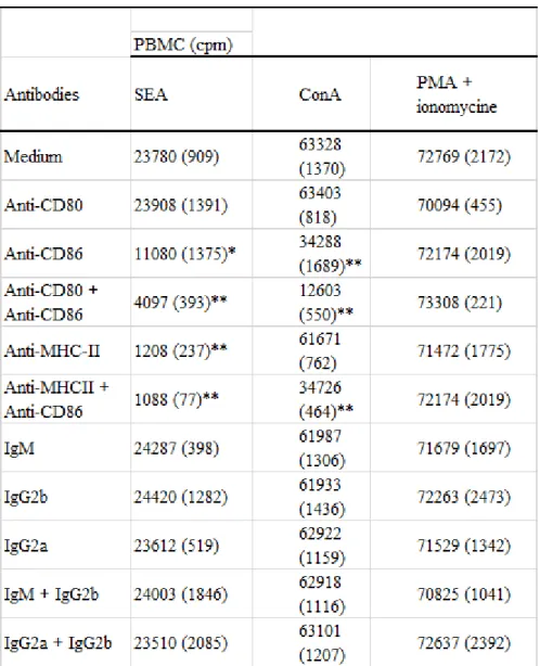
Preliminary experiments were devised in order to establish the activity of MoAbs CD80, anti-CD86 and anti-MHC-II in the inhibition of bovine T-cell proliferation in vitro. PBMCs were stimulated with SAgs or T-cell mitogens (ConA, PMA and/or mitomycine) in the presence of antibodies to CD80, CD86, MHC-II, combination of CD80 and CD86, or combination of CD86 and MHC-II. Isotype-matched mouse IgM, mouse IgG2a and mouse IgG2b were used as controls. Table 2 shows that the anti-CD86 but not anti-CD80 markedly inhibited SEA-mediated T-cell proliferation (P < 0.05), the combination of both anti-CD80 and anti-CD86 resulting in more significant inhibition (P < 0.01). Anti-MHC-II alone or in combination with anti-CD86 suppresses completely the proliferation (P < 0.01). The addition of anti-CD86 alone significantly inhibits ConA-mediated T-cell proliferation (P < 0.01), while anti-CD80 alone had no effect. A combination of both anti-CD80 and anti-CD86 strongly inhibited the proliferation induced by ConA (P < 0.01). In contrast to SAg, ConA-mediated T-cell response was not inhibited by the addition of anti-MHC-II. All combinations of isotype-matched controls remained ineffective. The specificity of the anti-CD80, anti-CD86 and anti-MHC-II-mediated inhibition was further verified by analysis of the blocking properties of the MoAbs in the costimulatory-independent response. As shown in Table 2, the proliferation response induced by PMA + ionomycine was not inhibited by MoAbs to CD80, CD86, MHC-II, the combination of CD80 and CD86 or the combination of CD86 and MHC-II. PMA or ionomycine alone did not induce the proliferation of PBMC (not shown).
To study the role of the DC costimulatory molecules on IL-2-dependent Ɣ𝛿 T-cell response, purified cells were cultured with SAgs SEA (Fig. 4A), SEB (Fig. 4B) or TSST-1 (Fig. 4C),
or with ConA (Fig. 4D), in the presence of anti-CD80 and/or anti-CD86 antibodies. Anti-CD86, but not anti-CD80, significantly inhibited the proliferation of WC1+ Ɣ𝛿 T cells in response to all SAgs tested and to ConA. The combination of both MoAbs almost completely suppresses the proliferative response. All isotype-matched MoAb controls remained ineffective.
Figure 3.
Légende de la figure. Proliferative response of WC1+ Ɣ𝛿 T cells to gradual doses of superantigens (SAgs) and concanavalin
A (ConA) in the presence of dendritic cells (DCs) or monocytes. WC1+ Ɣ𝛿 T cells (2 x 10⁵/well) were cultured in triplicate
with or without autologous Ɣ-irradiated DC or monocytes (2 x 10⁴ cells/well), in the presence of gradual doses of
staphylococcal enterotoxin A (SEA) (A), staphylococcal enterotoxin B (SEB) (B), toxic shock syndrome type-1 (TSST-1) (C) or ConA (D) with or without interleukin (IL)-2 addition (50 U/ml). Proliferation was assessed by thymidine incorporation during the last 18 h of 4 days of culture. The results are expressed as counts per minute (cpm), and each point represents the mean ± standard deviation (SD) from three independent experiments.
Discussion
The costimulation requirement for bovine Ɣ𝛿 T-cell proliferative response constitutes the aim of this study. We previously reported that the proliferation of purified bovine WC1+ Ɣ𝛿 T cells, in
response to bacterial SAgs (SEA, SEB and TSST-1) and T-cell mitogen (ConA), required the presence of APC and the addition of exogenous IL-2 [18]. The same observations have also been recorded with specific antigen [17]. To investigate how accessory cells mediate the proliferation of Ɣ𝛿 cells, we compared the efficiency of DC and monocytes to induce this response. DCs are APC with a strong capacity to stimulate T lymphocytes, owing to their high density of MHC-II and costimulatory molecules. Monocytes are also APC-identified by the low density of both MHC-II and costimulatory molecules [23]. Although DC and monocytes were able to present SAgs and induce Ɣ𝛿 T-cell proliferative response in the presence of exogenous IL-2, they showed a marked difference in their efficiency. The DC induced a much stronger Ɣ𝛿 T-cell response compared with monocytes.
The usefulness of the addition of IL-2 to the cultures to induce the proliferation of Ɣ𝛿 T cells has been documented by many authors [16-18]. WC1+ Ɣ𝛿 T cells have been found to express the gene
for IL-2R [8,17] but not for IL-2 on SAg-activated cells [18], indicating the need of exogenous IL-2 addition to SAgs- and ConA-mediated Ɣ𝛿 T-cell proliferation.
We and others [9] have previously reported that uncultured WC1+ Ɣ𝛿 T cells negatively purified (97%) [8] expressed CD28 and CTLA-4 mRNA. The interaction of CD80 and CD86 on APC with CD28 and CTLA-4 on T cells has been recognized as being of major importance in the stimulation of T-cell responses [24-26]. In addition, the level of costimulatory molecules and MHC antigen expression on APC has been suggested to reflect the amplitude of the T-cell response to SAgs [5,27]. Bovine DC and monocytes differ markedly in the expression of CD80, CD86 and MHC-II molecules, as shown in the FACS results. The efficiency of DC (MHC-IIhigh, CD80high and CD86high), compared with monocytes
(MHCIIlow, CD80low and CD86low), in inducing WC1+ Ɣ𝛿 T-cell response to SAgs could, therefore, be
related to the level of MHC-II antigen expression and/or to the expression of the costimulatory molecules. Using increasing doses of ConA and SAgs to stimulate purified Ɣ𝛿 T cells in the presence of either DC or monocytes resulted in different dose-dependent proliferation levels. The monocytes required higher doses of stimulus to induce detectable proliferative response of Ɣ𝛿 T cells to both SAgs and ConA. In contrast, DC induced significant responses of Ɣ𝛿 T cells at lower doses of stimulus, and the responses were even higher at optimal doses. ConA is known to mimic the cross-linking signal 1 provided by TCR interaction, resulting in the mitogenic stimulation of T cells independently of MHC-II antigen. Thus, the observed proliferation differences of Ɣ𝛿 T cells to ConA in the presence of monocytes or DC are rather due to the differences in the expression of CD80/CD86 molecules and not of the MHC-II antigens. By analogy, the pronounced expression of MHC-II on DC could be of lesser importance than the costimulatory molecules in the high induction of Ɣ𝛿 T cells observed in the presence of SAgs. The better Ɣ𝛿 T-cell response to SAgs and ConA in the presence of DC, in comparison with monocytes, suggests that the sensitivity of Ɣ𝛿 T cells to stimuli is a function of the expression level of CD80/CD86 costimulatory molecules on APC. The monocytes are generally regarded as less efficient APC for the activation of naive T cells in vitro [28]. This is attributed to the low expression of MHC-II and costimulatory molecules.
All in all, the important role attributed to macrophages in the initiation of the immune response in vivo [29] would reside in their phagocytotic capacity and the resulting cooperation with DC for the specific response of T cells against antigen [30].
To further assess the role of CD80 and CD86 molecules, anti-CD80 and/or anti-CD86 antibodies were added as blocking agents. Our data demonstrate that WC1+ Ɣ𝛿 T-cell proliferation induced by
SAgs and ConA in the presence of DC is dependent on the costimulatory signals. Indeed, while anti-CD86 alone inhibited the proliferation, the combination of both anti-CD80 and anti-anti-CD86 suppressed the response. Moreover, the finding that both anti-CD80 and anti-CD86 MoAbs did not inhibit APC-independent bovine T-cell proliferation induced by PMA + ionomycine reagents strongly supports this conclusion. The possibility that MoAb anti- CD80 could cross-link human MHC-II molecules has been evoked [31]. Our data show, however, that this antibody does not influence the MHC-II/SAg presentation, as the addition of bovine anti-MHC-II but not anti-CD80 suppresses SEA-mediated T-cell proliferation. Ɣ𝛿komizo et al. have shown that the bovine T-cell proliferation in response to SAg was MHC-II restricted [32]. The addition of anti-CD86 MoAb, but not
anti-CD80, in the cultures significantly inhibited the WC1+ Ɣ𝛿 T-cell proliferation, and the
combination of both antibodies suppressed the response indicating the dominant role of CD86 in Ɣ𝛿 T-cell activation. In vitro studies suggest that the CD86 molecule on professional APC provides an initial signal to induce the proliferation of T cells, whereas CD80 interacting with CD28 provided a neutral differentiation signal [33,34]. The results of Liensley et al. showing that CD80 was a low-affinity ligand for CD28 extended this hypothesis [35]. In the studies of other roles of the costimulatory molecules, several reports in vivo and in vitro [34,36-39] suggested that the engagement of CD86 provided an initial signal to induce naive T cells to become T-helper cells (Th2), whereas CD80 was believed to drive a neutral signal.
In conclusion, our data demonstrate that: (i) DC and monocytes presenting the SAgs stimulate the proliferation of WC1+ Ɣ𝛿 T cells; (ii) this response requires exogenous IL-2 in both instances; (iii) DCs
were more efficient than monocytes in inducing Ɣ𝛿 T-cell proliferation; and (iv) the costimulation delivered by the CD86 molecule or by the combination of CD80 and CD86 molecules expressed on APC was of primary significance for the activation of resting Ɣ𝛿 T cells.
Table 2. Specificity of anti-CD80, anti-CD86 and anti-major histocompatibility complex (MHC)-II-mediated
inhibition of bovine T-cell proliferation in vitro†
† Peripheral blood mononuclear cells (PBMCs) (2 x 10⁵ cells/well) were incubated with monoclonal antibodies (MoAbs)
anti-CD80, anti-CD86 and/or anti-MHC-II at a final concentration of 1 µg/ml and stimulated in vitro by concanavalin A (ConA) (10 µg/ml), staphylococcal enterotoxin A (SEA) (103 ng/ml) or combination of phorbol myristate acetate (PMA) (40
ng/ml) and ionomycine (40 ng/ml), and the proliferation was evaluated by [3H]-thyidime incorporation during the last 18 h of 4 days of culture. Isotype-matched mouse immunoglobulin (IgM) mouse IgG2b and mouse IgG2a were used as
controls. Data represent mean counts per minute (cpm) and standard deviation (SD) (in parentheses) of triplicate cultures. Proliferation of PBMC in the presence of medium alone was 852 ± 102 cpm. - *P < 0.05; - **P < 0.01 - significant difference between SEA or ConA-mediated T-cell response in the absence or presence of indicated monoclonal antibodies.
Figure 4
Blocking of CD80 and CD86 on dendritic cell (DC) affects differently the WC1+ Ɣ𝛿 T-cell proliferation. WC1+ Ɣ𝛿 T cells (2 x
105/well) were cultured in triplicate with autologous Ɣ-irradiated DC (2 x 10⁴ cells/well) with staphylococcal enterotoxin A
(SEA) (A), staphylococcal enterotoxin B (SEB) (B), toxic shock syndrome type-1 (TSST-1) (C) at 103 ng/ml or concanavalin A (ConA) (D) at 10 µg/ml and interleukin (IL)-2 (50 U/ml) in the presence or absence of anti-CD80 and/or anti-CD86 antibodies at a final concentration of 1 µg/ml. Isotype-matched mouse immunoglobulin (IgM) and mouse IgG2b were used as controls. Proliferation was assessed by thymidine incorporation during the last 18 h of 4 days of culture. The results are expressed as counts per minute (cpm), and each point represents the mean ± standard deviation (SD) from three independent experiments. NS, not significant; * P<0.05; ** P<0.01.
Acknowledments
We wish to gratefully thank the foundation 'Les amis de l'institut Pasteur de Bruxelles' for the financial support granted to one of us (F. Y.). Our thanks to Dr O. Denis, Unity of Mycobacterial Immunology, Pasteur Institute Brussels, Rue Engeland 642, B-1180 Brussels, Belgium, for reading the manuscript. The excellent technical assistance of G. Treutens, Goessel and F. Keuterickx is acknowledged.
References
1. Schwart RH. A cell culture model for T lymphocytes clonal anergy. Science 1990;248:1349-50.
2. Harding FA, Arthur JG, Gross JA, Raulet DH, Allison JP. CD28-mediated signalling co-stimulates murine T cells and prevents induction of anergy in T-cell clones. Nature 1992;356:607-9.
3. Mueller DL, Jenkins MK, Shwartz RH. Clonal expansion versus functional clonal inactivation: a costimulatory signalling pathway determines the outcome of T cell antigen receptor occupancy. Annu Rev Immunol 1989;7:445-80.
4. Renjifo X, Howard C, Kerkhofs P et al. Purification and characterization of bovine dendritic cells from peripheral blood. Vet Immunol Immunopathol 1997;60:77-88.
5. Muraille E, De Becker G, Bakkus M et al. Co-stimulation lowers the threshold for activation of naive T cells by bacterial superantigens. Int Immunol 1995;7:295-305.
6. Turka LA, Ledbetter JA, Lee K, June CH, Thompson CB. CD28 is an inducible T cell surface antigen that transduces a proliferative signal in CD3+ mature thymocytes. J Immunol 1990;144:1646-55.
7. Freeman GJ, Gribben JG, Boussiotis VA et al. Cloning of B7-2: a CTLA-4 counter-receptor that costimulates human T cell proliferation. Science 1993;262:909-10.
8. Fikri Y, Nyabenda J, Denis M, Pastoret PP. Purification and characterization of bovine WC1+ Ɣ𝛿 T
lymphocytes from peripheral blood. Vet Res 2000;31:229-39.
9. Hanrahan CF, Kimpton WG, Howard CJ et al. Cellular requirements for the activation and proliferation of ruminant Ɣ𝛿 T Cells. J Immunol 1997;159:4287-96.
10. Clevers H, MacHugh ND, Bensaid A et al. Identification of a bovine surface antigen uniquely expressed on CD4-CD8- T cell receptor Ɣ𝛿 T lymphocytes. Eur J Immunol 1990;20:809-17.
11. Morrison WI, Davis WC. Differentiation antigens expressed predominantly on CD4-CD8- T lymphocytes (WC1, WC2). Vet Immunol Immunopathol 1991;27:71-6.
12. Carr MM, Howard CJ, Sopp P, Manser JM, Parsons KR. Expression on porcine Ɣ𝛿 T lymphocytes of a phylogenetically conserved surface antigen previously restricted in expression to ruminant Ɣ𝛿 T lymphocytes. Immunology 1994;81:36-40.
13. Hein WR, Mackay CR. Prominence of Ɣ𝛿 T cells in the ruminant immune system. Immunol Today 1992;12:30-4.
14. Mackay CR, Maddox JF, Brandon MR. Three distinct subpopulation of sheep T lymphocytes. Eur J Immunol 1986;16:19-25.
15. Howard CJ, Sopp P, Parsons KR, Finch J. In vivo depletion of BoT4 (CD4) and non-T4/T8 lymphocyte subsets in cattle with monoclonal antibodies. Eur J Immunol 1989;19:757-63.
16. Brown WC, William CD, Sang HC, Dirk AED, Gary AS. Functional and phenotypic characterization of WC1+ Ɣ𝛿 T cells isolated from Babesia bovis-stimulated T cell line. Cell Immunol 1994;153:9-27.
17. Collins RA, Sopp P, Gelder KI, Morrison WI, Howard CJ. Bovine y/o TCR+ T lymphocytes are stimulated to proliferate by autologous T heileria annulata infected cells in the presence of interleukin-2. Scand J Immunol 1996;44:444-52.
18. Fikri Y, Denis O, Pastoret PP, Nyabenda J. Purified bovine WC1+ Ɣ𝛿 T lymphocytes are activated by
staphylococcal enterotoxins and toxic shock syndrome toxin-1 superantigens: proliferation response, TCR Vy profile and cytokines expression. Immunol Lett 2001;77:87-95.
19. Deringer JR, Ely RJ, Monday SR, Stauffacher CV, Bohach GA. Vꞵ-dependent stimulation of bovine and human T cells by host-specific staphylococcal enterotoxins. Infect Immun 1997;65:4048-54.
20. Marr JC, LƔ𝛿n JD, Roberson JR, Lupher M, David WC, Bohach GA. Characterization of novel Type C staphylococcal enterotoxin: biological and evolutionary implications. Infect Immun 1993;61:4254-62.
21. Renjifo X, Carine L, Gu· nther MK et al. Susceptibility of bovine antigen-presenting cells to infection by bovine herpesvirus 1 and in vitro presentation to T cells: two independent events. J Virol 1999;73:1286-98.
22. Goddeeris BM, Baldwin CL, Ole-MoiƔ𝛿i O, Morrison WI. Improved methods for purification and depletion of monocytes from bovine peripheral blood mononuclear cells - functional evaluation of monocytes in responses to lectins. J Immunol Methods 1986;89:58-66.
23. Hart DNJ. Dendritic cells: unique leukocyte populations which control the primary immune response. Blood 1997;3245-87.
24. Koulova L, Clark EA, Shu G et al. The CD28 ligand B7/BB1 provides costimulatory signal for alloactivation of CD4+ T cells. J Exp Med 1991;173-9.
25. Azuma M, Ito D, Yagida H et al. B70 antigens is a second ligand for CTLA-4 and CD28. Nature 1993;366:76-8.
26. Naedler LM, Thompson CB. The B7 and CD28 receptor families. Immunol Today 1994;15:321-31. 27. Bardwaj N, Friedman SM, Cole BC, Nisanian AJ. Dendritic cells are potent antigen presenting cells for
microbial superantigen. J Exp Med 1992;175:267-72.
28. Powers GD, Faherrty DA, Connaughton SE et al. Expression and functional analysis of murine B7 delineated by a novel monoclonal antibody. Cell Immunol 1994;153:298-311.
29. Szoka FC Jr. The macrophages as the principal antigen presenting cell for liposome-encapsulated antigen. Res Immunol 1992;143:186-8.
30. Nair S, Buiting AM, Rouse RJ, Van Rooijen N, Huang L, Rouse BT. Role of macrophages and dendritic cells in primary cytotoxic T lymphocyte responses. Int Immunol 1995;7:679-88.
31. Freeman GJ, Cardoso AA, Boussiotis VA et al. The BB1 monoclonal antibody recognizes both cell surface CD74 (MHC class II-associated invariant chain) as well as B7-1 (CD80), resolving the question regarding a third CD28/CTLA-4 counter - receptor. J Immunol 1998;161:2708-11.
32. Ɣ𝛿komizo Y, Mori Y, Shimoji Y et al. Proliferation response and cytokine production of peripheral blood mononuclear cells induced by superantigens staphylococcal enterotoxins and toxic shock syndrome toxin-1. J Vet Med Sci 1995;57:299-305.
33. Muraille E, De Smedt T, Thielemans K, Urbain J, Moser M, Leo O. Activation of murine T cells by bacterial superantigens requires B7-mediated costimulation. Cell Immunol 1995;162:315-20. 34. De Becker G, Moulin V, Tielemans F et al. Regulation of T helper cell differentiation in vivo by soluble
and membrane proteins provided by antigen-presenting cells. Eur J Immunol 1998;28:3161-71. 35. Linsley PS, Clark EA, Ledbetter JA. T-cell antigen CD28 mediates adhesion with B cells by interaction
with activation antigen B7/BB-1. Proc Natl Acad Sci U S A 1990;87:5031-6.
36. Ronchese F, Hausmann B, Gros GL. Interferon-y and interleukin - 4-producting T cells can be primed on dendritic cells in vivo and do not require the presence of B cells. Eur J Immunol 1994;24:1148-54. 37. Kuchroo VK, Das MP, Brown JA et al. B7-1 and B7-2 costimulatory molecules activate differentially
Th1/Th2 development pathways. Cell 1995;8:1549-60.
38. Ranger AM, Prabhu Das M, Kuchroo VK, Glimcher LH. B7-2 (CD86) is essential for the development of IL-4-producing T cells. Int Immunol 1996;8:1549-60.
39. Freeman GJ, Boussiotis VA, Anumanthan A et al. B7-1 and B7-2 do not deliver identical costimulatory signal, since B7-2 but not B7-1 preferentially costimulates the initial production of IL-4. Immunity 1995;2:523-32.