Connexin expression and gap junctional intercellular
communication in human first trimester trophoblast
L.Cronier
1
, N.Defamie
2
, L.Dupays
3
, M.The´veniau-Ruissy
3
, F.Goffin
4
, G.Pointis
2
and
A.Malassine´
5,6
1LBSC, CNRS UMR 6558, Universite´ de Poitiers, 86022 Poitiers cedex,
2INSERM EMI 00-09, 06107 Nice Cedex 02,
3
LGPD/IBDM, 13288 Marseille cedex 9,
4Departement d’Obstetriques et Gynecologie, CHR Citadelle, 4000 Lie`ge, Belgium and
5INSERM U427, Faculte´ de pharmacie, 4 Av. de l’observatoire, 75270 Paris cedex 6, France
6
To whom correspondence should be addressed. E-mail: [email protected]
Connexin (Cx) expression and gap junctional intercellular communication (GJIC) are involved in development and differentiation
processes, and recently mutation of connexin genes has been implicated in pathologies. In the human placenta, two distinct
differentiation pathways of cytotrophoblastic cells coexist and lead to a fusion phenotype (villous trophoblast) and a proliferative/
invasive phenotype (extravillous trophoblast). Here we characterized in situ and in vitro the expression of Cx transcripts and
proteins in the villous and extravillous trophoblast of first trimester placenta. In addition, the GJIC functionality was investigated
using the gap–fluorescence recovery after photobleaching (gap-FRAP) method. We demonstrated in the villous trophoblast the
presence of Cx43 mRNA and of Cx43 protein localized between cytotrophoblastic cells and between cytotrophoblastic cells and
syncytiotrophoblast. In vitro, a transient functional gap junctional intertrophoblastic communication was demonstrated during
the trophoblast fusion leading to the multinucleated syncytiotrophoblast. During the proliferative process of the extravillous
trophoblast, Cx40 is expressed in the proximal part of the cell columns. When cytotrophoblastic cells were cultured on Matrigel
®for 2 days,
α5β1 integrin expression was observed concomitant with the presence of Cx40 mRNA and of Cx40 protein between
the cells. No evidence for a GJIC was detected in this induced extravillous phenotype. In addition, Cx32 was detected between
some aggregated cells after 72 h of culture. Our data show that the presence of Cx43 allows an inter-trophoblastic GJIC and
is associated with the fusion process leading to the villous syncytiotrophoblast and that the presence of Cx40 does not allow
GJIC and is associated with the extravillous phenotype.
Key words: connexin/extravillous trophoblast/gap junction/placenta/villous trophoblast
Introduction
In mammals, embryonic development requires the formation of a
specific organ, the placenta, which is responsible for physiological
exchanges between the developing fetus and its mother in utero. In
the human, the placenta is characterized by an extensive invasion of
the trophoblast into the maternal uterus allowing direct contact of the
trophoblast with the maternal blood (haemomonochorial placentation)
(Aplin et al., 2000; Benirschke and Kaufmann, 2000). After the initial
phase of nidation, the human trophoblast proliferates and differentiates
along either the villous or the extravillous phenotype (Kaufmann and
Castellucci, 1997).
In the villous phenotype, the cytotrophoblastic cells of the floating
villi (in the intervillous space) remain attached to the villous basement
membrane, and form a monolayer of epithelial cells, which proliferate
and differentiate by fusion to form a syncytiotrophoblast covering
the entire surface of the villi. The syncytiotrophoblast is primarily
engaged in absorptive, exchange and specific endocrine functions.
In the extravillous phenotype, the cytotrophoblastic cells of the
anchoring villi proliferate, detach from the basement membrane and
aggregate into cell columns to attach to the uterine wall. From there,
individual cells migrate into the decidua and the myometrium,
remodelling the pregnant endometrium and its vasculature.
Alterna-tively, many of the extravillous trophoblasts (EVT) scattered through
the decidua and the myometrium differentiate into multinucleated
placental cells. EVT cells share many characteristics with tumour
cells, including similar mechanisms underlying their invasive ability
(Vicovac and Aplin, 1996; Kaufmann and Castellucci, 1997; Aplin
et al., 2000; Fisher, 2000). However, unlike tumour cells, proliferation,
migration and invasiveness of EVT cells are exquisitely regulated in
situ, both temporally and spatially, in order to maintain a healthy
uteroplacental homeostasis.
Gap junctions are clusters of transmembraneous channels composed
of hexameres of connexin (Cx) and provide a pathway for the
diffusion of ions and small molecules (for example, cAMP, cGMP,
inositol triphosphate, Ca
2⫹). Connexins represent a family of closely
related membrane proteins, which are encoded by a multigene family
(Willecke et al., 1991; Kumar and Gilula, 1996) of at least 21 members
in mammals. However, besides common topological features, these
junctional channels present a diversity in biophysical properties (such
as voltage-dependence and conductance) and some functional or
regulation characteristics are determined by the connexin type(s). It
is important to note that the presence of most connexins is not
restricted to one tissue and that most if not all tissues produce more
than one connexin (Bruzzone et al., 1996). Furthermore, differentiation
and physiological processes may induce spatiotemporal patterns of
connexin synthesis. The exchange of molecules through gap junctions
is involved in the control of cell proliferation, in the control of cell
and tissue differentiation, in metabolic cooperation and in spatial
compartmentalization during embryonic development. Many studies
strongly support the hypothesis that the loss of gap junctions and of
gap junctional intercellular communication (GJIC) plays an important
role in tumorigenesis, and that restoring gap junctional communication
in tumour cell lines can normalize the cell phenotype (Tanaka
et al., 2001).
Little is known about cell–cell communication in the trophoblast
during placental development. Earlier, ultrastructural studies have
reported the presence of gap junctions between trophoblastic layers
of haemochorial placentae (Cronier et al., 2001) and between
cyto-trophoblasts and syncytiotrophoblast of endotheliochorial placentae
(Malassine´ and Leiser, 1984). In rat placenta, immunolocalization
studies have indicated a switch in connexin gene expression (Cx31/
Cx26/Cx43) directly correlated to development (Reuss et al., 1996).
In addition, the importance of connexin expression and of GJIC has
been recently illustrated with transgenic mice. In Cx26-deficient mice,
transplacental uptake of glucose is decreased leading to embryonic
death (Gabriel et al., 1998) and Cx31 deficiency causes transient
placental dysmorphogenesis (Plum et al., 2001).
We have previously demonstrated in vitro that a GJIC and Cx43
expression are involved in the differentiation of term villous
tropho-blast (Cronier et al., 1997). Cx40 expression has also been detected
in the EVT of first trimester placenta (Winterhager et al., 1999,
2000), and Cx32 and Cx43 in the multinucleated cells of the placental
bed (Al-Lamki et al., 1999). Nevertheless, the functionality of
these trophoblastic transmembraneous channels during first trimester
placental development remains unknown.
Therefore, the aim of the present study was to characterize in situ
and in vitro the expression of connexin transcripts and proteins
in the villous and extravillous phenotypes. In addition, the GJIC
functionality was investigated using the fluorescence recovery after
photobleaching (FRAP) method. We demonstrated, in villous
tropho-blast, the presence of Cx43 transcripts and proteins allowing a
functional inter-trophoblastic communication. On the contrary, the
Cx40 expression in the extravillous phenotype was not associated
with a functional GJIC.
Table I. Antibodies used for immunohistochemistry
Antigens mAb Species Isotypes Dilution Source
Cytokeratin 07 OV.TL12/30 Mouse IgG1 1:200 Dako, Denmark
α5β1 integrin SAM1 Mouse IgG2A 1:200 Immunotech, France
Ki67 MIB1 Mouse IgG1 1:50 Immunotech, France
Cx43 2 Mouse IgG1 1:200 Transduction Labs, USA
Rabbit 1:50 Dr Gros, Marseille, France
Cx40 Rabbit 1:50 Dr Gros, Marseille, France
Cx33 Rabbit 1:50 Dr Pointis, Nice, France
Cx32 CX-2C2 Mouse IgG1 1:200 Zymed, USA
Cx26 CX-12-H10 Mouse IgG1 1:200 Zymed, USA
Mouse IgG 715-095-150 Donkey 1:100 Jackson Immunoresearch
(FITC) Laboratories, USA
Rabbit IgG 111–025–003 Goat 1:100 Jackson Immunoresearch
(TRITC) Laboratories, USA
Isotypic control 679.1Mc7 Mouse IgG1 1:50 or 1:200 Immunotech, France
Isotypic control U7.27 Mouse IgG2A 1:200 Immunotech, France
Preimmune serum Rabbit IgG 1:5 Dr Gros, Marseille, France
Preimmune serum Rabbit IgG 1:5 Dr Pointis, Nice, France
mAb⫽ monoclonal antibody; FITC ⫽ fluorescein isothiocyanate; TRITC ⫽ tetramethylrhodamine B isothiocyanate.
Materials and methods
Tissues
Placental tissues (n⫽ 15) (chorionic villi with attached decidua) from first trimester (8–12 weeks) were obtained after legal voluntary interruption of pregnancy. The tissue was washed in calcium and magnesium-free Earle’s balanced salt solution (EBSS) supplemented with gentamycin (50 µg/ml; Sigma Chemical Co., St Louis, MO, USA) and immediately transferred to the laboratory. Some samples of tissue were either fast-frozen in isopentane cooled in a liquid nitrogen bath and stored at –80°C or fixed for 3 h in 3% paraformaldehyde for in-situ hybridization. In other cases, chorionic villi were dissected, rinsed and minced for cell isolation.
Trophoblastic cell purification and cell culture
Minced tissue was transferred to pre-warmed EBSS, pH 7.4 containing 0.1% trypsin 1-250 (Sigma), 100 Kunitz units/ml DNase I (Sigma), 1 mmol/l CaCl2, 1 mmol/l MgSO4, 50 µg/ml gentamycin and subjected to two sequential digestions (10 min) at 37°C under gentle agitation. Cells were purified by Percoll gradient centrifugation (Kliman et al., 1986). For studies on the villous phenotype, a negative selection procedure was used to obtain a trophoblast preparation without contamination of other cells, by using a monoclonal anti-HLA A, B and C antibody (W6-32HL; Sera Lab, Crawley Down, UK), according to a published method (Schmon
et al., 1991). This antibody reacts with most cell types (e.g. macrophages,
fibroblasts, EVT) but not with villous cyto- or syncytiotrophoblast. Briefly, the isolated cells were transferred to culture dishes coated with the monoclonal antibody. After 15 min at 37°C, non-adherent cells were recovered by gently rocking the dishes and removing them with a pipette. Cells were then diluted to a final concentration of 0.5⫻106/ml in minimum essential medium (MEM) containing 10% fetal calf serum (FCS; Biowest, Nuaille´, France), 25 mmol/l glucose and 50µg/ml gentamycin and plated on 35 mm plastic dishes (Nunclon; Nunc, Roskilde, Denmark). After a 4 h incubation at 37°C in 5% CO2, non-adherent cells were removed by three washes with EBSS. To induce the extravillous phenotype, negative selection by anti-HLA A, B and C antibody was omitted and cells were plated on Matrigel-coated dishes (5 mg/ml; Becton Dickinson, le Pont de Claix, France). After a 4 h incubation at 37°C in 5% CO2, non-adherent cells were removed by three washes with EBSS.
The cells were cultured for 24, 48 and 72 h, and the medium was changed daily. Cytokeratin 07 immunocytochemistry was performed to confirm the cytotrophoblastic nature of the attached cells. The cells were positively stained at 95 and 85% after purification procedure for villous and extravillous trophoblast respectively.
Immunolocalization
For connexins and cytokeratin 07 detection, cultured cells and frozen sections were fixed for 10 min in methanol at –20°C, and for integrin detection, the specimens were fixed for 30 min in 3% (wt/vol) paraformaldehyde. They were then washed three times in PBS and processed using a method similar to that detailed elsewhere (Tabb et al., 1992). After incubation in a blocking solution consisting of 2% bovine serum albumin (BSA) and 1% Triton X-100 in PBS for 30 min at room temperature, specimens were washed three times in PBS and incubated overnight in an appropriate dilution of primary antibodies in PBS 1% BSA (Table I) for indirect immunofluorescence. After five further washes in PBS, the second antibodies conjugated to tetramethylrhodamine isothiocyanate (TRITC) and/or to fluorescein isothiocyanate (FITC) (Jackson
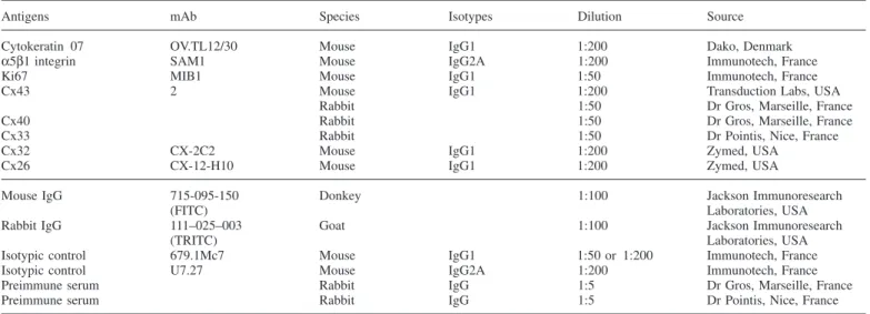
Figure 1. (A) Brightfield image of first trimester floating villous section immunostained for Cx43 and DAPI in (B). (B) Cx43 (green) immunostaining is observed in the trophoblastic layer between contacting cells and within the cytoplasm. (C) Transversal section of another floating villous at higher
magnification immunostained for Cx43. Cx43 punctate immunofluorescence is observed between two contiguous nuclei presumably of cytotrophoblastic cells and between other contacting nuclei (presumably cyto- and syncytiotrophoblast). Scale bars⫽ 20 µm. (D) Gap-FRAP experimentation. Typical computer-generated images of fluorescence distribution in villous trophoblastic cells cultured for 3 days measured during a gap-FRAP experiment. After a pre-bleach scan, the fluorescent dye was photobleached in some selected cells (polygons 1 and 2) by means of a strong laser illumination. Isolated cells (polygon 3) kept unbleached served as a control for the spontaneous fading of fluorescent emission. The evolution of fluorescence intensities was measured starting just after photobleaching for 12 min with a scanning period of 2 min. Corresponding curves of fluorescence evolution in selected cells: fluorescence recoveries in cells 1 and 2 follow a closely exponential time-course, reflecting the presence of open gap junctional channels. Note the low decrease of fluorescence intensity in the control unbleached cell (3) due to repeated scanning.
Immunoresearch Laboratories, Wet Grove, PA, USA) (1/50 and 1/100) were applied for 45 min at room temperature (Table I). After washing, samples were mounted in mounting medium with or without DAPI for nuclear staining (Vector Laboratories, Burlingame CA, USA) and examined on an Olympus Bx60 epifluorescence microscope. All controls, performed by omitting the primary antibody or by non-specific IgG of the same isotype, were negative.
Cx43 in-situ hybridization
Tissue sections (5µm thick) were collected on 3-aminopropyltriethoxysilane-coated slides and the in-situ hybridization procedure was performed as previously described (Sibony et al., 1995). The human 1 kb Cx43 cDNA (gift from Dr M. Mesnil, IARC, Lyon, France) was subcloned into the
EcoRI-restriction site of Bluescript KS⫹ vector. Cx43 antisense and Cx43 sense probes were obtained from linearized vectors with Hind III and Bam HI (Roche, Meylan, France). The radiolabelled probes were generated by in-vitro transcription and incorporation of [35S]uridine triphosphate (specific radioactivity, 1000 Ci/mmol; Amersham, UK). Two serial placental sections were mounted on the same histological slide. One received the antisense RNA probe, and the other received the RNA sense probe as negative control. Heart tissue sections were used as a positive control. Tissue sections were covered with 30–100µl hybridization buffer (50% formamide, 10% dextran sulphate, 1 mg/ml salmon sperm DNA, 70 mmol/l dithiothreitol and 20⫻standard saline citrate) containing the radiolabelled probe at a concentration of 10–15⫻103 c.p.m./µl. After the post-hybridization washings, the slides were dipped in NTB2 emulsion (Kodak, Rochester, NY, USA) and exposed at 4°C in dark boxes for 15 days. Slides were developed, fixed and counterstained with Toluidine Blue before examination.
RT–PCR
Total RNA was isolated by the RNeasy method (Qiagen, Hilden, Germany) from isolated highly purified cytotrophoblastic cells cultured on plastic and from isolated cytotrophoblastic cells cultured on Matrigel®. The total RNA was treated with RNase-free DNase I (Life Technologies, France) at 22°C for 5 min.
For Cx43 transcripts, total RNA (5µg) was transcribed into cDNA using oligo(dT)12–18as primer and superScriptII reverse transcriptase (Roche). One-fifth of the reaction mixture was amplified with Taq polymerase (Life Technologies) in a final volume of 50µl. For the semi-quantitative PCR, each cycle consisted of denaturation at 94°C for 1 min, primer annealing at 60°C for 1 min and primer extension at 72°C for 1 min. In order to quantify amplification in exponential phase, primers of GAPDH were added at cycle 8 and PCR products (10µl) were appropriated at cycles 23, 24, 25, 26 and 27. Primers were designed from previously published sequence data: the human GAPDH 5⬘ sense CTGCACCACCAACTGCTTAG-3⬘ and 5⬘ antisense AGGTCCACCACTGACACGTT-3⬘; human Cx43 5⬘ sense AGTCTATC-TTTGAGGTGGCC-3⬘ and 5⬘ antisense GGCTGTAATTCATGTCCAGC-3⬘. Reaction products were resolved on an agarose gel (1.5%) and stained with ethidium bromide. Sizes of the expected amplification products were 1148 base pairs (bp) for Cx43 and 275 bp for GAPDH.
For Cx40 transcripts, RNA samples (350 ng per assay) were reverse-transcribed using the Superscript II reverse transcriptase and oligo(dT)12–18 primer (Gibco–BRL, Cergy Pontoise, France). One-third of the cDNA was then used in each PCR reaction. For amplification of the endogenous Cx40 messenger, two primers were chosen in the coding region of the Cx40 human gene to amplify a 500 bp fragment: forward primer S40H, 5 ⬘-GCCACGCCATGCACACTGTG-3⬘, and reverse primer AS40H, 5⬘-GCTC-TGGACTATGCCCACAGAG-3⬘. Control PCR were performed on total RNA in order to exclude any genomic DNA contamination. Each PCR reaction was performed with 2.5 IU Taq DNA polymerase (Promega, Charbonnie`res, France) in a 50µl final volume in the presence of an appropriate 1⫻ buffer plus 25 mmol/l MgCl2, 50 ng of each primer and 20 mmol/l of each dNTP. Amplification was done in a UNOII Biometra thermal–cycler and consisted of 95°C for 3 min, 30⫻(94°C for 15 s, 60°C for 30 s, 72°C for 40 s) and 72°C for 5 min. After amplification, RT–PCR products were run on an ethidium bromide-stained 1.2% agarose gel and photographed.
Gap-FRAP method
The cell-to-cell diffusion of a fluorescent dye was measured by the gap-FRAP method (Wade et al., 1986), using an interactive laser cytometer (ACAS 570; Meridian Instruments, Okemos, MI, USA), which allows for convenient digital video imaging and analysis. After washing, cultured trophoblastic cells were loaded for 10 min at room temperature in saline solution containing the membrane-permeant molecule 6-carboxyfluorescein diacetate (Sigma: 7µg/ml in 0.25% dimethylsulphoxide). This lipophilic compound is hydrolysed by cytoplasmic esterases to 6-carboxyfluorescein, a hydrophilic derivative which accumulates inside the cells. After washing off the excess extracellular fluorogenic ester to avoid further loading, the fluorescence of some selected cells adjacent to others cells was photobleached by applying strong light pulses from an argon laser set at 488 nm. The fluorescence intensity was recorded in the bleached cells before and after photobleaching for 12 min (each time period⫽ 2 min). In each experiment, one labelled isolated cell,
Table II. First trimester connexin staining patterns at the fetal–maternal interface, as assessed by immunolocalization
Floating villi Anchoring villi Cell column CTB VCTB STB Proximal Distal Cytokeratin 07 ⫹⫹ ⫹⫹ ⫹⫹ ⫹⫹ Ki67 ⫹ ⫺ ⫹⫹ ⫺ Cx43 ⫹ ⫹ ⫺ ⫺ Cx40 ⫺ ⫺ ⫹⫹⫹ ⫺ Cx33 ⫺ ⫺ ⫺ ⫺ Cx32 ⫺ ⫺ ⫺ ⫺ Cx26 ⫺ ⫺ ⫺ ⫺
The intensity of staining was estimated by two observers and the results averaged.
VCTB⫽ villous cytotrophoblastic cell; STB ⫽ syncytiotrophoblast; CTB⫽ cytotrophoblastic cell.
left unbleached, served as a reference for the loss of fluorescence due to repeated scanning and dye leakage, and an isolated bleached cell served as a control. When the return of fluorescence followed a fast step-like course, reaching艌90% of the final steady state in ⬍30 s after photobleaching, this indicated that the diffusion of dye was neither prevented by the cell membrane nor limited by the presence of gap junctions. It was inferred that fusion of cell membranes had been completed and that the cellular elements were part of a true syncytium. When the bleached cells were interconnected by open gap junctional channels to unbleached contiguous cells, a fluorescence recovery following a slow exponential time course was measured (Figure 1D) (Cronier
et al., 1994b; Furger et al., 1996). Therefore, the analysis of the kinetic of
fluorescence recovery allows discrimination between aggregated cytotropho-blastic cells and syncytiotrophoblast. In our experimental conditions, GJIC was investigated (coupled cells or not) in a population of cells in contact, leading to a percentage of coupled cells (Figure 4B).
Results
Connexin expression and GJIC in the villous trophoblast
Among all the connexins tested, only Cx43 was detected in the
trophoblast of the floating villi (Table II). In situ, Cx43 immunostaining
was observed on most villi. Punctate Cx43 immunofluorescence
was observed between cytotrophoblastic cells, between cyto- and
syncytiotrophoblast and in the syncytiotrophoblastic cytoplasm
(Figure 1A–C). Cx43 immunofluorescence was also observed in the
mesenchymal cells of the villi. In controls performed by omission of
the primary antibody or by using non-specific IgG of the same
isotype, no immunoreactivity could be detected (data not shown).
In-situ hybridization with Cx43 antisense riboprobe revealed a high
accumulation of silver grains in the trophoblastic layers
(cytotropho-blastic cells and syncytiotrophoblast: 3.428
⫾ 0.006 grains/10 µm
2)
and in the mesenchymal core of the villi (0.563
⫾ 0.005 grains/
10
µm
2) (Figure 2B). Control sections incubated with the sense probe
did not reveal any specific hybridization (Figure 2A).
Villous cytotrophoblastic cells were cultured in 10% FCS on plastic
dishes. After 12 h, cells began to emit protrusions and pseudopodia
making initial contacts with neighbouring cells. Later, groups of
cytotrophoblastic cells in close apposition were observed, consistent
with an aggregation stage. Finally, after 72 h, large cellular masses
with central nuclei mount and expanding cytoplasm were seen, thus
confirming earlier studies (Dodeur et al., 1990; Tarrade et al., 2001).
Only Cx43 was detected in trophoblastic cells cultured on plastic
dishes. After 24 h, a strong punctate Cx43 immunofluorescence was
mainly observed at the intercellular contacts between pseudopodia of
Figure 2. Cx43 in-situ hybridization in first trimester villous section. (A) The hybridization signal is absent in control section with the RNA sense probe; (B) Localization of Cx43 mRNA in first trimester floating villous section hybridized with35S-labelled antisense Cx43 riboprobe. The hybridization signal is strong within the trophoblastic layer (TL: 3.428⫾ 0.006 grains/10 µm2) and is also present in the stroma of the villous core (VC: 0.563⫾ 0.005 grains/10 µm2) compared to the intervillous space (0.400⫾ 0.002 grains/10 µm2). Scale bars⫽ 20 µm.
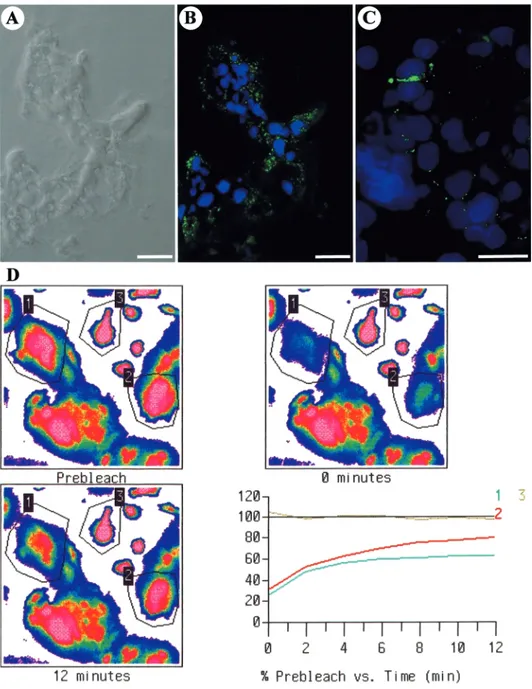
Figure 3. (A, B) Immunofluorescent localization of Cx43 protein in first trimester trophoblastic cells cultured on plastic dishes. Strong Cx43 spots are observed at the contact between pseudopodia after 24 h (A) and at the intercellular boundaries after 48 h (B). (C, D) Double immunofluorescence for Cx43 (D) and for nuclear staining with DAPI (C) after 72 h of culture. Cx43 punctate immunofluorescence is observed around the cluster of nuclei of a syncytiotrophoblast and at the border of a cytotrophoblastic cell which is still in contact with the syncytiotrophoblast. Scale bars⫽ 10 µm.
neighbouring cells (Figure 3A). After 48 h, Cx43 immunofluorescence
was detected at the intercellular boundaries between aggregated cells
(Figure 3B). After cellular fusion, the signal disappeared from the
cell membrane and appeared in intracytoplasmic spots (Figure 3C,
D). No immunoreactivity could be detected in controls. Cx43
expres-sion was confirmed by the presence of Cx43 transcripts in freshly
isolated cytotrophoblastic cells and in cytotrophoblastic cells cultured
for 72 h (Figure 4A). A single band for the Cx43 PCR product
was observed at the expected size (1148 bp) in similar levels in
cytotrophoblastic cells and syncytiotrophoblasts. No amplified DNA
fragments were identified in the absence of template (data not shown)
or in RNA samples incubated without RT (Figure 4A, lanes 1 and 3).
Functionality of the intercellular channels was demonstrated by
means of gap-FRAP. Typical changes in the fluorescence intensity of
selected cells within a field before and after the photobleaching
procedure are shown in Figure 1D, as well as the corresponding
fluorescence recovery curves. This slow exponential fluorescence
recovery characteristic of GJIC was recorded between
cytotropho-blastic cells, between cytotrophocytotropho-blastic cells and syncytiotrophoblasts
and between contiguous syncytiotrophoblasts. Gap junctional coupling
was present at all stages of differentiation (Figure 4B) in 5, 14.3 and
15.6% of the trials after 1, 2 and 3 days of culture respectively.
Connexin expression and GJIC in the extravillous trophoblast
of the cell column
Due to the use of curettage which disrupts the integrity of the
implantation site, the connexin immunostaining was mainly examined
in the cell columns of the anchoring villi. In situ, a strong punctate
Cx40 immunofluorescence was detected between the proliferative
cytotrophoblastic cells of the proximal part of the column cells
(Figure 5A and B). Expression became weak or absent in the distal
part of the column. Other connexins were not detected in the
extravillous trophoblasts of the cell columns (Table II).
Figure 4. (A) Expression of Cx43 mRNA in villous trophoblastic cells as detected by RT–PCR. The expected size of the PCR products is 1148 bp for Cx43. Lane 2: extracts from highly purified cytotrophoblastic cells immediately after cell isolation; lane 4: extracts from trophoblastic cells after 3 days of culture. Lanes 1 and 3 represent respective controls without RT. A 100 bp DNA ladder was used for size analysis. The results shown are representative of three independent experiments. (B) Evolution of functional intercellular communication measured by means of fluorescence recovery after the photobleaching method in cultured villous trophoblastic cells. The percentages of coupled cells were measured after 1, 2 or 3 days of culture on plastic dishes. The numbers of intercellular contacts analysed are indicated on top of the bars. GAPDH⫽ glyceraldehyde 6-phosphate dehydrogenase.
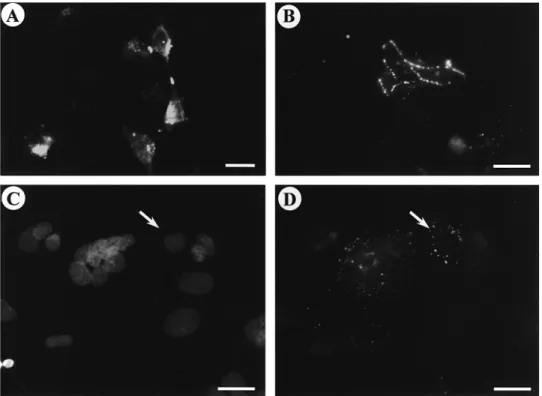
Figure 5. First trimester anchoring villi section double-immunolabelled for cytokeratin 07 (A) and Cx40 protein (B). Anticytokeratin antibody stained villous cytotrophoblastic cells (CTB), syncytiotrophoblast (STB), cytotrophoblastic cells of the cell columns (CC) and the invasive cytotrophoblastic cells. Punctate immunofluorescence for Cx40 is observed between the cytotrophoblastic cells of the proximal part of the cell column and on the membrane of some invasive cytotrophoblastic cells. Scale bar⫽ 20 µm. VC ⫽ villous core.
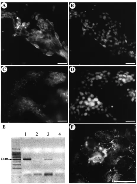
When first trimester isolated cytotrophoblastic cells were cultured
on Matrigel-coated dishes, 4 h after seeding ~60% of cells adhered.
They adhered as isolated or aggregated cells but never fused during
the first 2 days. During the first 3 days of culture, a cytokeratin 07
immunofluorescence was observed (data not shown). After 2 days of
culture,
α5β1 integrin immunofluorescence was also observed (Figure
6A, B). Furthermore, a strong punctate Cx40 immunofluorescence
was detected between contiguous cells (Figure 6C, D). After 3 days
of culture, a Cx32 immunofluorescence was also observed between
some aggregated cells (Figure 6F). Cx40 expression was confirmed
by the presence of Cx40 transcripts in freshly isolated
cytotropho-blastic cells and in cytotrophocytotropho-blastic cells cultured on Matrigel for
48 h (Figure 6E). A single band for the Cx40 RT–PCR product was
observed at the expected size (500 bp) in both specimens (Figure 6E;
lanes 1 and 3). No amplified DNA fragments were identified in the
absence of template (data not shown) or in RNA samples incubated
without RT (Figure 6E, lanes 2 and 4).
To elucidate the presence of a GJIC between cells cultured on
Matrigel, the gap-FRAP method was used on cytotrophoblastic cells
for 1, 2 or 3 days. GJIC was never detected between contacting cells
whatever the period investigated.
Discussion
This study demonstrated the presence of Cx43 (protein and transcript)
and of GJIC in first trimester villous trophoblast. Staining with
cytokeratin 07, a trophoblast-specific marker, clearly demonstrated
the trophoblastic origin of the Cx43 staining. The presence of
Cx43 transcripts in villous trophoblast was illustrated by the in-situ
hybridization signal and by RT–PCR in isolated and cultured cells.
Villous trophoblastic cells in culture are currently used to study
certain aspects of the dynamic process occurring during villous
differentiation (Kliman et al., 1986). The additional step of purification
with a monoclonal anti-HLA A, B and C rules out the contamination
by other cells containing Cx43 proteins and transcripts such as
endothelial and mesenchymal cells. In these conditions, RT–PCR
analysis showed that Cx43 transcripts are similarly present in freshly
purified cytotrophoblastic cells and in trophoblasts cultured for 3 days
(partly syncytiotrophoblast). This result is consistent with Northern
blotting analysis showing that the presence of Cx43 transcripts seemed
to stay constant during culture of trophoblastic cells (Winterhager
et al., 1999). It should be noted that numerous studies did not examine
functional coupling between the cells, but merely analysed the
presence of gap junction RNA/protein, whereas gap junctional
Figure 6. (A, B) Double immunofluorescence forα5β1 integrin (A) and for nuclear staining with DAPI (B) in first trimester cytotrophoblastic cells cultured on Matrigel for 48 h. A strong labelling is observed at the periphery of the aggregated cells. Scale bars⫽ 20 µm. (C, D) Double immunofluorescence for Cx40 (C) and for nuclear staining with DAPI (D) in first trimester cytotrophoblastic cells cultured on Matrigel for 48 h. Punctate immunofluorescence is observed at the border of aggregated cells. Scale bars⫽ 15 µm. (E) Detection of the Cx40 transcript in trophoblastic cells from different stages of development. Lane 1: 500 bp amplified Cx40 cDNA in freshly isolated cytotrophoblastic cells of first trimester placentae. Lane 3: 500 bp amplified Cx40 cDNA in cytotrophoblastic cells of first trimester placentae cultured on Matrigel. Lanes 2 and 4 are corresponding controls of lanes 1 and 3 with the absence of RT. (F) Immunofluorescent localization of Cx32 protein in first trimester trophoblastic cells cultured on Matrigel for 3 days. Cx32 spots are observed at the border of some aggregated cells. Scale bar⫽ 20 µm.
communication levels do not necessarily correlate with connexin
expression levels (Lo, 1999). Thus, in considering the role of gap
junction in differentiation, functional analysis of gap junctional
coupling is of critical importance. We have demonstrated for the first
time that the presence of Cx43 gap junctional channels allows a
functional cell–cell communication between all types of elements
in contact during the differentiation of the first trimester villous
trophoblastic cells. In culture, the quantitative evolution of GJIC
paralleled the morphological differentiation. These data are consistent
with our previous study with term trophoblastic cells showing that
the ability of villous trophoblastic cells to develop a transient GJIC
was a prerequisite for the formation of the syncytiotrophoblast
(Cronier et al., 1994a). In addition, another role for the presence of
gap junction channels between the two trophoblastic layers (cellular
and syncytial) of the first trimester villi could be the inter-trophoblastic
diffusion and transfer of small molecules (second messengers and
nutrients) as suggested for glucose in the labyrinth of the rat placenta
(Takata and Hirano, 1997; Gabriel et al., 1998).
In the extravillous pathway, the cytotrophoblastic cells proliferate,
migrate and invade the decidua and its vasculature. In the proximal
part of the cell column, Cx40 expression was demonstrated between
cytotrophoblastic cells. This localization correlates with the nuclear
expression of the proliferation marker Ki67 (Kaufmann and
Castellucci, 1997). To elucidate the functional role of Cx40 expression,
isolated cytotrophoblastic cells were cultured on Matrigel. We confirm
that in the presence of Matrigel, trophoblastic cells never fused during
the first 2 days and that the cells expressed the
α5β1 integrin after
2 days of culture (Tarrade et al., 2001). This fibronectin receptor
immunostaining confirms the extravillous phenotype of these cells
(Burrows et al., 1993; Damsky et al., 1994). A strong Cx40
immuno-fluorescence was observed between these contiguous cells. This result
is substantiated by the presence of Cx40 transcripts. Furthermore, a
recent ultrastructural study demonstrated the presence of gap junctions
in the proximal cytotrophoblastic cells of the columns (Enders et al.,
2001). The functionality of Cx40 expression was therefore investigated
by gap-FRAP. In our experimental conditions, no gap junctional
communication was demonstrated between cells. Interestingly, in
human choriocarcinoma cells expressing Cx40, an extremely low
level of electrical coupling has been measured (Hellmann et al.,
1996). This issue is important because some studies have suggested
that electrical coupling through gap junctions may be possible whereas
the transfer of fluorescent molecules is restricted (Warner, 1988; Dale
et al., 1991). It is interesting to note that Cx40 shows a restricted
expression pattern in mammals: heart, vascular smooth muscle cells
and vascular endothelium (Gros et al., 1994). Despite the fact that
recent electrophysiological studies support the hypothesis of an ionic
coupling in cell lines transfected by Cx40 and Cx43 (Cottrell and
Burt, 2001; Valiunas et al., 2001), it is currently admitted that Cx40
cannot form functional heterotypic channels with most other connexins
(Bruzzone et al., 1996). Thus, it could be hypothesized that cells of
the proximal part of the column cannot communicate with the villous
cytotrophoblastic cells (expressing Cx43), thus preventing them from
a villous phenotype differentiation process. A similar model has been
proposed in carcinogenesis (Krutovskikh and Yamasaki, 1997) where
the absence of communication between tumour cells and surrounding
normal cells maintained the dedifferentiated proliferative phenotype
of the transformed cell population.
In the distal cell column, when the cytotrophoblastic cells are
leaving the column, Cx40 immunostaining disappeared. According
to Winterhager et al. (1999), Cx40 expression is missing during
trophoblast migration and was re-expressed in trophoblastic aggregates
within the decidua. Furthermore, in the trophoblastic aggregates in
the placental bed, Cx32 and Cx43 immunostaining have been detected
(Al-Lamki et al., 1999; Cronier et al., 2001). These results support
the hypothesis of the implication of these connexins in the fusion
process leading to the multinucleated cells of the human placental
bed (Boyd and Hamilton, 1970).
Recently, principles concerning placental development have
emerged using gene knock out approaches in mice (Rossant and
Cross, 2001). Due to the striking diversity in placental structure,
caution must be exercised in extrapolating findings regarding placental
development from one species to another. In rat, a switch in connexin
gene expression (Cx31, Cx26, Cx43) was associated with trophoblast
development (Reuss et al., 1996) and in mice Cx26 and Cx31
deficiencies were shown to cause placental alterations (Gabriel et al.,
1998; Plum et al., 2001). In the study of human placenta, Cx26 and
Cx31 was not found to be expressed and a specific connexin expression
pattern (Cx43, Cx32, Cx40) was demonstrated during trophoblast
differentiation.
In conclusion, this study demonstrated the presence of Cx43
allowing an inter-trophoblastic GJIC and this appeared to be associated
with the fusion process leading to the villous syncytiotrophoblast.
Moreover, the presence of Cx40 was associated with the extravillous
phenotype but not with GJIC.
Acknowledgements
We would like to thank Dr D.Evain-Brion for helpful critical comments on this manuscript and Dr J.M.Gasc for his expert collaboration. We are grateful to the Clinique du Fief de Grimoire Poitiers and to the Department of Obstetrics and Gynecology at the St Vincent de Paul Hospital for providing us with the placental tissues. We would like to thank Dr D.Gros for his generous gift of Cx40 and Cx43 antibodies and Dr M.Mesnil for his gift of Cx43 cDNA. We also thank Dr A.Tarrade, Dr N.Pavlov, Dr L.Pavan, Dr T.Fournier and G.Bertin for their expert assistance. This research was partly supported by grants from EEC (contract QLG1 1999 CT 00510) to L.Dupays and M.The´veniau-Ruissy.
References
Al-Lamki, R.S.A.I., Skepper, J.N. and Burton, G.J. (1999) Are human placental bed giant cells merely aggregates of small mononuclear trophoblast cells? An ultrastructural and immunocytochemical study. Hum. Reprod., 14, 496–504.
Aplin, J., Haigh, T., Lacey, H. et al. (2000) Tissue interaction in the control of trophoblast invasion. J. Reprod. Fertil., 55, 57–64.
Benirschke, K. and Kaufmann, P. (eds) (2000) Pathology of the Human
Placenta, 4th edn. Springer Verlag, New York.
Boyd, J.D. and Hamilton, W.J. (1970) The Human Placenta. W.Heffer, Cambridge.
Bruzzone, R., White, T.W. and Paul, D.L. (1996) Connections with connexins: the molecular basis of a direct intercellular signaling. Eur. J. Biochem., 238, 1–27.
Burrows, T.D., King, A. and Loke, Y.W. (1993) Expression of integrins by human trophoblast and differential adhesion to laminin or fibronectin. Hum.
Reprod., 8, 475–488.
Cottrell, G.T. and Burt, J.M. (2001) Heterotypic gap junction channel formation between heteromeric and homomeric Cx40 and Cx43 connexons. Am. J.
Physiol., 281, C1559–C1567.
Cronier, L., Alsat, E., Herve´, J.C. et al. (1994a) Inhibition of gap junctional communication, trophoblast differentiation and hCS expression by heptanol in human trophoblast in culture. Placenta, 15, A10.
Cronier, L., Bastide, B., Herve´, J.C. et al. (1994b) Gap junctional communication during human trophoblast differentiation: influence of human chorionic gonadotropin. Endocrinology, 135, 402–408.
Cronier, L., Herve´, J.C., De´le`ze, J. and Malassine´, A. (1997) Regulation of gap junctional communication during human trophoblast differentiation.
Microsc. Res. Tech., 38, 21–28.
Cronier, L., Bastide, B., Defamie, N. et al. (2001) Involvement of gap junctional communication and connexin expression in trophoblast differentiation of the human placenta. Histol. Histopathol., 16, 285–295.
Dale, B., Santella, L. and Tosti, E. (1991) Gap-junctional permeability in early and cleavage-arrested ascidian embryos. Development, 112, 153–160. Damsky, C.H., Librach, C., Lim, K.H. et al. (1994) Integrin switching regulates
normal trophoblast invasion. Development, 120, 3657–3666.
Dodeur, M., Malassine´, A., Bellet, D. et al. (1990) Characterization and differentiation of human first trimester placenta trophoblastic cells in culture. Rep. Nutr. Dev., 30, 183–192.
Enders, A.C., Blankenship, T.N., Fazleabas, A.T. et al. (2001) Structure of anchoring villi and the trophoblastic shell in the human, baboon and macaque placenta. Placenta, 22, 284–303.
Fisher, S.J. (2000) The placenta dilemma. Semin. Reprod. Med., 18, 321–326. Furger, C., Cronier, L., Poirot, C. et al. (1996) Human granulosa cells in culture exhibit functional cyclic AMP-regulated gap junctions. Mol. Hum.
Reprod., 2, 541–548.
Gabriel, H.D., Jung, D., Bu¨tzler, C. et al. (1998) Transplacental uptake of glucose is decreased in embryonic lethal connexin26 deficient mice. J. Cell
Biol., 140, 1453–1461.
Gros, D., Jarry-Guichard, T., Ten Velde, I. et al. (1994) Restricted distribution of connexin40, a gap junctional protein, in mammalian heart. Circ. Res., 74, 839–851.
Hellmann, P., Winterhager, E. and Spray, D.C. (1996) Properties of connexin40 gap junction channels endogenously expressed and exogenously overexpressed in human choriocarcinoma cell lines. Eur. J. Physiol., 432, 501–509.
Kaufmann, P. and Castellucci, M. (1997) Extravillous trophoblast in the human placenta. A review. Trophoblast Res., 10, 21–65.
Kliman, H.J., Nestler, J.E., Sermasi, E. et al. (1986) Purification, characterization, and in vitro differentiation of cytotrophoblasts from human term placentae. Endocrinology, 118, 1567–1582.
Krutovskikh, V. and Yamasaki, H. (1997) The role of gap junctional intercellular communication (GJIC) disorders in experimental and human carcinogenesis. Histol. Histopathol., 12, 761–768.
Kumar, N.M. and Gilula, N.B. (1996) The gap junction communication channel. Cell, 84, 381–388.
Lo, C.W. (1999) Genes, gene knockouts, and mutations in the analysis of gap junctions. Dev. Genet., 24, 1–4.
Malassine´, A. and Leiser, R. (1984) Morphogenesis and fine structure of the near-term placenta of talpa europaea: I. Endotheliochorial labyrinth.
Placenta, 5, 145–158.
Plum, A., Winterhager, E., Pesch, J. et al. (2001) Connexin31-deficiency in mice causes transient placental dysmorphogenesis but does not impair hearing and skin differentiation. Dev. Biol., 231, 334–347.
Reuss, B., Hellmann, P., Dahl, E. et al. (1996) Connexins and E cadherin are differentially expressed during trophoblast invasion and placental differentiation. Dev. Dynam., 205, 172–182.
Rossant, J. and Cross, J.C. (2001) Placental development: lessons from mouse mutants. Nature Rev. Genet., 2, 538–548.
Schmon, B., Hartmann, M., Jones, C.J. and Desoye, G. (1991) Insulin and glucose do not affect the glycogen content in isolated and cultured trophoblast cells of human term placenta. J. Clin. Endocrinol., 73, 888–893.
Sibony, M., Commo, F., Callard, P. and Gasc, J.M. (1995) Enhancement of mRNA in situ hybridization signal by microwave heating. Lab. Invest., 73, 586–591.
Tabb, T., Thilander, G., Grover, A. et al. (1992) An immunocytologic study of the increase in myometrial gap junctions (and connexin43) in rats and humans during pregnancy. Am. J. Obstet. Gynecol., 167, 559–587.
Takata, K. and Hirano, H. (1997) Mechanism of glucose transport across the human and rat placental barrier: a review. Microsc. Res. Tech., 38, 145–152.
Tanaka, T., Yamasaki, H. and Mesnil, M. (2001) Induction of a bystander effect in HeLa cells by using a bigenic vector carrying viral thymidine kinase and connexin32 genes. Mol. Carcinogen., 30, 176–180.
Tarrade, A., Lai Kuen, R., Malassine´, A. et al. (2001) Characterization of human villous and extravillous trophoblast isolated from first trimester placenta. Lab. Invest., 81, 1199–1211.
Valiunas, V., Gemel, J., Brink, P.R. and Beyer, E.C. (2001) Gap junction channels formed by coexpressed connexin40 and connexin43. Am. J.
Physiol., 281, H1675–H1689.
Vicovac, L. and Aplin, J.D. (1996) Epithelial–mesenchymal transition during trophoblast differentiation. Acta Anat., 156, 202–216.
Wade, M.H., Trosko, J.E. and Schindler, M. (1986) A fluorescence photobleaching assay of gap junction-mediated communication between human cells. Science, 232, 525–528.
Warner, A. (1988) The gap junction. J. Cell Sci., 89, 1–7.
Willecke, K., Hennemann, H., Dahl, E. et al. (1991) The diversity of connexin genes encoding gap junctional proteins. Eur. J. Cell Biol., 6, 459–470. Winterhager, E., von Ostau, C., Gerke, M. et al. (1999) Connexin expression
patterns in human trophoblast cells during placental development. Placenta, 20, 627–638.
Winterhager, E., Kaufmann, P. and Gruemmer, R. (2000) Cell–cell-communication during placental development and possible implications for trophoblast proliferation and differentiation. Placenta, 21, S61–S68.
Submitted on December 6, 2001; resubmitted on April 22, 2002; accepted on July 19, 2002