Regulation of the memory T cell development and islet β-cell survival by DAPK-related apoptosis-inducing protein kinase 2 (Drak2)
Jianning Mao
Département de Médecine
Faculté de Médecine
Université de Montréal
Thèse présentée à la Faculté des études supérieures en vue de l’obtention du grade de
Philosophiae Doctor (Ph.D.) en Sciences Biomédicales
June , 2009
Université de Montréal Faculté des études supérieures
Cette thèse intitulée:
« Regulation of the memory T cell development and islet β-cell survival by DAPK-related apoptosis-inducing protein kinase 2 (Drak2) »
Présentée par:
Jianning Mao
A été évaluée par un jury composé des personnes suivantes:
Bertrand, Richard ……… Président du jury Wu, Jiangping ……… Directeur de recherché Luo, Hongyu ……… Codirectrice Labrecque, Nathalie ……… Membre du jury Di Battista, John ……… Examinateur externe Lajeunesse, Daniel ……… Représentant du doyen de la FES
SUMMARY
Drak2 is a member of the death-associated protein family, and is a serine threonine kinase. In Drak2 null mutant mice, T cells have no apparent defect in activation-induced apoptosisafter stimulation with anti-CD3 and anti-CD28, but havea lowered threshold to stimulation, compared with wild type(WT) T cells.
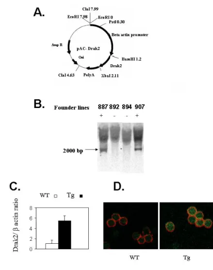
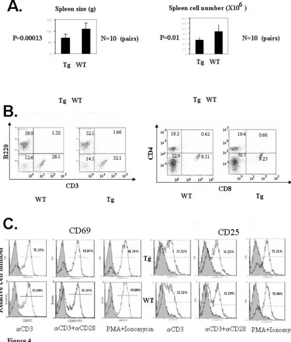
In our study, in situ hybridization analysis has revealed that Drak2 expression is ubiquitous at the mid-gestation stage in embryos, followed by more focal expression in various organs in the perinatal period and adulthood, notably in the thymus, spleen, lymph nodes, cerebellum, suprachiasmatic nuclei, pituitary, olfactory lobes, adrenal medulla, stomach, skin and testes. We generated Drak2 transgenic(Tg) mice using the human β-actin promoter. TheseTg mice showed normal T cell versus B cell and CD4 versus CD8populations in the spleen, but their spleen weight cellularity was lower in comparison with wild type mice. After TCR activation, the proliferation response in Drak2 Tg T cells was normal, althoughtheir interleukin (IL)-2 and IL-4 but not interferon-γ production was augmented. Activated Drak2 Tg T cells demonstrated significantlyenhanced apoptosis in the presence of exogenous IL-2. At the molecular level, Drak2 Tg T cells manifested a lower increaseof anti-apoptotic factors during activation; such a change probably rendered the cells vulnerable to subsequent IL-2 insults. The heightened apoptosis in Drak2 Tg T cells was associated withreduced numbers of T cells with the memory cell phenotype (CD62Llo) and repressed secondary T cell responses in delayed type
hypersensitivity.These results demonstrate that Drak2 expresses in the T cell compartmentbut is not T cell-specific; and it plays critical roles in T cellapoptosis and memory T cell development.
Further, we investigated the role of Drak2 in β-cell survival and diabetes. Drak2 mRNA and protein were rapidly induced in islet β-cells after exogenous inflammatory cytokine or free fatty acid stimulation, which is present endogenously in either type 1 or type 2 diabetes. Drak2 upregulation was accompanied by increased β-cell apoptosis. The β-cells apoptosis caused by the said stimuli was inhibited by Drak2 knockdown using siRNA. Conversely, transgenic (Tg) Drak2 overexpression led to aggravated β-cell apoptosis triggered by the stimuli. Drak2 overexpression in islets compromised the increase of anti-apoptotic factors, such as 2, Bcl-xL and Flip, upon the cytokine and free fatty acid stimulation. Further in vivo experiments demonstrated that Drak2 Tg mice were prone to type 1 diabetes in a multiple-low-dose streptozotocin-induced diabetes model, and they were also prone to type 2 diabetes in a diet-induced obesity model. Our data show that Drak2 is detrimental to β-cell survival.
We also investigated the signalling pathway of Drak2. We found that purified Drak2 could phosphorylate p70S6 kinase in an in vitro kinase assay. Drak2 overexpression in NIT-1 cells led to enhanced p70S6 kinase phosphorylation, while Drak2 knockdown in these cells reduced the phosphorylation. These mechanistic studies proved that p70S6 kinase was a bona fide Drak2 substrate in vitro and in vivo.
This study has discovered the important functions of Drak2 in T cell homeostasis and diabetes. We proved that p70S6 kinase was a substrate of Drak2. Our findings have broadened our
knowledge of Drak2 in the immune and endocrine system. Some of our findings, such as the roles of Drak2 in memory T cell development and β cell survival could be explored for clinical application in the areas of transplantation and diabetes.
Résumé
Drak2 est un membre de la famille des protéines associées à la mort et c’est une sérine/thréonine kinase. Chez les souris mutantes nulles Drak2, les cellules T ne présentent aucune défectuosité apparente en apoptose induite par activation,après stimulation avec anti-CD3 et anti-CD28, mais ont un seuil de stimulation réduit, comparées aux cellules T de type sauvage (TS).
Dans notre étude, l’analyse d’hybridation in situ a révélé que l’expression de Drak2 est ubiquiste au stade de la mi-gestation chez les embryons, suivie d’une expression plus focale dans les divers organes pendant la période périnatale et l’âge adulte, notamment dans le thymus, la rate, les ganglions lymphatiques, le cervelet, les noyaux suprachiasmatiques, la glande pituitaire, les lobes olfactifs, la médullaire surrénale, l’estomac, la peau et les testicules. Nous avons créé des souris transgéniques (Tg) Drak2 en utilisant le promoteur humain β-actine. Ces souris Tg montraient des ratios normaux entre cellules T versus B et entre cellules CD4 versus CD8, mais leur cellularité et leur poids spléniques étaient inférieurs comparé aux souris de type sauvage. Après activation TCR,la réponse proliférative des cellules T Tg Drak2 était normale, même si leur production d’interleukine (IL)-2 et IL-4 mais non d’interféron-γ était augmentée. Les cellules T Tg Drak2 activées ont démontré une apoptose significativement accrue en présence d’IL-2 exogène. Au niveau moléculaire, les cellules T Tg Drak2 ont manifesté une augmentation moins élevée des facteurs anti-apoptotiques durant l’activation; un tel changement a probablement rendu les cellules vulnérables aux attaques subséquentes d’IL-2. L’apoptose compromise dans les cellules T Tg Drak2 a été associée à un nombre réduit de cellules T ayant le phénotype des cellules mémoires (CD62Llo) et avec des réactions secondaires réprimées des cellules T dans l’hypersensibilité de type différé.Ces résultats démontrent que Drak2 s’exprime
dans le compartiment des cellules T mais n’est pas spécifique aux cellules T; et aussi qu’il joue des rôles déterminants dans l’apoptose des cellules T et dans le développement des cellules mémoires T.
En outre, nous avons recherché le rôle de Drak2 dans la survie des cellules β et le diabète. L’ARNm et la protéine Drak2 ont été rapidement induits dans les cellules β de l’îlot après stimulation exogène par les cytokines inflammatoires ou les acides gras libres et qui est présente de façon endogène dans le diabète, qu’il soit de type 1 ou de type 2. La régulation positive de Drak2 a été accompagnée d’une apoptose accrue des cellules β. L’apoptose des cellules β provoquée par les stimuli en question a été inhibée par la chute de Drak2 en utilisant petit ARNi. Inversement, la surexpression de Drak2 Tg a mené à l’apoptose aggravée des cellules β déclenchée par les stimuli. La surexpression de Drak2 dans les îlots a compromis l’augmentation des facteurs anti-apoptotiques, tels que Bcl-2, Bcl-xL et Flip, sur stimulation par la cytokine et les acides gras libres. De plus, les expériences in vivo ont démontré que les souris Tg Drak2 étaient sujettes au diabète de type 1 dans un modèle de diabète provoqué par de petites doses multiples de streptozotocine et qu’elles étaient aussi sujettes au diabète de type 2 dans un modèle d’obésité induite par la diète. Nos données montrent que Drak2 est défavorable à la survie des cellules β.
Nous avons aussi étudié la voie de transmission de Drak2. Nous avons trouvé que Drak2 purifiée pouvait phosphoryler p70S6 kinase dans une analyse kinase in vitro. La surexpression de Drak2 dans les cellules NIT-1 a entraîné l’augmentation de la phosphorylasation p70S6 kinase tandis que l’abaissement de Drak2 dans ces cellules a réduit la phosphorylation. Ces recherches mécanistes ont prouvé que p70S6 kinase était véritablement un substrat de Drak2 in vitro et in vivo.
Cette étude a découvert les fonctions importantes de Drak2 dans l’homéostasie des cellules T et le diabète. Nous avons prouvé que p70S6 kinase était un substrat de Drak2. Nos résultats ont approfondi nos connaissances de Drak2 à l’intérieur des systèmes immunitaire et endocrinien. Certaines de nos conclusions, comme les rôles de Drak2 dans le développement des cellules mémoires T et la survie des cellules β, pourraient être explorées pour des applications cliniques dans les domaines de la transplantation et du diabète.
TABLE OF CONTENTS
Summary………..…...………..……….…….……...….…..III Résumé……….………..………..…..…..……VI List of Figures……….…..……..……….…...XII List of Abbreviations……….…………...………..…..XV Acknowledgements………..….…….…………...………....…..XVIII I. INTRODUCTION……….………….………...…..….11. T cell homeostasis and apoptosis………..….……….……..….…...5
1-1 T cell apoptosis………..………...……….…..…….….5
1-1-1 Extrinsic cell-death-receptor and caspase-dependent apoptosis…...……...8
1-1-2 Intrinsic mitochondria and caspase-dependent apoptosis……….…...…9
1-1-3 Activation-induced cell death (AICD) ….………...…..…………...…11
1-1-4 Activated cell-autonomous death (ACAD)………...………...………...……13
1-2 T cell memory ….……….………….... 14
1-2-1 Clonal expansion………...……….. 16
1-2-2 Clonal contraction…………...………...….. 17
2. DAPK and Drak2………..………... 19
2-1 Death-associated protein kinase (DAPK) ……….………….…….. 19
2-2 DRAK2……….…….……….……….. 21
3-1 Type 1 diabetes……….……….…..….. 24
3-2 Type 2 diabetes…..……….………….……….. 30
3-2-1 Insulin resistance………....………..……….. 31
3-2-1-1 Adipokines……….…...………...………... 31
3-2-1-2 Inflammatory mediators………...……...…….……….. 33
3-2-1-3 Metabolic overload in the liver……….…...……….. 34
3-2-1-4 Metabolic overload in muscle………...………... 35
3-2-1-5 Relating metabolic overload to insulin signaling………....…….……….. 36
3-2-2 β-cell secretory dysfunction ……….………...…….. 37
3-2-2-1 Glucotoxicity……….………...……….. 37
3-2-2-2 Lipotoxicity……….……….……….…...…….. 38
3-2-3 Decreased β-cell mass………..…….………...….. 40
3-2-3-1 Adaptation of β-cell mass to metabolic load……...……….………... 40
3-2-3-2 Failure of β-cell mass to compensate for metabolic load……...………...….. 41
3-2-4 The role of IRS-2 signaling in β-cell survival and apoptosis……….. ...…... 41
4. Objectives of my work………..….……..…….……….…..44
References for introduction……….………....…...…... 46
II. ARTICLES………...………….………....…..64
Article 1: Transgenic Drak2 overexpression in mice leads to increased T-cell apoptosis and compromised memory T-cell development…...………..………….…..65
Article 2: Drak2 overexpression results in increased β-cell apoptosis after free fatty acid stimulation ...………...………..…....101
Article 3: Drak2 is upstream of p70S6 kinase: it implication in cytokine-induced islet
apoptosis, type 1 diabetes and islet transplantation………...……….………...128
III. DISCUSSION………..………….…...…..170
1. Expression of Drak2 ………..……..………..…….….171
2. The physiological role of Drak2 in T cell homeostasis………..………….172
3. Drak2 might work on a two-hit model………...…….………….….……..……….…173
4. The signalling pathway of Drak2………..………..…………..…..….174
4.1 Pathways upstream of Drak2………..……….…...……….174
4.2 Downstream of Drak2………..……….…………..…..….174
5. A proposed model of Drak2 signalling………..………..…...……….178
6. Summary and further perspectives………..………...……….….179
7. Contributions to science……….……..………..……….….181
LIST OF FIGURES
Introduction;
Fig 1. Innate and adaptive immune system………...……….…2
Fig 2. Intrinsic and extrinsic pathway of apoptosis………..………...……...7
Fig 3. Mammalian BCL-2 family member……….………...………11
Fig 4. Inflammatory cytokines and CD8+ T-cell homeostasis after infection…………..………15
Fig 5. Death-associated protein kinase (DAPK) family member………...……21
Fig 6. Numbers of people with diabetes for 2000 and 2010, and the percentage increase………...…23
Fig 7. ß-cell death in T1D and T2D….………..………24
Fig 8. The transcription factor and gene networks putatively involved in the cytokine-promoted ß-cell "decision" to undergo apoptosis………..….………...…………26
Fig 9. Cytokine-induced ß-cell apoptosis………..…….………....………..………27
Fig 10. Development of type 2 diabetes……….………..………31
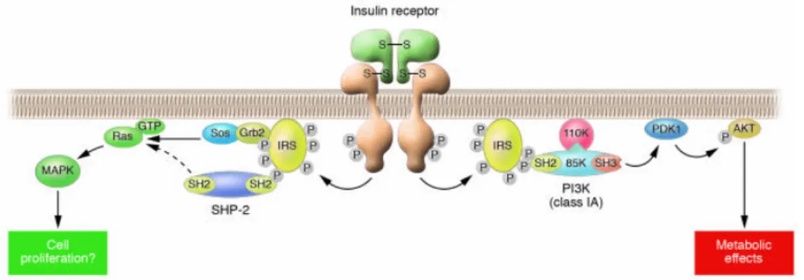
Fig 11. Insulin signaling in cells.……….………..………...…42
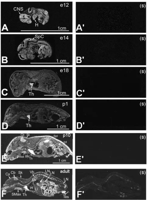
Article 1: Fig 1. Drak2 expression during ontogeny according to in situ hybridization………...….88
Fig 3. Generation and characterization of Drak2 Tg mice………..………….……….…92
Fig 4. Characterization of Drak2 Tg lymphoid organs and cells…..………….……...….….…..94
Fig 5. Lymphokine production and proliferation of Drak2 Tg T cells.…..…………..…………96
Fig 6. Drak2 T cells are prone to apoptosis….….……….…………...…..….….97
Fig 7. In vivo cellular and humoral immune responses of Drak2 Tg mice………....…...99
Article 2: Fig 1. Drak2 was rapidly augmented in islets treated with FFA……...………...……...120
Fig 2. Drak2 Tg islets were prone to apoptosis upon FFA stimulation…….…………..…..….121
Fig 3. Drak2 siRNA inhibited Drak2 protein upregulation and reduced apoptosis in NIT-1 cells upon FFA stimulation……….…...………..………123
Fig 4. Features of Drak2 Tg mice……….……….………….….. 124
Fig 5. Compromised anti-apoptotic factor upregulation in Drak2 Tg islet….…………...….125
Fig 6. Features of Drak2Tg mice after diet-induced obesity………..…....…126
Article 3: Fig 1. Drak2 mRNA was rapidly augmented in islets encountering inflammatory stimulation………154
Fig 2. Flow cytometry analysis of Drak2 protein upregulation in β-cells upon inflammatory stimuli………..……….…...…155
Fig 3. Drak 2 signalling is responsible for β-cell apoptosis………….…..…………..….…157
Fig 5. Drak2 Tg islets were prone to apoptosis upon inflammatory cytokine
stimulation………....161 Fig 6. p70S6 kinase phosphorylation by Drak 2 in vitro ………...….…164 Fig 7. Drak 2 phosphorylation p70S6 kinase in vivo………...………..166 Fig 8. Effect of Drak2 siRNA on p70S6 kinase phosphorylation and of rapamycin on β-cell apoptosis………..………..…..…168
Discussion;
Fig 1.The phosphorylation sites of mouse p70s6 kinase………...…………..……...…177 Fig 2. A proposed model of Drak2 signalling. ………...………...…...178
LIST OF ABBREVIATIONS
ACAD activated cell-autonomous death AICD activation-induced cell death AMPK 5'-AMP-activated protein kinase
APAF cytoplasmic apoptotic-protease-activating factor APC antigen presenting cell
ATF activating transcription factor
BIM BCL-2-interacting mediator of cell death CARD caspase-recruitment domain
CCR-7 CC-chemokine receptor-7
CHOP (CCAAT/enhancer binding protein) homologous protein CTL cytotoxic T lymphocytes
DAG diacylglycerols
DAPK death-associated protein kinase
DD death domain
DED death-effector domain
DISC death-inducing signalling complex
DR death receptor
DRAK DAPK-related apoptosis-inducing protein kinase EAE experimental autoimmune encephalomyelitis ER endoplasmic reticulum
ERK extracellular signal-regulated kinase FACS fluorescence-activated cell sorting
FasL Fas ligand
FFA free fatty acid
GAD glutamic acid decarboxylase GIIS glucose-induced insulin secretion
GLUT glucose transporter type
GPAT glycerol-3-phosphate acyltransferase ICAD inhibitor of the caspase-activated DNase IFN-γ Interferon-γ
IGF insulin-like growth factor IGT impaired glucose tolerance IL-12 Interleukin-12
IL-2 Interleukin-2
iNOS inducible nitric oxide synthase IRS insulin receptor substrate IκK-β IκB kinase catalytic subunit-β
JAK/STAT Janus Kinase-2/Signal Transducer and Activator of Transcription JNK c-Jun NH2-terminal kinase
LADA latent autoimmune diabetes of the adult LC-CoAs long-chain acyl CoAs
MAPK p38 mitogen-activated protein kinase MCP1 monocyte chemotactic protein-1 MHC major histocompatibility complex mTOR mammalian target of rapamycin NEFAs Non-esterified fatty acids
NF-κB Transcription factor nuclear factor-κB
NO nitric oxide
NOD nonobese diabetic
PI3K phosphatidylinositol 3-kinase
PKB phosphatidylinositol-3'-kinase (PI3'K)/protein kinase-B
PKC protein kinase C
PPAR peroxisome proliferator activated receptor PUMA p53-upregulated modulator of apoptosis RBP Retinol-binding protein
S6K ribosomal protein S6 kinase SOCS suppression of cytokine signalling TAC tricarboxylic acid
TCR T-cell receptor
TG triglyceride
TNF-α tumor necrosis factor-α
TRAIL TNF-related apoptosis-inducing ligand XBP-1 x-box binding protein-1
ACKNOWLEDGEMENTS
I would like to express my sincere gratitude to my supervisor Dr. Jiangping Wu and Dr. Hongyu Luo for their scientific guidance and encouragement throughout this study and during the preparation of this thesis.
I also would like to thank all the colleagues in the lab for their splendid co-operation.
Thanks also extended to all my friends and those who always support me in different ways.
Finally, I would like to thank all of my family members for their understanding, patience, and their great support, which are critically important for me to complete this study.
I. INTRODUCTION
Act Against Allergy web-site, SHS International Ltd
Fig.1. Innate and adaptive immune system: The mechanisms of innate immunity provide the initial defense against infections. Adaptive immune responses develop later and consist of activation of lymphocytes.
An immune system is a collection of mechanisms that kill pathogens and tumor cells, as well as distinguish them from the organism's own healthy cells and tissues. It can be divided into innate and adaptive immune system (Fig.1). The innate immune system is usually triggered when microbes are identified by pattern recognition receptors, which recognize components that are conserved among broad groups of microorganisms. Innate immune defenses are non-specific, and do not confer long-lasting immunity against a pathogen. The adaptive immune system is stronger and also has immunological memory, where each pathogen is "remembered" by a signature antigen. The adaptive immune response is antigen-specific and requires the recognition of specific “non-self” antigens during a process called antigen presentation. The cells of the adaptive immune system are special types of leukocytes,
called lymphocytes. B cells and T cells are the major types of lymphocytes and are derived from hematopoietic stem cells in the bone marrow. B cells are involved in the humoral immune response; T cells are involved in cell-mediated immune response. T lymphocytes consist of functionally distinct populations, the best defined of which are helper T cells and cytolytic, or cytotoxic, T lymphocytes (CTL). Most helper T cells are CD4+, and most CTLs are CD8+.
CD4+ lymphocytes, or helper T cells, are immune response mediators, and play an important role in establishing and maximizing the capabilities of the adaptive immune response.Helper T cells express T cell receptors (TCR) that recognize antigen bound to major histocompatibility complex (MHC) Class II molecules. The activation of CD4+ T cells requires engagement of the TCR and CD28 on the T cell by the MHC and B7 family members on the antigen presenting cell (APC). Both singals are required for production of an effective immune response. The activation of a naive helper T-cell causes it to release cytokines, which influences the activity of many cell types. Helper T cells can provide extra signals that "help" activate cytotoxic cells. Cytotoxic T cells induce the death of cells that are infected with viruses (and other pathogens), or are otherwise damaged or dysfunctional. Naive cytotoxic T cells are activated when their T-cell receptor (TCR) strongly interacts with a peptide-bound MHC class I molecule. Once activated, the CTL undergoes a process called clonal expansion in which it gains functionality, and divides rapidly, to produce effector cells. Activated CTL will then travel throughout the body in search of cells bearing that unique MHC Class I peptide. To limit extensive tissue damage during an infection, CTL activation is tightly controlled and generally requires a very strong
MHC/antigen activation signal, or additional activation signals provided by helper T-cells. Upon resolution of the infection, most of the effector cells will die and be cleared away by phagocytes, but a few of these cells will be retained as memory cells. T lymphocytes originate from hematopoietic stem cells in the bone marrow and seed the thymus. The thymus is the major site of maturation of T cells. The earliest thymocytes express neither CD4 nor CD8, and are therefore classed as
double-negative (CD4-CD8-) cells. As they progress through their development they become
double-positive thymocytes (CD4+CD8+), and finally mature to single-positive (CD4+CD8- or CD4-CD8+) thymocytes that are then released from the thymus to peripheral tissues. About 98% of thymocytes die during the development processes in the thymus by failing either positive selection or negative selection, whereas the other 2% survive and leave the thymus to become mature immunocompetent T cells.
Positive selection "selects for" T-cells capable of interacting with MHC. Double-positive thymocytes (CD4+/CD8+) that bind the MHC/antigen complex with adequate affinity will receive a vital "survival signal." The thymocytes with low affinity die by apoptosis, and are engulfed by macrophages.
Negative selection removes thymocytes that are capable of strongly binding with "self" peptides presented by MHC. Thymocytes that survive positive selection migrate towards the boundary of the thymic cortex and thymic medulla. While in the medulla, they are again presented with self-antigen in complex with MHC molecules on APCs such as dendritic cells and macrophages. Thymocytes that interact too strongly with the antigen receive an apoptotic signal that leads to cell death. The vast majority of all thymocytes end up dying during this process. This process prevents the
formation of self-reactive T cells that are capable of generating autoimmune diseases in the host.
(Molecular Biology of the Cell; Fourth Edition)
1. T cell homeostasis and apoptosis
T cells are the most versatile cells in the body. The homeostasis of T cells plays an important role in the immune system (1).
T cells rest in peripheral lymphoid organs until they encounter activating signals, such as foreign antigen presented by antigen-presenting cells. They will undergo clonal expansion and differenciation and gain the ability to enter sites of inflammation. To maintain homeostasis after clonal expansion, activated T cells must be removed once the invading antigen has been eliminated (2). Only a few T cells that have been exposed to the antigen remain and develop into memory T cells. These memory cells will respond rapidly to subsequent exposure to the same antigen and are resistant to death by apoptosis (3;4).
1-1 T cell apoptosis
The peripheral T cells go to apoptosis by several mechanisms. Caspases have a central role in the regulation and execution of most types of apoptotic cell death. All caspases are catalytically inactive zymogens and will undergo proteolytic processing after activation (5). The initiator caspases (also known as apical caspases) are activated first, then they activate the effector caspases. It is generally accepted that activation of
initiator caspases takes place in large protein complexes that bring together several caspase zymogens (6). All initiator caspases are characterized by the presence of a stretch of 80–100 amino acids, which is called death domain (DD). The DD superfamily includes the DD subfamily, the death-effector domain (DED) subfamily and the caspase-recruitment domain (CARD) subfamily, which enables the recruitment of caspase into the initiation complex. Following dimerization in the initiation complex, initiator caspases are activated. They then cleave and activate the effector caspases, mainly caspase-3, caspase-6 and caspase-7.
Activation of the caspase cascade results in the cleavage of a number of important cellular proteins, known as the 'cell-death substrates'. Cleavage of nuclear lamins results in chromatin condensation and nuclear shrinkage; cleavage of the inhibitor of the caspase-activated DNase (ICAD) results in the release of the endonuclease, which travels to the nucleus to fragment DNA. Cleavage of cytoskeletal proteins, such as actin, plectin, and gelsolin, leads to cell fragmentation, blebbing, the formation of apoptotic bodies and the destruction of the cell. The dying cells express 'eat-me' signals, such as phosphatidyl serine and different surface sugars, and are removed by phagocytes (7).
Youle, R. J. and Strasser, A. (2008) Nat.Rev.Mol.Cell Biol. 9, 47-59
Fig 2. Intrinsic and extrinsic pathway of apoptosis: The intrinsic pathway (left) starts with BH3-only
protein induction or post-translational activation, which results in the inactivation of some BCL-2 family members. This relieves inhibition of BAX and BAK activation, which in turn promotes apoptosis. Some BH3-only proteins, such as BIM and PUMA, may also be able to activate BAX and/or BAK (as shown by the dotted line). Once activated, BAX and BAK promote cytochrome c release and mitochondrial fission, which leads to the activation of APAF1 into an apoptosome and activates caspase-9 to activate caspase-3. Caspases in turn cleave a series of substrates, activate DNases and orchestrate the demolition of the cell. The extrinsic pathway (right) can bypass the mitochondrial step and activate caspase-8 directly, which leads to caspase-3 activation and cell demolition. The BCL-2 family regulates the intrinsic pathway and can modulate the extrinsic pathway when cleavage of BID communicates between the two pathways.
In lymphocytes, caspase activation and subsequent apoptosis may be induced by two distinct pathways. One is called the intrinsic, or mitochondrial, pathway of apoptosis,
which is associated with mitochondrial permeability changes. The other is called the extrinsic pathway of apoptosis, which is associated with signals from death receptors in the plasma membrane (Fig.2). Intrinsic apoptosis is passive cell death which results from loss of survival stimuli and extrinsic apoptosis is activation-induced cell death mediated by death receptors.
1-1-1 Extrinsic cell-death-receptor and caspase-dependent apoptosis
In the extrinsic apoptotic pathway, pro-apoptotic signals are delivered through cell-death-receptor-adaptor molecules to their ligands, such as tumour-necrosis factor (TNF), CD95 ligand (CD95L; also known as FASL) and TNF-related apoptosis-inducing ligand (TRAIL). The cell-death receptors, defined by the presence of a DD, include several members of the TNF receptor (TNFR) superfamily: TNFR1, CD95 (also known as FAS or APO-1), TRAIL receptor-1 (TRAILR1), TRAILR2, death receptor 3 (DR3) and DR6 (8-10). The transduction of the apoptotic signal from the cell-death receptors assembled a structure on the plasma membrane that is known as the DISC (death-inducing signalling complex). After the death domain is activated, further pro-apoptotic signals are delivered through caspase cascade. For example, CD95L bind to CD95, then leads to the formation of the CD95 DISC, which activate caspase-8 and caspase-10.
There are two types of CD95-mediated extrinsic apoptotic signalling pathways (11). Type I: Cells are characterized by high levels of CD95 DISC formation and high amounts of active caspase-8. Activated caspase-8 and caspase-10 directly lead to the activation of downstream effector caspase-3, caspase-6 and caspase-7.
Type II: There are lower levels of CD95 DISC formation, therefore, lower levels of active caspase-8. In this case, signalling requires an additional amplification loop, which involving the molecules from mitochondria. In this loop, caspase-8 cleaved Bid into truncated Bid (tBid). Subsequently, tBid releases cytochrome c from the mitochondria. The release of cytochrome c from the mitochondria results in formation of a large protein complex, called apoptosome. Apoptosome activates pro-caspase-9, which in turn allows caspase-9 to cleave the downstream effectors caspase-3, pro-caspase-6 and pro-caspase-7. CD95 signalling in Type II cells can be blocked by Bcl-2 family members, such as Bcl-Bcl-2 and Bcl-xL.
1-1-2 Intrinsic mitochondria and caspase-dependent apoptosis
In the intrinsic pathway of apoptosis, the caspase cascade can be triggered by several stimuli, including TCR stimulation, UV-irradiation, DNA damage, endoplasmic reticulum (ER) stress, hormones (such as glucocorticoids) and cytokine deprivation. This pathway crucially depends on permeabilization of the outer mitochondrial membrane (12;13). Mitochondrial membrane permeabilization results in the release of mitochondrial content, such as cytochrome c. Cytochrome c and cytoplasmic apoptotic-protease-activating factor 1 (APAF1) form the protein complex apoptosome. At apoptosome, the initiator pro-caspase-9 is recruited by the interaction of CARD with APAF1. Then caspase-9 is activated and leads to the activation of the effectors caspase-3, caspase-6 and caspase-7. Therefore, the extrinsic and intrinsic apoptotic pathways converge at the level of the effector caspases.
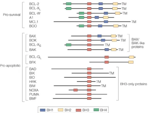
The release of apoptogenic factors from mitochondria is regulated by the interaction of a group of pro- and anti-apoptotic members from Bcl-2 family (14)(Fig.3). Structurally, members of the Bcl-2 family contain up to four conserved homology domains (BH-domains), and optionally a transmembrane segment. Bcl-2, Bcl-xL, Bcl-w, Mcl-1, A1, and CED-9, the homolog in C. elegans, share the BH domains 1–4 and promote cell survival, while mammalian Bax, Bak, Bok, and D. melanogaster DEBCL/DROB are related to Bcl-2 at BH1–3, and promote cell death. There is another sub-class of pro-apoptotic Bcl-2 family members, the BH3-only proteins: mammalian Bad, Bik, Blk, Hrk/DP5, Bid, Bim, Noxa, and C. elegans EGL-1. They share with their relatives only the short BH3 interaction domain (13). Apoptosis is initiated by the BH3-only Bcl-2 family members. And pro-apoptotic Bax and Bak act downstream in this pathway. Notably, Bid works as a conection between death-receptor pathway and the mitochondrial route of apoptosis. As mentioned before, upon CD95-ligation, Bid is cleaved by caspase-8 and migrates to the mitochondria to promote the release of cytochrome c. There is also evidence showing that Bid directly interactes with Bax and Bak.
Strasser, A. (2005) Nature Rev.Immunol. 5, 189-200
Fig 3. Mammalian 2 family member: The pro-survival family members — 2, XL,
BCL-W, A1, MCL1 (myeloid-cell leukaemia sequence 1) and BOO (BCL-2 homologue of ovary) — each have two or four distinct BCL-2-homology (BH) domains. The pro-apoptotic members of the BCL-2 family can be subdivided into at least two groups: the BAX /BAK-like proteins — BAX, BOK (BCL-2-related ovarian killer), BCL-XS and BAK; and the BH3-only proteins — BAD, BIK, BID, HRK
(harakiri), BIM, NOXA, PUMA and BMF (BCL-2-modifying factor) — which share with each other and other members of the BCL-2 family only the short (9–16 amino acid) BH3 domain.
After an infection is cleared, most T cells generated duringthe clonal expansion phase are removed through either of twoapoptotic pathways, termed activation-induced cell death (AICD) and activated cell autonomous death (ACAD). AICD is thought to depend mainly on an extrinsic form of apoptosis induced by the ligation of death receptors such as Fas on the surface of the T cell. ACAD is thought todepend on an intrinsic form of cell death which is regulated by Bcl family (15).
TCR restimulation of already expanded T cells in the absence of appropriate co-stimulation may also lead to the efficient induction of cell death. This is called activation-induced cell death (AICD) (16;17).
AICD involves stimulation through CD95 (18;19) TNFR1 (20) TRAILR (21-23) and other mechanisms independent of cell-death receptor (24). Some findings suggest that TNF is involved in the late phase of AICD (25). AICD can be mimicked by an in vitro model system using TCR-activated T cells cultured with IL-2 and restimulated through the TCR or by PMA and ionomycin (26).
After approximately 4 to 7 days of TCR restimulation in vitro culture results in the expression of CD95L (27-29). This induces CD95-mediated cell death either of the same cell (suicide) or of neighbouring cells (fratricide). The induction of CD95L expression requires calcium mobilization and production of reactive oxygen species (ROS) which is mediated by protein kinase Cθ (PKCθ) (30). Therefore, the CD95 system has been considered to be an important regulator of T-cell homeostasis. Mice with decreased levels of CD95 or mutations in the gene encoding CD95L (lpr or gld mice, respectively) produce autoantibodies and develop lymphoproliferative disease (31;32). However, specific deletion of the gene encoding CD95 in all T cells leads to lymphocyte depletion and pulmonary fibrosis-like disease (33). Therefore, CD95 inactivation in T cells alone is not sufficient for the pathogenesis of lymphoproliferation. These results indicate that in addition to the CD95 system, there are other CD95-independent AICD pathways that regulate T-cell homeostasis.
After TCR triggering, the expression of Bim is also increased, which suggests Bim is a mediator of AICD as well. Upregulation of Bim expression by TCR triggering
depends on the activities of p38 and JNK, and may also involve PKC and calcium signaling (24).
Non-caspase proteases have also been implicated in T-cell death. A serine protease called granzyme B plays a role in AICD of TH2 cells but not of TH1 cells (24). Granzyme B is an effector molecule of cytotoxic T cells which is located in intracellular lytic granules (34). Subsequently, granzyme B was found to be upregulated in response to TCR stimulation in TH2 cells and led to AICD which was independent of CD95–CD95L engagement. In granzyme-B-deficient mice, AICD was abrogated in TH2 cells. And the accumulation of TH2-cell-associated cytokines increased their susceptibility to allergen-induced asthma.
It has further been shown that in epithelial cells, lysosomes are also involved in the execution of cell death (35). Lysosomal proteases, such as cathepsin B, are activated by proteolytic processing as a result of lysosomal stress and are released into the cytosol. Cell death by cathepsins might involve the mitochondria (36;37), or cell-death-receptor stimulation (38).
1-1-4 Activated cell-autonomous death (ACAD)
Autonomous cell death occurs in the absence of appropriate survival signals such as cytokine deprivation or by neglect. This is called activated cell-autonomous death (ACAD) (39).
Bim is essential for ACAD. The expression of Bim or PUMA increased after cytokine deprivation (40). On the mitochondrial membrane, Bim or PUMA can bind Bcl-2 or Bcl-xL, which abrogates the inhibition of Bax or Bak and results in cytochrome c
release and the induction of T-cell death (41;42). During the expansion phase T cells can switch from a Bcl-xL high to a Bcl-xL low state, which allow the activation of the intrinsic apoptotic pathway (43). Bad also plays a role in activating or enhancing apoptosis in cells removed from required cytokines. In the presence of cytokines, Bad is phosphorylated by kinases such as Akt, PKA, and Rsk and is sequestered by 14-3-3 proteins in the cytosol. Upon removal of cytokine, Bad is dephosphorylated and translocates to the mitochondria, where it binds Bcl-2 and Bcl-xL and inhibits their antiapoptotic function (15).
1-2 T cell memory
Prior to contact with antigen, naive T cells congregate in the secondary lymphoid tissues (spleen, lymph nodes, and Peyer's patches) and migrate continuously from one lymphoid organ to another via blood and lymph (44). After encountering pathogens, naive T cells go through clonal expansion and differentiation into effector cells. In a typical viral infection, this will last 7–10 days until the pathogen is eliminated. After that, these activated T cells will undergo clonal shrinkage to keep T cell homeostasis. The disappearance of activated T cells at the end of the primary response involves two distinct mechanisms, death and homing to nonlymphoid tissues. First, many of the activated T cells leave the spleen and localize in nonlymphoid tissues, notably the lungs, liver, and gut (44). Second, in both lymphoid and nonlymphoid tissues, the majority of the T cells (90–95%) die within 5 days, those cells that survived become memory cells (45) (Fig.4).
Harty, J. T. and Badovinac, V. P. (2008) Nat.Rev.Immunol. 8, 107-119
Fig 4. Inflammatory cytokines and CD8+ T-cell homeostasis after infection: Antigenic peptides
presented naive CD8+ T cells trigger their proliferative expansion and differentiation into effector
CD8+ T cells.5–10% of CD8+ T cells detected at the peak of the expansion survive the contraction
phase and initiate the memory CD8+ T-cell pool. The contraction phase is diminished in the absence of
inflammation (for example the absence of IFN ) or when the balance and activation state of pro- and anti-apoptotic BCL-2 family members expressed by CD8+ T cells is altered. Diminished contraction
Based on the expression of homing molecules on their surface, memory T cells can be divided into two subsets. CD62Llow CCR7(CC-chemokine receptor 7)low are defined as effector memory T cells, while CD62Lhi CCR7hi are defined as central memory T cells (46-48).
Current evidence suggests that the survival of memory T cells is not dependent on persistent antigen. In fact, memory T cells are less dependent on antigen receptor engagement for survival than naive T cells. Instead, cytokines are important in maintaining memory T cell viability. After pathogen and antigen are cleared in acute infection, memory CD8+ T cells are maintained by signals through receptors that contain the common cytokine receptor γ-chain such as receptors for IL-7 and IL-15. IL-7 and IL-15 appear to deliver survival and proliferative signals to memory CD8+ T cells, respectively (49;50).
1-2-1 Clonal expansion
Naive T cell activation needs both peptide–MHC complexes (signal 1), and co-stimulatory signals (signal 2) (51;52). Moreover, activation of innate immune cells induces the production of pro-inflammatory cytokines, such as type I IFNs, IL-12 and IFNγ (53). Recent studies demonstrated that pro-inflammatory cytokines act directly on responding CD8+ T cells to affect crucial aspects of memory generation (signal 3). Early in vitro studies showed that the addition of IL-12 or type I IFNs to T-cell cultures enhanced the proliferation and survival of the activated T cells. These studies led to the concept that signal 3 was important for optimal CD8+ T-cell responses (54;55). T cells deficient in various cytokine receptors (for example, for type I IFNs,
IL-12 or IFNγ) have reduced expansion (56-58). Importantly, the absence of these pro-inflammatory cytokine receptors does not compromise proliferation of these gene-deficient TCR-transgenic T cells; instead it decreases their survival rates (56-58). How these cytokines act on the survival of activated T cells is not clear yet, but it’s possible that they might increase the expression of pro-survival molecules such as Bcl-3 or potentially alter the balance of pro-apoptotic and anti-apoptotic Bcl-2-family members to promote T-cell survival during proliferation (54;59).
1-2-2 Clonal contraction
Following acute or persistent infection, activated T-cells go to clonal contraction through twoapoptotic pathways, AICD and ACAD (60).
Early in vitro studies showed that effector, but not memory CD8+ T cells exhibited heightened sensitivity to AICD through CD95 and TNF-mediated cell-death pathways (61;62), whereas in vivo studies showed normal contraction of CD8+ T cells that lack both of these molecules (63;64). Current studies show that the balance and activation state of pro- and anti-apoptotic Bcl-2 family members modulate the death of activated T cells (39;65-68). Deletion of Bim severely compromises the death of superantigen-activated T cells in vivo (39) and results in reduced contraction of antigen-specific CD8+ T cells after herpesvirus infection (66). However, in Bim-deficient mice infected with LCMV, some antigen-specific CD8+ T-cells eventually contract. This suggests that additional pathways might contribute to the death of activated CD8+ T cells (14;69;70).
Inflammatory cytokines such as IFNγ and IL-12 can influence the contraction phase as well. CD8+ T cells failed to contract in BALB/c IFNγ-deficient mice after infection with an attenuated (actA-deficient) strain of L. monocytogenes that was otherwise cleared in wild-type and immunocompromised hosts (71). Also, BALB/c IFN γ-deficient mice and B6 IFNγ-deficient mice have reduced contraction after LCMV infection (71;72). After infection with L. monocytogenes, both CD8+ and CD4+ T cells exhibit abnormal contraction in IFNγ-deficient or IFNγR-deficient mice that were treated with antibiotics to eliminate the infection at day 4. So, persistent infection does not explain how IFNγ regulates CD8+ and CD4+ T-cell contraction after infection with L. monocytogenes (73;74). IFNγ might act indirectly through cells other than T cells to influence CD8+ T-cell contraction (72;75). Finally, the absence of the IL-12 receptor on antigen-specific T cells not only reduced expansion but also reduced contraction after infection (76). So, pro-inflammatory cytokines contribute important signals to regulate the contraction phase, perhaps by regulating Bcl-2-family members.
Recent studies also suggested that pro-inflammatory signals, specifically IL-12, control the amount of T-bet expression in the responding CD8+ T cells (77). In this model, relative T-bet expression is thought to differentiate short-lived effector CD8+ T cells (T-bethi) from memory precursor effector CD8+ T cells (T-betlow) and thus determines the cell’s fate. However, T-bet-deficient TCR-transgenic CD8+ T cells from P14 mice still contract by more than 80% from the peak of the response following LCMV infection (77) and the LCMV-specific CD8+ T-cell response
appears to undergo normal contraction in T-bet-deficient mice (78). So the link between T-bet and the contraction of the CD8+ T-cell response is still not clear.
How a precise fraction (5–10%) of the responding CD8+ T cells survives to initiate the memory pool is still not clear. Recent data demonstrate that naive CD8+ T cells undergo an initial asymmetric division after interaction with APCs (79). This raised the possibility that the segregation between long-term memory T cells versus short-lived effector-T-cell may be a very early event in the CD8+ T-cell response.
Many molecules are involved in T cell activation and apoptosis program. Through DNA microarray analysis of the mRNA expression profile of activation versus resting mouse T cells, we identified a group of differentially expressed genes, which are likely important in T-cell activation and apoptosis. Drak2 is a gene in this group.
2. DAPK and Drak2
2-1 Death-associated protein kinase (DAPK)
Death-associated protein kinase (DAPK) is a Ca2+/calmodulin (CaM)-regulated Ser/Thr kinase that mediates cell death (80-84). Increased DAPK activity, due to overexpression of the kinase, leads to pronounced death-associated cellular changes, which include membrane blebbing, cell rounding, detachment from extracellular matrix, and the formation of autophagic vesicles. Furthermore, DAPK activity is necessary for the induction of cell death by multiple death signals, including those
generated by death receptors, cytokines, matrix detachment, and oncogene-induced hyperproliferation (80).
DAPK belongs to a family of related death kinases, all of which share significant sequence and functional homology (85). This family consists of DAP (86) , DRP-1(DAPk-related protein 1, also known as DAPk2) (87), ZIP kinase [also known as Dlk (DAP-like kinase) or DAPk3] (88), DAPK2 (89), Drak1 and Drak2 (DAPK-related apoptosis-inducing protein kinase-1 and -2) (90)(Fig.5). Each DAPk family member contains at its N terminus a catalytic domain composed of the typical 11 subdomains found in all Ser/Thr kinases (80). Phylogenetically, the DAPK family is most closely related to the family of CaM-regulated kinases, in particular to myosin light chain kinase, which shares 44% identity within the corresponding catalytic domain (83). Beyond the common kinase domain, the family members differ in structure. DAP, DRP-1 and DAPK2 have a calmodulin regulatory domain following the kinase domains, ZIP, Drak1 and Drak2 do not (85). This domain serves to suppress catalytic activity by binding to the catalytic cleft, and functions as a pseudosubstrate. In addition, this domain undergoes autophosphorylation at Ser308, an inhibitory event that reduces its affinity to CaM and may further stabilize its docking within the substrate-binding site (80). The C terminus of DAPk contains a death domain, followed by a 17-aa tail rich in Ser residues, a feature common to other death domain-containing proteins (87). The 52-kDa ZIPk possesses a C-terminal leucine zipper motif, which mediates homodimerization and interactions between ZIPk and additional leucine zipper-containing proteins, such as activating transcription factor (ATF)4 (88).
Bialik, S. and Kimchi, A. (2006) Annu.Rev.Biochem. 75, 189-210
Fig 5. Death-associated protein kinase (DAPK) family member: The numbers above the proteins demarcate the amino acid position of each domain. The quantities within the kinase domains indicate the degree of amino acid identity to the kinase domain of DAPk.
2-2 DRAK2
Drak2 shares about 50% identity in the kinase domain with other members of the DAPK family (86). DAP, DAPK2, and DRP-1 are localized in the cytosol, ZIP kinase and Drak1 reside mainly in the nuclei, whereas Drak2 is found in both the cytosol and nuclei (90;91), suggesting different mechanisms of action. An in vitro kinaseassay revealed that both DRAKs are autophosphorylated and phosphorylate myosin light chain as an exogenous substrate (91). Furthermore, overexpressionof both DRAKs induces the morphological changes of apoptosis inNIH 3T3 cells, suggesting the role of DRAKs in apoptotic signaling (90). CHP interacts with the carboxyl-terminal
region of the kinase domain of DRAK2 and specifically inactivates DRAK2 kinase activity (92).
In Drak2−/− mice, Drak2−/− T cells did not demonstrate any defects in apoptosis or negative selection. However, T cells from Drak2−/− mice exhibited enhanced sensitivity to T cell receptor-mediated stimulation with a reduced requirement for costimulation. Drak2−/− mice were remarkably resistant to experimental autoimmune encephalomyelitis (EAE) (93). This study suggests that DRAK2 raises the threshold for T cell activation by negatively regulating signals through the TCR.
However, our study (to be elaborated later) demonstrated that the conclusion based on Drak2-/- mouse study was flawed, due to the superficial investigation by McGargill et al. As a matter of fact, Drak2 is actively involved in T-cell apoptosis and memory T cell development (94).
At the molecular level, apoptosis of T cells and other type of cells shares many common mechanisms. The important role of Drak2 in T-cell apoptosis led us to believe it is also essential in the survival of other type of cells, such as β-cells in the pancreatic islets, which are vital in maintaining normal blood glucose levels. Pathological changes in islets lead to diabetes.
3. Diabetes
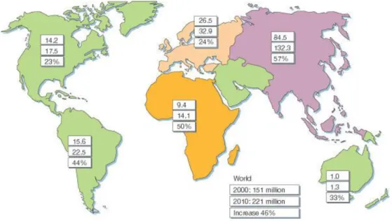
Diabetes mellitus is one of the most common endocrine disorders in the world (Fig.6). Now, almost 6% of the world’s population is affected by this disease. And the number will reach 300 million in 2025, within them, 97% will be type 2 diabetes (95).
Zimmet, P., Alberti, K. G., and Shaw, J. (2001) Nature 414, 782-787
Fig 6. Numbers of people with diabetes (in millions) for 2000 and 2010 (top and middle values,
respectively), and the percentage increase.
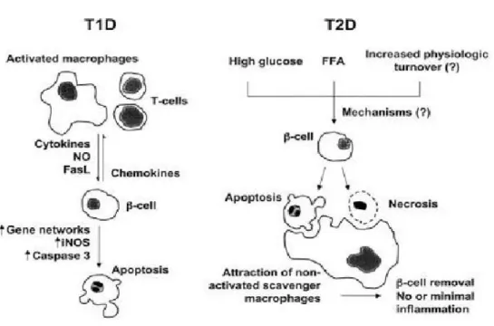
There are two main forms of diabetes, type 1 and type 2. Both types are characterized by progressive ß-cell failure (Fig.7). Type 1 diabetes is caused by anautoimmune assault against the ß-cells (96), while the cause of type 2 diabetes is more variable, including environmental factors such as physical inactivity, obesity and genetic factors. Environmental factors can modify the expression of susceptible diabetes genes, and cause ß-cellfailure and insulin resistance (96).
Cnop, M., Welsh, N., Jonas, J. C., Jorns, A., Lenzen, S., and Eizirik, D. L. (2005) Diabetes 54 Suppl 2, S97-107
Fig 7. ß-cell death in T1D and T2D: Overview of the putative sequence of events leading to β-cell
death in animal models of type 1 and type 2 diabetes. 3-1 Type 1 diabetes
In type 1 diabetes, β-cell mass is reduced by 70–80%at the time of diagnosis (97). Because of the variable degrees ofinsulitis and absence of detectable β-cell necrosis, it was suggested that β-cell loss occurs slowly over years. In antibody-positive individuals, progressivedecline in first-phase insulin secretion, impaired fasting or glucose tolerance are usually found long before the development of overt diabetes (97). For patients with long-term type 1 diabetes, some β-cell function remains (C-peptide secretion), although β-cell mass is usually decreased to less than 1% of normal (98).
The β-cell loss is due to autoimmune attack. Typically in type 1 diabetes, autoantibodies reacting with glutamic acid decarboxylase (GAD65), insulin, and
insulinoma antigen-2 can be found (99). Some adults with type 2 diabetes also express anti-islet autoantibodies (most often GAD65 autoantibodies). These patients are called LADA (latent autoimmune diabetes of the adult). They have both insulin resistance as well as anti-islet autoimmunity. So type 1 and type 2 diabetes can coexist (100). β-cell death in the course of insulitis is caused by direct contact with activated macrophages and T-cells, and/or exposure to soluble mediators secreted by these cells, including cytokines, nitric oxide (NO), and oxygen free radicals (101). The execution of β-cell death occurs through activation of mitogen-activated protein kinases, ER stress and by the release of mitochondrial death signals (96). In vitro exposure of β-cells to IL-1β, or to IL-1β + IFN-γ, causes functional changes similar to those observed in pre-diabetic patients, such as elevated proinsulin/insulin levels (102), and a preferential loss of first-phase insulin secretion in response to glucose.
Apoptosis is the main cause of β-cell death at the onsetof type 1 diabetes. It is a highly regulated process, which is activatedand/or modified by extracellular signals, intracellular ATPlevels, phosphorylation cascades, and expression of pro- and anti-apoptotic genes (101). Cytokines induce stress response genes that are either protective or deleterious for β-cellsurvival. There are around 700 genes identified to be up- or downregulated in purified rat β-cells orinsulin-producing cells after 1–24 hour of exposure to IL-1βand/or IFN-γ by microarray experiments (Fig.8). IL-1β activates the transcription factor nuclear factor (NF)-κB in rodent and human islet cells (101), and IκB protects pancreatic β-cells against cytokine-inducedapoptosis (103;104). NF-κB was also found to downregulate the expression of other transcription factors responsible for β-cell differentiation and function. NF-κB
regulates expressionof inducible nitric oxide synthase (iNOS) in β-cells, and 50% of the ß-cell genes modified after 12h of cytokine exposure are secondary to iNOS-mediated NO formation. So IL-1β induced NF-κB activationplays a crucial role in controlling multiple and distinct generegulatory networks in β cell. It affects the β-cell differentiation and ER Ca2+ homeostasis, attracts and activates immune cells,and directly contributes to β-cell apoptosis.
Cnop, M., Welsh, N., Jonas, J. C., Jorns, A., Lenzen, S., and Eizirik, D. L. (2005) Diabetes 54 Suppl 2, S97-107
Fig 8. The transcription factor and gene networks putatively involved in the cytokine-promoted ß-cell
"decision" to undergo apoptosis: The transcription factors NF-κB and STAT-1 are the main regulators of the pathways triggered by IL-1β and IFN-γ, respectively.
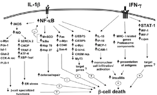
However, exposure of purified human or rodent β-cells to IL-1βalone is not sufficient to induce apoptosis, but when IL-1βis combined with IFN-γ, 50% of these cells undergo apoptosisafter 6–9 days (101). This suggests that IFN-γ signal transduction must synergize with IL-1β signaling to trigger β-cellapoptosis (Fig.9). IFN-γ binds to cell surface receptors and activates the tyrosine kinases JAK1 and JAK2. These
kinases phosphorylate the transcription factor STAT-1, which dimerizes and translocates to the nucleus to bind to γ-activated sitesof diverse genes (101). STAT-1 mediates the potentiating effect of IFN-γ on IL-1β–induced iNOS expression. Fluorescence-activated cell sorting (FACS)-purified β-cells from STAT-1–deficient mice (STAT-1–/–) are protected against IL-1β+ IFN-γ–induced apoptosis. Because excessive activation of JAK/STAT signaling may lead to cell death, STAT transcriptionalactivity is regulated by multiple negative feedback mechanisms.These include dephosphorylation of JAK and cytokine receptorsby SHP, and inhibition of JAK enzymatic activities by the suppressor of cytokine signaling (SOCS) family. Upregulation of SOCS-1 or SOCS-3 protects β-cells in vitro and in vivo against cytokine-induced death. SOCS-3 also protects insulin-producingcells against IL-1β– mediated apoptosis via NF-κBinhibition. The results indicate thatβ-cell fate after cytokine exposure depends on theduration and severity of perturbation of key β-cell gene networks. NF-κB and STAT-1 play important roles in this network.
Cnop, M., Welsh, N., Jonas, J. C., Jorns, A., Lenzen, S., and Eizirik, D. L. (2005) Diabetes 54 Suppl 2, S97-107
Fig 9. Cytokine-induced ß-cell apoptosis: Arrows indicate genes for which expression was modified by
cytokines in a time course microarray analysis. β-Cell apoptosis is probably mediated by three main pathways—namely JNK, ER stress, and liberation of pro-apoptotic proteins from the mitochondria.
Another question is to understand how the cytokine-activated geneexpression results in β-celldeath. The probable mechanisms include the following: 1) Fas/FasL pathway (105), 2) activation of the stress-activatedprotein kinases c-Jun NH2-terminal kinase (JNK), p38 mitogen-activated protein kinase (MAPK), and extracellular signal-regulated kinase(ERK); 3) triggering of ER stress; and 4) the release of deathsignals from the mitochondria.
Effector T cells induce apoptosis of β-cells. This then leads to absolute insulin deficiency and clinically manifest as diabetes, at least in the murine model of the autoimmune diabetes-NOD (nonobese diabetic) mice. T cell effector pathways involving Fas/Fas-ligand (Fas-FasL) interaction or the perforin/granzyme system are primarily responsible for the beta cell destruction. Perforin production from CD8+ T cells initiates the immune response, and then Fas/FasL interaction causes CD4+ T cell-induced beta cell death (100; 104).
JNK is a member of the MAPK family. Pancreatic β-cellsexposed to IL-1β have an early and sustained increasein JNK activity, which can be potentiated by IFN-γ or TNF-α (101;106).Cell-permeable peptide inhibitors of JNK prevent cytokine-induced apoptosis in insulin-producing cells (107), but this remainsto be confirmed in primary β-cells. p38 MAPK and ERKare also activated by cytokines, and pharmacological
probably by attenuating transcriptional activationof iNOS. In addition, the tumor suppressor p53 is activated inresponse to cytokine-induced NO production (110). It is conceivablethat stabilization of the pro-apoptotic protein p53 lies downstreamof the NO-induced activation of MAPKs.
Disruption of ER homeostasis, as induced by changes in ER Ca2+ concentrations, triggers accumulation of unfolded proteins andactivation of a specific stress response, which is called the ER stressresponse. This cellular response is a coordinated attempt to restore ER homeostasis and function, and it includes translational attenuation, upregulation of ER chaperones, and degradationof misfolded proteins. In prolonged and severe ER stress,the apoptosis program will be activated and executed by the transcriptionfactor CHOP (C/EBP(CCAAT/enhancer binding protein) homologous protein), MAPK JNK, and caspase-12.Because of their high rate of protein synthesis, β-cellsare particularly susceptible to ER stress (111). And NO induced ER stress response in β-cells leads toCHOP expression and apoptosis (112).
Mitochondria play animportant role in β-cell functionand survival (113), as well as in triggering apoptosis. Members of theBcl-2 protein family regulate the mitochondrial response topro-apoptotic signals (114), preventing release of mitochondrialproteins such as cytochrome c. When cytochrome c is liberated to the cytosol, it will sequentially activate caspase-9 and -3 and executecell death (115). Overexpression of Bcl-2 partially protects mouse (116) and human (117) islets againstcytokine-induced cell death, but does not prevent adenovirus-induced islet cell death (118) or spontaneous diabetes in NOD mice (119). Caspase-1 is induced in β-cells exposed to IL-1ß+ IFN-γ (120) , and blocking caspase-1 decreasesβ-cell apoptosis after 4 days
exposure to cytokine, but can not prevent their subsequent death by necrosisafter 9 days. These results suggest the possibility that during the early stage of cytokine exposure, nucleus, mitochondria, and ER could work togther to decide the fate of β-cell to undergo apoptosis or not. Other pro-apoptotic genes that are induced by cytokines, as detected by microarrayanalysis (121), includes Bid, Bak,and caspase-3.
3-2 Type 2 diabetes
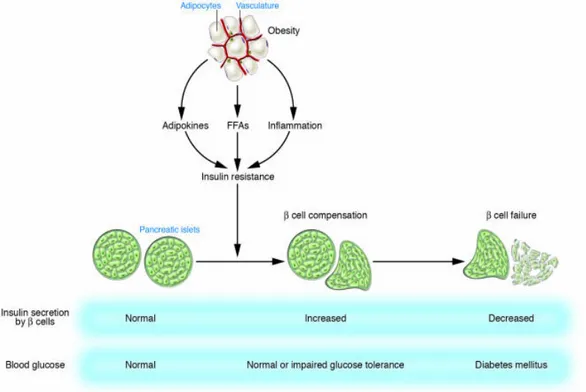
Modern lifestyle, with abundant nutrient supply and reduced physical activity, resulted in dramatic increases of type 2 diabetes (95). It occurs when the endocrine pancreas fails to secrete sufficient insulin to cope with the metabolic demand (122). Initially, reduced insulin sensitivity is the major problem, but at later stage, islet apoptosis also occurs (Fig.10). Three mechanisms are involved in type 2 diabetes: 1. Insulin risistance, 2. β-cell secretory dysfunction, 3. decreased β-cell mass. In type 2 diabetic subjects, initial pathological studies suggested a β-cell loss of 25–50%. However, recent studies showed a significant reduction in β-cell mass and a threefold increase in β-cell apoptosis (122).
Kasuga, M. (2006) J.Clin.Invest 116, 1756-1760
Fig 10. Development of type 2 diabetes: Insulin resistance associated with obesity is induced by
adipokines, FFAs, and chronic inflammation in adipose tissue. Pancreatic β cells compensate for insulin resistance by hypersecretion of insulin. However, at some point, β cell compensation is followed by β cell failure, and diabetes ensues.
3-2-1 Insulin resistance
Changes in lifestyle, such as consumption of a high-calorie diet and lack of exercise, have increased the global prevalence not only of diabetes but also of obesity. Between 60% and 90% of cases of type 2 diabetes now appear to be related to obesity. Insulin resistance, which is an impairment of insulin action that precedes the development of hyperglycemia is usually associated with obesity. It is therefore important to characterize the mechanisms of insulin resistance associated with obesity in order to develop approaches to prevent type 2 diabetes.
Adipokines are a group of hormones and cytokines that are produced by adipocytes. Adipocytes can store excess lipids and this fuction becomes saturated in obesity, resulting in abnormal redistribution of lipids to other organs and tissues. In the ob/ob mouse, which exhibits hyperphagia, hyperlipidaemia and insulin resistance, the mutated gene is the cytokine-related molecule leptin (123). Leptin and adiponectin have been categorized as 'anti-diabetogenic'; their common function is to decrease triglyceride (TG) synthesis, stimulate β-oxidation and enhance insulin action in both skeletal muscle and liver. These effects can be explained in part by their common ability to activate 5'-AMP-activated protein kinase (AMPK) (124). This enzyme responds to a fall in ATP and a rise in AMP levels by activating both glucose and fatty acid oxidation. Interestingly, leptin levels are increased and adiponectin levels are decreased in insulin-resistant obese humans and animals, which suggests that obesity leads to a state of leptin resistance and adiponectin deficiency.
Animals lacking white adipose tissue have severe hepatic and muscle insulin resistance, and exhibit large increases in TG stores in both tissues (125). This further proves the importance of adipose tissue in the normal regulation of insulin action. Transplantation of normal fat tissue into such mice restores insulin sensitivity (126). Leptin infusion ameliorates insulin resistance in these mice (127;128), while transplantation of fat from leptin-deficient mice into such animals fails to improve insulin sensitivity (129). These data suggest that leptin seems to be the major player in this model. Furthermore, leptin administration to humans with severe lipodystrophy partially reverses their insulin resistance and hyperlipidaemia (130).
Adipose cells also produce other peptide hormones, including adiponectin, retinol-binding protein-4 (RBP4) and resistin, and proinflammatory cytokines such as IL-6 and TNFα (124;131).
Mice with an adipose-specific knockout of the Glucose transporter type 4 (GLUT4) have impaired insulin sensitivity in muscle and liver (132). Circulating RBP4 levels are increased in these mice, and infusion or transgenic expression of RBP4 in normal mice causes insulin resistance (133). Interestingly, food deprivation (fasting) also causes a form of insulin resistance and is associated with a decrease in adipose GLUT4 expression (134). So the original purpose of adipocyte-derived insulin-desensitizing molecules, such as RBP4, TNFα and resistin, may have been to prevent hypoglycaemia in the fasted state, while in modern life style, with abundant nutrient supply, the function of these molecules has been subverted to pathophysiology (135). 3-2-1-2 Inflammatory mediators
Insulin resistance may also caused by inflammatory component (136). High-fat diets or obesity result in activation of the transcription factor NF-κB and its targets in the liver. Overexpression of a constitutively active version of the NF-κB-activating kinase, IκB kinase catalytic subunit-β (IκKβ), in the liver of normal rodents results in liver and muscle insulin resistance and diabetes. In addition, both high-fat feeding and IκKβ overexpression increase hepatic production of IL-6, IL-1β and TNFα; antibody-mediated neutralization of IL-6 in animals fed on a high-fat diet partially restores insulin sensitivity (137). Deletion of IκKβ in the liver can protect mice from diet-induced hepatic insulin resistance, while muscle and adipose insulin resistance still develop. However, myeloid cells specific IκKβ knockout mice remain globally
insulin sensitive (138). In rodents, after one week on high-fat feeding, adipocyte increases the expression of monocyte chemotactic protein-1 (MCP1), which recruits macrophage to adipocytes (139;140). This may be a mechanism by which inflammatory signalling is enhanced during the development of diabetes. Overall, evidence is accumulating that insulin resistance is at least partly caused by changes in hormone and cytokine production by the liver, adipose tissue and infiltrating immune cells in response to chronic exposure to lipids and other metabolic fuels.
3-2-1-3 Metabolic overload in the liver
Lipid species accumulation in the liver results in the redirection of long-chain acyl CoAs (LC-CoAs) into ER-localized and cytosolic lipid species, such as diacylglycerols (DAGs), ceramides and TGs. This is thought to be regulated mostly by glucose-induced increases of malonyl CoA. Malonyl CoA serves both as the immediate precursor of de novo lipogenesis and as an inhibitor of carnitine palmitoyltransferase-1 (CPT1), which is the rate-limiting enzyme for import of LC-CoAs into the mitochondria for β-oxidation (141). Indeed, infusion of lipids or ingestion of high-fat diets in rodents leads to the accumulation of TGs, LC-CoAs, DAGs and ceramides (142-144). Suppression of mitochondrial glycerol-3-phosphate acyltransferase-1 (GPAT1, the first enzyme in TG synthesis) or acetyl CoA carboxylase-2 (ACC2) activity results in increased fatty acid oxidation, lowered DAG levels and reversal of hepatic insulin resistance (145-147). Pharmacological inhibition of Ser palmitoyltransferase-1 (SPT1) or genetic knockout of dihydroceramide desaturase-1 (Degs1) — both of which are involved in the synthesis of ceramides from the saturated precursor palmitoyl CoA — prevented hepatic insulin resistance