Effects of cardiorespiratory exercise on motor skill learning and cognitive executive functions in Parkinson’s disease
par Catherine Duchesne
Département de psychologie, Université de Montréal
Faculté des arts et des sciences
Thèse présentée à la Faculté des arts et des sciences en vue de l’obtention du grade de doctorat (Ph. D.)
en psychologie -recherche et intervention- option neuropsychologie clinique
Décembre, 2016
II
La maladie de Parkinson (MP) est la deuxième maladie dégénérative la plus répandue au Canada. Elle se caractérise par des symptômes moteurs importants tels le tremblement de repos, la rigidité musculaire, l’instabilité posturale, la lenteur dans les mouvements ainsi que par des symptômes non moteurs, notamment une diminution du fonctionnement cognitif. Conséquemment, la nature hétérogène des symptômes de la MP dirige bien souvent l’individu atteint vers une sédentarité physique et mentale involontaire.
Les traitements pharmacologiques et neurochirurgicaux demeurent les approches thérapeutiques majoritairement choisies. Toutefois, de plus en plus d’études visant à examiner les impacts de l’exercice physique aérobique (EPA) ont démontré des bénéfices de ce traitement non pharmaceutique, entre autres, en améliorant les symptômes moteurs de la maladie. Néanmoins, l’effet de l’exercice sur la cognition et l’apprentissage moteur à travers la MP est encore méconnu. Ainsi, ce projet de thèse vise à étudier les changements cognitifs et moteurs suite à un entrainement cardiovasculaire. Une première étude visait à mesurer les changements comportementaux au niveau de la capacité aérobique, des fonctions cognitives dites exécutives et de l’apprentissage procédural moteur suite à l’EPA. Une deuxième étude utilisant l’imagerie par résonance magnétique fonctionnelle (IRMf) permettait ensuite d’identifier les corrélats neuronaux associés à l’effet de l’EPA sur l’apprentissage moteur.
20 participants en santé et 19 atteintes de la MP, âgées entre 40-80 ans, ont participé à un programme d’entrainement de 3 mois (3 fois/semaine) à une intensité élevée, débutant à 20 minutes (+5 minutes/semaine) pour atteindre 40 minutes d’EPA. Le niveau d’intensité de
III
(VO2peak, pression artérielle, fréquence cardiaque) et neuropsychologiques (« Stroop, trail making test » (TMT)) ont été prises en début et à la fin de l’entraînement. De plus, des sessions
d’acquisition de données cérébrales fonctionnelles grâce à l’IRMf ont été administrées durant la passation d’une tâche d’apprentissage moteur implicite (tache : « Serial Reaction Time Task »(SRT)).
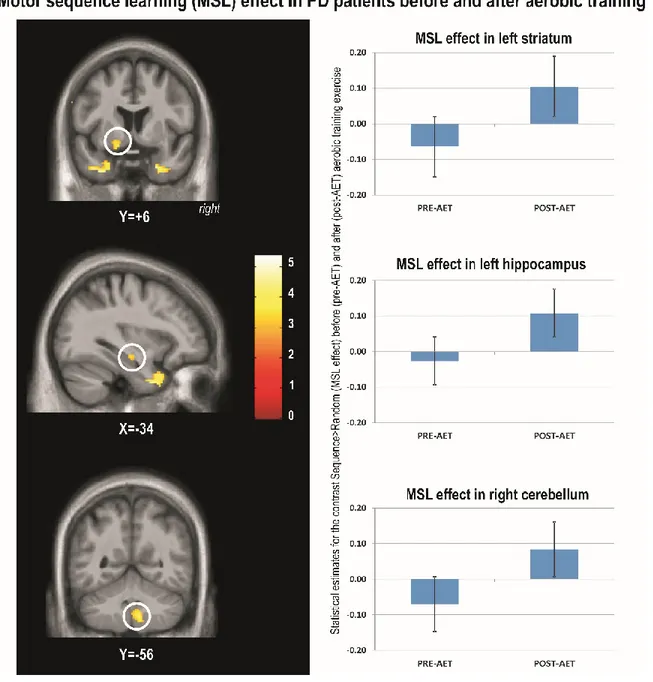
Les résultats ont montré que l’entraînement en EPA fut efficace car une amélioration significative de la capacité aérobique fut observée chez tous les participants. Au niveau comportemental, l’EPA a eu pour effet d’améliorer les capacités d’inhibition (Stroop) et d’apprentissage moteur (SRT), mais pas celle associée à la flexibilité mentale (TMT). Pour leur part, les données de neuroimagerie ont révélé une augmentation de l’activité fonctionnelle liée à l’amélioration de l’apprentissage moteur au niveau de l’hippocampe, du striatum et du cervelet, et ce en comparaison avec les sujets contrôles. De plus, les changements fonctionnels chez les individus atteints de la MP étaient corrélés au changement de la capacité aérobique : une relation positive fut liée à l’augmentation de l’activité de l’hippocampe et du striatum, tandis qu’une relation négative fut observée au niveau du cervelet.
Ce projet est l’un des premiers à mettre en lumière l’impact clinique d’un traitement non pharmaceutique visant à améliorer la nature motrice et cognitive des symptômes de la MP, ainsi que de proposer les mécanismes neurofonctionnels pouvant expliquer l’amélioration observés au niveau de l’apprentissage suivant l’entrainement en EPA. Ainsi, nous croyons que les résultats de cette étude serviront les milieux cliniques et la population de patients atteints
IV
Mots-clés : Maladie de Parkinson, exercice, cognition, moteur, fonctions exécutives, mémoire procédurale, neuroimagerie
V
Parkinson’s disease (PD) is the second most common neurodegenerative disorder in Canada. It is mainly characterized by important motor symptoms such as slow movement, tremor, rigidity and problems with locomotion, but non-motor symptoms (NMS) such as cognitive dysfunctions are also present early in the disease. Inadvertently, the heterogeneous nature of PD’s symptoms may lead to an unintentional sedentary behaviour both at the physical and mental level.
To date, the most common forms of PD treatments remain pharmacological and neurosurgical in nature. Most recently, however, studies have demonstrated benefits of aerobic exercise training (AET) as a non-pharmaceutical treatment with significant effects on PD’s motor symptoms. Nevertheless, the effects of exercise on cognitive and motor learning function in PD remain unknown. Thus, this thesis project aims at studying cognitive and motor changes following AET. Most specifically, the first study intended to assess behavioural changes related to aerobic capacity, cognitive (executive) functions and procedural learning following three months of AET. The second article used functional magnetic resonance imaging (fMRI) to identify the neural correlates associated with the effect of AET on motor learning.
Twenty healthy controls (HC) and 19 early PD individuals, aged 40-80 years old, participated in a supervised high intensity stationary recumbent bike training program (3 times/week; 12 weeks). Exercise prescription started at 20 minutes (+5 minutes/week up to 40 minutes) based on participants’ maximal volume of oxygen uptake (pre and post training). Several physical (VO2peak, blood pressure, heart rate) and cognitive (Stroop and Trail making
VI
Task (SRT) during the acquisition of functional neuroimaging data.
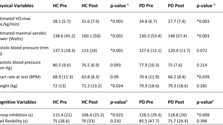
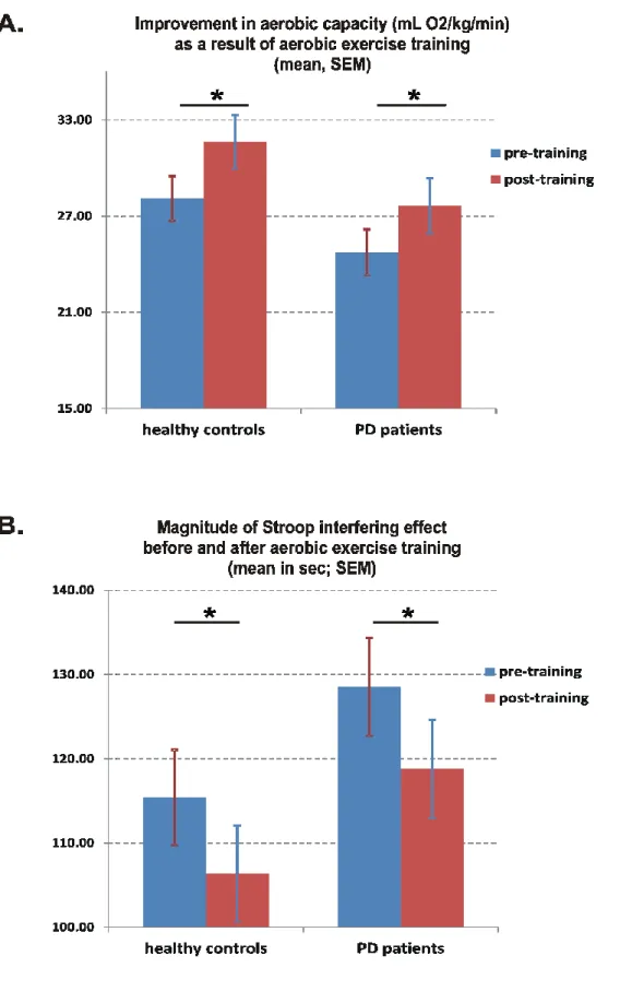
AET program was effective as indicated by a significant improvement in aerobic capacity in all participants. At the behavioural level, AET improved inhibition (Stroop) and motor learning (SRT), but not flexibility (TMT). Brain imaging data revealed pre-post MSL-related increases in functional activity in the hippocampus, striatum and cerebellum in PD patients as compared to controls. Importantly, functional brain changes in PD individuals correlated with changes in aerobic capacity: a positive relationship was found with increased activity in the hippocampus and striatum, while a negative relationship was observed with the cerebellar activity.
This project is one of the first to elucidate the clinical impact of such non-pharmaceutical treatment targeting motor and non-motor aspects of PD. Accordingly, it is believed that the results will be of use for clinical settings and for the population of patients with PD, as they give evidence in favour of an efficient and economical therapeutic solution for PD.
Keywords : Parkinson’s disease, aerobic exercise, cognition, motor, executive functions, procedural learning, neuroimaging
VII Abstract ... v List of Tables ... ix List of Figures ... x List of Abbreviation ... xi Remerciements ... xiii
Chapter I: Theoretical Background ... 1
1. Introduction ... 1
1.1. Parkinson’s Disease Etiology ... 1
1.2. Motor and Non-Motor Dysfunctions ... 1
2. Literature Review ... 5
2.1. Motor Skill Learning ... 5
2.2. Cognition ... 10
2.3. Treatment ... 13
2.4. Aerobic Exercise Training ... 16
3. Thesis’s Aim and Hypotheses... 24
Chapter II: Articles ... 2
Article 1: Enhancing both motor and cognitive functioning in Parkinson’s disease: Aerobic exercise as a rehabilitative intervention ... 2
Article 2: Influence of Aerobic Exercise Training on the Neural Correlates of Motor Learning in Parkinson’s Disease Individuals ... 65
Chapter III: General Discussion ... 27
1. Introduction ... 27
2. Summary of Results ... 104
3. Main scientific contributions ... 105
3.1. Parkinson’s Disease Cardiovascular Health in Comparison to Healthy Aging ... 106
3.2. Parkinson’s Disease Multidimensional Nature ... 108
3.2.1. Preservation of Cognitive Functions ... 109
3.2.2. Restoration of Motor Functions ... 110
VIII
5. Further Reflections on Parkinson’s Disease Heterogeneity and Exercise Variety ... 115
5.1. Perspective on Parkinson’s Disease Subtypes ... 116
5.1.1. Motor Symptoms ... 116
5.1.2. Non Motor Symptoms ... 117
5.2. Perspective on Exercise Diversity ... 119
5.2.1. Exercise as a Preventive Measure... 119
5.3. Exercise Prescription in Parkinson’s Disease Subtypes ... 121
6. Conclusion ... 123
IX
Table 1. Article 1: Demographics of the two groups of participants ... 59 Table 2. Article 1: Group average on physical and cognitive assessments before and after aerobic exercise training for the two groups of participants ... 60 Table 3. Article 2: Demographics of the two groups of participants ... 95 Table 4. Article 2: Functional imaging results of the changes in the main learning effect following aerobic exercise training in PD ... 96 Table 5. Demographics of PD group of participants. ... 112
X
Figure 1. A. Article 1: Diagram illustrating the experimental procedure. Figure 1. B. Diagram
illustrating the motor sequence learning task ... 61 Figure 2. A. Article 1: Aerobic capacity before and after 3 months of training. Figure 2. B. Magnitude of Stroop interference effect before and after aerobic exercise training ... 62 Figure 3. Article 1: Magnitude of motor sequence learning effect before and after aerobic exercise training ... 64 Figure 4. Article 2: Motor sequence learning effect in PD patients before and after aerobic training ... 97 Figure 5. Article 2: Changes in aerobic fitness predict MSL-related changes in functional brain activity in PD patients ... 98 Figure 6. Article 2: Greater MSL-related functional brain changes in PD patients than in healthy controls after AET ... 100 Figure 7. Article 2: Changes in aerobic fitness and MSL-related changes in functional brain activity ... 101 Figure 8: Improvement in aerobic capacity as a results of AET ... Erreur ! Signet non défini.
XI AET: Aerobic exercise training
BDNF: Brain derived neurotropic factors CC: Cortico-cerebellar
CS: Cortico-striatal
DBS: Deep brain stimulation EPA: Exercice physique aérobique FITT: Frequency, intensity, time and type fMRI: Functional magnetic resonance imaging HC: Healthy controls
IRMf: Imagerie par résonance magnétique fonctionnelle MCI: Mild cognitive impairment
MP: Maladie de Parkinson MSL: Motor sequence learning NMS: Non-motor symptoms PD: Parkinson’s disease QOL: Quality of life RT: Reaction time
SRT: Serial Reaction Time STN: Subthalamic nucleus TMT: Trail making test
UPDRS: Unified Parkinson’s disease rating scale VO2max: Maximal volume of oxygen uptake
XIII
Julien, merci de m’avoir fait confiance dans la mise en place d’un nouveau, beau et gros projet de recherche dans le labo. Ce défi m’a fait traverser toutes les gammes d’émotions, mais celle qui ressort davantage est la fierté.
Freja et Gen, à des moments clefs de mon parcours, vous m’avez partagé sans limites vos compétences et passions pour la recherche. Vous êtes des femmes extraordinaires, je vous remercie énormément.
Francine, Arnaud et Ovidiu, vous apportez au labo des ressources incommensurables et complémentaires. Merci de votre compétence et très grande générosité au cours de mon cheminement.
Catherine, je me souviens de t’avoir raconté ma première journée à la maternelle et de mon amie Catherine. Cette association naïve m’avait permis d’apaiser mes peurs et mes angoisses dans mes débuts de scolarisation. Je constate que mon amitié avec toi tout au long du cheminement doctoral m’a permis de nommer ses grandes émotions que nous fait vivre le monde en parallèle du doctorat en R-I. Je te remercie pour ta personnalité si attachante et colorée ainsi que ton écoute et ta présence si importantes durant mon cheminement. Ta compréhension et ton soutien à plusieurs niveaux ont rendu mon expérience doctorale humaine et je t’en suis fort reconnaissante.
Maman et papa, votre soutien et amour inconditionnel sont la base essentielle d’un si grand accomplissement. Cet amour a permis de faire fleurir mon autonomie, une capacité indispensable pour l’obtention du doctorat. Merci.
JB, la personne que l’on côtoie le plus souvent est malheureusement celle qu’on oublie de remercier à sa juste valeur. J’ai tant de reconnaissance pour toi. Tu as été le premier à m’encourager à m’inscrire au doctorat et jamais depuis tu n’as douté une seule fois de ma réussite. Tu as su t’adapter aux circonstances difficiles et m’offrir tous les besoins nécessaires pour vivre cette expérience harmonieusement. Merci du fond du cœur, je t’aime.
Chapter I: Theoretical Background
1. Introduction
1.1.
Parkinson’s Disease Etiology
Parkinson’s disease (PD) is the second most common neurodegenerative disorder after Alzheimer’s disease. It affects approximately 1% of the population over the age of 55 years and has a mean age onset of about 60 years (Hayes, Fung, Kimber et O’Sullivan, 2010). In Canada alone, PD is affecting more than 100,000 Canadians today, and unfortunately, current demographic trends predict that the number of cases will double by 2050 due to the expected increase in the size of our elderly population. As such, the development of knowledge and the implementation of new therapies to treat PD remain a major objective for our society. The primary treatment approaches of this disease remain pharmacological (e.g., L-Dopa and others) and neurosurgical in nature (e.g., deep brain stimulation). Yet, as these methods come with obvious risks and limitations due to adverse side effects, research dedicated to finding alternative means to alleviate the symptoms, and possibly to slow down the pathophysiological process of the disease, is of paramount importance. One of those treatments, which has gained empirical and clinical attention over recent years, is aerobic exercise training (AET). The present thesis aimed to investigate further the clinical benefits of such an alternate individualized therapeutic approach for patients with early PD.
1.2.
Motor and Non-Motor Dysfunctions
The most common form of Parkinsonism is called idiopathic (with unknown cause). PD is defined by a multidimensional spectrum of symptoms, involving the classical motor
1
symptoms (slow movement, tremor, rigidity and problems with locomotion) and non-motor symptoms (NMS), such as autonomic, sensory, sleep and neuropsychiatric dysfunctions. Although those symptoms are observed in patients, they are known to vary greatly, especially at the beginning of the disease process. Yet, different characteristics of PD have been identified, for instance, based on an early versus late onset of the disease, rapid versus slow disease progression, akinetic-rigidity versus tremor predominant (TD) or postural instability/gait difficulty (PIGD) vs tremor predominant (Chen et al., 2015; Lewis et al., 2011; Marras, 2015; Thenganatt et Jankovic, 2014). Most recently, subtyping has emerged in the literature to understand better the heterogeneous nature of PD. To date, motor subtyping has dominated the landscape of research (Marras et Chaudhuri, 2016), but mounting evidence for the relevance of non-motor features in PD subtypes (e.g. cognitive impairment, orthostatic hypotension, and rapid eye movement sleep behavior disorder—RBD) is rising (Fereshtehnejad et al., 2015). As such, PD is now defined by a multidimensional spectrum of motor and NMS reflecting a rather complex and heterogeneous nature of the disease’s process.
Certainly, the striatal dysfunction in PD is known to be responsible for the chronic, heterogeneous and progressive nature of this movement disorder. Specifically, PD is characterized by a significant loss of dopaminergic (DA) neurons in the basal ganglia, and more specifically in the substantia nigra pars compacta that projects to the striatum through the nigro-striatal pathway. Functionally, the dopaminergic system innervates the basal ganglia structures, hence facilitating movement control and organizing networks for associative motor learning and planning (Obeso et al., 2008). Structurally, the motor circuit of the basal ganglia
2
implicates predominantly the striatum (the putamen in particular), the subthalamic nucleus (STN), the globus pallidus pars interna (GPi) and externa (GPe), as well as the substantia nigra pars reticula (SNr), these structures communicating with cortex through the thalamus. Then, from a neuropathological point of view, the motor deficits in PD have been found to be due to an increased in neuronal activity in the STN and GPi/SNr, causing a cortico-striatal imbalance, which leads to excessive inhibition of thalamocortical and brainstem motor nuclei. Moreover, it is estimated that approximately 80% of striatal DA neurons have declined before classical motor symptoms of bradykinesia, rigidity and resting tremors appear (Stoessl, 2011).
Accordingly, before significant neuropathological (DA) degeneration manifests itself at the motor level, a pre-motor phase of the disease is characterised by NMS (Goldman et Postuma, 2014; Lee et Koh, 2015; Obeso et al., 2008; Rana, Ahmed, Chaudry et Vasan, 2015). The NMS of the disease are critical determinants of health-related quality of life and can antedate clinical motor manifestations in PD by years and even decades (Rana et al., 2015). This latter includes autonomic dysfunctions (e.g. orthostatic hypotension, constipation, urogenital dysfunction), sensory symptoms (e.g. olfactory dysfunction, abnormal sensations and pain), sleep disorders (e.g. insomnia, REM behaviour disorder, restless leg syndrome, excessive daytime somnolence) and neuropsychiatric dysfunctions (e.g. mood disorders, frontal executive dysfunction) (Poewe, 2008; Rana et al., 2015). Psychiatric NMS (e.g. hallucination, impulse control disorders) are also known as an outcome of dopamine replacement therapy (PD most common treatment) and may also occur at all stages of PD, more specifically when levodopa treatment has modified the striatal serotonin level within the brain (Lee et Koh, 2015).
3
Hence, PD is now recognized as a multi-system disorder with motor and non-motor features. The clinical nature of NMS reflects the widespread neurochemical and neuroanatomical changes that occur throughout the course of PD, involving not only the dopaminergic nigrostriatal system (as mentioned above), but the serotonergic and noradrenergic brainstem areas as well as the cholinergic frontal and brainstem regions (Goldman et Postuma, 2014). In fact, Braak and colleagues (2003) have suggested that the earliest pathological evidence of PD is found in the gastrointestinal system as well as the medulla and olfactory bulb, before years of anatomical manifestations (in the substancia nigra pars compacta and the cortex) converge with both motor and non-motor expressions.
To date, the clinical course of NMS in PD is of growing interest, but the literature is still in its infancy when compared to the plethora of studies on motor symptoms. Yet, we know that non-motor features of PD are common, associated with poor outcomes, and implicate several non-dopaminergic systems (Goldman et Postuma, 2014). However, we also know that improved therapeutics to treat NMS are awaiting. For instance, neuropsychiatric disorders, such as depression and anxiety are two of the most common NMS observed in de novo PD patients compared with the general population (de la Riva, Smith, Xie et Weintraub, 2014). Although de la Riva et al. (2014) found that early neuropsychiatric symptoms are stable (in severity) over the first two years of the disease, these NMS may have a deteriorating and devastating impact on quality of life and cognitive functions over the course of the disease. In fact, a systematic review on cognitive and neuroanatomical correlates of neuropsychiatric symptoms in PD revealed that executive dysfunctions are common in patients with depression at the moderate to severe stages of PD (Alzahrani et Venneri, 2015). Nevertheless, since
4
cognitive dysfunction is also a NMS, the apparent difficulty in determining whether the mood disorder precedes cognitive decline, or vice versa, remains of interest to future research. More specifically, the development of knowledge on underlying neurobiological mechanisms of NMS in PD may provide such understanding.
Besides, literature reviews on the cerebellum and PD provides further understanding on the motor and NMS of the disease. In fact, studies have shown that the cerebellum is not only involved in the pathophysiology of PD per se, but also in compensatory mechanisms (Lewis et al., 2013; Wu et Hallett, 2013). On the one hand, as the dopaminergic degeneration developed within the striatum, the hyperactivity of the cerebellum seems to contribute to several clinical symptoms (e.g. tremors) (Chen et al., 2015; Martinu et Monchi, 2013). On the other hand, studies suggest that the cortico-cerebellar (CC) system is also capable of compensating when the cortico-striatal (CS) pathway is functionally impaired, for example, during motor execution and motor learning processes (Bédard et Sanes, 2009; Doyon, 2008; Lewis et al., 2013; Sen, Kawaguchi, Truong, Lewis et Huang, 2010; Wu et Hallett, 2013). For instance, in a well-designed series of studies using a trial-and-error, reaching sequence learning paradigm as well as image subtraction and network-based statistical analyses of PET data, Eidelberg and colleagues (Carbon et Eidelberg, 2006; Eckert et Eidelberg, 2005; Eckert, Tang et Eidelberg, 2007) have shown that patients with Parkinson’s disease demonstrate abnormal task-related patterns of activations within the CS system compared with matched control participants. In fact, evidence indicates that they rely more on the CC system, as reported by Mentis et al. (2003) suggesting that PD patients need to activate a greater volume of the cerebellum to achieve equal performance levels on this trial-and-error reaching
5
sequence task; the latter findings being consistent with Doyon’s model (2005b) of the neural changes associated with motor learning. Altogether, these results suggest that the CC system is capable of compensating when the CS pathway is functionally impaired. Furthermore, as both the CS and the CC circuit systems influence cerebral cortical activity via independent and interconnected links (Bostan et Strick, 2010a), compensatory mechanisms in the cerebellum could be involved in motor as well as cognitive and affective functional roles of the cerebellum.
2. Literature Review
2.1.
Motor Skill Learning
Motor skill learning constitutes a type of procedural learning where new movements, either produced alone or in a sequence, come to be performed effortlessly through repeated practice. It is characterized by different learning processes (fast, slow, automatization), and the learning is known to be incremental, long-lasting and dependent upon anatomical and functional plasticity in motor-related brain structures, including the CS and CC systems (Doyon et al., 2009; Doyon et Benali, 2005a) as well as the cervical spinal cord (Vahdat et al., 2015). To study this phenomenon, researchers have most often used motor sequence learning (MSL) tasks designed to measure one’s ability to integrate simple stereotyped finger movement into a single motor representation. Learning of this type of ability can be explicit (i.e., the subject knows the sequence of movements to perform before practicing it until it becomes automatic) or entirely implicit in nature (i.e., without knowing that a sequence is embedded in the series of movements that the subject is performing). The latter is a process where knowledge is acquired simply through exposure, and will be the focus in this research project.
6
In this project, we investigated implicit motor learning, a process involved in many of our daily motor skills (e.g. typing). Quite often we learn specific sequential regularities through repeated practice, without necessarily intending to do so or without being able to articulate what exactly has been learned, hence suggesting that motor sequence skills can be acquired in an unconscious way (Destrebecqz, 2001; Stadler, 1998). Within the field of implicit motor sequence learning, the Serial Reaction Time (SRT) task originally developed by Nissen et Bullemer (1987b) has been by far the paradigm most extensively used to measure this type of memory. In the SRT task, a visual stimulus appears at one of the four horizontally aligned positions on a computer screen. Participants have to react as fast and as accurately as possible to the location of the stimulus by pressing the spatially corresponding key. Without their knowing, the succession of stimuli (and hence responses) follows a repeating sequence. With continued practice, reaction times (RTs) become much faster on trials following the sequence than on trials violating the sequence (i.e., that are presented at random). These RT differences between sequenced and non-sequenced (random) trials indicate that participants have learned the sequence. The SRT task is thus an optimal experimental paradigm to study implicit sequence learning given the relative simple experimental implementation, the facts that sequences are typically acquired rapidly and because it is an objective way to assess sequence learning (Clegg, DiGirolamo et Keele, 1998). This is also particularly true when one wishes to study subjects’ ability to learn a new motor sequence at several time points (e.g. before and after a program of aerobic exercise).
From a neuro-functional system viewpoint, the verdict with regards to the implicit learning capacity in PD is still inconclusive, especially as it relates to SRT literature. For instance,
7
a meta-analysis of SRT studies in PD patients concluded that implicit sequence learning is impaired in PD relative to healthy controls (Siegert, Taylor, Weatherall et Abernethy, 2006), while other studies concluded that implicit learning is preserved in the initial phase and progressively deteriorates over the course of the disease (Abbruzzese, 2009; Muslimović, Post, Speelman et Schmand, 2007). The most recent meta-analysis and meta-regression on SRT task performance, which analyzed 27 studies, concluded that worse sequence learning was observed in PD than in control matching group (Clark, 2014). Yet, PD implicit MSL literature is not unequivocal, as a set of studies has pointed out that task complexity, learning stage, disease severity, dopaminergic medication and sequence awareness as exacerbating factors that may interfere on learning capacity (Gamble et al., 2014; Ruitenberg, Duthoo, Santens, Notebaert, & Abrahamse, 2015). For instance, Gamble et al. (2014) conducted a study in patients with PD and healthy older adults who completed three sessions (10 blocks of 50 trials) of an implicit sequence learning task. Their analyses were based on a processing based model, which suggests that the hippocampus is involved in repeated encoding and reconsolidation (1st half of the block design), and may disengage as the striatal functions of the proceduralization process (2nd half of the block design) take over (Henke, 2010). Results demonstrated that both groups were capable of learning. When they compared first vs. second half of motor learning trials, however, a significant difference between groups was observed. While the control group showed learning throughout the task, the PD group showed learning only in first semi-portion of the task. They concluded that early PD’s patients had an intact hippocampal-dependent implicit sequence learning, but an impaired striatal dependent learning capacity (Gamble et al., 2014).
8
The latter findings are consistent with Doyon’s model of brain plasticity associated with MSL (Albouy et al., 2015; Doyon, Penhune, & Ungerleider, 2003 Doyon et al., 2009), as it proposes that in the fast learning phase, MSL recruits the striatum and the hippocampus, as well as the cerebellum, motor cortical regions, and prefrontal and parietal cortex. During this stage, dynamic functional interactions between these structures are thought to be critical for establishing the motor routine necessary to learn the new motor skill. Converging evidence from human neuroscience research carried out in Doyon’s laboratory and others has also stressed the role of the CS circuit during implicit MSL (see Doyon, 2008; Doyon et al., 2009; Doyon & Benali, 2005; Doyon et al., 2011; Doyon et al., 2003; Doyon & Ungerleider, 2002, for reviews). For example, numerous imaging studies using fMRI or positron emission tomography (PET) techniques found increased activation in the striatum during implicit SRT learning (Destrebecqz et al., 2005; Doyon, Owen, Petrides, Sziklas, & Evans, 1996; Peigneux et al., 2000; Rauch et al., 1997). 11C-Raclopride PET experiments have also shown significant endogenous dopamine release in the caudate and putamen during early stages of implicit SRT learning in young (Badgaiyan, Fischman, & Alpert, 2007) and older subjects (Garraux, Peigneux, Carson, & Hallett, 2007). Most recently, fMRI studies showed respective roles of the hippocampal and the striatal structures in MSL, suggesting that such complex learning memory process requires the recruitment of both systems as a function of spatial memory and motor task, respectively (Albouy et al., 2015; Albouy, King, Maquet, & Doyon, 2013).
Yet to our knowledge, evidence related to the brain structures involved during MSL in PD is limited in the literature. In fact, there have been only two studies that investigated brain activation patterns between PD and healthy controls (HC) during implicit SRT learning
9
(Schendan, Tinaz, Maher, & Stern, 2013; Werheid, Zysset, Müller, Reuter, & von Cramon, 2003). In Werheid et al. (2003) study, results suggested that rule learning (seq ˃rand) was possible in both groups and that this correlated with highly similar fronto-median and posterior cingulate activation for both groups, although striatal activation was only present in HC subjects. Their study, however, was limited by the facts that only seven subjects were included in each group, most subjects reported awareness of the sequence, the early phase of sequence learning was not scanned, shorter duration of random blocks compared to sequence was used, and the fMRI acquisition excluded the cerebellum; an important structure of the motor system involved in motor sequence learning as well. In Schendan et al. (2013), the study aimed at comparing functional brain response patterns in the medial temporal lobe (MTL) and basal ganglia (regions of interests; ROIs) during the early acquisition phase of an implicit sequence learning task (Schendan, Searl, Melrose, & Stern, 2003) in three groups of participants: PD patients, healthy matched elderly controls and younger control participants. The results showed that the sequence-specific learning (seq ˃ rand) of higher-order associations were reduced with age, and changed even further in the PD group, as revealed by reduced intensity and extent of sequence learning-related activation in fronto-striatal and MTL circuits. For the PD group only, the sequence-specific learning (reaction time) varied across trials, which corresponded to an over-recruitment of brain regions (ROI’s) throughout the SRT task in both conditions (random and sequence). In addition, PD showed changes in MTL beyond the changes in normal aging: right MTL hypoactivation (exacerbation of aging pattern) and left MTL hyperactivation. They concluded that PD affects sequence learning and memory functions, as the MTL and striatal brain changes were differently involved compared to normal
10
aging brain processes. Unfortunately, the cerebellum was not included in the analysis, and thus no conclusion can be drawn on whether or not such important motor brain structure was involved during the learning process. In sum, in PD patients, regions similar to those observed with aging seem to be recruited during MSL functioning, but altered brain mechanisms of the CS and MTL are observed. Moreover, to develop a complete understanding of such functional process in PD, studies should include the cerebellum while investigating motor tasks, as it has been linked to motor skill learning functions. Overall, it thus seems that PD patients are typically capable of learning a new motor sequence, but that their learning ability is impaired and less efficient than matched healthy older subjects.
2.2.
Cognition
It is now well documented that a phase of mild cognitive impairment (MCI) may precede the motor deficits in PD (Aarsland, Brønnick et Fladby, 2011; Beyer, Janvin, Larsen et Aarsland, 2007; Braak, Rüb et Del Tredici, 2006; Ibarretxe-Bilbao, Tolosa, Junque et Marti, 2009; Lin et Wu, 2015; Monchi, Hanganu et Bellec, 2016). For example, Aarsland and colleagues (2011) have conducted a multicenter pooled data analysis to determine the frequency and profile of MCI in PD, which was defined by one or more cognitive domains (verbal memory, visuospatial, and attentional/executive) that were at least 1.5 standard deviations below the mean of normative data (Aarsland et al., 2011). A total of 25.8 % of PD patients were classified as having MCI, and among those, MCI was mainly found in male gender, and was associated with depression, and severe motor symptoms.
A recent study conducted by Weintraub et al. (2015) examined the prevalence and correlates of cognitive impairment and neuropsychiatric symptoms in early PD compared to
11
healthy controls. While 22% of PD individuals were cognitively impaired according to the Montreal Cognitive Assessment (MoCA) cut-off, only 9% met detailed neuropsychological testing criteria for MCI. In addition, the PD patients were more depressed and experienced more anxiety than healthy controls. Yet, no association was found between cognitive and neuropsychiatric symptoms. As such, these results highlight that at an early stage of PD, cognitive and neuropsychiatric dysfunctions may coexist silently. However, at moderate to severe stages of PD, a systematic review on cognitive and neuroanatomical correlates of neuropsychiatric symptoms revealed that cognitive dysfunctions in patients with depression are substantial (Alzahrani et Venneri, 2015). Such findings thus suggest that to reduce risk of developing a more severe state of cognitive decline such as PD dementia, signs of cognitive decline are important to detect at an early phase of the disease.
Yet, executive dysfunctions have most often been reported in studies investigating cognition in PD, and again, the symptoms have been found to manifest themselves as an early feature of the disease (Cools, Barker, Sahakian et Robbins, 2001; Elgh et al., 2009; Monchi et al., 2016; Monchi et al., 2004; Muslimovic, Post, Speelman, De Haan et Schmand, 2009; Muslimović et al., 2007; Obeso et al., 2011). PD patients have been found to exhibit problems in tasks requiring inhibition, as reflected by a delay observed in the Stroop interfering effect (Obeso et al., 2011), as well as difficulties in performing cognitive flexibility tasks, such as the Wisconsin card sorting task (Monchi, Petrides, Mejia-Constain et Strafella, 2007). For example, Monchi et al. (2007) conducted an fMRI study and they found that early PD patients have less activation in fronto-striatal than normal controls while executing a behavioural shifting task. Such finding is consistent with a plethora of studies that have reported executive dysfunctions
12
in this clinical population (see (Antonelli, Ray et Strafella, 2010; Christopher et Strafella, 2013; Hirano, Shinotoh et Eidelberg, 2012; Lin et Wu, 2015; Monchi et al., 2016) for reviews).
Although the executive functions seem to be the predominant cognitive domains affected in PD, the pattern of cognitive dysfunction in this disease is complex and heterogeneous, as some patients also exhibit impairments in memory, language, or visual-spatial functions (Janvin, Larsen, Aarsland et Hugdahl, 2006; Janvin, Larsen, Salmon, et al., 2006; Verreyt, Nys, Santens et Vingerhoets, 2011; Watson et Leverenz, 2010). Although conjectural, such heterogeneity has been thought to be due to the variability in the neuropathological substrates involved. For instance, it is common that at disease onset, patients with PD report that only one side of the body is affected (Djaldetti, Ziv et Melamed, 2006; Toth, Rajput et Rajput, 2004), the latter condition being associated with an asymmetric dopaminergic degeneration in the brain (Eidelberg et al., 1990). Indeed, Verreyt et al. (2011) have shown that patients with right-sided motor-symptoms predominance exhibit problems with language-related tasks and verbal memory, whereas patients with left-sided motor symptoms perform worse on tasks of spatial attention, visuospatial orienting and memory. Moreover, a recent literature review on cognitive dysfunction in PD conducted by Barker et Williams-Gray (2014) highlighted two different cognitive syndromes observed in early PD patients resulting from different aetiologies. One syndrome describes the “Frontal executive impairments” with fronto-striatal malfunctioning and dopaminergic deficits (both in the frontal lobes and the striatum). The other syndrome defines the “Posterior cortical impairments” with posterior cortical Lewy bodies and non-dopaminergic deficits. Although
13
these profiles of cognitive decline are still debatable, it provides some reflections on possible pathophysiology associated with cognitive decline observed in PD.
In sum, cognitive impairment is one of the most common and devastating non-motor symptoms of this disease and studies agree to say that the cognitive decline varies greatly, suggesting a heterogeneous pathophysiology in PD. Yet, limited amount of studies have investigated the effects of non-pharmacological interventions, and of chronic aerobic exercise in particular as a treatment to improve cognition in this patient population. Considering that aerobic exercise has demonstrated improvement in cognitive abilities (e.g. enhanced executive functions) in healthy elderly individuals ((Predovan, Fraser, Renaud et Bherer, 2012) see), and several other reviews on this ((Audiffren, André et Albinet, 2011; Bherer, Erickson et Liu-Ambrose, 2013b; Colcombe et Kramer, 2003; Voss, Nagamatsu, Liu-Ambrose et Kramer, 2011) for reviews), it seems natural to investigate the effects of such a treatment approach on cognitive functioning in PD. The next section will review the theoretical background for PD treatment with an emphasis on physical exercise, and will develop further on its benefits and effects on the brain.
2.3.
Treatment
Dopaminergic-derived treatments have been used for many decades to stop symptoms of basal ganglia dysfunction; the most common approach involving drugs (e.g. levodopa, dopamine agonists, and monoamine oxidase [MAO-B] inhibitors). Although dopaminergic drugs are effective to overcome motor symptoms by reducing striatal neuronal output activity, antiparkinsonian medication is also causing side effects (e.g. dizziness, dyskinesia, involuntary body movement, dry mouth); and may even hinder other NMS, such as cognitive processes
14
(Ruitenberg, Duthoo, Santens, Notebaert et Abrahamse, 2015). As a result, strict regimen of drug intake regulates patient’s daily living.
When drug measures fail, subthalamic nucleus deep brain stimulation (STN DBS) is another effective treatment for PD. In fact, motor function and quality of life (QOL) can be improved substantially in some patients with PD by STN DBS treatment. Yet, it has been reported that on some occasions, patients can develop cognitive and emotional difficulties after surgery (Smeding, Speelman, Huizenga, Schuurman et Schmand, 2011; Troster, 2011). For instance, Smeding and colleagues conducted a study assessing neuropsychological status and quality of life before and post DBS treatment; a year after surgery a profile of cognitive decline was found. Despite motor and quality of life gains, declines in verbal fluency, verbal memory, visuospatial reasoning, attention and processing speed were reported. As such, DBS provides important functional benefits, but at the cost of possible cognitive deteriorations.
Apart from drug and DBS interventions, alternative treatments, such as physical activity, have recently been investigated in PD. Such a new approach is based on an increasing number of studies in animals and humans, which indicate that physical exercise may attenuate symptoms of the disease, and even exert a neuroprotective effect (Hirsch et Farley, 2009b; Hirsch, Iyer et Sanjak, 2016). In animals, for example, mice with PD model forced to exercise on treadmill did not develop behavioural deficits and significantly preserved nigrostriatal neuronal connections as well as striatal dopamine levels compared to sedentary mice (Pothakos, Kurz et Lau, 2009; Shin, Jeong, An, Lee et Sung, 2016; Yoon et al., 2007). Most recently, findings also suggest that exercise may provide beneficial effects to PD's patients by facilitating synaptic plasticity and increasing dendritic spines (Shin et al., 2016). Although the
15
exact mechanism of action remains unknown, Petzinger et al., (2007) have found that a similar intervention does not change the animals’ dopamine levels, nor the amount of neurons in the animals’ brains, but improves efficiency of brain dopamine cells more in mice that exercise compared to those that did not. These results suggest that exercise improves neurotransmission efficiency by modifying the areas of the brain (e.g., the substantia nigra and basal ganglia) where dopamine signals are received (Petzinger et al., 2007). As movement is modulated through dopamine, these findings also suggest an interaction between behaviour and cerebral neuronal viability in the striatum after cardiovascular exercise.
In PD patients, studies have demonstrated positive effects of aerobic exercise on functional capacities, such as gait or dexterity (Herman, Giladi, Gruendlinger et Hausdorff, 2007; Monteiro et al., 2016; Ridgel, Vitek et Alberts, 2009; Uhrbrand, Stenager, Pedersen et Dalgas, 2015; van Eijkeren et al., 2008). For instance, Ridgel et al. (2009) conducted a study where ten PD patients were randomly assigned to either a control group involved in voluntary-exercise (VE) (instructed to pedal at preferred rates), or a forced-voluntary-exercise bicycle program of high intensity (30% of VE rates) for the duration of two months, three times a week. Measures of the Unified Parkinson’s Disease Rating Scale (UPDRS) motor scores and functional bimanual dexterity were taken pre-exercise, immediately post-exercise and four months after the end of the program. The results showed that clinical measure of rigidity and bradykinesia as well as biomechanical measures of dexterity were significantly improved in the FE group compared to the VE group, but four months later the pattern of results was then similar between groups. The authors concluded that overall, to gain benefits across disease’s symptoms, the high intensity and the chronicity of aerobic training were important.
16
2.4.
Aerobic Exercise Training
Physical activity training can be administered in various ways. On one hand, some studies (Alberts et al., 2016; Coles et Tomporowski, 2008; Dietrich et Audiffren, 2011; Hillman, Kamijo et Scudder, 2011; Hillman et al., 2009; Hillman, Snook et Jerome, 2003; Roig, Skriver, Lundbye-Jensen, Kiens et Nielsen, 2012; Snow et al., 2016) have been interested in the effects of acute training, which corresponds to the short-term effect of one single session of exercise limited in time (e.g. 20 minutes of treadmill exercise). The results have shown that acute effects of exercise on the brain are transitory and that they modify the subject’s behavioural and neuronal activity for a certain period of time (Dietrich et Audiffren, 2011). For instance, Roig et al. (2012) have shown that an acute bout of cardiovascular exercise can improve long-term retention of a novel motor skill and that the subjects’ capacity to learn seems to be modulated by the intensity of acute aerobic exercise (Snow et al., 2016). On the other hand, chronic exercise, which corresponds to a long-term training period (e.g. three months of bicycle training), has been shown to induce long-lasting effects by modifying the subject’s behavioural activity and by inducing brain plasticity. For instance, improved cognitive efficiency of brain structures (e.g. increased density of gray matter and volume of the hippocampus) and functions (e.g. executive functions, memory) have been observed in older adult following chronic aerobic exercise training (AET) (Bherer et al., 2013b).
AET is defined as a structured and repetitive form of exercise with the goal of improving or maintaining physical fitness (Bherer et al., 2013b; Dishman, 2006). In the aging brain, a plethora of studies has observed plastic brain changes following AET ((Bherer et al., 2013b; Erickson, Leckie et Weinstein, 2014; Gregory, Gill et Petrella, 2013) for reviews). In recent
17
years, there has also been a growing interest in exploring the impact of adding AET to the patient regular treatment regimen, most specifically in PD (Fisher et al., 2008; Herman, Giladi et Hausdorff, 2009; Hirsch et al., 2016; Monteiro et al., 2016). Such studies have explored the role of AET at the behavioural level (e.g. functional aspects) and overall, these reports have revealed that AET ameliorates functional capacity and quality of life (QoL) in PD’s patients (Uhrbrand et al., 2015). However, little is known about the functional and structural substrates associated with the therapeutic benefits of AET on performance in motor and cognitive functions. One of the first human studies identifying possible brain mechanisms following AET in PD was conducted by Fisher et al. (2008), who carried out a two-month study where they divided PD patients in three groups to compare the effects of cardiorespiratory intensity exercise levels on PD motor symptoms, functional capacities and neuronal activity using transcranial magnetic stimulation (TMS). People in the high-intensity group exercised three times a week, an hour each time, on a body-weight supported (with harness) treadmill. The second group did low-intensity balance and stretching exercises, while the third group did no organized exercise at all. Over the course of 24 sessions, those in the high-intensity group walked and ran faster than those in the other groups, working up to speeds of five to eight miles an hour (Fisher et al., 2008). They also took longer strides, and had better posture and bigger arm swings, hence indicating that both walking and balance capacities had improved. In addition to functional capacity improvement, corticomotor excitability measured by cortical silent period (CSP) duration in response to a single-pulse TMS was normalized. They concluded that alteration in both dopaminergic and glutamatergic neurotransmission, induced by activity-dependent (exercise at elevated intensity), normalizes basal ganglia hyper excitability.
18
While exercise has been a beneficial rehabilitative treatment for motor and cognitive functions in various clinical populations (Bertram, Brixius et Brinkmann, 2016; Hasan, Rancourt, Austin et Ploughman, 2016; Kaltsatou et al., 2015; Liu-Ambrose, Eng, et al., 2010; Motl et Sandroff, 2015; Pedersen et Saltin, 2015; Quaney et al., 2009; Volkers et Scherder, 2011), and elderly individuals (Bherer, 2015; Colcombe et al., 2006; Duzel, van Praag et Sendtner, 2016; Gregory et al., 2013; Huang, Fang, Li et Chen, 2016; Kirk-Sanchez et McGough, 2014; Langlois et al., 2012) little is known in regards to the effect of physical activity in PD as it relates to cognition and motor learning. An extensive review of the literature on the effect of exercise in the elderly population and other clinical populations was conducted to implement our study. The next parts will discuss the underlying brain mechanisms and the benefits of such therapeutic effects already observed in older adults. The literature related to the brain, physical exercise and PD will then be presented.
A significant amount of epidemiological, cross-sectional and interventional studies suggests that physical activity has a prophylactic effect (Duzel et al., 2016; Paillard, Rolland et de Souto Barreto, 2015; Vogel et al., 2009). There is increasing evidence that physical activity executed on a regular basis at moderate intensity will decrease risks to sustain certain diseases such as diabetes, obesity, cancer (e.g. breast cancer), osteoporosis, and cardiovascular disease. In the last two decades, studies have shown interest in understanding such effect, specifically on the aging brain. One of the first epidemiological Canadian study (Canadian Study of Health Aging: 1991-1992 and 1996-1997) conducted on 4615 subjects aged 65 years or older identified regular exercise as a protective factor against Alzheimer’s disease (Lindsay et al., 2002). They estimated that a physically active senior had 31% lower risk to develop such
19
neurodegenerative disease. In the same line, other pioneer studies have reported that: 1) dementia is less present in seniors who are physically active at least three times a week or more (Larson et al., 2006), 2) non-active older adults are showing more significant cognitive decline compared to people who are regularly active on a daily basis (van Gelder et al., 2004), and 3) risks of cognitive decline are higher for seniors practising less than an hour of exercise every day (Schuit, Feskens, Launer et Kromhout, 2001). Such findings have contributed to the development of knowledge on the benefits of chronic exercise on cognition. Most recently, a literature review by Bauman, Merom, Bull, Buchner et Fiatarone Singh (2016) updated the epidemiological evidence for physical activity in aging as it relates to reduce cardio-metabolic risk, reduced risks of falls, grown evidence on improved cognitive function and functional capacity, and reduced risk of depression, anxiety, and dementia. The range of benefits of physical activity in aging in regards to chronic disease prevention and risk reduction includes reducing all-cause mortality risk, preventing cardiovascular disease and diabetes, as well as evidence of benefits on lipid levels, hypertension and reducing the risks of cancer (e.g. breast and colon). Moreover, immediate outcomes on psychological (e.g. reduce anxiety and depression, increase well-being) and functional status (e.g. muscle strength and bone density maintenance, activity of daily living and cognitive function improvement) have been reported (Bauman et al., 2016). In sum, physically active seniors are less inclined than sedentary older adults to develop the normal physiological effects of aging, such as cognitive weakening, as exercise seems to exert a protective effect on the aging brain.
While epidemiological studies have reported a positive influence of physical activity on the brain, cross-sectional investigations have helped to identify the relationship between
20
physical activity (e.g. fitness level) and other outcome variables (e.g. cognition). For instance, Colcombe et al. 2004 was one of the first to conduct an fMRI study to explore the relationship between older adults’ cardiovascular fitness (estimated measure of maximal oxygen uptake, VO₂max) and cognitive functions, measured by the flanker test. Following fitness assessment, participants (N=41) were divided into a low-fit or high-fit group; then, both groups were asked to complete the cognitive task while being scanned. The results demonstrated that the high-fit group showed greater task-related activity than the low-high-fit group in regions of the prefrontal and parietal cortices; regions that are involved in spatial selection and inhibitory functioning. These results thus demonstrate that high fitness levels act on the brain, hence promoting cognitive efficiency. To date, a substantial amount of studies have also investigated the relationship(s) between cardiovascular fitness and brain structures or functions (Erickson et al., 2014; Gordon et al., 2008; Kleemeyer et al., 2016; Maass et al., 2016; McAuley, Szabo, Mailey, Erickson, Voss, White, Wójcicki, et al., 2011; Peters et al., 2009; Renaud, Bherer et Maquestiaux, 2010; Voelcker-Rehage, Godde et Staudinger, 2010; Voss, Erickson, et al., 2010). A recent review exploring the association among physical activity, cardiorespiratory fitness, and exercise on gray matter volume in older adults reinforced the link between higher cardiorespiratory fitness levels with greater gray matter volume in the prefrontal cortex, and highlighted the association with the hippocampus volume (Erickson et al., 2014). Furthermore, cardiorespiratory fitness levels have recently been associated with the size of the basal ganglia (Verstynen et al., 2012), but further studies need to be conducted as investigations are at their infancy.
21
Interventional studies have also demonstrated the effect of exercise on the aging brain. (Ballesteros, Kraft, Santana et Tziraki, 2015; Bherer, Erickson et Liu-Ambrose, 2013a; Colcombe et al., 2004; Erickson et al., 2014; Kramer, Erickson et Colcombe, 2006; Langlois et al., 2013; Renaud, Maquestiaux, Joncas, Kergoat et Bherer, 2010; Voelcker-Rehage, Godde et Staudinger, 2011). Once again, the aging and exercise literature have shown positive effects of exercise on brain structure, function and cognition. For instance, in a randomized control-trial with 120 older adults, Erickson et al. (2011) studied the effect of a chronic program of aerobic exercise at moderate intensity (three times a week for six months) on seniors’ brain. A change in fitness levels was associated with an increased size of hippocampus (2%), and this change was correlated at the behavioural level by a significant improvement in spatial memory. The increased hippocampal volume was also associated with greater serum levels of brain derived neurotropic factors (BDNF), a mediator of neurogenesis in the dentate gyrus, but also a substance involved in exercise dependent plasticity (Gomez-Pinilla, Zhuang, Feng, Ying et Fan, 2011). This suggests that chronic aerobic exercise in seniors is able to prevent cognitive impairment and even reverse hippocampal volume loss normally seen in late adulthood.
Although there is a significant amount of evidence supporting the benefits of exercise on the brain, and specifically on cognitive abilities, there is still a paucity of literature to determine exercise-dependent changes on other types of brain functions, such as motor learning. We know that motor learning in seniors is preserved (Bennett, Madden, Vaidya, Howard Jr et Howard; King, Fogel, Albouy et Doyon, 2013; Rieckmann, Fischer et Backman, 2010), and somehow affected in PD (Dan, King, Doyon et Chan, 2015; Muslimović et al., 2007; Siegert et al., 2006; Werheid, Zysset, Muller, Reuter et von Cramon, 2003), but to our
22
knowledge our articles are the first studies to present results on the effect of exercise on cognitive and motor learning processes.
In PD, studying the effect of aerobic exercise on the motor system seems obvious. Epidemiologic evidence suggests that moderate exercise may act as a protective factor in PD (Chen, Zhang, Schwarzschild, Hernan et Ascherio, 2005; Hirsch et al., 2016; Xu et al., 2010). Animal studies also demonstrate a neuroprotective effect following vigorous exercise in rodent models of parkinson’s disease by inducing brain neurotrophic and glial-derived neurotrophic factors expression, and by promoting angiogenesis or compensatory strategy to dopaminergic loss (Al-Jarrah, Jamous, Al Zailaey et Bweir, 2010; Fisher et al., 2008; Huang et al., 2012; Lau, Patki, Das-Panja, Le et Ahmad, 2011; Shin et al., 2016; Smith et Zigmond, 2003; Vucckovic et al., 2010; Wang, Guo, Myers, Heintz et Holschneider, 2015a; Wang, Guo, Myers, Heintz, Peng, et al., 2015; Yoon et al., 2007). To our knowledge, no studies in neuroimaging have been conducted to underlie the effect of cardiovascular exercise in PD despite strong evidence of brain plasticity in the elderly and chronic population.
Structures and functions of the brain modified by chronic exercise have been explained by four major neurophysiological mechanisms: 1) cortical vascularization (increased cerebral perfusion and blood flow; phenomena called angiogenesis), 2) synaptic plasticity (cerebral connectivity network modifies itself by creating new connexion, called synaptogenesis, or by reinforcing actual connexions due to new sensory and motor experiences), 3) neurogenesis (creation of new neuron in cerebral connectivity network, mostly observed in the dentate gyrus and hippocampus), 4) cerebral catecholamine or dopaminergic hypothesis changes release of dopamine and noradrenaline leading to more efficient dopaminergic central
23
receptors (Audiffren et al., 2011; Ryan et Nolan, 2016; Svensson, Lexell et Deierborg, 2015). These four neurophysiological hypotheses are hypothetically supported by the same molecular mechanism known as the neurotrophic hypothesis. Release of neurotrophins, due to chronic exercise would increase cerebral plasticity, mainly by participating in angiogenesis, synaptogenesis, neurogenesis, and neurotransmitters’ synthesis. The three major neurotrophins involved in exercise dependent plasticity are Brain-Derived Neurotrophic Factor (BDNF), vascular endothelial-derived growth factor (VEGF) and Insulin-Like Growth Factor 1 (IGF-1). In PD, the dopaminergic (changes in the release) hypothesis seems intuitive, as we know, the undeniable implication of this neurotransmitter in the progression of the disease. Moreover, exercise can induce synaptic plasticity in the CS and CC systems, as we are aware of an existing compensatory mechanism in PD.
Although different types of exercise exist (e.g. resistance training, flexibility, coordination), aerobic training has shown the most unequivocal effects on brain health across the life span (Prakash, Voss, Erickson et Kramer, 2015; Voss et al., 2011). To date, this form also seems to have the most positive effect in PD by improving physical functioning, health-related quality of life, functional capacities and motor performance (Ahlskog, 2011; Gracies, 2010; Monteiro et al., 2016; Shu et al., 2014; Speelman et al., 2011b) while having also a promising effect on improving the NMS of PD (Reynolds, Otto, Ellis et Cronin-Golomb, 2016). Nevertheless, to our knowledge, no studies have investigated the effect of such treatment on motor learning and executive functions in PD. Hence, this thesis aimed to fill the gap in the literature by investigating the effect of aerobic exercise on cognition and motor learning in PD. Specifically, we have been interested to study the impact of aerobic exercise on executive
24
functions, such as inhibition and flexibility, as measured by reliable and valid cognitive tasks, the Stroop and the Trail making (A and B) tests. Most importantly, we studied the effect of aerobic exercise on motor skill learning in PD, and identify the neural correlates involved in an implicit motor sequence learning task (SRTT).
3. Thesis’s Aim and Hypotheses
The principal aim of the present thesis was to investigate the effects of cardiorespiratory (VO₂max and maximal aerobic power) exercise in PD patients on cognition (neuropsychological testing) and motor learning (SRT-fMRI), and then to identify the neural substrates mediating the effects of exercise on motor sequence learning. Secondary outcomes included functional capacities (United Parkinson’s Disease Rating Scale, UPDRS) and other health indicators (blood pressure, weight, heart rate). We determined: 1) whether 3 months of aerobic exercise training (AET) improves results of VO₂max and maximal aerobic power in PD and in healthy control (HC), 2) whether 3 months of cardiorespiratory training increase cognitive abilities (executing functions) in PD and in HC, 3) whether 3 months of AET ameliorates the learning of a new motor sequence of finger movements in PD and HC, and 4) the neurofunctional mechanisms mediating the effects of AET on motor sequence learning (MSL). Specifically following Doyon’s model of MSL, we verified whether AET improves MSL through augmented activity in the cortico-striatal (CS) system or via a compensatory mechanism involving the cortico-cerebellar (CC) system, and 5) whether the improvements following AET in PD were specific to the aerobic nature of the training program.
Chapter II: Articles
Article 1: Enhancing both motor and cognitive functioning in
Parkinson’s disease: Aerobic exercise as a rehabilitative intervention
Statement of contribution for all authors:Catherine Duchesne: Study conception and design, acquisition of data, analysis and interpretation of data, drafting of manuscript, critical revision
Ovidiu Lungu: Analysis and interpretation of data, drafting of manuscript, critical revision Alexandra Nadeau: Acquisition of data, analysis and interpretation of data, critical revision Marie-Ève Robillard: Acquisition of data, critical revision
Arnaud Boré: Acquisition of data, critical revision Florian Bobeuf: Acquisition of data, critical revision
Anne-Louise Lafontaine: Acquisition of data, critical revision
Freja Gheysen: Acquisition of data, analysis and interpretation of data, critical revision Louis Bherer: Analysis and interpretation of data, critical revision
Julien Doyon: Study conception and design, analysis and interpretation of data, drafting of manuscript, critical revision
Published: Brain and Cognition
TITLE: Enhancing both motor and cognitive functioning in Parkinson’s disease: Aerobic exercise as a rehabilitative intervention
AUTHORS: Duchesne C., M a,b,c; Lungu O. PhD.a,b,d,e; Nadeau A., MSc a,b,c; Robillard. M. E., MA a,b; Boré A., MSc a,b; Bobeuf F., PhD a; Lafontaine A.L., MD, MSc, FRCPC f, Gheysen F., PhD g; Bherer L.,PhD a,h; Doyon J., PhD a,b,c
a) Centre de Recherche de l’Institut Universitaire de Gériatrie de Montréal, 4565 Queen-Mary road Montréal, Québec, H3W 1W5, Canada
b) Unité de Neuroimagerie Fonctionelle, 4565 Queen-Mary road Montréal, Québec, Montréal, Québec, H3W 1W5, Canada
27
c) Département de psychologie, Université de Montréal, Montréal, Pavillon Marie-Victorin, 90 avenue Vincent d'Indy, Montréal, Québec, H2V 2S9, Canada
d) Département de psychiatrie, Université de Montréal, Pavillon Roger-Gaudry, 2900 Édouard-Montpetit Blvd., Montréal, Québec, H3T 1J4, Canada
e) Centre for Research in Aging, Donald Berman Maimonides Geriatric Centre, 5795 Caldwell Ave., Montréal, Québec, H4W 1W3, Canada
f) McGill Movement Disorder Clinic, McGill University, 3801 University Street, Montréal, Québec, H3A 2B4, Canada
g) Department of Movement and Sport Sciences, Department of Experimental Psychology, Ghent University, Campus HILO, Watersportlaan 2, Ghent, 9000, Belgium h) PERFORM Centre, Concordia University, Loyola Campus, 7200 Sherbrooke St. W.,
Montréal, Québec, H4B 1R6, Canada
Corresponding author: Julien Doyon, PhD
Scientific Director Director
Functional Neuroimaging Unit Quebec Bio-Imaging Network
28 ABSTRACT
Background: Aerobic exercise training (AET) has been shown to provide health benefits in individuals with Parkinson’s disease (PD). However, it is yet unknown to what extent AET also improves cognitive and procedural learning capacities, which ensure an optimal daily functioning. Objective: In the current study, we assessed the effects of a 3-month AET program on executive functions (EF), implicit motor sequence learning (MSL) capacity, as well as on different health-related outcome indicators. Methods: Twenty healthy controls (HC) and 19 early PD individuals participated in a supervised, high-intensity, stationary recumbent bike-training program (3 times/week for 12 weeks). Exercise prescription started at 20 minutes (+5 minutes/week up to 40 minutes) based on participant’s maximal aerobic power. Before and after AET, EF tests assessed participants’ inhibition and flexibility functions, whereas implicit MSL capacity was evaluated using a version of the Serial Reaction Time Task. Results: The AET program was effective as indicated by significant improvement in aerobic capacity in all participants. Most importantly, AET improved inhibition but not flexibility, and motor learning skill, in both groups. Conclusion: Our results suggest that AET can be a valuable non-pharmacological intervention to promote physical fitness in early PD, but also better cognitive and procedural functioning.
Key Words: Parkinson’s disease, aerobic exercise, cognition, motor, executive functions,
procedural learning (6)
29 1. INTRODUCTION
Parkinson’s disease (PD) is a neurodegenerative disorder, which manifests through motor symptoms including tremor, rigidity, slowness of movement (bradykinesia) and gait difficulties. Neuropathologically, PD is a multisystem disorder characterized not only by nigrostriatal dopaminergic cell loss in the basal ganglia, but also by disorders of mesocortical dopaminergic, noradrenergic, and other systems (Jellinger, 2012). These lesions lead to disruptions of motor program selection by the striatal circuitry, affecting in turn, the entire cortico-striatal system (Amano, Roemmich, Skinner et Hass, 2013), and individuals’ motor learning capacity (Stefanova, Kostic, Ziropadja, Markovic et Ocic, 2000). In addition, there is evidence that degeneration in PD is present in multiple systems even from the onset, as it can be observed in early cognitive dysfunction, mainly in processes and tasks that require executive functions, abilities directing and coordinating the execution of human behaviours (Kudlicka, Clare et Hindle, 2011). As a result, cognitive dysfunctions aggravate motor symptoms, and in turn compromise activities of daily living and the quality of life (Dirnberger et Jahanshahi, 2013). Overall, the heterogeneous nature of the disease raises difficulties in finding treatments to alleviate these multicomponent manifestations. Thus, much effort is devoted nowadays to researching complementary, non-pharmacological interventions, which can help improve both motor and cognitive symptoms of PD. Physical exercise constitutes such an alternative intervention.
Among many different types of physical exercising (e.g., resistance training, flexibility, coordination etc.), aerobic exercise training (AET) has been the most studied and has shown unequivocal health benefits across the life span (Voss et al., 2011), as well as in different clinical
30
populations, such as PD. Specifically in PD, AET has been found to improve physical functioning, quality of life, and functional capacities (Ahlskog, 2011; Goodwin, Richards, Taylor, Taylor et Campbell, 2008; Gracies, 2010; Herman et al., 2009; Nadeau, Pourcher et Corbeil, 2014; Petzinger et al., 2013a; Speelman et al., 2011b). For instance, progressive treadmill training has revealed mobility gains following 6 weeks of AET, resulting in improvements in both activities of daily living and quality of life in people with PD (Herman et al., 2007). Another cardiorespiratory exercise study in the same population has demonstrated improvements in motor functions and bimanual dexterity after only two months of intense supervised bicycle training (Ridgel et al., 2009). While much work has been done to show the benefits of AET on functional capacities in PD, such as gait, the evidence for such an effect on cognition and motor learning capacity is still scarce (Murray, Sacheli, Eng et Stoessl, 2014). Furthermore, additional investigations are still needed to demonstrate that such treatment can benefit cognition and motor functioning in parallel, and to identify the underlying brain mechanisms by which AET benefit PD individuals in both areas.
There is now overwhelming evidence that the adult brain is very plastic and that this cerebral plasticity is maintained or increased through exercise in elderly and other frail populations (Bherer et al., 2013b). The structural and functional changes produced by chronic exercise have been explained by various neurophysiological mechanisms, including the synaptic plasticity (e.g., synaptogenesis, reinforcing the existing connexions due to repeated associations between new sensory and motor experiences) and a change in dopaminergic neurotransmission (Audiffren et al., 2011). Given that the latter mechanisms suggest the existence of a connection between the brain circuitry involved in PD (i.e., dopaminergic) and