R E S E A R C H
Open Access
Assessment of left ventricular volumes and
primary mitral regurgitation severity by 2D
echocardiography and cardiovascular magnetic
resonance
Caroline M Van De Heyning
1,2, Julien Magne
1, Luc A Piérard
1, Pierre-Julien Bruyère
3, Laurent Davin
1,
Catherine De Maeyer
2, Bernard P Paelinck
2, Christiaan J Vrints
2and Patrizio Lancellotti
1,4*Abstract
Background: Two-dimensional transthoracic echocardiography (2DTTE) remains the first-line diagnostic imaging tool to assess primary mitral regurgitation although cardiovascular magnetic resonance (CMR) has proven to establish left ventricular function more accurately and might evaluate mitral regurgitation severity more reliably. We sought to compare routine evaluation of left ventricular function and mitral regurgitation severity by 2DTTE with assessment by CMR in moderate to severe primary mitral regurgitation without overt left ventricular dysfunction. Methods: We prospectively included 38 patients (79% of male, age 57 ± 14 years) with at least moderate primary mitral regurgitation, a left ventricular ejection fraction≥60% and a left ventricular end-systolic diameter ≤45 mm. Patients with evidence of coronary artery disease, arrhythmias or significant concomitant valvular disease were excluded. All patients were scheduled for 2DTTE and CMR.
Results: Left ventricular end-diastolic and end-systolic volumes were significantly underestimated by 2DTTE in comparison with CMR, although there was a strong correlation (Pearson r = 0.81, p < 0.00001 and r = 0.7, p < 0.00001, respectively). Measurement of the regurgitant orifice was similar between 2DTTE PISA method and planimetry by CMR (47 ± 24 vs. 42 ± 16 mm2, p = 0.12) with a strong correlation between both imaging techniques (Pearson r = 0.76, p < 0.0001). By contrast, assessment of the regurgitant volume by 2DTTE and by phase contrast velocity mapping by CMR showed poor agreement.
Conclusions: In moderate to severe primary mitral regurgitation without overt left ventricular dysfunction, 2DTTE significantly underestimates left ventricular remodelling in comparison to CMR. Measurement of the regurgitant orifice with planimetry by CMR shows good agreement with the PISA method by 2DTTE and thus may be a valuable alternative to assess mitral regurgitation severity.
Keywords: Valve, Mitral regurgitation, Left ventricular function, Echocardiography, Cardiac magnetic resonance Background
Careful assessment and follow-up of asymptomatic pa-tients with moderate to severe primary mitral regurgita-tion (MR) is mandatory to define the optimal timing for surgery [1,2]. Echocardiography is recommended as the first-line imaging modality for diagnosing MR and more
specifically for establishing its aetiology and mechanism, for quantifying its severity and for evaluating its reper-cussion on left ventricular (LV) function [3]. Cardiovas-cular magnetic resonance (CMR) imaging is the method of choice in patients with inadequate echogenicity [4]. Although there are limited data in primary MR, CMR has proved to be a more accurate non-invasive tool than echocardiography to measure LV dimensions [5]. Fur-thermore, the regurgitant volume (RVol) obtained by phase contrast velocity mapping by CMR has been re-cently accepted as an accurate non-invasive parameter
* Correspondence:plancellotti@chu.ulg.ac.be
1
Department of Cardiology, University of Liège Hospital, GIGA Cardiovascular Sciences, Heart Valve Clinic, CHU Sart Tilman, Liège, Belgium
4
Department of Cardiology, University Hospital, Sart-Tilman, B-4000 Liège, Belgium Full list of author information is available at the end of the article
© 2013 Van De Heyning et al.; licensee BioMed Central Ltd. This is an Open Access article distributed under the terms of the Creative Commons Attribution License (http://creativecommons.org/licenses/by/2.0), which permits unrestricted use, distribution, and reproduction in any medium, provided the original work is properly cited. The Creative Commons Public Domain Dedication waiver (http://creativecommons.org/publicdomain/zero/1.0/) applies to the data made available in this article, unless otherwise stated.
to evaluate MR severity [6,7]. Planimetry of the anatomic regurgitant orifice has also been validated in a small series of patients and is an attractive new method to as-sess MR severity [8].
We sought to compare routine evaluation of LV func-tion and MR severity by two-dimensional transthoracic echocardiography (2D TTE) and CMR in patients with asymptomatic moderate to severe primary MR and with-out overt LV dysfunction.
Methods Study population
The present study concerned 41 prospectively included patients in 2 Belgian centres (CHU Sart Tilman Liège and University of Antwerp Hospital) with at least mod-erate primary MR (effective regurgitant orifice [ERO]≥ 20 mm2and/or a RVol≥ 30 ml) and without overt signs of LV dysfunction or dilatation (LV ejection fraction (EF)≥60% and LV end-systolic diameter ≤45 mm) after in-formed consent. Patients with poor echogenicity, history of coronary artery disease, persistent arrhythmias or other significant concomitant valvular heart disease (> mild mi-tral/aortic stenosis or regurgitation) were excluded. More-over, we did not enrol patients with CMR-incompatible devices or claustrophobia. The ethical committee of the 2 centres approved the study protocol (Ethisch comité Uni-versitair Ziekenhuis Antwerpen and Comité d'éthique Hospitalo-Facultaire Universitaire de Liège).
Transthoracic echocardiography
We performed a comprehensive 2D TTE in all patients using Vivid 7 or Vivid 9 cardiovascular ultrasound system
(GE Healthcare, Little Chalfont, UK). All data obtained by echocardiography were analyzed off-line with an EchoPAC workstation (GE Vingmed Ultrasound AS, Horten, Norway). End-systolic and end-diastolic LV diameters were measured with M-mode in the para-sternal long-axis view according to current recommenda-tions [9] with subsequent calculation of the LVEF by the Teichholz formula. LV volumes and LVEF were quantified by modified Simpson’s method in the apical four- and two-chamber view (Figure 1A). The severity of MR was assessed as recommended [3]. The ERO was quantified using the proximal isovelocity surface area (PISA) method (Figure 2A). RVol was assessed by the PISA method, as well as by the Doppler volumetric method. Moderate pri-mary MR was defined as an ERO between 20 mm2 and 40 mm3and/or a RVol between 30 ml and 60 ml while an ERO≥ 40 mm2and/or a RVol≥ 60 ml defined severe MR.
Cardiovascular magnetic resonance
CMR imaging was performed using a 1.5-T scanner (Symphony TIM, Siemens, Erlangen, Germany). Breath-hold ECG-gated steady-state free precession sequences in standard long-axis and multiple parallel short-axis slices were used for the assessment of end-systolic and end-diastolic LV dimensions and volumes (Figure 1B). LV stroke volume (end-diastolic LV volume– end-systolic LV volume) and the LVEF ([end-systolic volume/end-diastolic volume] × 100) were calculated. In 22 patients of the study group, we measured the anatomic regurgitant orifice (ARO) by planimetry of the regurgitant orifice in a slice parallel to the valvular plane and perpendicular to the regurgitant jet at mid-systole as previously
A
B
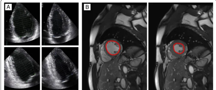
Figure 1 Measurement of the ejection fraction by 2D TTE and CMR. A. The LVEF by 2D TTE was obtained by the modified Simpson’s method in the apical four- and two-chamber view. B. The LVEF by CMR was calculated by assessment of end-diastolic and end-systolic LV volumes in multiple parallel short-axis slices.
described by Buchner et al. [8] (Figure 2B). The ante-grade LV stroke volume was obtained by phase con-trast velocity mapping at the ascending aorta [6]. CMR RVol was calculated as the difference between the LV stroke volume and the antegrade LV stroke volume. All CMR data were assessed by agreement of 2 readers (PJB and LD) who were blinded to echocardiographic data.
Statistic analysis
All data were analysed with SPSS version 20 (IBM Statis-tics). Results are expressed as mean ± SD or percentage unless otherwise specified. Comparisons of LV dimen-sions, ERO/ARO and RVol obtained by 2D TTE and CMR were made using the paired Student t test and Pearson correlation. Bland-Altman analysis was per-formed and the intraclass correlation coefficient was calculated to assess the agreement of both imaging methods. Values of p < 0.05 were considered significant.
Results
All enrolled patients underwent CMR within a period of 1 month after 2DTTE, in 2 patients CMR followed 2DTTE within 2 months but follow-up 2DTTE con-firmed stable LV dimensions in these patients. 3 patients had limited image quality on CMR and were excluded from further analysis. The baseline characteristics of the remaining study population are summarised in Table 1. Among the 38 patients included, 79% were males and the origin of MR was predominantly degenerative (92%). Severe MR, as assessed by the PISA method, was found in 45% of patients.
Comparison of measurement by 2D TTE and CMR re-garding LV dimensions and parameters of MR severity are represented in Table 2. Measurements of linear
LV dimensions by both imaging modalities were statisti-cally similar (LV end-diastolic dimension: 53 ± 6 mm by transthoracic echocardiography vs. 53 ± 8 mm by CMR, p = 0.91; average bias −0.1 mm, 95% confidence inter-val −8.9 to +8.7 mm and LV end-systolic dimension: 36 ± 5 mm vs. 36 ± 6 mm, p = 0.95; average bias 0 mm, 95% confidence interval −5.7 to +5.7 mm). LV volumes were overall significantly underestimated by modified Simpson’s method in comparison with CMR, although LV end-diastolic and end-systolic volumes showed a strong correlation by Pearson correlation analysis (Figure 3A and C). Bland-Altman analysis confirmed general under-estimation by 2D TTE of both LV end-diastolic volume (average bias +28 ml, 95% confidence interval −53 to +109 ml) and LV end-systolic volume (average bias +20 ml, 95% confidence interval−25 to + 66 ml) (Figure 3B
A
B
ARO
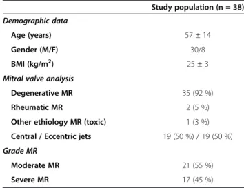
Figure 2 Measurement of the regurgitant orifice by 2D TTE and CMR. A. Acquisition of PISA radius and continuous wave Doppler of the MR jet allows calculation of the effective regurgitant orifice. The PISA radius is measured at mid-systole using the first aliasing with a reduced Nyquist limit (15–40 m/s). B. The anatomic regurgitant orifice can be measured by planimetry on a slice parallel to the valvular plane obtained by cardiovascular magnetic resonance.
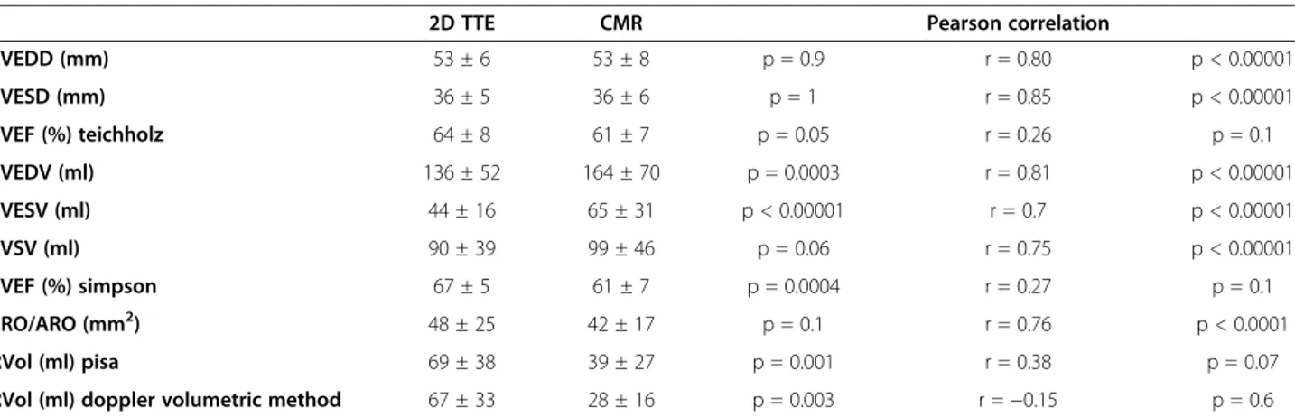
Table 1 Baseline characteristics of the study population
Study population (n = 38) Demographic data
Age (years) 57 ± 14
Gender (M/F) 30/8
BMI (kg/m2) 25 ± 3
Mitral valve analysis
Degenerative MR 35 (92 %)
Rheumatic MR 2 (5 %)
Other ethiology MR (toxic) 1 (3 %) Central / Eccentric jets 19 (50 %) / 19 (50 %) Grade MR
Moderate MR 21 (55 %)
Severe MR 17 (45 %)
Table 2 Comparison of 2D TTE and CMR measurements of LV dimensions and MR severity
2D TTE CMR Pearson correlation
LVEDD (mm) 53 ± 6 53 ± 8 p = 0.9 r = 0.80 p < 0.00001 LVESD (mm) 36 ± 5 36 ± 6 p = 1 r = 0.85 p < 0.00001 LVEF (%) teichholz 64 ± 8 61 ± 7 p = 0.05 r = 0.26 p = 0.1 LVEDV (ml) 136 ± 52 164 ± 70 p = 0.0003 r = 0.81 p < 0.00001 LVESV (ml) 44 ± 16 65 ± 31 p < 0.00001 r = 0.7 p < 0.00001 LVSV (ml) 90 ± 39 99 ± 46 p = 0.06 r = 0.75 p < 0.00001 LVEF (%) simpson 67 ± 5 61 ± 7 p = 0.0004 r = 0.27 p = 0.1 ERO/ARO (mm2) 48 ± 25 42 ± 17 p = 0.1 r = 0.76 p < 0.0001 RVol (ml) pisa 69 ± 38 39 ± 27 p = 0.001 r = 0.38 p = 0.07
RVol (ml) doppler volumetric method 67 ± 33 28 ± 16 p = 0.003 r =−0.15 p = 0.6 MR = mitral regurgitation; LVEDD = left ventricular end-diastolic diameter; LVESD = left ventricular end-systolic volume; LVEF = left ventricular ejection fraction; LVEDV = left ventricular end-diastolic volume; LVESV = left ventricular end-systolic volume; LVSV = left ventricular stroke volume; ERO = effective regurgitant orifice; ARO = anatomical regurgitant orifice; RVol = regurgitant volume.
Figure 3 Comparison of measurements of LV diastolic and systolic volumes by 2D TTE and CMR. A. Measurement of LV end-diastolic volumes by both imaging methods showed a strong correlation by Pearson correlation analysis (r = 0.81, p < 0.00001). B. Bland-Altman analysis indicated general underestimation of the LV end-diastolic volume by 2D TTE in comparison with CMR (average bias +28 ml, 95% confidence interval−53 to +109 ml). C. Measurement of LV end-systolic volumes by both imaging methods showed as well a good correlation (Pearson r = 0.7, p < 0.00001). D. Bland-Altman analysis revealed general underestimation of the LV end-systolic volume by 2D TTE compared to CMR (average bias +20 ml, 95 % confidence interval−25 to +66 ml).
and D). The LVEF was significantly overestimated by 2D TTE in comparison with CMR and there was no significant correlation between LVEF measurements of the two imaging modalities.
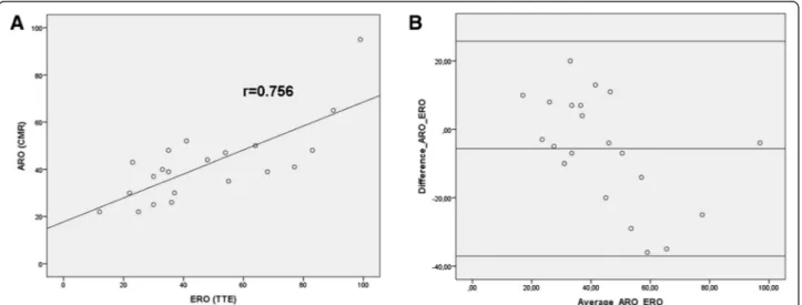
Measurement of the ARO was feasible in 21 out of 22 patients (95%). ERO calculated by the PISA method with 2D TTE was similar to ARO measured by planimetry with CMR (47 ± 24 vs. 42 ± 16 mm2, p = 0.12). Pearson correlation analysis showed a strong correlation bet-ween both imaging techniques of the regurgitant orifice (r = 0.76, p < 0.0001) (Figure 4A). Furthermore Bland-Altman analysis showed good agreement between 2D TTE PISA method and CMR planimetry of the ARO (average bias −5.7 mm2, 95% confidence interval −37 to +26 mm2), less accordance was observed in some pa-tients with an ERO ≥50 mm2 (Figure 4B). In addition, the intraclass correlation coefficient (=0.7, p = 0.0001) confirmed strong agreement between both methods to quantify the regurgitant orifice. By contrast, RVol cal-culated by either of the two 2D TTE methods was signi-ficantly higher in comparison with RVol obtained by phase contrast velocity mapping by CMR (PISA method vs. CMR: 69 ± 38 vs. 39 ± 27 ml, p = 0.001; r = 0.45, p = 0.07; Doppler volumetric method vs. CMR: 67 ± 33 vs. 28 ± 16 ml, p = 0.003; r =−0.14, p = 0.6). Assessment of regurgitant volume by PISA and Doppler volumetric method by 2D TTE was similar (74 ± 40 vs. 67 ± 33 ml, p = 0.6). Of note, 20% of patients with moderate MR ac-cording to ERO as assessed by the PISA method were reclassified as severe MR according to planimetry of ARO, likewise 20% of patients with severe MR by 2D TTE were reclassified as moderate MR by CMR.
Discussion
LV dimensions and volumes
An important finding of the present study is that LV volumes are significantly underestimated by 2D TTE in patients with moderate to severe primary MR in com-parison with CMR, which is widely accepted as the most accurate non-invasive imaging tool to assess LV shape and sizes [10]. This finding might be of importance since adequate assessment of LV contractility and dimensions is crucial for clinical decision making in these patients. It is well known that LV volumes may be significantly underestimated (even up to 50%) by 2D TTE in com-parison with invasive ventriculography or CMR in pa-tients with normal and decreased LV function due to poor image quality, apical foreshortening and geomet-rical assumptions [5,11,12]. However, there are poor data about the degree of underestimation of LV volumes by 2D TTE in patients with primary MR with good echo-genicity and without overt LV dysfunction; our data showed a mean difference of 28 ml regarding the LV end-diastolic volume and 20 ml regarding the LV-end systolic volume. Interestingly, there was a strong correl-ation between measurements of LV volumes by the two imaging methods, which may suggest that 2D TTE may still be useful to evaluate the progression of LV remodel-ling in a single patient. At present, guidelines use linear LV dimensions (LV end-systolic diameter) to define LV dilatation in primary MR as a criterion for surgery [4,13]. Our study showed very good agreement be-tween 2D TTE and CMR regarding the measurement of linear LV dimensions. However, a recent CMR study by Schiros et al. [14] showed that the LV end-systolic
Figure 4 Comparison of ERO measured by 2D TTE and ARO measured by CMR. A. Pearson correlation analysis showed a strong correlation between 2D TTE PISA method and CMR planimetry of the ARO (r = 0.76, p < 0.0001). B. Bland-Altman analysis showed good agreement between both imaging techniques of the regurgitant orifice (average bias−5.7 mm2, 95% confidence interval−37 to +26 mm2), less accordance was
diameter measured in the parasternal long-axis at the mitral valve leaflet tips may underestimate the LV volume due to spherical mid to apical remodeling. Further studies are needed to determine whether assessment of LV volumes has a benefit over LV linear dimensions for risk stratification in patients with asymptomatic primary MR.
LV ejection fraction
Both Teichholz formula and modified Simpson’s method by 2D TTE seemed to overestimate LVEF in comparison to CMR. Furthermore, there was no significant correl-ation between both imaging modalities regarding quanti-fication of the LVEF. However, our study population consisted of subjects with a preserved LV function and, as a consequence, a narrow range of LVEF (60-75%). Furthermore, 2D TTE is known to have a higher inter-observer variability than CMR, with a mean percentage of error that can be up to 10-15% [12] which implies that a large number of patients should have been in-cluded to detect any correlation between the two im-aging modalities. Of note, 33% of our patients with severe MR and preserved LV function (LVEF ≥60%) by 2D TTE Simpson’s method had a mild decreased LVEF (50-59%) by CMR and therefore already have a class I recommendation for surgery [4,13]. These observations are in line with another small CMR study in patients with moderate to severe primary MR [15] and suggest that more accurate assessment of LVEF by CMR might be indicated in asymptomatic severe MR to determine optimal timing for surgery.
Severity of MR
The PISA method with calculation of ERO and RVol is strongly recommended to assess MR severity [3]. How-ever, several studies have used RVol obtained by phase contrast velocity mapping by CMR as a reference me-thod to compare various 2D and 3D echocardiographic techniques to evaluate MR severity [6,8,16,17].
To our knowledge, only one study by Buchneret al. in 35 patients with mitral regurgitation described planim-etry of the ARO and found a good agreement with cal-culation of the ERO by the PISA method [8]. In their study, 66% of patients had less than grade III mitral regurgitation, 66% had primary MR and 43% had a LVEF <50%. Their data showed that ARO was slightly higher than ERO, presumably because ERO represents the narrowest flow stream, which is located distal from the anatomic orifice. Our study confirms good agreement between the two methods in 21 patients with moder-ate to severe primary MR without overt LV dysfunc-tion. We observed a rather higher ERO than ARO for most ERO values >50 mm2, although this finding has little clinical implications since most of these cases
were graded as ‘severe’ by both methods. Thus, plan-imetry of the ARO by CMR seems to be a valuable alternative in patients with primary MR and poor echo-genicity or eccentric jets which are difficult to assess with the PISA method.
In our study, RVol derived from CMR showed only moderate correlation with RVol obtained by the PISA method as well as with the Doppler volumetric method. Furthermore, RVol might have been overestimated by the PISA method in comparison to CMR, which is in line with a recent study by Hamada et al. [17]. This might be due to difficult determination of the - often dynamic - radius of the hemispheric contour of the flow convergence zone, especially in eccentric jets. By con-trast, other studies found rather an underestimation of RVol by 2D and 3D echocardiographic techniques in comparison with CMR [18,19], indicating that in any case measurements of RVol by CMR or by 2D TTE are not interchangeable.
Limitations
We did not perform more advanced echocardiographic techniques like 3D or contrast enhanced echocardiog-raphy, which are known to correlate better with CMR regarding LV volumes, LVEF and RVol [12,17-20] than 2D TTE. However, notwithstanding the well known benefits of these techniques, 2D TTE remains the most widely used imaging method in daily practice for the assessment of patients with primary MR. Fur-thermore, there was no comparison with invasive measurements of LV function or MR severity by an-giography, but this latter approach is not any more recommended.
In most patients 2DTTE and CMR were not per-formed on the same date but within 1 month, which might influence the measurement of load-dependent parameters, in particular end-diastolic dimensions. How-ever, we did not include patients with clinical or echo-cardiographic signs of heart failure and there were no patients with acute onset of symptoms during the study period. Furthermore, underestimation of LV volumes on 2DTTE was also seen in patients in whom both exams were performed on the same date.
Conclusion
In moderate to severe primary MR without overt LV dysfunction, 2D TTE seems to significantly underesti-mate LV remodelling and overestiunderesti-mate LVEF in com-parison to CMR. These findings might provide more evidence for routine assessment of LV size and function by CMR in asymptomatic primary MR in order to better define optimal timing for surgery. However, further studies are needed to determine whether the increase in LV volume is a more powerful predictor of the outcome
than the end-systolic diameter. Moreover, the measure-ment of the regurgitant orifice with planimetry by CMR shows very good agreement with the validated PISA method by 2D TTE and thus may be a valuable alterna-tive in patients with poor echogenicity or barely assess-able eccentric jets.
Abbreviations
MR:Mitral regurgitation; LVEF: Left ventricular ejection fraction; 2DTTE: Two-dimensional transthoracic echocardiography; CMR: Cardiovascular magnetic resonance; ERO: Effective regurgitant orifice; ARO: Anatomic regurgitant orifice; RVol: Regurgitant volume; PISA: Proximal isovelocity surface area.
Competing interests
The authors declare that they have no competing interests. Authors’ contributions
CVDH is the first author, analysed off-line the echocardiographic data, and performed the statistical analysis. JM verified the echocardiographic data and statistical analysis and helped to draft the manuscript. LP and PL designed and coordinated the study in the University of Liege Hospital, acquired the echocardiographic data and helped to draft the manuscript. PB and LD acquired the CMR images in the University of Liege Hospital and performed the measurements of all CMR data. CDM acquired the echocardiographic data in the University of Antwerp Hospital. BP acquired the CMR images in the University of Antwerp Hospital. CV coordinated the study in the University of Antwerp Hospital and helped to draft the manuscript. All authors read and approved the final manuscript.
Author details
1Department of Cardiology, University of Liège Hospital, GIGA Cardiovascular
Sciences, Heart Valve Clinic, CHU Sart Tilman, Liège, Belgium.2Department of Cardiology, University of Antwerp Hospital, Edegem, Belgium.3Department
of Radiology, University of Liège Hospital, Thoracic and Cardiovascular Imaging, CHU Sart Tilman, Liège, Belgium.4Department of Cardiology,
University Hospital, Sart-Tilman, B-4000 Liège, Belgium.
Received: 2 October 2013 Accepted: 18 November 2013 Published: 27 December 2013
References
1. Van de Heyning CM, Magne J, Vrints CJ, Piérard L, Lancellotti P: The role of multi-imaging modality in primary mitral regurgitation. Eur Heart J Cardiovasc Imaging 2012, 13:139–151.
2. Itagaki S, Adams DH, Anyanwu AC: Triggers for surgical referral in degenerative mitral valve regurgitation. Circ J 2012, 77:28–34.
3. Lancellotti P, Tribouilloy C, Hagendorff A, Popescu BA, Edvardsen T, Pierard LA, Badano L, Zamorano JL: Recommendations for the echocardiographic assessment of native valvular regurgitation: an executive summary from the European Association of Cardiovascular Imaging. Eur Heart J Cardiovasc Imaging 2013, 14:611–644.
4. Vahanian A, Alfieri O, Andreotti F, Antunes MJ, Barón-Esquivias G, Baumgartner H, Borger MA, Carrel TP, De Bonis M, Evangelista A, Falk V, Iung B, Lancellotti P, Pierard L, Price S, Schäfers HJ, Schuler G, Stepinska J, Swedberg K, Takkenberg J, Von Oppell UO, Windecker S, Zamorano JL, Zembala M: Guidelines on the management of valvular heart disease (version 2012): The Joint Task Force on the Management of Valvular Heart Disease of the European Society of Cardiology (ESC) and the European Association for Cardio-Thoracic Surgery (EACTS). Eur Heart J 2012, 2012(33):2451–2496.
5. Mogelvang J, Stokholm KH, Saunamaki K, Reimer A, Stubgaard M, Thomsen C, Fritz-Hansen P, Henriksen O: Assessment of left ventricular volumes by magnetic resonance in comparison with radionuclide angiography, contrast angiography and echocardiography. Eur Heart J 1992, 13:1677–1683.
6. Fujita N, Chazouilleres AF, Hartiala JJ, O'Sullivan M, Heidenreich P, Kaplan JD, Sakuma H, Foster E, Caputo GR, Higgins CB: Quantification of mitral regurgitation by velocity-encoded cine nuclear magnetic resonance imaging. J Am Coll Cardiol 1994, 23:951–958.
7. Grayburn PA, Weissman NJ, Zamorano JL: Quantitation of mitral regurgitation. Circulation 2012, 126:2005–2017.
8. Buchner S, Debl K, Poschenrieder F, Feuerbach S, Riegger GA, Luchner A, Djavidani B: Cardiovascular magnetic resonance for direct assessment of anatomic regurgitant orifice in mitral regurgitation. Circ Cardiovasc Imaging 2008, 1:148–155.
9. Lang RM, Bierig M, Devereux RB, Flachskampf FA, Foster E, Pellikka PA, Picard MH, Roman MJ, Seward J, Shanewise J, Solomon S, Spencer KT, St John Sutton M, Stewart W: Recommendations for chamber quantification. Eur J Echocardiogr 2006, 7:79–108.
10. Epstein FH: MRI of left ventricular function. J Nucl Cardiol 2007, 14:729–744. 11. Bellenger NG, Burgess MI, Ray SG, Lahiri A, Coats AJ, Cleland JG, Pennell DJ:
Comparison of left ventricular ejection fraction and volumes in heart failure by echocardiography, radionuclide ventriculography and cardiovascular magnetic resonance; are they interchangeable? Eur Heart J 2000, 21:1387–1396.
12. Hoffmann R, von Bardeleben S, ten Cate F, Borges AC, Kasprzak J, Firschke C, Lafitte S, Al-saadi N, Kuntz-Hehner S, Engelhardt M, Becher H, Vanoverschelde JL: Assessment of systolic left ventricular function: a multi-centre comparison of cineventriculography, cardiac magnetic resonance imaging, unenhanced and contrast-enhanced echocardiography. Eur Heart J 2005, 26:607–616.
13. Bonow RO, Carabello BA, Chatterjee K, de Leon AC Jr, Faxon DP, Freed MD, Gaasch WH, Lytle BW, Nishimura RA, O'Gara PT, O'Rourke RA, Otto CM, Shah PM, Shanewise JS: 2008 focused update incorporated into the ACC/AHA 2006 guidelines for the management of patients with valvular heart disease: a report of the American College of Cardiology/American Heart Association Task Force on Practice Guidelines (Writing Committee to revise the 1998 guidelines for the management of patients with valvular heart disease). J Am Coll Cardiol 2008, 52:e1–e142.
14. Schiros CG, Dell'Italia LJ, Gladden JD, Clark D, Aban I, Gupta H, Lloyd SG, McGiffin DC, Perry G, Denney TS, Ahmed MI: Magnetic resonance imaging with 3-dimensional analysis of left ventricular remodeling in isolated mitral regurgitation: implications beyond dimensions. Circulation 2012, 125:2334–2342.
15. Ozdogan O, Yuksel A, Gurgun C, Kayikcioglu M, Yavuzgil O, Cinar CS: Assessment of cardiac remodeling in asymptomatic mitral regurgitation for surgery timing: a comparative study of echocardiography and magnetic resonance imaging. Cardiovasc Ultrasound 2010, 8:32. 16. Buck T, Plicht B, Hunold P, Mucci RA, Erbel R, Levine RA: Broad-beam
spectral Doppler sonification of the vena contracta using matrix-array technology: a new solution for semi-automated quantification of mitral regurgitant flow volume and orifice area. J Am Coll Cardiol 2005, 45:770–779.
17. Hamada S, Altiok E, Frick M, Almalla M, Becker M, Marx N, Hoffmann R: Comparison of accuracy of mitral valve regurgitation volume determined by three-dimensional transesophageal echocardiography versus cardiac magnetic resonance imaging. Am J Cardiol 2012, 110:1015–1020.
18. Plicht B, Kahlert P, Goldwasser R, Janosi RA, Hunold P, Erbel R, Buck T: Direct quantification of mitral regurgitant flow volume by real-time three-dimensional echocardiography using dealiasing of color Doppler flow at the vena contracta. J Am Soc Echocardiogr 2008, 21:1337–1346. 19. Shanks M, Siebelink HM, Delgado V, van de Veire NR, Ng AC, Sieders A,
Schuijf JD, Lamb HJ, Ajmone Marsan N, Westenberg JJ, Kroft LJ, de Roos A, Bax JJ: Quantitative assessment of mitral regurgitation: comparison between three-dimensional transesophageal echocardiography and magnetic resonance imaging. Circ Cardiovasc Imaging 2010, 3:694–700. 20. Lang RM, Mor-Avi V, Sugeng L, Nieman PS, Sahn DJ: Three-dimensional echocardiography: the benefits of the additional dimension. J Am Coll Cardiol 2006, 48:2053–2069.
doi:10.1186/1476-7120-11-46
Cite this article as: Van De Heyning et al.: Assessment of left ventricular volumes and primary mitral regurgitation severity by 2D echocardiography and cardiovascular magnetic resonance. Cardiovascular Ultrasound 2013 11:46.