Olivier Detry, Arnaud De Roover, Pierre Honoré, Michel Meurisse
INTRODUCTION
Fulminant hepatic failure (FHF) is an infrequent but dreadful disease, defined by the appearance of hepatic encephalopathy within 8 wk after the onset of jaundice in patients with no known chronic liver disease[1]
. Most FHF patients rapidly develop electrolyte, metabolic, and coagulation abnormalities[2]
. They frequently suffer from acute renal insufficiency and/or adult respiratory distress syndrome (ARDS), leading to multiple organ failure (MOF)[3]
. They are very sensitive to infection, and frequently develop a sepsis-like syndrome, with systemic hypotension, low peripheral resistance and increased cardiac output. Modern intensive care units (ICU) have learned to treat all these conditions, and have prolonged FHF patient survival. However, in absence of liver transplantation (LT), the mortality rate of FHF patients remains high (60%-80%). Causes of death of these patients are mainly MOF, sepsis, and/or brain edema leading to intracranial hypertension and secondary brain death.
Brain edema in FHF patients is a relatively recent concept. In a 1944 report of 125 autopsies of military patients dying from what was called fatal hepatitis (previously named idiopathic acute yellow atrophy of the liver), Lucké noted little alteration in the brain, except edema, but he did not describe cerebral herniation[4]
. He hypothesized that the cerebral changes of fatal hepatitis might be attributed to loss of detoxifying function of the liver. The first reports of brain edema and cerebral herniation as complications of FHF were published in the 1970’s[5,6]
and were criticized at that time. Widespread recognition that brain edema and intracranial hypertension are complications of FHF only occurred in the 1980’s[7,8]
. It is also very interesting to note that brain edema and intracranial hypertension are not recognized common features of terminal chronic liver failure, despite some case reports or small series[9,10]
. The recent recognition of brain edema in FHF patients could be due to the advances in FHF patient care. Previously, FHF patients were dying from early hepatocellular insufficiency complications, mainly hemorrhage or sepsis[4]
. Improvements in ICU techniques have lengthened the survival of FHF patients. The longer course of the disease may have allowed the development of brain edema, possibly a later complication of FHF (Figure 1). Significant advances in the understanding of FHF brain edema have been made this last decade, but the exact pathophysiological mechanisms underlying development of brain edema and intracranial hypertension in FHF are still not entirely clear and are
Brain edema and intracranial hypertension in fulminant
hepatic failure: Pathophysiology and management
Olivier Detry, Arnaud De Roover, Pierre Honoré, Michel Meurisse, Department of Abdominal Surgery and Transplantation, University of Liège, CHU Sart Tilman B35, B4000 Liège, Belgium
Correspondence to: Dr. Olivier Detry, Department of Abdominal Surgery and Transplantation, CHU Sart Tilman B35, B4000 Liège, Belgium. oli.detry@chu.ulg.ac.be
Telephone: +3243667645 Fax: +3243667069 Received: 2006-05-16 Accepted: 2006-06-14
Abstract
Intracranial hypertension is a major cause of morbidity and mortality of patients suffering from fulminant hepatic failure. The etiology of this intracranial hypertension is not fully determined, and is probably multifactorial, combining a cytotoxic brain edema due to the astrocytic accumulation of glutamine, and an increase in cerebral blood volume and cerebral blood fl ow, in part due to infl ammation, to glutamine and to toxic products of the diseased liver. Validated methods to control intracranial hypertension in fulminant hepatic failure patients mainly include mannitol, hypertonic saline, indomethacin, thiopental, and hyperventilation. However all these measures are often not sufficient in absence of liver transplantation, the only curative treatment of intracranial hypertension in fulminant hepatic failure to date. Induced moderate hypothermia seems very promising in this setting, but has to be validated by a controlled, randomized study. Artificial liver support systems have been under investigation for many decades. The bioartifi cial liver, based on both detoxification and swine liver cells, has shown some effi cacy on reduction of intracranial pressure but did not show survival benefi t in a controlled, randomized study. The Molecular Adsorbents Recirculating System (MARS) has shown some efficacy in decreasing intracranial pressure in an animal model of liver failure, but has still to be evaluated in a phase Ⅲ trial.
© 2006 The WJG Press. All rights reserved.
Key words: Intracranial hypertension; Fulminant hepatic failure; Brain edema
Detry O, Roover AD, Honoré P, Meurisse M. Brain edema and intracranial hypertension in fulminant hepatic failure: Pathophysiology and management. World J Gastroenterol 2006; 12(46): 7405-7412
http://www.wjgnet.com/1007-9327/12/7405.asp
wjg@wjgnet.com © 2006 The WJG Press. All rights reserved.
likely to be multifactorial[11]
. The aim of this paper is to review the pathophysiology of intracranial hypertension in FHF in order to improve understanding and management of this complication.
PATHOPHYSIOLOGY OF INTRACRANIAL
HYPERTENSION IN FHF
Normal intracranial pressure (ICP) is 5 to 10 mmHg and intracranial hypertension becomes clinically relevant when ICP exceeds 20 mmHg. The main complication of severe intracranial hypertension in FHF patients is transtentorial herniation[12]
. This herniation may induce (1) compression of the posterior cerebral artery, leading to medial temporal, thalamic, and occipital lobe infarction; (2) cerebral aqueduct and subarachnoid space compression, causing obstructive hydrocephalus; and (3) brain stem compression, resulting in brain stem ischemia, haemorrhage, and death[12]
. Additionally, severe intracranial hypertension compromises cerebral perfusion pressure (CPP). By defi nition, CPP is the difference between mean arterial pressure (MAP) and cerebral venous pressure. As cerebral venous pressure can be approximated by ICP, CPP equals MAP minus ICP[12,13]
. An increase in ICP reduces CPP, and thus a decrease in cerebral blood fl ow (CBF). This reduction in CBF may cause cerebral ischemia or infarction, resulting in neurological deficits in FHF survivors.
A rise in ICP is the mechanical consequence of an increase in the intracranial volume. The central nervous system (CNS) is protected by the skull, which is rigid and incompressible. Inside the skull, 3 different compartments can be defined: the brain, the cerebrospinal fluid (CSF) and the blood. If the volume of one of these elements increases, the volume of another compartment might decrease, resulting in some intracranial compensation capacity or compliance. If the increase in volume exceeds this compliance, any further addition of volume leads to a rise in ICP.
It is generally accepted that CSF volume is not expanded in FHF. During episodes of intracranial hypertension in FHF, ventricular spaces measured by computed tomography (CT) were either unchanged or compressed, suggesting an increase in the brain tissue or blood volume[14,15]
. In animal models and in FHF patients there is increased brain volume, secondary to edema[5,16,17]
. In this environment even a small increase in cerebral blood volume could significantly increase ICP. In fact, these two phenomena have been proposed to account for intracranial hypertension in FHF: (1) brain edema due to osmotic astrocyte swelling secondary to ammonia-induced accumulation of glutamine (ammonia-glutamine hypothesis); (2) alteration of CBF regulation with increase of the intracranial blood volume.
Brain edema in FHF (Figure 2)
Both vasogenic and cytotoxic mechanisms are implicated in the development of cerebral edema. Vasogenic brain edema occurs as a result of the disruption of the blood-brain barrier (BBB), allowing uncontrolled access of
plasma components and water to the extracellular cerebral compartment. Cytotoxic edema is the consequence of impaired cellular osmoregulation in the brain, resulting in an increase of cellular water. In FHF, evidence from experimental animals and postmortem human brain supports aspects of each of these mechanisms, but it is now well established that brain edema in FHF is mainly cytotoxic[18]
. In several models of FHF, compounds to which the BBB is normally impermeable (Evans Blue or a-aminoisobutyric acid) were detected in the brain in increased concentrations[19-21]
. These observations suggest that BBB permeability is altered in FHF, which is consistent with a vasogenic mechanism. However, the ability of mannitol to reduce ICP in patients with FHF indicates that the BBB remains largely intact[8]
. Furthermore, electron microscopic studies of human brain tissue in FHF revealed no alteration in the integrity of tight junctions[22]
. Moreover, consistent with cytotoxic edema, marked intracellular swelling of perivascular astrocytes was observed. Recent magnetic resonance imaging of brain of FHF patients confir med the predominant cytotoxic character of FHF brain edema[23]
. This suggests that FHF brain edema primarily develops within the cellular component of the brain as a cytotoxic edema, and changes in the permeability of the BBB may represent secondary events that could exacerbate edema or intracranial hypertension.
The ammonia-glutamine hypothesis: Cor tical
astrocyte swelling is the most common obser-vation A
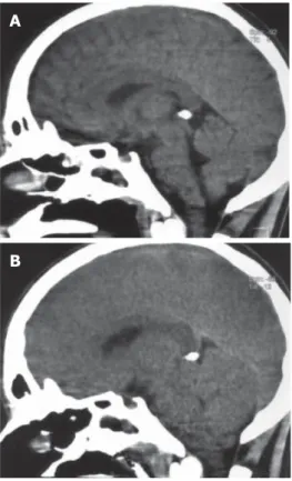
B
Figure 1 Computed tomography (CT) (sagittal sections) of the brain of a 17-year
old man who developed fulminant hepatic failure. A: first CT performed in encephalopathy stage IV; B: control CT performed when the patient developed unilateral fixed midriasis, showing diffuse edema, compression of the fourth ventricle and of the brain stem. The patient developed brain stem death while waiting liver transplantation, despite medical therapy and MARS treatment.
in neuropathological studies of brain edema in FHF. Astrocytes are the most numerous cell type in the brain and occupy about one-third of the cortical volume[24]
. Astrocytes have several critical metabolic functions involved in the maintenance and regulation of the extracellular microenvironment. They participate in water regulation in the brain, detoxify ammonia and maintain normal levels of extracellular glutamate. Hyperammonemia is prevalent in acute liver failure. In the brain, ammonia is detoxified to glutamine via the amidation of glutamate by an astrocytic enzyme, glutamine synthetase. Glutamine leaves the astrocyte by passive diffusion into the extracellular space where it is taken up by neurons and converted to glutamate. There is clear evidence of increased brain glutamine concentrations in animal FHF models[25]
and in postmortem samples of FHF[26]
. Brusilow fi rst proposed that this glutamine accumulation is the link between hyperammonemia and edema via altered osmoregulation[27]
. In astrocyte cultures, ammonia induces cell swelling[17,28]
. Brain swelling and intracranial hypertension have been documented in humans with hyperammonemic conditions[29]
. In rats, a continuous infusion of ammonia is associated with brain edema or intracranial hypertension[30] which is reduced by inhibition
of glutamine synthesis with methionine-sulfoximine[31-33]
. The relationship between hyperammonemia, glutamine and ICP was recently demonstrated in humans[34]
. Although there is much clinical and experimental evidence in support of the glutamine hypothesis, prevention of brain edema in FHF by inhibition of glutamine synthesis has not been successful in humans.
Increase in cerebral blood flow and intracranial blood volume
Another phenomenon that has also been involved in
intracranial hypertension in FHF is the increase of intracranial blood volume and CBF. Some reports describe decreased CBF in patients suffering from acute liver failure[35,36]
, but most have found high CBF associated with intracranial hypertension in FHF[37,38]
. Paulson suggests that an impairment of vascular autoregulation in the brain could be responsible for this increase in CBF and blood volume[39]
. Impaired autoregulation of CBF has been reported in animal models of FHF[40]
and in patients with FHF[41]
. The exact cause of this increase of CBF in FHF is not known. Nitric oxide (NO) has been implicated but it is possible that the increased NO in the brain of FHF patients is secondary to an increase in CBF, rather than a primary event[18,42]
. Inflammation markers (IL-1 β, TNF α, IL-6) and systemic infl ammatory response have been associated with increased CBF and ICP in FHF[37,43]
, and poor outcome[44]
. The association of systemic inflammation and impaired regulation of CBF might be related to the role of the necrotic liver in intracranial hypertension in FHF. The observation that brain edema and intracranial hypertension are complications of FHF and not of chronic liver disease lead to the hypothesis that these phenomena may in part result from products of the acutely necrotic liver. There is experimental and clinical evidence to support this theory, the “toxic liver hypothesis”. In a rat model, cerebral edema was significantly lower in anhepatic than FHF animals[45]
. In pigs, no elevation of ICP was observed after total hepatectomy, whereas a rise in ICP was observed in pigs with FHF secondary to ischemia[46]
. Several human observations reinforced this hypothesis. During LT for FHF, it was established that ICP normalizes during the anhepatic phase and may increase during the dissection of the diseased liver and during graft reperfusion[47,48]
. The removal of the diseased liver has been linked to ICP normalization and to marked and sustained reduction of several pro-inflammatory cytokines in a case report[49]
. Moreover some patients underwent prolonged period (up to 72 h) of anhepatic state without neurological sequelae[50,51]
. Although these fi ndings are suggestive, the role of products from the necrotic liver in cerebral edema and intracranial hypertension is still unknown.
The respective role of all of these phenomena in the development of intracranial hypertension in FHF remains to be determined. It can be hypothesized that brain edema (increase in brain volume) secondary to osmotic effect of glutamine in astrocytes, and cerebral hyperemia (increase in blood volume) secondary to vasodilation (cytokines, products of the necrotic liver, glutamine, others…) may contribute to intracranial hypertension leading to brain stem herniation and brain stem death in FHF. During all these FHF phenomena, the brain may respond by altering the expression of genes coding for various proteins whose role may be critical to some CNS functions, including the maintenance of cell volume and neurotransmission. Cerebral gene expression during FHF is modified as demonstrated by differential display in rat models[52,53]
. Some genes have been more specifically studied, as GLUT-1[54]
, aquaporin IV[55]
, GLT-1[52,56]
and others[57]
. The exact role of this gene expression observed during FHF is still to be determined.
Fulminant hepatic failure
Ammonia-glutamine Toxic liver hypothesis hypothesis
Toxic products of the failing liver Infl ammatory cytokines
Defi cit in liver detoxifi cation of ammonia
Systemic hyperammonemia
Astrocyte accumulation of glutamine Astrocyte swelling
Increase in cerebral blood fl ow
Cytotoxic brain edema
Increase in brain volume Increase in intracranial blood volume Increase in intracranial volume
Intracranial hypertension brain stem death
↓
↓
↓
↓
↓
↓
↓
↓
↓
↓
↓
↓
↓
↓
↓
↓ ↓
Figure 2 Schematic representation of the hypotheses explaining intracranial
D I A G N O S I S O F I N T R A C R A N I A L
HYPERTENSION IN FHF PATIENTS
Intracranial hypertension should clinically be suspected in FHF patients with systemic hypertension (sustained ≥ 160 mmHg or paroxysms ≥ 200 mmHg), aggravation of hepatic encephalopathy, abnormal pupillary signs, or signs of decerebration. However, most of these clinical signs are not specifi c, and may be developed by patients in hepatic grade IV encephalopathy without intracranial hypertension. It was reported that brain CT is unreliable in the diagnosis of intracranial hypertension in FHF patients[14]
, and there have been no reports of the value of brain magnetic resonance imaging in FHF patients for confirming the diagnosis of intracranial hypertension. The most accurate method of diagnosing intracranial hypertension is ICP monitoring. Although the advantages of this monitoring in FHF patients have not yet been demonstrated by a randomized study, ICP monitoring may be very helpful in establishing the presence of intracranial hypertension and in guiding specifi c therapy. Intracranial hypertension in FHF patients may suddenly rise from normal to life threatening levels within minutes. In this situation, continuous ICP monitoring may allow rapid and specifi c management. Several groups have included ICP monitoring in the protocol of FHF patient management[14,47,58]
. The main argument against ICP monitoring is the enhanced risk of complications in FHF patients, mainly infection and hemorrhage. In a national survey of 262 FHF patients, the complication rate of ICP monitoring was 10%. In this series, intracranial hemorrhages were the cause of death in 7 patients and the epidural transducers had the lowest complication rate (3.7%)[59]
. The complication rate of ICP monitoring in FHF patients was reported to be lower in a recent multicenter report, but is still signifi cant[60]
.
M A N A G E M E N T O F I N T R A C R A N I A L
HYPERTENSION IN FHF PATIENTS
General management of FHF patients is beyond the scope of this review and was presented elsewhere[2]
. The goal in the medical management of FHF patients with intracranial hypertension is to maintain ICP below 20 mmHg and CPP above 70 mmHg. Cerebral ischemia occurs if CPP is less than 40 to 50 mmHg, and LT should be contraindicated if CPP remains below 40 mmHg for two hours[61]
. This goal requires intense medical management and nursing. FHF patients should be admitted to ICU in an institution with an active liver transplant program. FHF patients should be monitored with peripheral arterial catheters. Vital signs, urinary output, arterial blood gases and central hemodynamic parameters should be continuously monitored. FHF patients are treated with standard supportive measures to correct electrolyte, metabolic, respiratory and hemodynamic abnormalities. Hypovolemia may exacerbate hypotension, and may reduce CPP, inducing brain ischemia. Systemic hypertension may also be deleterious by increasing ICP[62]
. In this case, ß-blockers may be more useful than nitroprusside or calcium-channel inhibitors, because of their potential risk of brain blood vessel dilatation. Patient positioning and nursing is also
important in the care of FHF patients with intracranial hypertension. The head should be in the midline because neck rotation or fl exion may compromise jugular venous drainage and increase ICP. Head and chest elevation may lower ICP by enhancing CSF drainage and maximizing cerebral venous output[12]
. However, the efficacy of this positioning in FHF patients is yet to be proven and further elevations to 40° and 60° may paradoxically increase ICP[63]
. Environmental stimulation should be maintained at a minimal level. Most of the FHF patients with encephalopathy grade III/IV are endotracheally intubated to provide airway protection and/or ventilation support. This ventilation may promote some ICP fluctuation. Moreover, positive end-expiratory pressure (PEEP) may increase ICP when mean airway pressures are increased and should be used carefully[12]
. Straining against the mechanical ventilator may increase intrathoracic pressure and reduce venous outflow from the head. Coughing, which is a frequent reflex to tracheal tube aspiration, should be avoided for the same reason. Therefore, if necessary, the patient is sedated and/or paralyzed with nondepolarizing neuromuscular blockers.
Specifi c treatment of intracranial hypertension in FHF patients is aimed at the culprit underlying pathophysiology, for example therapy to reduce brain volume or lower ICP by reducing intracranial blood volume and CBF.
Treatments To decrease brain volume
As hyperammonemia is considered responsible for the cytotoxic brain edema, it seems logical to try to reduce ammonia in FHF patients. There is no randomized study on the effects of lactulose in FHF. In a retrospective study, lactulose administration did not change the outcome of treated patients, and the routine use of lactulose is not recommended[64]
. The use of intravenous mannitol improved the survival and decreased the ICP level in a controlled trial[8]
. Mannitol (0.5 to 1 g/kg iv every 6 h, blood osmolarity < 310 mosmol/L) increases blood osmolarity, thereby inducing fluid movement from brain to blood. Therefore, the effi cacy of mannitol depends on an intact BBB. The efficacy of mannitol to reduce ICP may be affected by acute renal failure and oliguria. In order to be able to use mannitol repeatedly, fl uid can be taken off with hemofi ltration (up to 500 mL), which by itself reduces ICP[64]
. Hypertonic saline has also been evaluated in a small controlled trial to prevent the occurrence of intracranial hypertension. Intravenous hypertonic saline (30%) to maintain serum sodium between 145-155 mmol/ L was compared with an untreated group. The treated group suffered smaller increases of ICP[65]
. By extension, hyponatremia should be avoided in FHF patients.
Treatments To decrease cerebral blood flow and intracranial blood volume
Hyperventilation: In non-FHF patients, hyperventilation
induces ICP reduction through vasoconstriction of the brain blood vessels[12]
. The duration of this ICP reduction varies, and ICP usually returns to baseline within hours of hyperventilation. In FHF patients, this effect of hyperventilation on intracranial hypertension is not clear[7]
autoregulation[66]
. From these observations, it can be stated that hyperventilation may reduce ICP acutely but should not be used over a prolonged period[64]
. Indomethacin induces cerebral vasoconstriction through inhibition of endothelial cyclooxygenase pathway, alterations in extracellular pH and reduction in cerebral temperature[64]
. Indomethacin has been shown to reduce ICP in an animal model[67]
and a small cohort of 12 patients[68]
, and should be evaluated in a randomized controlled study before wider use.
Thiopental sodium has also been studied in FHF patients. Its
administration results in cerebral vasoconstriction possibly by inhibition of nitric synthase. There is only one small study in the literature involving 13 patients, that showed benefi cial effects of thiopental on ICP but its effi cacy and side effects have to be evaluated in further randomized studies[69]
. Propofol in a dose of 6 mg/kg per hour reduces CBF through metabolic suppression[64]
and was used in a small report[70]
. It could be the sedative of choice in FHF patients.
Liver transplantation
LT has emerged as the most important advance in the treatment of FHF[58,71-74]
. To date, transplantation of a functioning graft is the best treatment to achieve control of brain edema and intracranial hypertension. For this reason, every FHF patient should be referred to a transplant center and listed for LT if transplant criteria are met (Table 1)[75,76]
. However, some FHF patients in Grade Ⅳ encephalopathy may develop severe cerebral injury or brain death during the perioperative period, and these complications are believed to be secondary to perioperative ICP elevation or CPP reduction. For example, in a study from Paul Brousse’s group, 13 patients among 116 (11.2%) who underwent LT for FHF developed brain death during or after the procedure, and 2 others suffered from neurological sequelae[74]
. It was demonstrated that during LT, the dissection phase and the graft reperfusion are particularly at risk of ICP elevation, and that the anhepatic phase seems to be more favorable with ICP normalization[47,48]
.
FUTURE PROSPECTS IN THE TREATMENT
OF INTRACRANIAL HYPERTENSION OF
FHF
Hypothermia
The effects of moderate hypothermia (32℃-34℃) on ICP in FHF patients are currently being investigated. In rat models of FHF, hypothermia lowered brain edema measured by a gravimetric technique, and prolonged survival[77,78]
. Several reports have demonstrated that hypothermia causes a signifi cant decrease of ICP in FHF patients[79,80]
. Hypothermia could be a very effi cient therapy for patients with uncontrolled intracranial hypertension[81]
and a multicenter, randomized, controlled trial should be conducted to defi nitively assess the role of hypothermia in FHF patient management.
Liver assist systems
For more than 50 years, many research groups have attempted to support patients in acute liver failure as a bridge to LT or to recovery of adequate native liver function. Development of such a system presents a unique challenge as it has to reproduce an array of complex liver functions. Moreover, the results of this system has to be evaluated in very sick and unstable patients, in whom large, randomized, controlled trials are very difficult. Plasma exchange, plasmapheresis, blood exchange, hemodialysis, hemofi ltration, circulation, and cross-hemodialysis have all been tried without any benefit to patient survival[82]
. Ex vivo whole (animal and/or human) liver perfusion might be promising in small series, but is limited by several drawbacks that are beyond the scope of this review[82]
. Recent advances in semipermeable membranes and hollow-fiber technologies, as well as improved techniques of hepatocyte isolation, have allowed the development of new liver support systems, that may be classifi ed as non-biological blood detoxifi cation systems and liver assist systems with biological components[82]
. Two systems have achieved relatively large use in humans. The Molecular Adsorbents Recirculating System (MARS), which is based on the selective removal of albumin-bound toxins from the blood, is commercially available. In a small, randomized trial MARS was shown to improve survival of patients suffering from acute-on-chronic liver failure[83]
, but controlled clinical data for the use of MARS in FHF patients is lacking, especially its effect on ICP. Recently, it was demonstrated that MARS may attenuate (but not normalize) ICP in a pig model with ischemic liver failure[84]
. MARS has still to prove its value in FHF patients in controlled trials.
Multiple liver assist systems with biological components have been tried, in order to construct a liver support that may provide not only detoxification, but also biotransformation and missing liver synthetic function[82]
. A lot of systems were based on isolated or cultured hepatocytes and liver tissue slices placed in a variety of perfusion bioreactors[82]
. Only one system has completed a controlled trial, the bioartifi cial liver (BAL). The BAL design is based both on a detoxification part and on a cartridge containing porcine hepatocyte and is explained elsewhere[82]
. The BAL has shown some
Table 1 Liver transplantation criteria in patients with fulminant hepatic failure
Clichy criteria (Hepatology 1991; 14: 49A) Grade Ⅲ or Ⅳ encephalopathy and
- factor Ⅴ level < 20% (patients younger than 30) - factor Ⅴ level < 30% (patients older than 30)
King’s College criteria (Modifi ed from Gastroenterology 1989; 97: 439-445) Paracetamol intoxication:
- pH < 7.3 or
- INR > 4 and serum creatinin > 300 μmol/L (> 34 mg/L) and grade Ⅲ or Ⅳ encephalopathy
Other causes: - INR > 4 or
- 3 of the following criteria: - age < 10 or > 40 years
- etiology: NonA nonB hepatitis, halothane hepatitis,
idiosyncratic drug reactions
- delay between jaundice and encephalopathy > 7 d - INR > 3.5
effi cacy to decrease ICP in FHF patients[85]
and in patients suffering from acetaminophen-induced liver failure[86]
. However a controlled, randomized trial did not show any improvement in survival in the BAL treated group[87]
and the Circe company that produced the BAL, has stopped its activity. In conclusion, despite years of scientifi c efforts, there is no (bio)artifi cial system that has proved its effi cacy on ICP control. The MARS seems promising but has still to prove its role on ICP in FHF patients.
Hepatocyte Transplantation
Transplantation of isolated hepatocytes has been shown to provide metabolic support and improve survival in various experimental models of acute liver failure including 90% hepatectomy[88,89]
, D-galactosamine[90]
, acetaminophen[91]
and ischemic models[92]
. Hepatocyte transplantation also has been shown to improve chronic encephalopathy, induced by an end-to-side portocaval shunt in rats[93]
. In a pig model of ischemic liver failure, the intrasplenic transplantation of hepatocytes allowed the transplanted animal to maintain normal ICP, compared to the treated group[46]
.
In two clinical reports, 12 patients were transplanted with a very small number of hepatocytes (0.01%-0.4% of the liver mass) which were infused either intraperitoneally or intrasplenically[94,95]
. Although both studies reported improvement in neurologic status and survival of the transplanted patients, the limited number of patients and the lack of appropriate controls do not allow reliable conclusions to be reached. More experiments in large animal models are needed in order to investigate the “neuro-protective” potential of transplanted hepatocytes. In addition, three major problems need to be solved before clinical application of hepatocyte transplantation can be established: (1) how to harvest and store the maximum number of functional hepatocytes from human liver (e.g. hepatic resection specimens, organs rejected for transplantation, etc); (2) how to safely perform transplantation of a significant amount of hepatocytes (at least 5% of the liver mass) considering the anatomic limitations and the severe metabolic disturbances of FHF patients (e.g. coagulopathy); (3) how to determine the optimal timing of hepatocellular transplantation in the course of FHF.
REFERENCES
1 Trey C, Davidson CS. The management of fulminant hepatic
failure. Prog Liver Dis 1970; 3: 282-298
2 Detry O, Honore P, Meurisse M, Jacquet N. Management of
fulminant hepatic failure. Acta Chir Belg 1998; 98: 235-240 3 Rahman T, Hodgson H. Clinical management of acute hepatic
failure. Intensive Care Med 2001; 27: 467-476
4 Lucké B. The pathology of fatal epidemic hepatitis. Am J Pathol
1944; 20: 471-525
5 Ware AJ, D’Agostino AN, Combes B. Cerebral edema: a major
complication of massive hepatic necrosis. Gastroenterology 1971; 61: 877-884
6 Gazzard BG, Portmann B, Murray-Lyon IM, Williams R.
Causes of death in fulminant hepatic failure and relationship to quantitative histological assessment of parenchymal damage. Q J Med 1975; 44: 615-626
7 Ede RJ, Gimson AE, Bihari D, Williams R. Controlled
hyperventilation in the prevention of cerebral oedema in
fulminant hepatic failure. J Hepatol 1986; 2: 43-51
8 Canalese J, Gimson AE, Davis C, Mellon PJ, Davis M,
Williams R. Controlled trial of dexamethasone and mannitol for the cerebral oedema of fulminant hepatic failure. Gut 1982;
23: 625-629
9 Donovan JP, Schafer DF, Shaw BW Jr, Sorrell MF. Cerebral
oedema and increased intracranial pressure in chronic liver disease. Lancet 1998; 351: 719-721
10 Crippin JS, Gross JB Jr, Lindor KD. Increased intracranial pressure and hepatic encephalopathy in chronic liver disease.
Am J Gastroenterol 1992; 87: 879-882
11 Jalan R. Pathophysiological basis of therapy of raised intracranial pressure in acute liver failure. Neurochem Int 2005;
47: 78-83
12 Bingaman WE, Frank JI. Malignant cerebral edema and intracranial hypertension. Neurol Clin 1995; 13: 479-509 13 Czosnyka M, Pickard JD. Monitoring and interpretation of
intracranial pressure. J Neurol Neurosurg Psychiatry 2004; 75: 813-821
14 Munoz SJ, Robinson M, Northrup B, Bell R, Moritz M, Jarrell B, Martin P, Maddrey WC. Elevated intracranial pressure and computed tomography of the brain in fulminant hepatocellular failure. Hepatology 1991; 13: 209-212
15 Wijdicks EF, Plevak DJ, Rakela J, Wiesner RH. Clinical and radiologic features of cerebral edema in fulminant hepatic failure. Mayo Clin Proc 1995; 70: 119-124
16 Traber PG, Ganger DR, Blei AT. Brain edema in rabbits w i t h g a l a c t o s a m i n e - i n d u c e d f u l m i n a n t h e p a t i t i s . Regional differences and effects on intracranial pressure.
Gastroenterology 1986; 91: 1347-1356
17 Ganz R, Swain M, Traber P, DalCanto M, Butterworth RF, Blei AT. Ammonia-induced swelling of rat cerebral cortical slices: implications for the pathogenesis of brain edema in acute hepatic failure. Metab Brain Dis 1989; 4: 213-223
18 Blei AT. The pathophysiology of brain edema in acute liver failure. Neurochem Int 2005; 47: 71-77
19 Larsen FS. Cerebral circulation in liver failure: Ohm’s law in force. Semin Liver Dis 1996; 16: 281-292
20 Dixit V, Chang TM. Brain edema and the blood brain barrier in galactosamine-induced fulminant hepatic failure rats. An animal model for evaluation of liver support systems. ASAIO
Trans 1990; 36: 21-27
21 Horowitz ME, Schafer DF, Molnar P, Jones EA, Blasberg RG, Patlak CS, Waggoner J, Fenstermacher JD. Increased blood-brain transfer in a rabbit model of acute liver failure.
Gastroenterology 1983; 84: 1003-1011
22 Kato M, Hughes RD, Keays RT, Williams R. Electron microscopic study of brain capillaries in cerebral edema from fulminant hepatic failure. Hepatology 1992; 15: 1060-1066 23 Ranjan P, Mishra AM, Kale R, Saraswat VA, Gupta RK.
Cytotoxic edema is responsible for raised intracranial pressure in fulminant hepatic failure: in vivo demonstration using diffusion-weighted MRI in human subjects. Metab Brain Dis 2005; 20: 181-192
24 Norenberg MD. Astrocytic-ammonia interactions in hepatic encephalopathy. Semin Liver Dis 1996; 16: 245-253
25 Swain M, Butterworth RF, Blei AT. Ammonia and related amino acids in the pathogenesis of brain edema in acute ischemic liver failure in rats. Hepatology 1992; 15: 449-453 26 Record CO, Buxton B, Chase RA, Curzon G, Murray-Lyon IM,
Williams R. Plasma and brain amino acids in fulminant hepatic failure and their relationship to hepatic encephalopathy. Eur J
Clin Invest 1976; 6: 387-394
27 Brusilow SW, Traystman R. Hepatic encephalopathy. N Engl J
Med 1986; 314: 786-787; author reply 787
28 Norenberg MD, Baker L, Norenberg LO, Blicharska J, Bruce-Gregorios JH, Neary JT. Ammonia-induced astrocyte swelling in primary culture. Neurochem Res 1991; 16: 833-836
29 Brusilow SW, Danney M, Waber LJ, Batshaw M, Burton B, Levitsky L, Roth K, McKeethren C, Ward J. Treatment of episodic hyperammonemia in children with inborn errors of urea synthesis. N Engl J Med 1984; 310: 1630-1634
Ammonia-induced brain edema and intracranial hypertension in rats after portacaval anastomosis. Hepatology 1994; 19: 1437-1444 31 Willard-Mack CL, Koehler RC, Hirata T, Cork LC, Takahashi
H, Traystman RJ, Brusilow SW. Inhibition of glutamine synthetase reduces ammonia-induced astrocyte swelling in rat. Neuroscience 1996; 71: 589-599
32 Takahashi H, Koehler RC, Brusilow SW, Traystman RJ. Inhibition of brain glutamine accumulation prevents cerebral edema in hyperammonemic rats. Am J Physiol 1991; 261: H825-H829
33 Tanigami H, Rebel A, Martin LJ, Chen TY, Brusilow SW, Traystman RJ, Koehler RC. Effect of glutamine synthetase inhibition on astrocyte swelling and altered astroglial protein expression during hyperammonemia in rats. Neuroscience 2005; 131: 437-449
34 Tofteng F, Hauerberg J, Hansen BA, Pedersen CB, Jorgensen L, Larsen FS. Persistent arterial hyperammonemia increases the concentration of glutamine and alanine in the brain and correlates with intracranial pressure in patients with fulminant hepatic failure. J Cereb Blood Flow Metab 2006; 26: 21-27 35 Almdal T, Schroeder T, Ranek L. Cerebral blood flow and
liver function in patients with encephalopathy due to acute and chronic liver diseases. Scand J Gastroenterol 1989; 24: 299-303
36 Wendon JA, Harrison PM, Keays R, Williams R. Cerebral blood flow and metabolism in fulminant liver failure.
Hepatology 1994; 19: 1407-1413
37 Jalan R, Olde Damink SW, Hayes PC, Deutz NE, Lee A. Pathogenesis of intracranial hypertension in acute liver failure: inflammation, ammonia and cerebral blood flow. J Hepatol 2004; 41: 613-620
38 Aggarwal S, Kramer D, Yonas H, Obrist W, Kang Y, Martin M, Policare R. Cerebral hemodynamic and metabolic changes in fulminant hepatic failure: a retrospective study. Hepatology 1994; 19: 80-87
39 Paulson OB, Strandgaard S, Edvinsson L. Cerebral autoregu-lation. Cerebrovasc Brain Metab Rev 1990; 2: 161-192
40 Larsen FS, Knudsen GM, Paulson OB, Vilstrup H. Cerebral blood fl ow autoregulation is absent in rats with thioacetamide-induced hepatic failure. J Hepatol 1994; 21: 491-495
41 Larsen FS, Ejlersen E, Hansen BA, Knudsen GM, Tygstrup N, Secher NH. Functional loss of cerebral blood flow autoregulation in patients with fulminant hepatic failure. J
Hepatol 1995; 23: 212-217
42 Larsen FS, Gottstein J, Blei AT. Cerebral hyperemia and nitric oxide synthase in rats with ammonia-induced brain edema. J
Hepatol 2001; 34: 548-554
43 Vaquero J, Polson J, Chung C, Helenowski I, Schiodt FV, Reisch J, Lee WM, Blei AT. Infection and the progression of hepatic encephalopathy in acute liver failure. Gastroenterology 2003; 125: 755-764
44 Rolando N, Wade J, Davalos M, Wendon J, Philpott-Howard J, Williams R. The systemic infl ammatory response syndrome in acute liver failure. Hepatology 2000; 32: 734-739
45 Olafsson S, Gottstein J, Blei AT. Brain edema and intracranial hypertension in rats after total hepatectomy. Gastroenterology 1995; 108: 1097-1103
46 Arkadopoulos N, Chen SC, Khalili TM, Detry O, Hewitt WR, Lilja H, Kamachi H, Petrovic L, Mullon CJ, Demetriou AA, Rozga J. Transplantation of hepatocytes for prevention of intracranial hypertension in pigs with ischemic liver failure.
Cell Transplant 1998; 7: 357-363
47 Detry O, Arkadopoulos N, Ting P, Kahaku E, Margulies J, Arnaout W, Colquhoun SD, Rozga J, Demetriou AA. Intracranial pressure during liver transplantation for fulminant hepatic failure. Transplantation 1999; 67: 767-770 48 Jalan R, Olde Damink SW, Deutz NE, Davies NA, Garden
OJ, Madhavan KK, Hayes PC, Lee A. Moderate hypothermia prevents cerebral hyperemia and increase in intracranial pressure in patients undergoing liver transplantation for acute liver failure. Transplantation 2003; 75: 2034-2039
49 Jalan R, Pollok A, Shah SH, Madhavan K, Simpson KJ. Liver derived pro-inflammatory cytokines may be important in
producing intracranial hypertension in acute liver failure. J
Hepatol 2002; 37: 536-538
50 Kim HB, Maller E, Redd D, Hebra A, Davidoff A, Buzby M, Hoffman MA. Orthotopic liver transplantation for inflammatory myofibroblastic tumor of the liver hilum. J
Pediatr Surg 1996; 31: 840-842
51 Hammer GB, So SK, Al-Uzri A, Conley SB, Concepcion W, Cox KL, Berquist WE, Esquivel CO. Continuous venovenous hemofiltration with dialysis in combination with total hepatectomy and portocaval shunting. Bridge to liver transplantation. Transplantation 1996; 62: 130-132
52 Margulies J, Detry O, Rozga J, Demetriou AA. Differential display of gene expression in cerebral edema induced by fulminant hepatic failure. Surg Forum 1998; 45: 504-506 53 Desjardins P, Belanger M, Butterworth RF. Alterations in
expression of genes coding for key astrocytic proteins in acute liver failure. J Neurosci Res 2001; 66: 967-971
54 Belanger M, Desjardins P, Chatauret N, Butterworth RF. Selectively increased expression of the astrocytic/endothelial glucose transporter protein GLUT1 in acute liver failure. Glia 2006; 53: 557-562
55 Margulies JE, Thompson RC, Wycoff K, Detry O, Demetriou AA. Aquaporin-4 water channel plays a role in the pathogenesis of cerebral edema in fulminant hepatic failure.
Surg Forum 1999; 46: 518-520
56 Knecht K, Michalak A, Rose C, Rothstein JD, Butterworth RF. Decreased glutamate transporter (GLT-1) expression in frontal cortex of rats with acute liver failure. Neurosci Lett 1997; 229: 201-203
57 Butterworth RF. Molecular neurobiology of acute liver failure.
Semin Liver Dis 2003; 23: 251-258
58 Lidofsky SD, Bass NM, Prager MC, Washington DE, Read AE, Wright TL, Ascher NL, Roberts JP, Scharschmidt BF, Lake JR. Intracranial pressure monitoring and liver transplantation for fulminant hepatic failure. Hepatology 1992; 16: 1-7
59 Blei AT, Olafsson S, Webster S, Levy R. Complications of intracranial pressure monitoring in fulminant hepatic failure.
Lancet 1993; 341: 157-158
60 Vaquero J, Fontana RJ, Larson AM, Bass NM, Davern TJ, Shakil AO, Han S, Harrison ME, Stravitz TR, Munoz S, Brown R, Lee WM, Blei AT. Complications and use of intracranial pressure monitoring in patients with acute liver failure and severe encephalopathy. Liver Transpl 2005; 11: 1581-1589 61 Hoofnagle JH, Carithers RL Jr, Shapiro C, Ascher N.
Fulminant hepatic failure: summary of a workshop. Hepatology 1995; 21: 240-252
62 Ede RJ, Williams R. Hepatic encephalopathy and cerebral edema. Sem Liver Dis 1986; 6: 107-118
63 Davenport A, Will EJ, Davison AM. Effect of posture on intracranial pressure and cerebral perfusion pressure in patients with fulminant hepatic and renal failure after acetaminophen self-poisoning. Crit Care Med 1990; 18: 286-289 64 Jalan R. Acute liver failure: current management and future
prospects. J Hepatol 2005; 42 Suppl: S115-S123
65 Murphy N, Auzinger G, Bernel W, Wendon J. The effect of hypertonic sodium chloride on intracranial pressure in patients with acute liver failure. Hepatology 2004; 39: 464-470 66 S t r a u s s G , H a n s e n B A , K n u d s e n G M , L a r s e n F S .
Hyperventilation restores cerebral blood fl ow autoregulation in patients with acute liver failure. J Hepatol 1998; 28: 199-203 67 Chung C, Gottstein J, Blei AT. Indomethacin prevents the
development of experimental ammonia-induced brain edema in rats after portacaval anastomosis. Hepatology 2001; 34: 249-254
68 Tofteng F, Larsen FS. The effect of indomethacin on intracranial pressure, cerebral perfusion and extracellular lactate and glutamate concentrations in patients with fulminant hepatic failure. J Cereb Blood Flow Metab 2004; 24: 798-804
69 Forbes A, Alexander GJ, O’Grady JG, Keays R, Gullan R, Dawling S, Williams R. Thiopental infusion in the treatment of intracranial hypertension complicating fulminant hepatic failure. Hepatology 1989; 10: 306-310
70 Wijdicks EF, Nyberg SL. Propofol to control intracranial pressure in fulminant hepatic failure. Transplant Proc 2002; 34: 1220-1222
71 Devictor D, Desplanques L, Debray D, Ozier Y, Dubousset AM, Valayer J, Houssin D, Bernard O, Huault G. Emergency liver transplantation for fulminant Liver failure in infants and children. Hepatology 1992; 16: 1156-1162
72 A s c h e r N L , L a k e J R , E m o n d J C , R o b e r t s J P . L i v e r transplantation for fulminant hepatic failure. Arch Surg 1993;
128: 677-682
73 Brems JJ, Hiatt JR, Ramming KP, Quinones-Baldrich WJ, Busuttil RW. Fulminant hepatic failure: the role of liver transplantation as primary therapy. Am J Surg 1987; 154: 137-141
74 Bismuth H, Samuel D, Castaing D, Adam R, Saliba F, Johann M, Azoulay D, Ducot B, Chiche L. Orthotopic liver transplantation in fulminant and subfulminant hepatitis. The Paul Brousse experience. Ann Surg 1995; 222: 109-119
75 Bernuau J, Goudeau A, Poynard T, Dubois F, Lesage G, Yvonnet B, Degott C, Bezeaud A, Rueff B, Benhamou JP. Multivariate analysis of prognostic factors in fulminant hepatitis B. Hepatology 1986; 6: 648-651
76 O’Grady JG, Alexander GJ, Hayllar KM, Williams R. Early indicators of prognosis in fulminant hepatic failure.
Gastroenterology 1989; 97: 439-445
77 Traber P, DalCanto M, Ganger D, Blei AT. Effect of body temperature on brain edema and encephalopathy in the rat after hepatic devascularization. Gastroenterology 1989; 96: 885-891
78 Eguchi S, Kamlot A, Ljubimova J, Hewitt WR, Lebow LT, Demetriou AA, Rozga J. Fulminant hepatic failure in rats: survival and effect on blood chemistry and liver regeneration.
Hepatology 1996; 24: 1452-1459
79 Jalan R, Damink SW, Deutz NE, Lee A, Hayes PC. Moderate hypothermia for uncontrolled intracranial hypertension in acute liver failure. Lancet 1999; 354: 1164-1168
80 Jalan R, Olde Damink SW, Deutz NE, Hayes PC, Lee A. Moderate hypothermia in patients with acute liver failure and uncontrolled intracranial hypertension. Gastroenterology 2004;
127: 1338-1346
81 Jalan R, Rose C. Hypothermia in acute liver failure. Metab
Brain Dis 2004; 19: 215-221
82 Arkadopoulos N, Detry O, Rozga J, Demetriou AA. Liver assist systems: state of the art. Int J Artif Organs 1998; 21: 781-787
83 Mitzner SR, Stange J, Klammt S, Risler T, Erley CM, Bader BD, Berger ED, Lauchart W, Peszynski P, Freytag J, Hickstein H, Loock J, Lohr JM, Liebe S, Emmrich J, Korten G, Schmidt R. Improvement of hepatorenal syndrome with extracorporeal albumin dialysis MARS: results of a prospective, randomized, controlled clinical trial. Liver Transpl 2000; 6: 277-286
84 Sen S, Rose C, Ytrebo LM, Davies NA, Nedredal GI, Drevland SS, Kjonno M, Prinzen FW, Hodges SJ, Deutz NE, Williams R, Butterworth RF, Revhaug A, Jalan R. Effect of albumin dialysis
on intracranial pressure increase in pigs with acute liver failure: a randomized study. Crit Care Med 2006; 34: 158-164 85 Watanabe FD, Mullon CJ, Hewitt WR, Arkadopoulos N,
Kahaku E, Eguchi S, Khalili T, Arnaout W, Shackleton CR, Rozga J, Solomon B, Demetriou AA. Clinical experience with a bioartifi cial liver in the treatment of severe liver failure. A phase I clinical trial. Ann Surg 1997; 225: 484-491; discussion 491-494
86 Detry O, Arkadopoulos N, Ting P, Kahaku E, Watanabe FD, Rozga J, Demetriou AA. Clinical use of a bioartifi cial liver in the treatment of acetaminophen-induced fulminant hepatic failure. Am Surg 1999; 65: 934-938
87 Demetriou AA, Brown RS, Jr., Busuttil RW, Fair J, McGuire BM, Rosenthal P, Am Esch JS, 2nd, Lerut J, Nyberg SL, Salizzoni M, Fagan EA, de Hemptinne B, Broelsch CE, Muraca M, Salmeron JM, Rabkin JM, Metselaar HJ, Pratt D, De La Mata M, McChesney LP, Everson GT, Lavin PT, Stevens AC, Pitkin Z, Solomon BA. Prospective, randomized, multicenter, controlled trial of a bioartificial liver in treating acute liver failure. Ann Surg 2004; 239: 660-667 ; discussion 667-670 88 Demetriou AA, Reisner A, Sanchez J, Levenson SM, Moscioni
AD, Chowdhury JR. Transplantation of microcarrier-attached hepatocytes into 90% partially hepatectomized rats. Hepatology 1988; 8: 1006-1009
89 Aoki T, Jin Z, Nishino N, Kato H, Shimizu Y, Niiya T, Murai N, Enami Y, Mitamura K, Koizumi T, Yasuda D, Izumida Y, Avital I, Umehara Y, Demetriou AA, Rozga J, Kusano M. Intrasplenic transplantation of encapsulated hepatocytes decreases mortality and improves liver functions in fulminant hepatic failure from 90% partial hepatectomy in rats.
Transplantation 2005; 79: 783-790
90 Hirai S, Kasai S, Mito M. Encapsulated hepatocyte transplantation for the treatment of D-galactosamine-induced acute hepatic failure in rats. Eur Surg Res 1993; 25: 193-202 91 Nguyen TH, Mai G, Villiger P, Oberholzer J, Salmon P, Morel P,
Buhler L, Trono D. Treatment of acetaminophen-induced acute liver failure in the mouse with conditionally immortalized human hepatocytes. J Hepatol 2005; 43: 1031-1037
92 Sandbichler P, Then P, Vogel W, Erhart R, Dietze O, Philadelphy H, Fridrich L, Klima G, Margreiter R. Hepatocellular transplantation into the lung for temporary support of acute liver failure in the rat. Gastroenterology 1992;
102: 605-609
93 Ribeiro J, Nordlinger B, Ballet F, Cynober L, Coudray-Lucas C, Baudrimont M, Legendre C, Delelo R, Panis Y. Intrasplenic hepatocellular transplantation corrects hepatic encephalopathy in portacaval-shunted rats. Hepatology 1992; 15: 12-18
94 Habibullah CM, Syed IH, Qamar A, Taher-Uz Z. Human fetal hepatocyte transplantation in patients with fulminant hepatic failure. Transplantation 1994; 58: 951-952
95 Strom SC, Fisher RA, Thompson MT, Sanyal AJ, Cole PE, Ham JM, Posner MP. Hepatocyte transplantation as a bridge to orthotopic liver transplantation in terminal liver failure.
Transplantation 1997; 63: 559-569