H
ⴙ
-ATPase Are Differentially Expressed
during Nephrogenesis
Franc¸ois Jouret,* Ce´line Auzanneau,
†Huguette Debaix,* Ge-Hong Sun Wada,
‡Chrystel Pretto,
†Etienne Marbaix,
†Fiona E. Karet,
§Pierre J. Courtoy,
†and
Olivier Devuyst*
*Division of Nephrology and
†ICP Cell Unit, Universite´ catholique de Louvain, Brussels, Belgium;
‡Doshisha
University, Kyoto, Japan; and
§Department of Medical Genetics, Cambridge University, Cambridge, United Kingdom
The vacuolar Hⴙ-ATPase (V-ATPase) is a ubiquitous multisubunit pump that is responsible for acidification of intracellular organelles. In the kidney, a particular form of V-ATPase, made of specific subunits isoforms, has been located at the plasma membrane of intercalated cells (IC). Mutations in genes encoding IC-specific subunits cause infant distal renal tubular acidosis (dRTA), suggesting that the segmental distribution of these subunits is acquired at birth or during early infancy. However, the comparative ontogeny of the IC-specific versus the ubiquitous subunits of V-ATPase and the mechanisms involved in their segmental expression remain unknown. Real-time reverse transcription–PCR, in situ hybridization, immu-noblotting, immunostaining, and subcellular fractionation analyses characterized the expression and distribution of V-ATPase subunits, transcription factors, and differentiation markers during mouse nephrogenesis. Ubiquitous A, E1, B2, G1, and C1 subunits showed an early (embryonic day 13.5 [E13.5]) and stable expression throughout nephrogenesis, followed by a slight increase around birth. The developmental pattern of a1 was bimodal, with early induction, gradual decrease during organo-genesis, and neonatal increase. These patterns contrasted with the later (from E15.5) and progressive expression of IC-specific a4, B1, G3, and C2 subunits, after the induction of the forkhead transcription factor Foxi1. From E15.5, Foxi1 mRNA was detected in IC, where it co-distributed with B1 in late nephrogenesis. Immunostaining showed that the distribution of ubiquitous E1 and B2 was acquired from E15.5, whereas a4 was located in IC during late nephrogenesis. Subcellular fractionation showed that in both fetal and mature (cortex and medulla) kidneys, E1 and a4 were located in endosomes. These data demonstrate a differential expression and a coordinate regulation of IC-specific versus ubiquitous V-ATPase subunits during nephrogenesis. They provide new insights into the complex regulation of V-ATPase subunits, the maturation of IC along the nephron, and the pathophysiology of hereditary dRTA.
J Am Soc Nephrol 16: 3235–3246, 2005. doi: 10.1681/ASN.2004110935
T
he vacuolar-type proton ATPase (V-ATPase) is a ubiq-uitous multisubunit pump that is essential for the acid-ification of intracellular organelles by coupling ATP hydrolysis to transmembrane proton transport (1). The V-ATPase has been located in a variety of intracellular compart-ments, including clathrin-coated vesicles, endosomes, and ly-sosomes. Acidification of these intracellular organelles is a prerequisite for a number of important intracellular processes, such as ligand-receptor dissociation, receptor recycling and ligand degradation, storage, or intracellular targeting (2). In addition to its intracellular distribution, V-ATPase has been located in the plasma membrane of specialized epithelial cells of the kidney and epididymis, as well as in the ruffled bordermembrane of osteoclasts, where it functions in urinary or se-men acidification and in bone resorption, respectively (3).
The structure of V-ATPase includes at least 13 different subunits that form two functional domains, V0 and V1 (Figure 1) (3,4). According to quantitative amino acid analysis, the transmem-brane V0 domain contains five different subunits that are orga-nized into a complex that is responsible for proton translocation. The cytosolic V1 domain, which involves eight subunits, catalyzes ATP hydrolysis and provides the energy necessary for active proton transport. When located to the plasma membrane, the overall structure of V-ATPase is very similar, but specific isoforms of V0 and V1 subunits are present (5).
The subunit isoforms of the V-ATPase are encoded by distinct genes, with tissue-specific expression patterns. In the kidney, the V-ATPase is essentially distributed in two cell types that are involved in acid–base homeostasis: the proximal tubule cells (PTC) and the intercalated cells (IC) of the collecting duct (CD) (5,6). In PTC, the V-ATPase has been located on the apical side, where it participates in HCO3⫺ reabsorption (7). In addition, V-ATPase is responsible for correct vesicular acidification along Received November 12, 2004. Accepted July 26, 2005.
Published online ahead of print. Publication date available at www.jasn.org.
Address correspondence to:Dr. Olivier Devuyst, Division of Nephrology, Uni-versite´ catholique de Louvain, 10 Avenue Hippocrate, Brussels, Belgium B-1200. Phone:⫹32-2-764-1855; Fax: ⫹32-2-764-5455; E-mail: [email protected]
the endocytic pathway, which is essential for receptor-mediated endocytosis and proper intracellular membrane targeting (8). In IC, a specialized form of V-ATPase, made of tissue-specific V0 and V1 subunits isoforms, mediates transepithelial proton transport, ensuring correct urinary acidification (7).
Despite the pivotal role of V-ATPase in acid– base homeosta-sis, the ontogeny of its subunits in the nephron remains unknown. During pre- and postnatal nephrogenesis, the ex-pression and maturation of several transporters implicated in acid– base homeostasis is tightly regulated to compensate the acid-generating process of growth (9,10). Term neonates are indeed characterized by lower values of blood pH than adults, reflecting an incomplete kidney maturation to reabsorb HCO3⫺ (11). The targeted disruption of the V0 c subunit in mouse leads to early fetal lethality by lack of postimplantation development (12). By contrast, mutations in ATP6V0A4 and ATP6V1B1 genes, encoding IC-specific V0 a4 and V1 B1 subunits, respec-tively (13–15), have been associated with early-onset cases of distal renal tubular acidosis (dRTA), which suggests that the segmental distribution of IC-specific isoforms of V-ATPase could be acquired during early infancy.
In this study, we used real-time reverse transcription–PCR (RT-PCR) and in situ hybridization, coupled with immunoblot-ting and immunostaining analyses, to establish the comparative ontogeny of ubiquitous versus IC-specific subunits of V-ATPase during mouse nephrogenesis. In addition, on the basis of the structural model of V-ATPase (4), we assessed the relative mRNA expression of alternate isoforms in the adult mouse kidney. Finally, the subcellular distribution of V-ATPase sub-units in the developing and mature kidney was compared with endosomal and lysosomal markers by analytical subcellular fractionation.
Materials and Methods
Mouse Kidney Samples
All samples were obtained from CD-1 mice (Iffa Credo, Brussels, Belgium), in accordance with National Institutes of Health guidelines
for the care and use of laboratory animals and with the approval of the Committee for Animal Rights of the Universite´ catholique de Louvain. Fetuses were removed and microdissected to isolate kidneys. Embryos from four different litters (average 12 embryos per litter) were collected daily from embryonic day 13.5 (E13.5) until day 5 after birth (newborn). Comparative studies were performed with eight 12-wk-old male kid-neys.
Real-Time RT-PCR
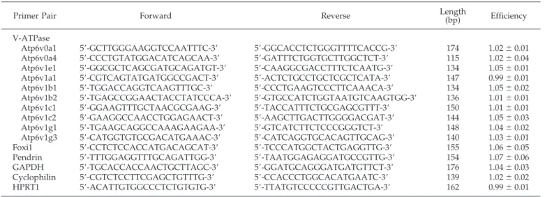
Mouse kidney samples were homogenized in Trizol (Invitrogen, Merelbeke, Belgium) to extract total RNA. Total RNA samples (2.7g) were treated with DNase I (Invitrogen) and reverse-transcribed into cDNA using SuperScript II RNase H Reverse Transcriptase (Invitro-gen). Specific primers for V-ATPase subunits, Foxi1, Pendrin (PDS), glyceraldehyde-3-phosphate dehydrogenase (GAPDH), cyclophilin, and hypoxanthine guanine phosphoribosyl transferase (HPRT1) were designed using Beacon Designer 2.0 (Premier Biosoft International, Palo Alto, CA) and are summarized in Table 1. Real-time PCR analyses were performed in duplicate with 200 nM of both sense and antisense primers in a final volume of 25l using 1 unit of Platinum TaqDNA Polymerase, 2 mM MgSO4, 400M dNTP, and SYBR Green I
(Molec-ular Probe, Leiden, The Netherlands) diluted to 1:105. The PCR mixture
contained 10 nM fluorescein for initial well-to-well fluorescence nor-malization. PCR conditions were settled as incubation at 94°C for 3 min followed by 40 cycles of 30 s at 95°C, 30 s at 61°C, and 1 min at 72°C. The melting temperature of PCR product was checked at the end of each PCR by recording SYBR green fluorescence increase upon slowly renaturating DNA. For each assay, standard curves were prepared by serial four-fold dilutions of mouse adult kidney cDNA. The efficiencies of the amplifications with each primer set were calculated from the slope of the standard curve [efficiency⫽ (10⫺1/slope)⫺ 1] and were
close to 100% (Table 1).
The relative mRNA expression of the various isoforms (A, a1, B2, E1, G1, C1, a4, B1, G3, and C2) was investigated in adult male kidneys (n⫽ 8), after normalization to HPRT1, cyclophilin, and GAPDH [Ratio⫽ 2⌬Ct (Reporter ⫺ Target Gene)]. The relative changes in mRNA levels of these alternate isoforms during ontogeny were determined by comparison with the adult mRNA level, after adjustment to GAPDH [Ratio ⫽ (Efficiencytarget gene)⌬Ct(Adult ⫺ Sample)/(EfficiencyGAPDH)⌬Ct (Adult ⫺ Sample)]
Figure 1. Structural model of the vacuolar type H⫹-ATPase (V-ATPase). The transmembrane V0 domain of the mammalian V-ATPase is organized into a complex of a(c)4 to 5c⬙d, whereas the cytosolic V1 domain has a stoichiometry of A3B3CDEFG2H2. The ubiquitous and the intercalated cell (IC)-specific alternate isoforms of V0 and V1 subunits are colored. In the plasma membrane of murine IC, a4, d2, B1, C2, and G3 substitute ubiquitous a1, d1, B2, C1, and G1, respectively. This model was modified from the yeast model to represent the mammalian V-ATPase complex (adapted with permission from reference 5). Note that the presence of up to three copies of each E and G per V-ATPase complex has been suggested from analysis of Coomassie staining (4) and that the existence of the c⬘ subunit in mammals is uncertain (42).
(16). Real-time RT-PCR results were confirmed by using another set of primers for each of V-ATPase subunits.
In parallel with the relative quantification described above, we cloned DNA plasmids for each subunit and GAPDH to determine copy numbers of V-ATPase subunits transcripts. The PCR products were cloned into a pTZ57R vector (Fermentas GmbH, Vilnius, Lithuania) and transformed in JM 107 Competent Cells (MBI Fermentas). LB-broth cultures (150 ml) of single colonies were grown up overnight skating at 200 rpm at 37°C. Plasmid purification was performed with the High Purity Plasmid Systems (Gentaur, Brussels, Belgium) according to the manufacturer’s recommendations. Standard curves were generated from serial 10-fold dilutions of the purified plasmid that contained the appropriate cDNA, and regression analyses of the threshold cycle (Ct) values of standard dilution series were used to determine the amplifi-cation efficiency (data not shown). The absolute amount of mRNA molecules of V-ATPase subunits in mouse adult kidneys (n⫽ 8) was determined by extrapolating the Ct values from the standard curves, according to the molecular weight of the plasmid (1.9⫻ 106g/mol) and
the Avogadro’s number (1 mol⫽ 6.022 ⫻ 1023molecules), and then
normalized to the GAPDH level in each sample. Each experiment was run in duplicate, with highly reproducible results.
In Situ Hybridization
Kidney samples were fixed in 4% formaldehyde (Boehringer In-gelheim, Heidelberg, Germany) in 0.1 M phosphate buffer (pH 7.4) before embedding in paraffin as described (17). Five-micrometer-thick sections were deparaffinized, rehydrated. and treated for 45 min at 37°C with 1 mg/ml proteinase-K (Sigma, St. Louis, MO) in 100 mM Tris-HCl (pH 8.0) and 50 mM EDTA. Sections then were acetylated for 10 min in 0.25% acetic anhydride and 0.1 M triethanolamine (pH 8.0) and prehybridized for 1 h at 52°C (Foxi1) or 56°C (B1) in the hybrid-ization mixture that contained 40% (vol/vol) formamide, 50% (wt/vol) dextran sulfate, 2% (vol/vol) Denhardt’s solution, and 2⫻ SSC (1⫻ SSC⫽ 150 mM NaCl and 15 mM sodium citrate) in a humidified chamber. A mixture of two Foxi1- or B1-specific oligonucleotide probes (Foxi1 5⬘-AGGCTGGGTGACCTTCGAAGGCTGGATAAAGGGAACCGGG-3⬘, 5⬘-GAGTGGCTGTGAGCACAGCCTCTGGCACTGCTCGCTCCTCT-3⬘; B1 5⬘-AACATGGGTTCAAGCCATAAAGGAGAAGGCTGCAGCTGG-3⬘, 5⬘-TCCATTTCAGTTACATCTTTGGAGGCGGGGCTTCTATAAT-3⬘)
or of two control oligonucleotides with the complementary sequence was prepared by fluorescein-dUTP labeling of the 3⬘ ends (Amersham Bio-sciences, Piscataway, NJ). The sections then were hybridized overnight at 52°C (Foxi1) or 56°C (B1) with the hybridization mixture supplemented with 1.5 ng/ml labeled probes and washed with decreasing concentrations of SSC (4⫻, 2⫻, and 1⫻). All washes were carried out at room temperature for 2⫻ 10 min. The detection of hybridized fluorescein-labeled probes was performed according to the immunostaining procedure, described below.
Antibodies
Immunoblotting and immunostaining analyses were performed with well-characterized antibodies, including mAb against the E1 subunit of the V-ATPase (a gift of Dr. S. Gluck, University of California, San Francisco, CA) (6);-actin (Sigma); fluorescein (Dako, Glostrup, Den-mark); ␣1 Na⫹/K⫹-ATPase (Upstate Biotechnology Inc., Waltham, MA); rabbit polyclonal antibodies against a4 subunit (13); B1, B2, G1, and G3 subunits (17); A subunit (Wako Chemicals GmbH, Neuss, Germany); C1 subunit (Santa Cruz Biotechnology, Santa Cruz, CA); Rab5a (Santa Cruz Biotechnology); aquaporin-2 (AQP2; Sigma); podo-cin (a gift from C. Antignac, Inserm U574, Necker Hospital, Paris, France) (18); sheep polyclonal antibodies against megalin (a gift from P.J. Verroust, Saint-Antoine Medical Faculty, Paris, France) (8); type II carbonic anhydrase (CAII; Serotec, Oxford, UK); goat polyclonal anti-bodies against cathepsin D (Santa Cruz Biotechnology); and chicken polyclonal antibodies against pendrin (a gift from P. Kopp, Northwest-ern University, Chicago, IL).
Analytical Subcellular Fractionation
Kidneys from approximately 40 fetuses at E16.5 and dissected cortex and medulla from five adult mice were minced in 0.25 M sucrose and 3 mM imidazole buffer (pH 7.4) that contained Complete protease inhibitors (Roche, Vilvoorde, Belgium) and homogenized in a Potter-Elvehjem tissue homogenizer (Thomas Scientific, Swedesboro, NJ). A low-speed “nuclear” fraction was pelletted at 700⫻ g for 10 min and extracted twice by resuspension sedimentation. Pooled postnuclear supernatants were sedimented further at 100,000⫻ g for 60 min in a 50Ti fixed-angle rotor (Beckman, Palo Alto, CA). The supernatant (S), which represents the cytosolic fraction, was collected for further
char-Table 1. Primers used for real-time RT-PCR
aPrimer Pair Forward Reverse Length(bp) Efficiency V-ATPase
Atp6v0a1 5⬘-GCTTGGGAAGGTCCAATTTC-3⬘ 5⬘-GGCACCTCTGGGTTTTCACCG-3⬘ 174 1.02⫾ 0.01 Atp6v0a4 5⬘-CCCTGTATGGACATCAGCAA-3⬘ 5⬘-GATTTCTGGTGCTTGGCTCT-3⬘ 115 1.02⫾ 0.04 Atp6v1e1 5⬘-GGCGCTCAGCGATGCAGATGT-3⬘ 5⬘-CAAGGCGACCTTTCTCAATG-3⬘ 134 1.05⫾ 0.01 Atp6v1a1 5⬘-CGTCAGTATGATGGCCGACT-3⬘ 5⬘-ACTCTGCCTGCTCGCTCATA-3⬘ 147 0.99⫾ 0.01 Atp6v1b1 5⬘-TGGACCAGGTCAAGTTTGC-3⬘ 5⬘-CCCTGAAGTCCCTTCAAACA-3⬘ 134 1.05⫾ 0.02 Atp6v1b2 5⬘-TGAGCCGGAACTACCTATCCCA-3⬘ 5⬘-GTGCCATCTGGTAATGTCAAGTGG-3⬘ 136 1.01⫾ 0.01 Atp6v1c1 5⬘-GGAAGTTTGCTAACGCGAAG-3⬘ 5⬘-TACCATTTCTGCGAGCGTTT-3⬘ 150 1.01⫾ 0.01 Atp6v1c2 5⬘-GAAGGCCAACCTGGAGAACT-3⬘ 5⬘-AAGCTTGACTTGGGGACGAT-3⬘ 144 1.05⫾ 0.03 Atp6v1g1 5⬘-TGAAGCAGGCCAAAGAAGAA-3⬘ 5⬘-GTCATCTTCTCCCGGGTCT-3⬘ 148 1.04⫾ 0.02 Atp6v1g3 5⬘-CATGGTGTGCGACATGAAAC-3⬘ 5⬘-CATCAGGTGCACAGTTGCAG-3⬘ 140 1.03⫾ 0.01 Foxi1 5⬘-CCTCTCCACCATGACAGCAT-3⬘ 5⬘-TCCCATGGCTACTGAGGTTG-3⬘ 155 1.06⫾ 0.05 Pendrin 5⬘-TTTGGAGGTTTGCAGATTGG-3⬘ 5⬘-TAATGGAGAGGATGCCGTTG-3⬘ 154 1.07⫾ 0.06 GAPDH 5⬘-TGCACCACCAACTGCTTAGC-3⬘ 5⬘-GGATGCAGGGATGATGTTCT-3⬘ 176 1.04⫾ 0.03 Cyclophilin 5⬘-CGTCTCCTTCGAGCTGTTTG-3⬘ 5⬘-CCACCCTGGCACATGAATC-3⬘ 139 1.02⫾ 0.02 HPRT1 5⬘-ACATTGTGGCCCTCTGTGTG-3⬘ 5⬘-TTATGTCCCCCGTTGACTGA-3⬘ 162 0.99⫾ 0.01
a
V-ATPase, vacuolar H⫹-ATPase; GAPDH, glyceraldehyde-3-phosphate dehydrogenase; HPRT1, hypoxanthine guanine phosphoribosyl transferase.
acterization. The high-speed pellet (MLP fraction of postnuclear parti-cles) was resuspended in 1 ml of homogenization buffer, mixed with 7 ml of 16% (vol/vol) Percoll (average final density 1.048 g/ml), layered over a 250-l Percoll cushion, and centrifuged at 60,000 ⫻ g for 30 min in a 50Ti rotor into a self-generating gradient, after which 10 fractions of approximately 750l each, excluding the packed cushion, were collected from the bottom. Each fraction was analyzed for density (refractometry), total protein content (Lowry assay), and amount of the indicated antigens (immunoblotting).
Immunoblotting
Membrane extraction and immunoblotting were performed as de-scribed previously (19). Briefly, kidney samples were homogenized in ice-cold buffer [300 mM sucrose and 25 mM N-2-hydroxyethylpipera-zine-N⬘-2-ethanesulfonic acid made to pH 7.0 with 1 M tris(hydroxym-ethyl)aminomethane (Tris)] that contained Complete protease inhibi-tors (Roche). The homogenate was centrifuged at 1000⫻ g for 15 min at 4°C, and the resulting supernatant was centrifuged at 100,000⫻ g for 120 min at 4°C. The pellet was suspended in ice-cold homogenization buffer, before determination of protein concentration and storage at ⫺80°C. After resolution by SDS-PAGE and blotting on nitrocellulose, the membranes were incubated overnight at 4°C with primary antibod-ies, washed, incubated for 1 h at room temperature with appropriate peroxidase-labeled antibodies (Dako), washed again, and visualized with enhanced chemiluminescence. Normalization for-actin was ob-tained after stripping the blots and reprobing with the anti–-actin antibody. Specificity of the immunoblot was determined by incubation with nonimmune rabbit or mouse IgG (Vector Laboratories, Burlin-game, CA) or control ascites fluid (Sigma). Densitometry analysis was performed with a Canon CanoScan8000F using the NIH Image V1.60 software. All immunoblots were performed at least in duplicate.
Immunostaining
After blocking endogenous peroxidase for 30 min with 0.3% hydrogen peroxide, sections were incubated with 10% normal goat serum for 60 min, before incubation with the primary antibodies diluted in PBS that con-tained 2% BSA for 60 min. After washing in 50 mM Tris-HCl, sections were incubated successively with biotinylated secondary anti-IgG antibodies, avidin-biotin peroxidase, and diaminobenzidine or aminoethylcarbazole (Vector Laboratories). For immunofluorescence studies, secondary anti-bodies were detected with Texas red–conjugated and FITC-conjugated avidin (Vector Laboratories). Sections were viewed under a Leica DMR coupled to a Leica DC300 digital camera (Leica, Heerbrugg, Switzerland). The specificity of immunostaining was tested by incubation (1) in the absence of primary antiserum and (2) with nonimmune rabbit serum or control rabbit or mouse IgG (Vector Laboratories).
Results
Relative Expression of V-ATPase Subunits in Adult
Mouse Kidney
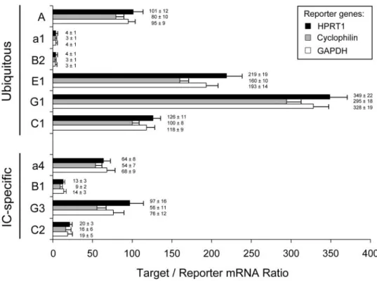
The recently updated structure of the V-ATPase contains at least 13 different subunits that form two functional domains, V0 and V1 (Figure 1). Before considering the expression of isoforms during mouse nephrogenesis, we performed compar-ative real-time RT-PCR quantification, with different reporter genes, to establish the relative mRNA expression of ubiquitous and IC-specific subunits in adult mouse kidney (Figure 2). These data were essentially confirmed by absolute real-time RT-PCR using standard curves of cloned DNA plasmids for the V-ATPase subunits and GAPDH (data not shown). These stud-ies revealed striking differences in terms of relative expression
Figure 2. mRNA quantification of V-ATPase subunits. Real-time reverse transcription–PCR (RT-PCR) quantification of ubiquitous (A, a1, B2, E1, G1, and C1) and IC-specific (a4, B1, G3, and C2) subunits in mouse adult kidney (n⫽ 8). The mRNA expression levels (mean ⫾ SEM) were compared after adjustment to HPRT1 (⫻50), cyclophilin (⫻103
), or glyceraldehyde-3-phosphate dehydrogenase (GAPDH;⫻2.103) as reporter genes. HPRT1, hypoxanthine guanine phosphoribosyl transferase.
at the mRNA level that could not be explained by the structural stoichiometry of the V-ATPase complex shown in Figure 1. For instance, the A subunit was 20-fold more abundant than B2, although these subunits are considered to form the A3B3 cata-lytic core of the V1 domain. Moreover, comparative analysis of alternate isoforms inside ubiquitous (a1, B2, C1, and G1) or IC-specific (a4, B1, C2, and G3) pumps did not show a constant stoichiometry. These results suggest that the assembly of the V-ATPase complex results from important posttranscriptional modifications and/or reflects significant differences in the turn-over of its various components.
Expression of V-ATPase Subunits mRNA in Developing
Mouse Kidney
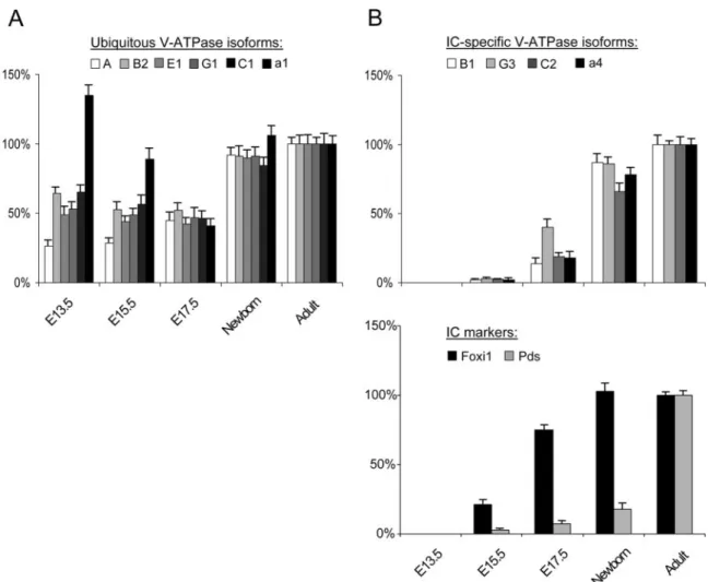
The differential expression patterns of ubiquitous and IC-spe-cific V-ATPase subunits during mouse nephrogenesis were inves-tigated using real-time RT-PCR analyses (Figure 3). The ubiqui-tous A, B2, E1, G1, and C1 subunits showed an early (E13.5) and
stable expression (approximately 40% of the adult level) through-out nephrogenesis, reaching the adult level at birth (Figure 3A). Of note, the a1 isoform showed an earlier induction at E13.5 (approx-imately 135%) followed by a gradual decrease during late ontog-eny and a second increase around birth. This particular develop-mental pattern is similar to that described for the endosomal kidney-specific chloride channel, ClC-5, and for the V0 d1 V-ATPase subunit (19,20). In contrast, the corresponding IC-spe-cific B1, G3, C2, and a4 subunits were barely detected at E15.5 and showed a progressive induction during late ontogeny, like the anion transporter pendrin (Figure 3B). It must be noted that the forkhead transcription factor Foxi1, which is a member of the HFH/winged helix family, was detected from E15.5 (Figure 3B). These data show that the early expression of ubiquitous subunits of V-ATPase definitively contrasts with the later and more progressive appearance of Foxi1, pendrin, and IC-specific subunits.
Figure 3. Expression of V-ATPase subunits in mouse developing kidney. Real-time RT-PCR quantification for mRNA expression of ubiquitous A, B2, E1, G1, C1, and a1 subunits (A) and IC-specific B1, G3, C2, and a4 isoforms, as well as Foxi1 and pendrin (pds) (B), during mouse nephrogenesis. The mRNA levels were first adjusted to GAPDH at every developmental stage, then normalized to the adult level set at 100% using the following formula: [Ratio ⫽ (Efficiencytarget)⌬Ct (Adult ⫺ Sample)/ (EfficiencyGAPDH)⌬Ct (Adult ⫺ Sample)]. The early (embryonic day 13.5 [E13.5]) and stable expression of ubiquitous subunits during nephrogenesis clearly contrasts with the later and progressive appearance of IC-specific isoforms and pds, which follows the induction of the transcription factor Foxi1 at E15.5. Note that the a1 subunit is characterized by a bimodal developmental pattern.
Distribution of Forkhead Transcription Factor Foxi1 in
Developing Mouse Kidney
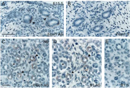
The winged helix transcriptional activator Foxi1 has been located to the distal part of the nephron from E16.5 during mouse nephrogenesis (21) and recently implicated in IC differ-entiation along the CD (22). To investigate the putative link between Foxi1 and IC-specific V-ATPase isoforms expression, we performed in situ hybridizations on mouse developing kid-ney with fluorescein-labeled oligonucleotides directed against Foxi1 or B1 (Figure 4). At E15.5, Foxi1 was detected in some scattered cells of the medullary CD, whereas no signal was found in glomeruli, PT, and differentiating structures in the cortex (Figure 4A). At later stages, Foxi1 mRNA was identified in a subset of cells within distinct tubules in cortex and medulla (Figure 4C). In these cells, Foxi1 co-distributed with the IC-specific B1 V-ATPase subunit (Figure 4D). Altogether, the real-time RT-PCR and in situ hybridization data during mouse nephrogenesis strongly support the role of Foxi1 in the differ-entiation of IC along the CD.
Expression of V-ATPase Subunits in Developing
Mouse Kidney
Well-characterized antibodies were used to evaluate the ex-pression of ubiquitous and IC-specific V-ATPase subunits in mature and developing kidney (Figure 5). Postnuclear particles and high-speed supernatant, resolved by differential centrifu-gation of adult kidney samples, were analyzed by immunoblot-ting. In the adult kidney, approximately 20% of V1 subunits, i.e., A, B2, E1 and G1, were detected in the cytosolic fraction, whereas the transmembrane a4, as well as the V1 C1 subunit, was associated only with membranes (Figure 5A). During nephrogenesis (Figure 5B), ubiquitous V-ATPase subunits (A,
B2, E1, G1, and C1) were detected as early as E13.5 and grad-ually increased from E16.5 until birth. In contrast, the IC-specific subunit isoforms (a4, B1, and G3) and the anion ex-changer pendrin were detected only from E16.5 and increased progressively during late nephrogenesis.
Segmental Distribution of V-ATPase Subunits in
Developing Mouse Kidney
The segmental distribution of ubiquitous and IC-specific sub-units in the developing mouse kidney was investigated using well-characterized antibodies against E1, B2, and a4 (Figure 6). No immunostaining for the IC-specific a4 subunit was detected at E14.5, by which time dispersed glomeruli and developing PT were observed in the inner cortex (Figure 6A). At E15.5, the immuno-reactive signal was located in the apical area of scattered cells along the cortical (Figure 6B) and medullary CD (Figure 6C), whereas PT, ureteric buds, glomeruli, and differentiating struc-tures remained unlabeled. By contrast, at this fetal stage, the distribution of ubiquitous E1 and B2 was acquired in PTC (Figure 6, D and I), as well as the distribution of E1 in IC (Figure 6D, inset). From E16.5, apical staining for a4 was detected in scattered IC (Figure 6, E, G, and H), clearly identified by co-distribution with CAII (Figure 6, F and G) but negative staining for AQP2 (Figure 6H). The distribution pattern of a4 in mouse developing kidney was restricted to IC (Figure 6J), similar to that observed in adult kidney (Figure 6K). The specificity of a4 staining was demon-strated with preimmune rabbit antibodies (Figure 6L). These re-sults show that, in mouse developing kidney, the a4 subunit is detected progressively during IC differentiation, whereas the dis-tribution of ubiquitous E1 and B2 includes both PTC and IC of the CD at an early stage.
Figure 4. Distribution of Foxi1 and V-ATPase B1 subunit mRNA in mouse developing kidney. In situ hybridization for Foxi1 (A and C: antisense probe; B: sense); B1 V-ATPase subunit (D: antisense probe; E: sense) in the developing mouse E15.5 (A and B) and neonatal (C through E) kidney. At E15.5, Foxi1 mRNA is specifically detected in scattered cells within medullary collecting ducts (CD; A, arrowheads). In the newborn kidney, Foxi1 and B1 co-distribute in distinct cells within cortical CD (C and D, arrowheads on serial sections). No signal was detected when sense probes were used on consecutive sections (E). Bars⫽ 50m in A and B; 20m in C through E.
Subcellular Fractionation in Percoll Gradients
In addition to its intracellular location where it mediates vesicle acidification, the V-ATPase is located at the cell surface of renal IC. We investigated the subcellular distribution of ubiquitous E1 and IC-specific a4 subunits in the mature and developing mouse kid-ney (Figure 7). Adult kidkid-neys were dissected into cortex and medulla, and appropriate markers were analyzed by immuno-blotting to validate the dissection (Figure 7A). Podocin and pen-drin, as well as E1 and a4, were considerably enriched in the cortex, whereas AQP2 was detected mostly in the medulla (Figure 7B). These results confirm that the two parts of adult kidneys were purified adequately, as indicated by an enrichment ratio ⬎30 (podocin versus AQP2). Next, the subcellular distribution of E1 and a4 in fetal kidney and adult cortex and medulla was com-pared (Figure 7C). Percoll gradients resolved a low-density peak (fractions 2 to 4) that comprised all (fetus) or the bulk (adult) of the early endosomal marker Rab5a from a high-density peak (frac-tions 9 and 10), including most of the lysosomal marker cathepsin D. In the three tissue samples, both V-ATPase subunits peaked in the low-density fractions, suggesting their major association with endosomes and not with lysosomes.
Discussion
In this study, we showed that ubiquitous and IC-specific isoforms of V-ATPase subunits are differentially expressed in mature kidney and during mouse nephrogenesis. The early expression of the ubiquitous A, B2, E1, G1, C1, and a1 subunits contrasts with the later and more progressive appearance of the IC-specific isoforms B1, G3, C2, and a4. The forkhead transcrip-tion factor Foxi1 mRNA was detected in IC from E15.5, with co-distribution with the B1 subunit in late nephrogenesis. The segmental distribution of E1 and B2 was acquired in PTC at E15.5, whereas a4 co-distributed with CAII in the IC during late nephrogenesis. Subcellular fractionation studies indicated a preferential location of both ubiquitous and IC-specific sub-units to endosomes in both developing and mature kidneys.
The subunit composition of the bovine V-ATPase, previously determined by quantitative amino acid studies (1), was updated recently using electron microscopy and single-molecule image analysis (4). These investigations led to a structural model of the mammalian V-ATPase, which seems to be similar to the F1F0-ATP synthase in yeast (4). As a preliminary to the ontogeny analysis, real-time RT-PCR analyses in the adult kidney showed that the Figure 5. Expression of V-ATPase subunits in mouse developing kidney. (A) Relative expression of V1 A, B2, E1, G1, and C1, and V0 a4 in membrane (MLP) and cytosolic (S) compartments of adult mouse kidney. Equal loads (20g) of postnuclear particles (MLP 37% of total homogenate proteins) and high-speed supernatant (S 29% of total homogenate proteins) were analyzed. Densitometry analyses show that approximately 20% of V1 subunits are not associated with membranes at steady state. The transmembrane V0 a4 subunit, as well as the V1 C1, are not detected in the cytosolic fraction. (B) Representative immunoblots for ubiquitous A, B2, E1, G1, C1, and IC-specific a4, B1, and G3 V-ATPase subunits and pds in fetal (from E13.5 to E18.5), newborn, and adult mouse kidneys. A total of 20 g of protein was loaded in each lane. Blots were probed with antibodies against A (1:1000), B2 (1:1000), E1 (1:100), G1 (1:500), C1 (1:1000), a4 (1:5000), B1 (1:1000), and G3 (1:500) subunits; pendrin (1:2000); and, after stripping,-actin (1:10,000). Ubiquitous subunits are detected as early as E13.5 and gradually increase during late nephrogenesis, whereas the developmental pattern of IC-specific isoforms and pendrin is characterized by a later expression from E16.5.
relative mRNA expression of V-ATPase subunits does not corre-spond to the expected stoichiometry. These results were con-firmed by absolute mRNA quantification using standard curves from serial dilution of purified plasmids (data not shown). The ratio between the two methods varied between 0.8 and 1.8 accord-ing to the subunits, in agreement with previous comparative studies (23). This variability is attributed to distinct primer effi-ciency and, overall, cumulative errors introduced by the addi-tional steps of the absolute mRNA quantification (spectrophotom-etry, calculations of molecular weight) (24). Thus, the relative method, using primer sets of similar efficiency (Table 1) and three distinct internal standards, was preferred to compare the abun-dance of V-ATPase subunit transcripts in one single kidney sam-ple. The comparative mRNA quantification of subunits demon-strates a variable expression from ubiquitous to IC-specific V-ATPase complexes. Although minimal and/or limited correla-tion between the mRNA and protein levels is most often reported,
the degree of correlation could vary according to subcellular lo-calization or functional category (25). In the case of the renal V-ATPase complex, a first hypothesis could be significant varia-tions in the turnover of the subunits that compose the complex, as well as a differential regulation of subunit mRNA stability (26). Because in vivo regulation of V-ATPase, which involves the re-versible dissociation of V1 from V0 domain (27), does not require new protein synthesis but an efficient microtubular system (28), a second hypothesis could be posttranscriptional modifications dur-ing subunit synthesis and V-ATPase assembly. For instance, rear-rangements during the formation of V0 and V1 domains might stabilize each of these subunits by tight interactions and/or mu-tual chaperoning. Of interest, a selective posttranscriptional con-trol of protein expression levels among loci of the V-ATPase complex has been shown to operate in Saccharomyces cerevisiae (29). The molecular events that occur in renal tubular cells after the onset of glomerular filtration, which starts at E14 in mouse Figure 6. Segmental distribution of E1 and a4 V-ATPase subunits in mouse developing kidney. Immunostaining for a4 (all panels except D, F, I, and L), E1 (D), B2 (I), type II carbonic anhydrase (F and G), aquaporin 2 (AQP2; H), megalin (I and J), and negative control (L) in developing mouse kidney at E14.5 (A), E15.5 (B through D), E16.5 (E through G and I), and E18.5 (H and J) and in adult mouse kidney (K and L). No specific staining for a4 is observed at E14.5 (A). From E15.5, a4 is detected in the apical area of scattered cells of cortical (B) and medullary (C) CD, whereas proximal tubule cells (PTC) remain unstained. At this stage, the ubiquitous E1 and B2 are already expressed in developing PTC (D and I) and in IC of the medullary and cortical (D, inset) CD. At E16.5, apical staining for a4 is still present in scattered IC of the CD (E, arrowheads), clearly identified by co-staining positive for type II carbonic anhydrase (CAII; F, arrowheads, and G) but negative for AQP2 (H). This segmental distribution restricted to IC in mouse fetal kidney (J) is similar to that observed in the adult kidney (K). No staining for a4 subunit was observed with preimmune rabbit serum (L). Magnification,⫻300 in A through F, K, and L; ⫻450 in G through J.
(30), remain incompletely elucidated. Recent evidence suggests that different steps of kidney organogenesis are reflected by clusters of differentially regulated genes, including transporters and ions-motive ATPases, under the control of specific tran-scription activators (9,31). In rats (32) and humans (33), PT endocytic activity is effective during nephrogenesis or imme-diately after birth. Both the receptor-mediated endocytic path-way and the apical targeting within PTC (8) depend on a correct vacuolar acidification, which is mediated by V-ATPase
in PTC. Our real-time RT-PCR and immunoblotting analyses show an early and stable expression of ubiquitous isoforms of V-ATPase subunits during nephrogenesis, which is in keeping with its essential role for early mouse development (12). It is interesting that the endosomal a1 subunit shows an early in-duction, a progressive decrease during late ontogeny, and a novel induction around birth. Such a biphasic developmental pattern is also described for the renal chloride channel ClC-5 that co-locates with V-ATPase in PT endosomes (34), as well as Figure 7. Localization and subcellular distribution of V-ATPase subunits in fetal and adult (cortex and medulla) mouse kidneys. (A) Representative immunoblots for podocin, IC-specific a4 and ubiquitous E1 V-ATPase subunits, pendrin,-actin, Na⫹/K⫹ -ATPase, and AQP2 in cortex and medulla of adult mouse kidney. Thirty micrograms of protein were loaded in each lane. Podocin (1:5000), a4 (1:5000), E1 (1:100), and pendrin (1:1000) are highly enriched in the dissected cortex and depleted in the dissected medulla;-actin (1:10,000) and Na⫹/K⫹-ATPase (1:5000) are equally expressed in both tissues; AQP2 (1:2000) is detected mostly in the dissected medulla. (B) The relative enrichment of these markers, based on densitometry. (C) Percoll gradients of total fetal (E16.5) and adult (cortex and medulla) mouse kidneys. Distributions after centrifugation are presented in comparison with the initial concentration (C/Ci) so that values ⬎1 reflect organelle enrichment and values ⬍1 reflect organelle depletion. For immunoblotting analyses, equal volumes were loaded in each lane. The gradients resolve a low-density peak (fractions 2 to 4), including early endosomes (Rab5a), from a high-density peak (fractions 9 and 10), enriched in lysosomes (cathepsin D). The two V-ATPase subunits peak in the low-density fractions, which suggests their major association with endosomes and not with lysosomes in both developing and adult (cortex and medulla) kidneys.
for the V0 d1 subunit (20). ClC-5 is mutated in Dent’s disease, an X-linked renal tubular disorder characterized by a major and early defect in the PT endocytic pathway (35). Thus, besides the stable expression of ubiquitous subunits, ClC-5 and a1 isoform, which are considered to target correctly V-ATPase complex to appropriate intracellular compartments (36), both are ex-pressed simultaneously in PTC as early as the glomerular fil-tration starts. These essential components of the endocytotic process, responsible for the reabsorption of amino acids, vita-mins, and minerals, may indeed be required early for correct growth and development. Analytical subcellular fractionation clearly resolved the two V-ATPase subunits from lysosomes, as assessed by the cathepsin D distribution, and rather pointed to a primary endosomal location, as shown by co-distribution with Rab5a. This low-density pattern is identical to what we recently reported for ClC-5 (8). Thus, kidney endosomes seem to be well equipped with electrogenic proton pumps and a chloride conductance, both in the adult and the fetus.
The IC constitute approximately one third of the cells that line the mouse CD (37). Although both␣- and -IC are characterized by high CAII activity, they differ by the selective polarity of V-ATPase and type 1 anion exchanger, allowing them to partici-pate in proton (␣ type) or bicarbonate ( type) urinary secretion (7). The differentiation and maturation of the various cell types within the CD is poorly understood. Studies in the rat have shown that V-ATPase and CAII are detected simultaneously in IC at the end of gestation (38). During the first weeks of life, IC continue their maturation but are partially removed from specific parts of the CD by apoptosis or luminal extrusion (39). Recently, the winged helix transcriptional factor Foxi1, previously located in mouse fetal distal nephron (21), was shown to play an essential role in IC differentiation from epithelial precursor cells in the CD (22). In addition, Foxi1 is regarded as an upstream regulator of the anion transporter pendrin during inner ear development (40). Indeed, mice that lack Foxi1 showed a lack of differentiation of the distal nephron, leading to dRTA (22), as well as a sensorineural deafness resembling that of Pendred syndrome (40). Our descrip-tion of the developmental expression of Foxi1, IC-specific V-ATPase isoforms, and pendrin provides additional information on CD maturation in mouse. Real-time RT-PCR showed that IC-specific subunits B1, G3, C2, and a4, as well as pendrin, are expressed later than the ubiquitous isoforms, with a progressive induction after Foxi1 appearance (E15.5). In situ hybridization showed that at E15.5, Foxi1 is expressed in scattered cells of the medullary CD. At later stages, Foxi1 is identified in distinct cells within tubules in the cortex and outer medulla, in which it co-distributes with the IC-specific B1 subunit. In silico studies (http:// mordor.cgb.ki.se/cgi-bin/CONSITE/consite) indicate that gene promoters of IC-specific markers, such as type 1 anion exchanger, AE4, pendrin, or IC-specific V-ATPase subunits, exhibit Foxi1 DNA binding consensus sequence (21,22). Immunoblot studies showed a coordinate expression of the IC-specific subunits and pendrin at approximately E16.5, with immunolocalization of a4 and CAII in IC. Of note, subcellular fractionation showed that, in both fetal (E16.5) and adult kidneys, a4 and E1 subunits are located mostly in endosomes. This observation corroborates a dynamic process that occurs in IC, which involves regulated
exo-cytic insertion of V-ATPase from a vesicular pool to the plasma membrane (41). Taken together, the mRNA and protein data support the case for a differential but coordinate regulation of IC-specific versus ubiquitous V-ATPase subunits during kidney development.
The comparative ontogeny of V-ATPase subunits helps us un-derstand the pathophysiology of inherited renal disorders such as Dent’s disease and dRTA. Dent’s disease, which is due to inacti-vating mutations in CLCN5 that presumably lead to impaired endosomal acidification in PTC, is characterized by low molecular weight proteinuria that can be detected within the first weeks of life (35). These clinical findings, which suggest that the molecular mechanisms that ensure endosomal acidification must be ac-quired at birth (19), have their counterpart in our demonstration of a parallel induction of the endosomal a1 isoform and ClC-5 that culminates at birth. Given the essential role of V-ATPase in early embryogenesis and its large tissue distribution, no mutation of any ubiquitous V-ATPase subunits has ever been reported, being definitively not compatible with life (12). In contrast, mutations in IC-specific isoforms have been associated with human dRTA (13– 15). Some well-documented patients show clinical manifestations of dRTA during infancy, which suggests that the segmental ex-pression of these IC-specific isoforms of V-ATPase must be ac-quired early, as indeed documented here.
In conclusion, our data demonstrate that ubiquitous and IC-specific isoforms of V-ATPase subunits are differentially ex-pressed during mouse nephrogenesis. These results provide new insights into the segmental distribution of V-ATPase complexes in the kidney and represent a useful tool to investigate further any newly discovered subunit isoforms. The deciphering of the com-plex maturation of PTC and IC along the distal nephron also helps us understand the pathophysiology of renal tubular disorders.
Acknowledgments
These investigations were supported by the Universite´ catholique de Louvain, the Fonds National de la Recherche Scientifique (FNRS), the Forton Foundation, Concerted Research Actions, Inter-University At-traction Poles, and the EuReGene integrated project of the European Community (FP6). F.J. is a research fellow of the FNRS, and C.P. is a research fellow of the FRIA.
We are grateful to Y. Cnops, M. Leruth, L. Wenderickx, and P. Henriet for excellent technical help and discussion and to M. Futai, P. Gailly, A. Goffinet, N. Tajeddine, and M.-F. van den Hove for material.
References
1. Nishi T, Forgac M: The vacuolar H⫹-ATPases: Nature’s most versatile proton pumps. Nat Rev Mol Cell Biol 3: 94 –103, 2002
2. Nelson N, Harvey WR: Vacuolar and plasma membrane proton-adenosinetriphosphatases. Physiol Rev 79: 361–385, 1999
3. Wagner CA, Finberg KE, Breton S, Marshansky V, Brown D, Geibel JP: Renal vacuolar H⫹-ATPase. Physiol Rev 84: 1263–1314, 2004
4. Wilkens S, Takao I, Forgac M: Three-dimensional structure of the vacuolar ATPase—Localization of subunit H by difference imaging and chemical cross-linking. J Biol Chem 279: 41942– 41949, 2004
5. Borthwick KJ, Karet FE: Inherited disorders of the H⫹-ATPase. Curr Opin Nephrol Hypertens 11: 563–568, 2002 6. Brown D, Hirsch S, Gluck S: Localization of a
proton-pumping ATPase in rat kidney. J Clin Invest 82: 2114 –2126, 1988
7. Giebisch G, Windhager E: Transport of acids and bases. In: Medical Physiology, edited by Boron WF, Boulpaep EL, Phil-adelphia, Saunders, 2004, pp 845– 860
8. Christensen EI, Devuyst O, Dom G, Nielsen R, Van der Smissen P, Verroust P, Leruth M, Guggino WB, Courtoy PJ: Loss of chloride channel, ClC-5, impairs endocytosis by defective trafficking of megalin and cubilin in kidney prox-imal tubules. Proc Natl Acad Sci U S A 100: 8472– 8477, 2003 9. Schwab K, Patterson LT, Aronow BJ, Luckas R, Liang HC, Potter SS: A catalogue of gene expression in the developing kidney. Kidney Int 64: 1588 –1604, 2003
10. Bonnici B, Wagner CA: Postnatal expression of transport proteins involved in acid-base transport in mouse kidney. Pflugers Arch 448: 16 –28, 2004
11. Satlin LM, Woda CB, Schwartz GJ: Development of function in the metanephric kidney. In: The Kidney, from Normal Development to Congenital Disease, edited by Vize PD, Woolf AS, Bard JBL, Orlando, Academic Press (Elsevier), 2003, pp 267–326
12. Inoue H, Noumi T, Nagata M, Murakami H, Kanazawa H: Targeted disruption of the gene encoding the proteolipid subunit of mouse vacuolar H⫹-ATPase leads to early em-bryonic lethality. Biochem Biophys Acta 1413: 130 –138, 1999 13. Smith AN, Skaug J, Choate KA, Nayir A, Bakkaloglu A, Ozen S, Hulton SA, Sanjad SA, Al-Sabban EA, Lifton RP, Scherer SW, Karet FE: Mutations in ATP6N1B, encoding a new kidney vacuolar proton pump 116-kDa subunit, cause recessive distal renal tubular acidosis with preserved hear-ing. Nat Genet 26: 71–75, 2000
14. Karet FE, Finberg KE, Nelson RD, Nayir A, Mocan H, Sanjad SA, Rodriguez-Soriano J, Santos F, Cremers CW, Di Pietro A, Hoffbrand BI, Winiarski J, Bakkaloglu A, Ozen S, Dusunsel R, Goodyer P, Hulton SA, Wu DK, Skvorak AB, Morton CC, Cunningham MJ, Jha V, Lifton RP: Mutations in the gene encoding B1 subunit of H⫹-ATPase cause renal tubular acidosis with sensorineural deafness. Nat Genet 21: 84 –90, 1999
15. Stover EH, Borthwick KJ, Bavalia C, Eady N, Fritz DM, Rungroj N, Giersch AB, Morton CC, Axon PR, Akil I, Al-Sabban EA, Baguley DM, Bianca S, Bakkaloglu A, Bir-can Z, Chauveau D, Clermont MJ, Guala A, Hulton SA, Kroes H, Li Volti G, Mir S, Mocan H, Nayir A, Ozen S, Rodriguez Soriano J, Sanjad SA, Tasic V, Taylor CM, To-paloglu R, Smith AN, Karet FE: Novel ATP6V1B1 and ATP6V0A4 mutations in autosomal recessive distal renal tubular acidosis with new evidence for hearing loss. J Med Genet 39: 796 – 803, 2002
16. Pfaffi MW: A new mathematical model for relative quan-tification in real-time RT-PCR. Nucleic Acids Res 29: 2004 – 2007, 2001
17. Sun-Wada GH, Murata Y, Namba M, Yamamoto A, Wada Y, Futai M: Mouse proton pump ATPase C subunit iso-forms (C2-a and C2-b) specifically expressed in kidney and lung. J Biol Chem 278: 44843– 44851, 2003
18. Roselli S, Gribouval O, Boute N, Sich M, Benessy F, Attie T, Gubler MC, Antignac C: Podocin localizes in the kidney to the slit diaphragm area. Am J Pathol 160: 131–139, 2002 19. Jouret F, Igarashi T, Gofflot F, Wilson PD, Karet FE,
Thak-ker RV, Devuyst O: Comparative ontogeny, processing, and segmental distribution of the renal chloride channel, ClC-5. Kidney Int 65: 198 –208, 2004
20. Smith AN, Jouret F, Bord S, Borthwick KJ, Al-Lamki RS, Wagner CA, Ireland DC, Cormier-Daire V, Frattini A, Villa A, Kornak U, Devuyst O, Karet FE: Vacuolar H⫹-ATPase d2 subunit: Molecular characterization, developmental regulation, and localization to specialized proton pumps in kidney and bone. J Am Soc Nephrol 16: 1245–1256, 2005 21. Overdier DG, Ye H, Peterson RS, Clevidence DE, Costa
RH: The winged helix transcriptional activator, HFH-3, is expressed in the distal tubules of embryonic and adult mouse kidney. J Biol Chem 272: 13725–13730, 1997 22. Blomqvist SR, Vidarsson H, Fitzgerald S, Johansson BR,
Ollers-tam A, Brown R, Persson AE, Bergstrom GG, Enerback S: Distal renal tubular acidosis in mice that lack the forkhead transcrip-tion factor, Foxi1. J Clin Invest 113: 1560–1570, 2004
23. Rose’Meyer RB, Mellick AS, Garnham BG, Harrison GJ, Massa HM, Griffiths LR: The measurement of adenosine and estrogen receptor expression in rat brains following ovariectomy using quantitative PCR analysis. Brain Res Brain Res Protoc 11: 9 –18, 2003
24. Freeman WM, Walker SJ, Vrana KE: Quantitative RT-PCR: Pitfalls and potential. Biotechniques 26: 112–122, 1999 25. Greenbaum D, Colangelo C, Williams K, Gerstein M:
Com-paring protein abundance and mRNA expression levels on a genomic scale. Genome Biol 4: 117, 2003
26. Wang SP, Krits I, Bai S, Lee BS: Regulation of enhanced vacuolar H⫹-ATPase expression in macrophages. J Biol Chem 277: 8827– 8834, 2002
27. Kane PM: Disassembly and reassembly of the yeast vacu-olar H⫹-ATPase in vivo. J Biol Chem 270: 17025–17032, 1995 28. Xu T, Forgac M: Microtubules are involved in glucose-dependent dissociation of the yeast vacuolar H⫹-ATPase in vivo. J Biol Chem 276: 24855–24861, 2001
29. Washburn MP, Koller A, Oshiro G, Ulaszek RR, Plouffe D, Deciu C, Winzeler E, Yates JR 3rd: Protein pathway and complex clustering of correlated mRNA and protein ex-pression analyses in Saccharomyces cerevisiae. Proc Natl Acad Sci U S A 100: 3107–3112, 2003
30. Loughna S, Landels E, Woolf AS: Growth factor control of developing kidney endothelial cells. Exp Nephrol 4: 112– 118, 1996
31. Stuart RO, Bush KT, Nigam SK: Changes in global gene expres-sion patterns during development and maturation of the rat kidney. Proc Natl Acad Sci U S A 98: 5649–5654, 2001
32. Smaoui H, Schaeverbeke M, Mallie JP, Schaeverbeke J: Transplacental effects of gentamicin on endocytosis in rat renal proximal tubule cells. Pediatr Nephrol 8: 447– 450, 1994 33. Mussap M, Fanos V, Piccoli A, Zaninotto M, Padovani EM, Plebani M: Low molecular mass proteins and urinary en-zymes in amniotic fluid of healthy pregnant women at progressive stages of gestation. Clin Biochem 29: 51–56, 1996 34. Devuyst O, Christie PT, Courtoy PJ, Beauwens R, Thakker RV: Intra-renal and subcellular distribution of the human chloride channel, ClC-5, reveals a pathophysiological basis for Dent’s disease. Hum Mol Genet 8: 247–257, 1999 35. Scheinman SJ: X-linked hypercalciuric nephrolithiasis:
Clinical syndromes and chloride channel mutations. Kid-ney Int 53: 3–17, 1998
36. Manolson MF, Wu B, Proteau D, Taillon BE, Roberts BT, Hoyt MA, Jones EW: STV1 gene encodes functional
homo-logue of 95-kDa yeast vacuolar H⫹-ATPase subunit Vph1p. J Biol Chem 269: 14064 –14074, 1994
37. Teng-umnuay P, Verlander JW, Yuan W, Tisher CC, Mad-sen KM: Identification of distinct subpopulations of inter-calated cells in the mouse collecting duct. J Am Soc Nephrol 7: 260 –274, 1996
38. Kim J, Tisher CC, Madsen KM: Differentiation of interca-lated cells in developing rat kidney: An immunohisto-chemical study. Am J Physiol 266: F977–F990, 1994 39. Kim J, Cha JH, Tisher CC, Madsen KM: Role of apoptotic and
nonapoptotic cell death in removal of intercalated cells from developing rat kidney. Am J Physiol 270: F575–F592, 1996 40. Hulander M, Kiernan AE, Blomqvist SR, Carlsson P,
Sam-uelsson EJ, Johansson BR, Steel KP, Enerback S: Lack of pendrin expression leads to deafness and expansion of the endolymphatic compartment in inner ears of Foxi1 null mutant mice. Development 130: 2013–2025, 2003
41. Brown D, Breton S: H⫹-ATPase-dependent luminal acidi-fication in the kidney collecting duct and the epididymis/ vas deferens: Vesicle recycling and transcytotic pathways. J Exp Biol 203: 137–145, 2000
42. Smith AN, Borthwick KJ, Karet FE: Molecular cloning and characterization of novel tissue-specific isoforms of the human vacuolar H(⫹)-ATPase C, G and d subunits, and their evaluation in autosomal recessive distal renal tubular acidosis. Gene 297: 169 –177, 2002
![[PDF] Manuel complet pour apprendre à utiliser Joomla | Cours informatique](data:image/gif;base64,R0lGODlhAQABAIAAAP///wAAACH5BAEAAAAALAAAAAABAAEAAAICRAEAOw==)