HAL Id: tel-01929004
https://tel.archives-ouvertes.fr/tel-01929004
Submitted on 20 Nov 2018HAL is a multi-disciplinary open access archive for the deposit and dissemination of sci-entific research documents, whether they are pub-lished or not. The documents may come from teaching and research institutions in France or abroad, or from public or private research centers.
L’archive ouverte pluridisciplinaire HAL, est destinée au dépôt et à la diffusion de documents scientifiques de niveau recherche, publiés ou non, émanant des établissements d’enseignement et de recherche français ou étrangers, des laboratoires publics ou privés.
homeostasis and oxidatives stress
Thi-Hong-Liên Han
To cite this version:
Thi-Hong-Liên Han. Functional studies of Frataxin : relations with metal homeostasis and oxidatives stress. Other. Université Sorbonne Paris Cité, 2016. English. �NNT : 2016USPCC288�. �tel-01929004�
UNIVERSITE SORBONNE PARIS CITE
UNIVERSITE PARIS.DIDEROT
Ecole Doctorale 388 : Chimie Physique et Chimie Analytique de Paris Centre Laboratoire ITODYS – UMR 7086
THESE DE DOCTORAT
Spécialité : Physico-chimie
HAN Thi Hong Lien
Titre
Etudes de la fonction de la frataxine :
Relations avec l’homéostasie métallique et le stress oxydant.
Thèse dirigée par Nguyet-Thanh Ha-DuongSoutenue le 12 octobre 2016
Jury
Mme Anne-Laure Bulteau Rapportrice
M. Peter Faller Rapporteur
M. Jean-Michel Camadro Examinateur
M. Jean-Michel El Hage Chahine Co-directeur Mme Nguyet-Thanh Ha Duong Directrice de thèse
Je tiens à remercier Nguyêt-Thanh HA-DUONG, ma directrice de thèse qui m’a encadrée tout au long de mes trois années de thèse. Je te remercie pour ta patience, ta disponibilité, nos nombreuses discussions sur la science et également sur la vie quotidienne. Merci de m’avoir toujours comprise et encouragée pour que je grandisse et évolue. Pour moi, tu es non simplement un « cô-giáo » mais une grande amie que j’ai la chance de connaître.
Un grand remerciement à Jean-Michel EL HAGE CHAHINE, mon co-directeur de thèse qui est aussi chef de l’équipe « Métaux, Chélateur, Protéine ». Je te remercie de m’avoir accueillie au sein de l’équipe. Tous tes conseils, tes explications ainsi que tes connaissances de la physicochimie m’ont beaucoup apportée.
J’exprime ma sincère reconnaissance à Jean-Michel CAMADRO, notre collaborateur à l’Institut Jaques Monod. Ces travaux de thèse ne pourraient jamais être achevés sans les suggestions de ton côté, tes idées de recherche, tes expériences ainsi que tes connaissances très approfondies sur les études de l’ataxie de Friedreich et la frataxine.
J’adresse mes remerciements à l’ensemble des membres du jury : Anne-Laure BULTEAU, Peter FALLER, et Sylvie MICHEL d’avoir accepté de juger mon travail Mes travaux de thèse ont été effectués au sein dulaboratoire ITODYS et l’Institut Jaques Monod. Je remercie François Maurel – le directeur du laboratoire ITODYS et Giuseppe Baldacci – le directeur de l’Institut Jaques Monod de m’avoir accueillie dans vos laboratoires. Je remercie Pierre-François Quenin, Brigitte Eftassiou, Brigitte Delavaquerie et Äyméric Noël pour tous vos aides administratifs et informatiques pendant mes trois années de thèse. Je remercie aussi les équipes de techniciens des deux instituts pour leurs aides indispensables.
J’aimerais exprimer mes gratitudes à l’ensemble des personnes que j’ai eu l’occasion de rencontrer au cours de cette thèse et avec lesquels je pouvais travailler, apprendre, discuter et partager des connaissances. Tout d’abord, merci à tout l’équipe de la plateforme de Spectrométrie de Masse de l’Institut Jaques Monod : Thibault LEGER et Camille GARCIA pour les analyses qui m’ont permis d’identifier mes protéines – les matières essentielles de ma thèse. Je remercie Emmanuelle LESUISSE pour tous les renseignements en utilisant du système FPLC Akta Purifier. Merci à Valérie SERRE et Lorène TELOT pour les conseils et explications pour que je puisse obtenir avec succès les tous premiers PCR et plasmides recombinants de ma vie. Je remercie Françoise AUCHERE, Océane VANPARIS, Rachel GERGONDEY, Arthur BOURDAIS, Sophie HALLEZ et tous les autres collègues de l’équipe « Mitochondrie, métaux et stress oxidatif » pour leur disponibilité et leur patience, sans oublier les bons moments que nous avons partagés.
Je reconnais les bienfaits de Myriana HEMADI et Nawal SERRADJI. Merci pour vos commentaires et discussions pendant les réunions de l’équipe. Vos amitiés et encouragements ont donné une très bonne ambiance de travail dans notre équipe. Je remercie aussi Hélène Piraux, Jun Hai, Sylvie, Eva qui sont des collègues très sympathiques avec lesquels j’ai eu l’occasion de travailler.
Quelques manipulations dans ces travaux ont été effectuées par les stagiaires. Je remercie donc Yvan Ahyi, Gordon Paul, Thien Thanh Nguyen, Yingchuan Zhu pour leurs participations.
Je ne peux pas oublier mes chères copines du bureau 456. Merci Annette, Alina, Sarrah, Christelle, Atieh pour tous les partages, discussions et même des papotages. Merci pour votre amitié, vos encouragements surtout les derniers mois. Je suis heureuse de vous connaître et d’être dans le même bureau avec vous.
Je remercie mes chers amis Mai, Quynh, Vân, Thùy, Khuê, Thuân, Vy, Quyên, Nghi, Linh, Ha Anh, Kiêu Ly. Nous venons de différentes régions du Vietnam et
Je terminerai par la gratitude que je voue à ma famille, mes parents, ma grande sœur et mon petit frère et qui sont toujours à mes côtés et m’encouragent. Enfin, merci à toi, Nam, pour tout ton soutien, ton réconfort de près ou loin, merci simplement pour ta présence à mes côtés.
CHAPTER I: Bibliography 1. Friedreich’s ataxia ... 1 1.1. Clinical features ... 1 1.1.1. Neuropathology ... 2 1.1.2. Cardiac system ... 2 1.1.3. Skeletal muscle ... 2 1.2. Cellular metabolism ... 2 1.2.1. Genetic mechanism ... 2
1.2.2. Perturbation in mitochondrial processes ... 5
1.3. Pharmacotherapy for Friedreich ataxia ... 22
1.3.1. Iron chelators ... 23
1.3.2. Antioxidants and/or stimulant of mitochondrial biogenesis ... 23
1.3.3. Frataxin level modifiers ... 24
2. Frataxin and functions hypothesis ... 27
2.1. Structure ... 27
2.2. Stability ... 29
2.3. Hypothesis about function ... 30
2.3.1. Iron-binding: iron storage and/or mitochondrial iron-uptake control ... 30
2.3.2. In iron-sulfur cluster assembly ... 31
2.3.3. Heme synthesis ... 37
2.3.4. In response to oxidative stress ... 37
2.4. Functional interactions reported for frataxin ... 38
3. Objectives ... 42
CHAPTER II: Proteins production and purification 1. Yeast frataxin homologue 1 – Yfh1 ... 55
1.1. Expression ... 55
2.1. Optimization of the expression ... 63
2.2. Purification ... 67
2.3. Characterization ... 69
2.4 Verification of the presence of the two cofactors ... 72
3. Discussion ... 75
References ... 78
CHAPTER III: Mechanism of frataxin-metal interactions 1. Introduction ... 81
2. Results ... 83
2.1. Iron (II) binding ... 83
2.1.1. Thermodynamics of iron(II) uptake ... 83
2.1.2. Kinetics of Iron(II) uptake ... 84
2.2. Iron(III) binding ... 88
2.2.1. Thermodynamics ... 88
2.2.2. Kinetics of Iron(III) uptake ... 91
2.3. Copper(II) binding ... 93
2.3.1. Thermodynamics of copper(II) uptake ... 93
2.3.2. Kinetics of Copper(II) uptake... 96
2.4. Copper(I) binding ... 104
2.4.1. Thermodynamics of copper(I) uptake ... 104
2.4.2. Kinetics of Copper(I) uptake ... 106
2.5. Manganese (II) and zinc (II) binding ... 108
3. Discussion ... 111
3.1. Metal-Yfh1 interaction ... 111
effects of frataxin on their activity
1. Introduction ... 121
1.1. CuZn-superoxide dismutase (SOD1) ... 123
1.2. Manganese superoxide dismutase (SOD2) ... 125
1.3. Yhb1- S.cerevisiae flavohemoglobin ... 128
2. Results ... 130
2.1. CuZnSOD (SOD1) ... 130
2.1.1. Yfh1-SOD1 molecular interaction ... 130
2.1.2. Enzymatic activity of SOD1 in presence of Yfh1 ... 134
2.2. MnSOD (SOD2) ... 137
2.2.1. Yfh1- SOD2 interaction ... 137
2.2.2. Enzymatic activity of SOD2... 138
2.3. Yhb1 ... 139 2.3.1. Yfh1-Yhb1 interaction ... 139 2.3.2. Enzymatic activity: ... 143 3. Discussion ... 146 3.1. Protein-protein interaction... 146 3.2. Enzymatic activity ... 150 References ... 153
CONCLUSION AND PERSPECTIVES………... 157
CHAPTER V: Materials and methods 1. Materials ... 161
1.1. Chemical materials ... 161
1.2. Biological materials ... 161
2.3. Protein expression ... 163
2.4. Crude extracts preparation ... 164
2.5. Purification ... 164
2.5.1. Anion exchange chromatography ... 164
2.5.2. Size-exclusion chromatography ... 165
2.5.3. Concentration and dialysis ... 165
2.6. Protein identification by electrophoresis ... 165
2.6.1. Coomassie stain ... 166
2.6.2. Heme- stain ... 167
2.7. Western blot ... 168
2.8. Mass spectroscopy (MS) ... 168
2.8.1. In-gel digestion by Trypsin: ... 169
2.8.2. Zip-tip procedures: ... 169
2.8.3. MALDI TOF TOF analysis ... 170
2.8.4. ESI analysis ... 170
2.9. HPLC-Size exclusion chromatography ... 171
2.10. Dosage ... 171 2.10.1. Protein concentration ... 171 2.10.2. Heme quantification ... 172 2.10.3. FAD quantification ... 173 3. Thermodynamic study... 173 3.1. Spectrofluorimetric measurements ... 173
3.2. Isothermal titration calorimetric ... 173
4. Kinetic study ... 174
5.2. Yhb1 enzymatic activity essay ... 180 References………181
∆yfh1: yeast strain depleted of frataxin
∆hFxn: mammalian cells depleted of frataxin
δ-ALA: δ-aminolevulinic acid
ACN: acetonitrile
BL21(DE3): competent cells E. coli
CyaY: bacterial frataxin homologue
CIA: cytosolic iron-sulfur cluster assembly
CV: column volume (chromatography)
Dfh: Drosophila frataxin homologue
DN: dentate nuclei
DRG: dorsal root ganglia
ESI: electrospray ionization
FA: Friedreich’s ataxia
FAD: Flavin adenine dinucleotide
FXN: gene which encodes frataxin (mammalian)
GSH: reduced glutathione
hFxn: mammalian frataxin
IPTG: Isopropyl β-D-1-thiogalactopyranoside
ISP: iron-sulfur proteins
ISCU: iron-sulfur cluster assembly scaffold (mammanlian)
Isu1: iron-sulfur cluster assembly scaffold (yeast)
ITC: Isothermal titration calorimetric
Kd: Dissociation constant
Km: Michealis Menten constant
kobs : observed rate constant of forward reaction
k-obs : observed rate constant of backward reaction
LB: Luria Bertani medium
LBE5052: Auto induction medium
MALDI: matrix-assisted laser desorption/ionization
MCK: muscle creatine kinase (mouse)
MWCO: molecular weight cut-off
NFS1: cysteine desulfurase
NTA: nitriolotriacetic acid
OD600: optical density at 600 nm
OXPHOS: oxidative phosphorylation
PAGE: Polyacrylamide gel electrophoresis contains
PDB: protein data bank
ROS: reactive oxygen species
SDS: sodium dodecyl sulfate
SOD: Superoxide dismutase
Tau (τ): relaxation time
TB: Terrific Broth medium
TCA: Trichloroacetic acid
TFA: Trifluoroacetic acid
WST-1: (2-(4-Iodophenyl)-3-(4-nitrophenyl)-5-(2,4-disulfophenyl)-2H-tetrazolium monosodium salt
XO: Xanthine oxidase
Yfh1: yeast frataxin homologue (S. cerevisieae)
CHAPTER I. BIBLIOGRAPHY
1.
Friedreich’s ataxia
1.1.
Clinical features
Ataxia means a neurological sign consisting of the lack in voluntary coordination of muscle movements that includes gait abnormality. Friedreich’s ataxia (FA) is the most prevalent form of hereditary ataxia in the Caucasian population (Martelli and Puccio 2014). FA was initially described by Nikolaus Friedreich. Clinical features as ataxia, dysarthria, sensory loss, muscle weakness, scoliosis, foot deformation and cardiac symptoms were present, and the age of onset was around puberty (Table I.1). Often as young as teenagers, patients become wheelchair-bound and unable to independently perform daily activities. This disease is the consequence of a mutation localized in the region 9q13 of chromosome, which induces the abnomalities in cellular metabolism and manifests in different tissues under a diversity of clinical features.
Table I.1: Clinical features of Friedreich’s ataxia
Clinical features of Friedreich’s ataxia Progressive ataxia (legs, arms, and speech) Areflexia (up-going toe sign)
Dysarthria
Atrophy of the spinal cord
Loss of position and vibratory sense Extensor plantar responses
Heart disease (abnormalities on ECG) Vision loss
Eye movements (fixation instability) Hearing loss
Foot deformity Scoliosis
1.1.1. Neuropathology
Friedreich’s ataxia is a neurodegenerative disorder involving both the peripheral and central nervous systems. FA neuropathology includes atrophy of the dorsal root ganglia (DRG) with a progressive destruction of the larger neurons and thicker myelinated axons, which accounts for the thinning of the dorsal root and sensory nerve neuropathy. The impact of DRG degeneration on the downstream structures of the spinal cord and brainstem underlies secondary damage, with atrophy of the Clarke and dorsal column, gracile and cuneate nucleus and spinocerebellar tract. Atrophy of Betz cells and the cortico-spinal tract is the second purely intrinsic central nervous system lesion of FA (Koeppen A. H. and Mazurkiewicz 2013).
1.1.2. Cardiac system
Besides the typical neurological involvement, FA is also associated with a progressive hypertrophic cardiomyopathy (thickening of ventricular walls). The common cause of death in FA patients is arrhythmias or cardiac failure. The cardiac morphology of patients with FA is variable. The cardiac symptoms may develop early in life but the degree and timing of cardiac involvement correlates poorly with the level of neurological disability. The histological changes in the left ventricle mainly consist of cellular hypertrophy, diffuse fibrosis, and focal myocardial necrosis (Weidemann et al. 2013).
1.1.3. Skeletal muscle
FA patients are wheelchair dependent and unable to perform daily activities. Clinically, they show progressive and symmetrical loss of muscle strength particularly affecting the lower limbs. Morphological and histological studies of patients muscle biopsy indicated atrophy in the neurogenic muscle as well as the absence of muscle regeneration. They also showed reduced capillary density in skeletal muscle, which may be the result of the immobility as a consequence of ataxia (Nachbauer et al. 2012).
1.2.
Cellular metabolism
1.2.1. Genetic mechanism
Patients with FA have guanine-adenine-adenine (GAA) trinucleotide repeat expansion in the first intron of FXN gene located in chromosome 9q13 (Campuzano et al. 1996), which
encoded for a mitochondrial protein so-called frataxin (Figure I.1). This protein is highly conserved during the evolution. It has been found in all organisms from bacteria to mammalian. Thus, in order to study this disease, the FA type mutant or complete deletion mutant has been created in multiple models from E. coli to human cell lines. Herein, the nomenclature of different frataxin homologues is unified as below:
- Bacteria frataxin homologue: CyaY - Yeast frataxin: Yfh1
- Drosophile frataxin: Dfh - Mammalian frataxin: hFxn
Figure I.1: FXN gene in the 9q13 chromosome.
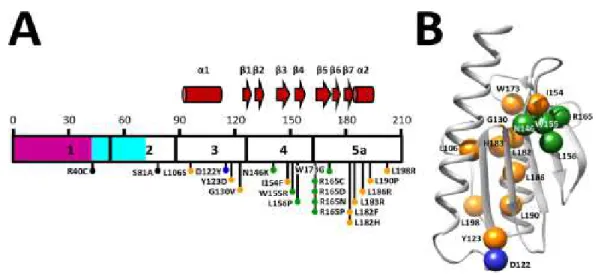
In healthy individuals, the number of repeats ranges from 6-36, whereas in FA patients, it is in the 70-1700 range, most commonly in 600-900 (Campuzano et al. 1996, Pandolfo and Pastore 2009). The length of (GAA) repeat expansion inversely correlates with the ages of onset and the severity of the disease. Long expansion leads to an early onset, severe clinically illness and death in young adult life, whereas patients with short expansion have a later onset and milder signs, some even are not diagnosed during their life time (Koeppen A. H. 2011). In 96 % of patient, this repeat expansion is homozygous, but in 2-4 % of patients it is heterozygous with missense mutations present in the coding region of one allele and expansion in the other. Until now, 44 different mutations have been described in FA heterozygous form, including point mutations, as well as insertion or deletion mutations (Figure I.2a) (follow Human Gene Mutation Database, Cardiff, UK). No patient homozygous for point mutations has been described (Galea et al. 2016).
The GAA expansion leads to silence mRNA transcription through a mechanism involving modifications of the chromatin structure of the locus, resulting in expression of a structurally and functionally normal frataxin but at levels that are estimated at ~5-30 % of normal
(Gottesfeld 2007). The missense mutations can lead to an amino acid residue change (Figure I.2b).
Figure I.2: Diseases-associated FXN point mutations and corresponding positions in human
frataxin molecule. (A) Exons 1 to 5a are numbered and delineated within the frataxin sequence.The mitochondrial transit peptide is colored purple, the intermediate N-terminal tail is light blue, and
mature frataxin is white. Secondary structure motifs are shown above the sequence, where the α
-helices are illustrated as cylinders and β-strands as arrows. Black dots represent residues at sites
cleaved during frataxin maturation (B) Illustration of human frataxin molecule highlights residues that are mutated in FA (Galea et al. 2016).
In most cases, heterozygous patients are clinically indistinguishable from patients that are homozygous for the GAA expansion, but a few missense mutations in compound heterozygous patients cause atypical or milder clinical presentations (Cossée et al. 1999) (Gellera et al. 2007). It was shown in knockout animal model that the complete absence of frataxin leads to early embryonic death (Cossée et al. 2000).
The nuclear-encoded human frataxin protein is synthesized in the nucleus as a 210 amino acid precursor (23 kDa) with an N-terminal mitochondrial-targeting sequence and it undergoes two steps of cleavage to achieve activity site. The insufficient level of frataxin in mitochondria causes defects in mitochondrial and cellular metabolism as: iron-sulfur cluster synthesis, heme synthesis and anti-oxidative stress. Most of these alterations concern “made-in mitochondrion” processes, thus the mitochondria-rich tissues such as the heart and the brain are most affected. The levels of FXN mRNA and frataxin protein show specificity for tissue that partially correlates with tissue-specific effects of its deletion pathology (Table I.2).
Table I.2: Tissue-specific FA pathology (Anzovino et al. 2014).
Alteration Heart Neurons Skeletal muscle
Cytosolic iron deficiency Yes ? No
Mitochondrial iron loading Yes No No
Perturbed iron-sulfur cluster synthesis Yes Yes ?
Autophagy/apotosis Yes Yes ?
Oxydative stress ? Yes ?
Inflammatory response ? Yes ?
1.2.2. Perturbation in mitochondrial processes
The deficiency of frataxin causes mitochondrial dysfunction (Vázquez-Manrique et al. 2005), (Gonzalez-Cabo and Palau 2013), which has a direct effect on the pathophysiology of the disease. The mitochondria are long known as the powerhouses of the cell. They generate ATP by oxidative phosphorylation via respiration chain (complexes I-IV). The generation of reactive oxygen species (ROS) is closely associated with ATP production so mitochondria are considered as the major source of ROS. Mitochondrion is also known as a major site for the metabolism of transition metals, including iron, which is essential for metabolic processes critical for cell viability. Important processes such as [Fe-S] cluster assembly and heme biosynthesis occur in this organelle. The dysfunction of mitochondria in FA patients or FA models organisms involves all the processes listed above.
1.2.2.1. Transition metal homeostasis 1.2.2.1.1. Physiological condition
Transition metal ions are particularly required for the correct function of all mitochondrial machineries. They are frequently used as cofactors for enzymes and oxygen-carrying proteins that take advantage of their propensity to gain and lose single electron. These metalloproteins participate in all vital processes of mitochondria as ATP production or detoxification of ROS.
zinc are inserted into peptides during the folding or post-translation process. Hence, mitochondria have its own mechanism of storage corresponding to each metal (Atkinson and Winge 2009, McCormick et al. 2015).
(i) Fe:
The mitochondrion is a major site of cellular iron utilization, and inversely, iron influences many aspects of mitochondrial metabolism. Iron is imported into mitochondria from cytosol by crossing the inner mitochondrial membrane through the mitochondrial iron importers (mitoferrins Mrs3/4). Within the mitochondrion, iron participates in three major metabolic pathways: (i) iron-sulfur cluster (ISC) assembly, (ii) heme biogenesis and (iii) mitochondrial iron storage. All of these processes give mitochondrion a central role in modulating iron homeostasis.
(a) Iron-sulfur cluster
Figure I.3: Iron prosthetic groups.
(b) Heme b
- [Fe-S] cluster biogenesis: Iron sulfur clusters (ISC) (Fig I.3a) are ancient and vital
prosthetic groups that are found in a wide range of proteins in the mitochondria, cytosol and nucleus. Many of these proteins play major roles in the metabolism, such as catalysis, electron-transfer in redox reactions and regulation of gene expression. ISC assembly machineries include at least 10 proteins. That comprise essentially protein scaffold, sulfur donor, electron transporter, iron chaperone and cluster transporter. Importantly, mammalian ISC biogenesis occurs in two different but functionally connected compartments: mitochondrial (ISC system) and extra-mitochondrial (CIA system). The inorganic de novo [Fe-S] cluster is synthesized within the mitochondrion by ISC assembly apparatus. They are then inserted into mitochondrial apo-proteins or passed through a process which generates an unknown molecule (X-S) that is exported by the ABC transporter Atm1 to the cytosol to
support the cytosol iron sulfur cluster proteins (ISP) biogenesis and to be utilized elsewhere (Figure I.4) (Lill et al. 2015).
A
B
Figure I.4: Illustration of [Fe-S] cluster (ISC) biogenesis in eukaryote cells. (A) Three distinct machineries assist the maturation of Fe/S proteins in non-green eukaryotes. The mitochondrial ISC assembly machineries consist of 17 known proteins and mature all organelle ISPs. The ISC system cooperates with the ISC export machinery and the CIA machinery, which is in charge for the biogenesis of cytosolic and nuclear ISPs. The central component of ISC export machinery is the mitochondrial inner membrane ABC transporter Atm1, which exports a sulfur- and glutathione
de novo [2Fe-2S] cluster is assembled in the scaffold protein Isu1 with the participation of cysteine desulfurase complex Nfs1-Isd11 as sulfur supplier, an electron donor ferredoxin Yah1 and frataxin Yfh1 with unknown function. Iron for this step is imported from cytosol by mitoferrins Mrs3/4. Step 2: [2Fe-2S] cluster is released from Isu1. The cluster is transferred from Isu1 to mitochondrial glutaredoxin Grx5 that binds the [2Fe-2S] group in a glutathione dependent manner. The dedicated Hsp70 chaperone Ssq1 bind to the conserved LPPV motif of Isu1(ATP-hydrolysis dependent association) and facilitate the transfer of [2Fe-2S] from Isu1 to Grx5. Jac1 is a co-chaperone which assists the recruitment of Ssq1 to Isu1. The ADP-ATP exchange by Mge1 helps to dissociate Ssq1 from Isu1 and thus closes the chaperone cycle. From here, the Grx5-bound [2Fe-2S] clusters are transmitted to one of three pathways: (i) be transferred to mitochondrial apo-proteins without further assistance; (ii) pass through an unknown process which generates a sulfur or possibly glutathione-containing molecule (X-S) that is exported by the ABC transporter Atm1 to the cytosol to support the cytosol ISPs biogenesis; and (iii) shift to the third step. Step 3: All mitochondrial [4Fe-4S] proteins are assembled. Specialized ISC factors [Iba57-Isa1-Isa2] first facilitate the synthesis of [4Fe-4S] cluster from Grx5-bound [2Fe-2S] cluster. Then, the target specific insertion of [4Fe-4S] cluster into apo-proteins is archieved by various dedicated ISC targeting factors (Ind1, Nfu1, Aim1). (Lill et al. 2015)
- Heme biogenesis: Heme is a prosthetic group that contains a metal, iron is the most
common, in the central position of porphyrin ring (Fig I. 3b). Iron atom can uptake or release another smaller ligand e.g. O2. Heme is an essential cofactor of important proteins, such as
hemoglobin and myoglobin. The heme biosynthetic pathway includes eights enzymes, it begins and ends in mitochondria but the intermediate steps occur in the cytoplasm (Ponka 1997). In the final step, an iron atom is inserted into protoporphyrin IX by ferrochelatase. In the case of iron unavailability or deficiency, it is replaced by Zn(II) to form Zn-protoporphyrin, which can be an indicator for iron deficiency diseases. Ferrochelatase requires one [2Fe-2S] cluster to govern its catalytic activity, it is possible that any disruption in ISC synthesis would also adversely affect the heme synthesis (Anzovino et al. 2014).
- Mitochondrial iron storage: Iron excess from heme or ISC synthesis is sequestered
by mitochondrial ferritin (Levi et al. 2001). Since reactive oxygen species are generated in mitochondria, it is essential that iron is stored in an inert form to prevent adventitious redox cycling reactions (Richardson Des R. et al. 2010). Ferritin stores iron by inducing its oxidation and deposes it inside the cavity in a ferric oxo-hydroxide core, which is structurally similar to the mineral ferrihydrite. Ferritin is highly expressed in tissues with high metabolic activity and oxygen consumption, such as testis, brain, and heart. Non-ferritin iron or labile iron pool is reactive and probably toxic for cell. Mitochondria from iron-sufficient cells contain labile iron in low-molecular-mass complexes at about 100-150 µM (McCormick et al. 2015).
The three pathways described above contribute to mitochondria the role of a gatekeeper for iron metabolism. Every disruption in these pathways can affect iron homeostasis in both mitochondria and cytoplasm.
(ii)Cu:
Copper is required within the mitochondrion for the function of two metalloproteins: Cytochrome c oxidase (CcO) and Cu-Zn superoxide dismutase (SOD1). CcO locates in the inner membrane. CcO is the terminal complex in respiratory chains, which reduces O2 to two
water molecules and transfers two protons into intermembrane space. Two subunits CuA and
CuB of CcO containing copper are encoded by mitochondrial genome. Hence, the insertion of
Cu certainly occurs in intermembrane space. SOD1 is localized and functionalized mainly in cytosol, with however 1-5 % is found in the intermembrane space. It is one of three common superoxide dismutases that catalytically convert superoxide to oxygen and hydrogen peroxide. SOD1 contains copper and zinc, where copper plays enzymatic role and zinc stabilizes the structure of protein. SOD1 is imported into mitochondrial as apo-protein; the metalation of SOD1 depends on a copper chaperone Ccs1.
There is very little, if any, free Cu within the cells. Copper is bound to proteins or complexed to glutathione or other small molecules. McCormick et al., have indicated that 22 % of total mitochondrial copper (~16 µM of 71 µM) exist in a complex with an unknown low-molecular mass molecule (CuL). But the remaining question is whether the copper metalation of SOD1 and CcO is supplied by this CuL or from the cytosol (McCormick et al. 2015).The mechanism of copper import into the mitochondrion is unknown. Recently, Cobine PA et al. identified in yeast model two proteins of the mitochondria carrier family involving in mitochondrial copper import Pic2 and Mrs3. Mrs3 is an iron carrier that suggests a connection between copper and iron homeostasis (Vest et al. 2013, Vest et al. 2016). These studies also gave evidences for the direct interaction between CuL and Pic2/Mrs3.
Subsequently, copper is an important trace element for redox activity. It can also induce the generation of ROS via Fenton-like reaction. Therefore, cellular uptake, storage as well as copper export necessitate a tight regulation in order to guarantee sufficient copper supply for copper-containing enzyme but also to prevent copper-induced oxidative stress.
In yeast, the only known function of Mn(II) in mitochondria is that of a cofactor in superoxide dismutase SOD2, the matrix superoxide dismutase. In contrast to Cu-dependent superoxide dismutase, SOD2 plays essential role in anti-oxidative stress since the complete loss of the enzyme SOD2 results in neonatal lethality in mice (Li Yibing et al. 1995) that cannot be rescued by CuZnSOD (Copin et al. 2000). Mammalian mitochondria contain the second Mn enzyme, which is type II arginase. It catalyzes the synthesis of ornithine and may regulate the production of NO by nitric oxide synthase (Topal et al. 2006).
Mitochondrial Mn labile pool exists majorly as a complex with an 1100 Da molecular weight
(~2 µM) (McCormick et al. 2015). The mechanism of metalation for MnSOD is unknown. Apo-SOD2 has specific affinity to FeII. In iron excess condition, SOD2 risks to be irreversibly inactivated by the mismetalation of iron into apo-SOD2.
(iv) Zn:
Zinc is the most commonly utilized metal cofactors (9 % of all enzymes). In mitochondria, zinc is the catalytic cofactor of a range of proteases, which include inner membrane associated protease (mAAA, iAAA, Oma1 proteases of the inner membrane, and the intermembrane space protease Atp23) and mitochondrial processing protease (functions in cleavage of the N-terminal mitochondrial targeting sequences); Zn(II) is likely associated with the yeast mitoribosome. The dominant non-protein zinc in mitochondrial is at ~110 µM (McCormick et al. 2015). A subunit of cytochrome c oxidase (Cox4) and SOD1 require Zn for is stability.
1.2.2.1.2. Impairment of metal metabolism in FA model
Early characterization of the pathophysiology in individuals with FA provided obvious evidence of a link between frataxin deficiency and cellular metal metabolism dysregulation (Table I.3). However, their occurrences depend on the nature of the tissues.
Table I.3: Mitochondrial metals: function and the impaired metabolism in FA model.
Metal Functions Metabolism in FA
Fe - Respiratory chain with 4 enzymes complexes I, II, III, IV
- [Fe-S] cluster synthesis
- Reduce in ATP generation.
- Deficiency in [Fe-S] cluster protein activity.
- Heme synthesis
- Ferritin storage
of heme.
- Iron accumulation in non-ferritin form.
Cu - CuZn-superoxide dismutase (SOD1) – intermembrane space (IMS).
- Cytochrome c oxidase (CcO) - inner membrane proteins, complex IV of respiratory chain.
- SOD1 is highly induced but decreased in activity (oxidative damage and lack of Cu for the metalation).
- Reduce in activity of CcO.
Mn MnSOD (SOD2) – superoxide
dismutase in mitochondrial matrix.
Increase in protein amount but decrease in activity of SOD2.
Zn - Cofactor of proteinases
- Required structural stability SOD1 and Cox4, a subunit of CcO
Uncharacterized
In cardiac tissues, studies on autopsies of FA patients reported independent proofs of mitochondrial iron overload. The accumulation of iron in cardiomyocytes is restricted to small regions and progresses from granules in heart fibers to coarse aggregates in phagocytized cardiomyocytes (Lamarche et al. 1980). Cytosolic and mitochondrial ferritin, which are marker of iron excess, co-localize extensively in heart tissues. Hepcidine, a hepatic hormone controls systemic iron distribution, has been found in non-hepatic tissues including heart, which is an indicator of iron dysmetabolism (Koeppen A. H. et al. 2012). Rotig et al. was the first group to report the deficient activity of the ISC proteins of mitochondrial respiratory complexes I-III and aconitase in FA heart biopsies but not in other tissues (Rotig et al. 1997). The impairment of ATP synthesis in calf muscle and reduced biochemical activities of complex I and IV in the skeletal of FA patients were also reported. However, it was suggested that they were due to a quantitative decrease in the number of mitochondria rather than a selective mitochondrial dysfunction (Nachbauer et al. 2012).
However, iron overload does not occur typically in the nervous system. Dorsal root ganglion (DRG) are a primary target of FA with a remarkable lesion (Koeppen A. H. and Mazurkiewicz 2013). The assay of total Fe in DRG of FA patients was not increased. The accumulation of ferritin was only found in satellite cells surrounding dying DRG neurons
nucleus (DN) is an iron-rich structure that contains relatively high amounts of copper and zinc. The net amount of Fe, Cu or Zn does not increase or decrease significantly in the DN of FA patients, but the distributions of these metals are changed in affected DN. The Cu and Zn rich regions broaden and overlap extensively with the Fe-rich region. Copper is supposed to exist mainly in protein-bound forms in the DN of FA. The atrophy of the DN in FA correlates with Cu and Zn redistribution rather than with Fe dysregulation only (Koeppen A. H. et al. 2007, Koeppen A. H. et al. 2012).
A conditional mouse locally depleted frataxin within the heart and skeletal muscle, named the muscle creatine kinase (MCK) conditional - hFxn null mice (herein referred to as MCK-hFxn-knockout (KO) mice) is an effective model to study extensively the cellular mechanism of FA. Similar to patient cardiac tissues, mitochondrial iron accumulation as phosphorus and sulphur iron, cytosolic iron deficiency and low levels of cytosolic ferritin has been observed in MCK-hFxn-KO mouse (Huang et al. 2009, Puccio et al. 2001, Whitnall et al. 2008, Whitnall et al. 2012). However, no detectable iron deposits were observed in the complete hFxn-KO mouse or in the central nervous system of the ‘neuron-specific’ hFxn-KO mouse models (Santos et al. 2010). Also in yeast strain lack of yeast frataxin homolog (∆yfh1), iron was found in amorphous nanoparticles of ferric phosphate in mitochondria (Babcock et al. 1997, Foury and Cazzalini 1997, Lesuisse et al. 2003).
The inactivated iron-sulfur cluster proteins (ISPs) were found in cardiomyocytes of MCK-hFxn-KO mouse (Puccio et al. 2001) and ∆yfh1 (Foury 1999, Rotig et al. 1997). Importantly, it was emphasized that the inactivation of iron-sulfur enzymes is the early phenomenon and intra-mitochondrial iron accumulation is the later (Poburski et al. 2016, Puccio et al. 2001, Stehling et al. 2004). FA patients do not manifest anaemia when frataxin expression is reduced, but heme production is attenuated (Huang et al. 2009). The enzymes involved in heme synthesis are altered (Boddaert et al. 2007, Yoon and Cowan 2004). Zn-protoporphyrine IX is formed instead of heme in ∆yfh1 (Lesuisse et al. 2003).
The absence of frataxin in the heart and skeletal muscle leads to iron loading in liver, spleen and kidney because of the induction of hepcidin, hepatic hormone of iron metabolism (Whitnall et al. 2012). Biochemical studies on the iron metabolism of MCK-hFxn-KO mouse showed two alterations. First, there are the global down-regulation of molecules involved in ISC biogenesis, iron storage and heme synthesis while up-regulation of cytosolic heme catabolism. Second, the expression of molecules involved in cellular and mitochondrial iron
uptake tends to an increase in iron uptake targeting mitochondria, as well as a decrease in iron release (Huang et al. 2009). This implies a systemic perturbation of iron metabolism.
In contrast to the eukaryotic systems, a complete loss of the bacterial frataxin ortholog (∆cyaY) does not exhibit a typical phenotype. It does not exhibit any modification in iron content or in the sensitivity to oxidant (Li D.S. et al. 1999).
Despite tissue-specific variations in frataxin-mediated iron handling, the dysregulation of iron metabolism due to frataxin deficiency remains the most prominent feature.
1.2.2.2. Mitochondrial redox control and oxidative stress 1.2.2.2.1. Generation of ROS/RNS in healthy mitochondrion
Redox dependent processes affect most cellular functions. Mitochondria are in the center of these processes as they generate reactive species (RS) that drive redox-sensitive events and respond to RS-mediated changes in the cellular redox state. Reactive species are not only harmful they are also important signaling molecules with potential therapeutic effect. The term reactive species relates to many kinds, such as reactive oxygen species (ROS: O2•-, H2O2,
OH•-), RS of nitrogen (RNS), carbon, sulfur and halogen, etc. Herein, we only mention ROS and RNS, the two most important RS in biological system. The components of RNS and ROS are listed in Figure I.5.
Figure I.5: Reactive oxygen and nitrogen intermediate production in mammalian cells. ROS include
superoxide anion (O2•-)- the one electron reduction product of oxygen, hydrogen peroxide (H2O2),
hydroxyl radical (OH•-). Nitric oxide (NO) is generated from the conversion of arginine to
L-citrulline. The reduction step-by-step of NO generates nitrite (NO2-, nitrogen dioxide (NO2•) and
nitrate (NO3-). The reaction of NO with cysteine sulphydryl (R-SH) can result in either S-nitrosylation
(R-SNO) or oxidation to the sulphenic acid (R-SOH). In reaction with superoxide anion, the highly
reactive radical would be formed, include peroxynitrite anion (ONOO-) and peroxynitrous acid
(ONOOH). ONOOH spontaneously decomposes through a series of species that resemble the reactive
radical hydroxyl (OH•) and nitrogen dioxide (NO
2•) (Fang 2004).
(i) Reactive oxygen species (ROS)
In living organism, under aerobic condition, 90 % of the consumed oxygen is reduced directly to water by 4 electrons per O2 molecule, catalyzed by cytochrome c oxidase – the complex IV
in electron transport chain. The 10% is reduced systematic as detailed in ROS branch in figure I.5, the incompletely reduced products including O2•- (superoxide anion), H2O2 (hydrogen
peroxide), OH•- (hydroxyl radical) are called reactive oxygen species (ROS) (Fang 2004, Lushchak 2014).
In mitochondria, about 1-2 % of molecular oxygen is converted to superoxide anion during the oxidative phosphorylation by complexes I and III of the electron-transport chain, where 70% of O2•- are from the Q cycle (ubiquinol QH2 ubiquinone Q) as a part of electron
transfer to complex III (Figure I.6). This superoxide can be released into the mitochondrial matrix or intermembrane space since the complex III- coenzyme Q10 binding site localize within the inner membrane (Handy and Loscalzo 2012).
Superoxide radical is not a particularly strong reductant or oxidant (Abreu and Cabelli 2010). In physiological conditions, it seems unreactive with the amino acids. It is however highly reactive with some transition metal complexes, such as Cu, Mn, Fe because of the Haber-Weiss reaction (equation I.1). The damages induced by superoxide anion O2•- are more
important on soluble metalloproteins such as [Fe-S] proteins (equations I.2 & I.3). Nevertheless, O2•- is converted into hydrogen peroxide H2O2 by superoxide dismutases
(SODs). H2O2 is not a free radical but it is ROS as it is more reactive than an oxygen
molecule, it can react with reduced cations of transition metal as Fe2+ or Cu+ in Fenton-like
reaction (equation I.4) and generates hydroxyl radical OH•- and hydroxyl anion HO-.
Hydrogen peroxide and others ROS/RNS can furthermore inhibit aconitase - an ISC enzyme of Kreb’s cycle, thus slow down glucose consumption, respiration and ATP synthesis (Brazzolotto et al. 1999, Gardner 1997, Lushchak 2014). H2O2 excess is scavenged by
catalase- a heme containing protein (Handy and Loscalzo 2012).
Haber-Weiss reaction: Fe3+ + O2•- → Fe2+ + O2 (I.1)
Inactivation [Fe-S] cluster: [4Fe-4S]2+ + O2•- → [4Fe-4S]-OO• (I.2)
[4Fe-4S]-OO• + 2 H+→ [3Fe-4S] + H2O2 +Fe3+ (I.3)
Fenton reaction: Fe2+(aq) + H2O2 → Fe3+(aq) + OH−(aq) + OH•- (I.4)
Hydroxyl radical can acquire one more electron and a proton to yield H2O. In biological
systems, this reaction mainly occurs through the substraction of hydrogen atom from proteins, lipids or DNA (Stadtman and Levine 2003).
It has been established that 90 % of ROS are produced in mitochondria (Herrero et al. 2008). Every post-translational modification of mitochondrial subunits can either promote or attenuate the generation of ROS. The other sources of ROS include: diverse oxidases (xanthine oxydases, NADPH oxidase, etc.) and the auto-oxidation of different small molecules (epinephrine, norepinephrine and xenobiotics, etc.) (Lushchak 2014).
(ii) Reactive nitrogen species (RNS)
nervous system and glial regulated pathways. Under physiological conditions, it contributes to regulating proliferation, survival, and differentiation of neurons. Nitric oxide is involved in synaptic activity, neural plasticity, and cognitive function (i.e., memory). The imbalance nitric oxide metabolism can contribute to neuronal cell death and various neurodegenerative disorders (as Alzheimer’s diseases, Parkinson’s diseases etc.) (Bradley and Steinert 2016).
An increase in the production of NO during the neuroinflammation in the presence of ROS, especially superoxide anion, yields highly reactive peroxynitrite (ONOO-). Peroxynitrite can
react directly with proteins that contain transition metal centers. Therefore, it can modify proteins such as hemoglobin, myoglobin, and cytochrome c by oxidizing ferrous heme into its corresponding ferric forms. Peroxynitrite may also be able to change protein structure through the reaction with various amino acids in the peptide chain. The most common reaction is tyrosine nitration (nitrotyrosination Tyr Tyr-NO3) which is considered as a biomarker of
nitrosative stress. In another hand, NO itself can be added covalently into a cysteine thiol/sulfhydryl (RSH) (S-nitrosylation RSH R-SNO) (Figure I.5). All of these reactions affect protein structure and function, cause changes in the catalytic activity of enzymes, alter cytoskeletal organization, and impair cell signal transduction (Castro et al. 2011).
1.2.2.2.2. Oxidative defense mechanism
As the excess of reactive species (RS) can induce unexpected changes in the structure and function of biomolecule, it is then necessary for RS to be kept in a range compatible with normal cellular function. The imbalance between the production of ROS/RNS and the protection against ROS/RNS is defined as “oxidative stress” and/or “nitrosative stress”. The oxidative/nitrosative stress induces damage to DNA and RNA (incorporation of an oxidized base during DNA polymerization, or oxidization of the integrated base), proteins (protein-protein cross-linking, fragmentations, unfolding, etc.) and lipid (formation of lipid peroxides (LOOH) which can also damage DNA and protein). Hence, the RS steady-state level must be strictly controlled.
Figure I.7: ROS/RNS production and cellular antioxidant defense enzymes. Copper-dependent superoxide dismutase (SOD1), which locates in intermembrane space, is responsible for the dismutation of superoxide anion while manganese-dependent superoxide dismutase (SOD2) achieves
the same purpose in the mitochondrial matrix. These processes generate hydrogen peroxide (H2O2)
which can react with labile iron pool and produce OH−and OH•-. H
2O2 and other peroxides are
detoxified by glutathione peroxidases (GPXs), which oxidize glutathione.Glutathione (GSH/GSSG) is
a tripeptide synthesized in two steps from glutamic acid, cysteine, and glycine. GSSG is reduced to GSH by glutathione reductase (GR) by using electrons from NADPH. NADPH is regenerated by the pentose phosphate pathway enzymes, glucose phosphate dehydrogenase (G6PDH), and
6-phosphogluconate dehydrogenase. Other enzymes that scavenge H2O2 and peroxides are catalases
and peroxiredoxins (PRXs). Peroxiredoxins can also scavenge ONOO-. The cellular thiol redox status
is maintained by the thioredoxin (TRX)/thioredoxin reductase (TR) and glutathione/glutaredoxin systems by reducing the oxidized sulfhydryl groups of proteins (Santos et al. 2010).
Living organisms possess multilevel and complicated antioxidant system operating either to eliminate RS, or minimize their negative effects (Figure I.7). Antioxidants can be classified according to their molecular mass: low molecular mass (< 1000 Da) and high molecular mass (> 1000 Da).
The group of low molecular mass antioxidants includes molecules such as vitamins C (ascorbic acid) and E (tocopherol), carotenoids, anthocyanins, polyphenols, and uric acid. Most of them are supplied as food or supplement components. However, the most important antioxidant in this group is glutathione (GSH) (L-γ-glutamyl-L-cysteinylglycine), a tripeptide synthesized in most living organisms from glutamic acid, cysteine, and glycine. GSH is used to control RS level either via direct interaction, or serving as a cofactor for RS-detoxifying
includes enzymes such as: superoxide dismutase SOD, catalase and glutathione-dependent peroxidase, etc. (Indo et al. 2015).
(i) GSH and glutathione-dependent antioxidants
Under the conditions of oxidative/nitrosative stress, the thiols in cysteine residues (-SH) within proteins are among the most susceptible oxidant-sensitive targets that can undergo various reversible and irreversible redox alterations affecting protein activity or structure
(Figure I.8). The tripeptide glutathione is present in cells at the millimolar concentrations (∼1– 10 mM). It shows high negative redox potential (high electron-donating capacity). It is the reason for which GSH can protect protein thiol groups from oxidation either directly as a free radical scavenger, or indirectly as co-substrate for a number of important enzymatic systems. Glutathione can also form a mixed-disulfide bridge with an accessible free thiol group on a protein (S-glutathionylation- P-SSG). Glutathionylated proteins may thus be used as a biomarker for oxidative stress.
Glutathione-dependent peroxidases (GPx) reduce H2O2 to form GSSG, and also lipid
peroxides (L-OOH) – a reactive species (equation I.5 & I.6).
2GSH + H2O2 → GSSG+ 2H2O (I.5)
2GSH + LOOH → LOH + GSSG + H2O (I.6)
Figure I.8: Illustration of the modification of proteins thiol groups under oxidative/nitrosative
stress.
The reduced form of glutathione, GSH, is the quantitatively most important buffering system against oxidative stress in mammals. The physiological intracellular milieu is a reducing
SH H2O2 SOH S – S S N S S SO2H SO3H SNO S—SG
environment with a GSH/GSSG ≥ 100. Maintaining optimal GSH/GSSG ratios in the cell is critical to cell survival and is important for regulating the redox state of protein thiols. In mitochondria, this ratio is higher than in cytosol, which minimizes protein glutathionylation by thiol disulfide exchange (Lushchak 2014). The concentration of reduced GSH and ratio GSH/GSSG allow evaluating the buffering capacity of medium.
(ii) SODs
SOD family consists of metalloproteins, whose active site uses copper-zinc, manganese, iron, or nickel, are ubiquitous components of cellular antioxidant system. These proteins catalyze the dismutation of O2•- to oxygen and hydrogen peroxide. All known SODs require a redox
active transition metal in the active site in order to accomplish the catalytic breakdown of superoxide anion. A generic mechanism for the metalloenzyme dependent dismutation steps is below (equation I.7 & I.8).
M(n+1)+-SOD + O2•- → Mn+-SOD + O2 (I.7)
Mn+-SOD + O2•- + 2H+ → M(n+1)+-SOD + H2O2 (I.8)
(*) M= Cu, Mn, Fe, Ni
CuZn-dependent SOD (SOD1) and Mn-dependent SOD (SOD2) are the only members of SOD family found within mitochondria (Culotta et al. 2006). SOD1 is an intracellular enzyme, which localizes throughout the cell cytoplasm, nucleus and microsomes; in mitochondria it has been detected only in intermembrane space. Whereas, MnSOD is the only mitochondrial matrix superoxide dismutase. SOD2 has a more crucial role in the inactivation of mitochondrial superoxide anion than SOD1. Indeed, SOD2 deficiency causes early neonatal death in knockout mice while the total absence of SOD1 does not (Li Yibing et al. 1995). Both SOD1 and SOD2 are imported into the mitochondria as apo-peptide and must undergo a step of post-translation coupled with the metalation.
The structure and function of SOD1 and SOD2 will be discussed in details in chapter IV. 1.2.2.2.3. FA cases
Cortopassi 2015). On the other hand, although anti-oxidative therapie (discussed in 1.3) can reduce the symptoms of FA, they never can cure the disease (Santos et al. 2010). Therefore, the involvement of oxidative stress in FA remains controversal. The main research related to the oxidative deal with three aspects: (i) the evidences of oxidative damage in FA patients as well as in frataxin depletion models; (ii) increase of ROS/RNS production; (iii) FA models become highly sensitive to oxidative stress.
(i) Oxidative stress markers
The loss of mitochondrial function and the appearance of the markers of oxidative damage in nearly all FA models have been examined. Products of DNA oxidative damage have been found in urine (Schulz et al. 2000); both mitochondrial and nuclear DNA damage are detected in peripheral blood cells (Haugen et al. 2010); shortened telomere and abnormal glutathionylation have been reported by study autopsy (Anjomani Virmouni et al. 2015). By investigating different human cell lines with FA, the increased level of glutathionylated cytoskeletal proteins was found in fibroblast and spinal cord of patients with FA together with a significant rise of dynamic tubulin and neurofilaments (Sparaco et al. 2009).
Frataxin deficient pancreatic islets in mice show decreased proliferation and increased apoptosis due to the increased ROS (Ristow et al. 2003). The disruption of frataxin expression in murine hepatocytes reduces life span and develops multiple hepatic tumors. Also, biomarkers of lipid oxidation and oxidized glutathione (GSSG) were found to be significantly high in liver specimen (Thierbach et al. 2005). In mice mutant with the (GAA) repeat expansion, oxidized proteins were found in cerebrum, cerebellum, heart and skeletal muscle and increased lipid peroxidation was also detected in cerebrum and heart samples (Al-Mahdawi et al. 2006). In yeast models, frataxin depletion led to the oxidization of mitochondrial proteins, mitochondrial DNA lesions and accumulations and nuclear DNA damage (Karthikeyan et al. 2003). Furthermore, frataxin deficiency had little effect when yeast cells were grown anaerobically, but a shift to aerobic growth resulted in loss of aconitase activity and oxidative protein damage (Bulteau et al. 2007). Moreover, oxidative stress in ∆yfh1 mutants increased the proportion of fragmented mitochondria as compared to wild type (Lefevre et al. 2012).
The increase of ROS as superoxide and hydrogen peroxide was reported in FA lymphoblasts by use of fluorescent probes (Napoli et al. 2006) and in frataxin-depletion pancreatic islets (Ristow et al. 2003).
The increased generation of ROS in FA was suggested to be a result of deficiency in ISC biosynthesis and iron accumulation. Indeed, in mitochondria of FA models, hydrogen peroxide H2O2 is overproduced because of the insufficient activity of complexes I-III of the
oxidative phosphorylation chain and cytosolic aconitase. Iron is also overloaded in FA mitochondria in non-protein iron pool. The presence of H2O2 together with iron accumulation
favor the Fenton-like reaction that generates ROS (equation I.4) (Babcock et al. 1997, Calabrese et al. 2005, Napoli et al. 2006, Radisky et al. 1999).
Conversely, this hypothesis can be contradicted by the following results. A study reported that the iron accumulated in frataxin deficient yeast is in an oxidized and insoluble form and thus unable to participate in Fenton-like reaction (Seguin et al. 2011). Recently, a time-resolved functional analysis on modified murine fibroblast revealed that a full depletion of frataxin induced a massive increase in ROS production before the iron accumulation. This result excludes away the primary role of iron overload for oxidative stress (Poburski et al. 2016).
(iii) Increase oxidant sensitivity and oxidant protection deficiency
The increase of oxidant sensitivity has been shown by independent studies in various model: FA patient cells (Wong et al. 1999), yeast (Santos et al. 2010), C. elegans (Vázquez-Manrique et al. 2005), Drosophila (Runko et al. 2008), mouse (Al-Mahdawi et al. 2006). In these studies, the depletion of frataxin aggravates the sensitivity to a variety of pro-oxidants, including H2O2 and other peroxides as well as iron. The mechanism of the aggravation is
complex. However a critical aspect is that the defense against oxidative stress is altered in FA models.
Biochemical studies have shown the impairment of glutathione homeostasis in FA patients’ blood plasma and lymphocytes, FA mouse model, DRG cells and frataxin deficient S. cerevisiae. It was found that free reduced glutathione (GSH) decreased while glutathionyl-protein increased in blood of patients with Friedreich’s ataxia (Piemonte et al. 2001) or in
showed that glutathione imbalance progressively increases with a significant rise of all oxidized forms of glutathione, including protein bound one (Carletti et al. 2014). The isolated mitochondria from ∆yfh1 cells and lymphoblasts of FA patients showed evidence for a severe mitochondrial glutathione-dependent oxidative stress, with a low GSH/GSSG ratio (1.7 within the mitochondria ∆yfh1), and thiol modifications of key mitochondrial enzymes (Bulteau et al. 2012).
A continuous oxidative damage due to an impaired response to oxidative stress may contribute further to mitochondrial dysfunction and cell degeneration in FA. As mentioned before, SODs are the most important defenses against ROS as they catalyze the disproportionation of superoxide anion to hydrogen peroxide. Their activity requires however redox active metal ions. In heart cells of MCK-hFxn KO mouse, MnSOD expression was induced early but reduced when respiratory chain enzyme activities are extremely decreased and iron accumulation is visible in mitochondria. Similarly, ∆hFxn cerebellum has a lower
MnSOD expression level (Seznec et al. 2005). In frataxin-deficient mouse model of FA,
microarray and neurotic growth experiments in DRG tissue identified decreased transcripts encoding the antioxidants, including peroxiredoxins, glutaredoxins, and glutathione S-transferase (Shan et al. 2013). In ∆yfh1 cells, both of CuZnSOD and MnSOD are highly induced but their specific activity (meaning dividing the enzymatic activity with the protein amount) is decreased. CuZnSOD activity can be restored in copper supplement medium.
Whereas, the matrix mitochondrial MnSOD is always inactivated even in manganese
supplemented culture (Irazusta et al. 2010).
1.3.
Pharmacotherapy for Friedreich ataxia
Currently, there is no treatment for FA as no drug therapy showed any efficacy in slowing
disease progression. The palliative and symptomatic treatments, such as β-blockers, angiotensin-converting-enzyme inhibitors, surgery for cardiac manifestations and physical therapy are applied only to improve the quality of life. A number of active ingredients are under investigation since the identification of the frataxin responsible gene. The current efforts in developing therapeutic strategies deal with three approaches: iron chelators, antioxidants and/or stimulants of mitochondrial biogenesis, and frataxin level modifiers (Table I.4).
1.3.1. Iron chelators
Dysregulation of metabolic iron and its accumulation in mitochondria are typical hallmark of FA patients. Several iron chelators that target to the mitochondria have been avaluated including deferoxamine and deferiprone. They successfully protected the mitochondria and reduced ROS damage to mitochondrial proteins. However, the utilization of iron chelators presents an important risk mainly because of many side effects such as the decrease of the mRNA levels of both aconitase and frataxin (Pandolfo and Hausmann 2013).
1.3.2. Antioxidants and/or stimulant of mitochondrial biogenesis
Even if the role of oxidative stress in FA pathology remains controversial, it is clear that patients with FA present increased oxidative stress, resulting in DNA damage (Schulz et al. 2000), increased levels of lipid peroxidation (Abeti et al. 2015), and impaired ROS defenses (Irazusta et al. 2010). A lot of work has been done in evaluating the potential of antioxidants in FA therapy.
Until now, the first drug who reached phase III in clinical trials is Idebenone, an analogue of Co-enzyme Q10 (Figure I.8). Co-enzyme Q10 is a small lipophilic molecule present within the inner mitochondrial membrane in association with the electron transport chain (ETC) complexes, which transfers electrons between complexes I and II, and from oxidation of fatty acids and branched chain amino acids to complex III resulting in the ultimate production of ATP (Figure I.9).
Figure I.9: Chemical structures of co‐enzyme Q10 and Idebenone. They show the common reducible benzoquinone nucleus and lipophilic side chains.
Idebedone can act as an electron carrier within the ETC and has similar antioxidant properties to co-enzyme Q10. Idebenone has been reported to increase oxidative phosphorylation and
and FA symptoms, to decrease oxidative stress, lipid peroxidation and to slow the progression of heart diseases (Rustin et al. 1999, Schulz et al. 2000). However, in 2011, Idebenone failed its Phase III trial because it was found not to significantly improve lifespan and cardiac outcomes in patients. Idebenone is currently not licensed in Europe or the United States for use in FA. Therefore, further works are being undertaken with other CoQ10 analogs but no study has yet been performed in FA patients (Parkinson et al. 2013).
1.3.3. Frataxin level modifiers
One of the most promising treatment strategies for FA therapy is to increase the intracellular content of frataxin, thus preventing the cascade of protein deficiency that leads to the clinical syndrome. The level of frataxin expression can be increased by the compounds that combat the FXN gene silencing and/or a delivery system which introduce frataxin to cells.
Erythropoietin (EPO) is a 30 400 Da glycoprotein that was initially recognized as a regulator of red cell production. rHuEPO, which has the same biological effects as endogenous erythropoietin, is a 165 amino acid glycoprotein synthesized by recombinant DNA technology. Sturm et al., 2005 was the first group to report that rHuEPO can increase the quantity of frataxin, in addition to its classical neuro- and cardio protective effects (Sturm et al. 2005). It is suggested that EPO promotes the translation of mRNA into frataxin without a concurrent rise in frataxin mRNA level (Acquaviva et al. 2008). Other compounds have also shown success in increasing frataxin protein levels, such as histone deacetylase inhibitors BML-201 and 106. These two compounds are in pre-clinical phase (Richardson Timothy E. et al. 2013).
In addition, varieties of systems that allow the introduction of frataxin or vector expressing human FXN have been developed. For example, transactivator of transcription (TAT) from the human immunodeficiency virus (HIV) is a short peptide able to efficiently guide the delivery of fused proteins across cellular and intracellular membranes. TAT fusion proteins containing a mitochondrial targeting sequence can translocate through the mitochondrial membranes, with appropriate processing and persistence of the fusion protein within mitochondria. A TAT/h-frataxin fusion protein able to localize within the mitochondria is currently being developed and tested in mammalian model. Also, gene therapy is being experienced with several successful applications (Evans-Galea et al. 2014).
Agent Groups (+) Effects, (-) Side effects Present stage
Idebenone Antioxidant
Analogue of Co Q10
(+) as free radical scavenger.
(+) facilitate electron transport in ETC, thus enhance ATP production.
(-) adequately improve cardiac outcomes in patients.
Phase III (failed in phase III study since 2011)
Mitoquinone Antioxidant
Analogue of CoQ10 specifically targeting at mitochondria
(+) decrease oxidative damage; 800 times more potent in preventing cellular death than idebenone
(-) does not enhance ATP production.
Phase II
Deferiprone Deferoxamine
Iron chelator (+) detoxify labile iron pool
(-) side effects: agranulocytosis,
musculoskeletal pain, dizziness, nausea vomiting, gastrointestinal discomfort and elevated hepatic enzymes, etc.
(-) Decrease mRNA levels of both frataxin and aconitase (deferoxamine); and activity of aconitase (deferiprone). Phase II 17β-Estradiol (E2) and methylene blue (MB) Combination of
antioxidant and iron chelator
(+) E2: neuroprotective effects; attenuate ROS, prevent lipids and proteins damage; stabilize mitochondrial membrane potential, maintain activity of ETC chain, aerobic respiration and favorable balance of anti/pro apoptotic proteins.
(+) MB: neuroprotection and antioxidant (similar to idebenone)
Preclinical
Erythropoietin Increase frataxin
expression
(+) Promoting the translation of mRNA into frataxin without a concurrent rise in frataxin mRNA level.
Phase II
Pioglitazone Stimulant
mitochondrial
Agonist of the peroxisome proliferator-activated receptor gamma
(+) increase fatty acid oxidation and mitochondrial function; decrease ROS accumulation and inflammation.
Phase III
Adeno-associated virus vector
Gene-therapy Preclinical
2.
Frataxin and functions hypothesis
2.1.
Structure
Frataxin is a small globular protein, highly conserved in most organisms from prokaryotes to eukaryotes, in eukaryote cells it locates in the mitochondrion. All frataxin orthologues have an unique fold that combines two terminal α-helices to form one plane, five antiparallel β-strands that construct the second plane of the protein and a sixth (or seventh in hFxn) β-strand that intersects the planes to give an overall planar “α–β sandwich” structure motif (Figure I.10) (Bencze et al. 2006).
Figure I.10: Frataxin’s structure. Top: ribbon diagram for yeast, human and bacterial frataxin. Middle: electropotential plots for proteins in same orientation. Bottom: electropotential plots for proteins rotated −90 degrees around the y-axis compared to top display. Structure figures made using solution structures of Yfh1 (PDB ID# 2GA5), hFxn (PDB ID# 1LY7) and CyaY (PDB ID# 1SOY) frataxins. The negatively charged amino acids are red, the neutrally charged ones are grey and the positively charges ones are blue (Bencze et al. 2006)
The strong structural similarity between frataxin orthologues results from the fact that these proteins share an extremely high degree of amino acid sequence similarity (Figure I.11). Various biochemical studies have reported the importance of conserved residues in both of two regions: the α1/β1 acidic ridge and the conserved β-sheet surface in stabilizing structure as well as in frataxin’s functions (Bencze et al. 2007, Dhe-Paganon et al. 2000).