Summary
Disorders of consciousness represent a major challenge in clinical practice. The last decade of neuroscience research brought new insights about brain function and neural correlates of these pathological states of consciousness. Although behavioural evaluation still remains the gold standard, conscious behaviours are too often missed, leading to unwanted grey zones between conscious and unconscious patients. In order to increase the chances of de-tecting the signs of consciousness, scientists now focus on the development and validation of neuroimaging and electrophysiological paradigms in non-communicative patients. Recent insights in this field also raise new questions of medical ethics. Indeed, for conscious patients, legal questions will occur about treatment plans, rehabilitation and communication strategies while for the unconscious patients, end-of-life decisions will take place after the patients’ condition is stated as “permanent” or “irreversible”.
Key words: coma; vegetative state/unresponsive wakefulness syndrome; minimally conscious state; neuroimaging; treatment; ethical issues
Coma and related disorders
For a long time, the primary seat of consciousness was be-lieved to be localised only in the cerebral cortex [1]. It was only very recently that scientists were confronted with a great discovery. In 1949 neuroscientists found, through animal intracranial electrophysiological experiments, that the activation of the brain stem reticular formation was associated with the level of arousal [2]. However, like other revolutionary discoveries, the idea was then abandoned some years later following many controversies. Fortunately, due to our recent advancements in consciousness research, these subcortical correlates of consciousness were brought back. In fact, we do not argue anymore that a preserved reticular formation system is essential for normal vigilance, while the intralaminar nuclei of the thalami are implicated in higher order brain processes since the thalamus is the first centre for the integration and filtering of sensory inputs [3, 4]. These subcortical structures are working closely together
Coma and related disorders
Vanessa Charland-Verville, Dina Habbal, Steven Laureys, Olivia Gosseries
Coma Science Group, Cyclotron Research Center and Neurology Department, CHU Sart-Tilman Hospital and University of Liège, Belgium
Funding / potential competing interests: This work was funded by the Belgian National Funds for Scientific Research (FNRS); Fonds Léon Fredericq; James S. McDonnell Foundation; Mind Science Foundation; European Commission (Mindbridge, DISCOS, DECODER & COST); Concerted Research Action (ARC 06/11-340); Public Utility Foundation “Université Européenne du Travail” and “Fondazione Europea di Ricerca Biomedica”.
VCV is Research Fellow, OG is Postodoctoral Fellow and SL is Research Director at FNRS.
Correspondence: Olivia Gosseries, PhD
Coma Science Group, Cyclotron Research Center and Neurology Department
University and University Hospital of Liège Sart-Tilman B30
B-4000 Liège Belgium
ogosseries[at]ulg.ac.be
with a set on fronto-parietal regions that are shown to be functionally impaired in patients with disorders of con-sciousness (DOCs) [5–7].
Following a brain insult, patients may experience differ-ent stages/levels of altered and/or fluctuating arousal and awareness. Since the middle of the 20th century, the
inven-tion of the mechanical ventilainven-tion made it possible for many patients to “survive” their brain damage. In fact, before that era, extensive lesions almost always led to a fatal prognosis [8]. Therefore, this revolutionary invention called for a redefinition of death, which was previously only based on the cessation of cardiac and respiratory functions, to then include death based on brain function criterion (i.e., brain death or irreversible coma with absence of brainstem reflexes). As discussed above, DOCs can be clinically visual-ised on a functional con tinuum encompassing vigilance (i.e., the level of arousal) and awareness (i.e., the level of conscious perception) [9, 10]. Awareness can in turn be subdivided in external (e.g., stimulus-dependent thoughts, sensation and perception of external environmental sti-muli) and internal awareness (e.g., stimulus-independent thoughts; inner speech, mental imagery, daydreaming and mind wandering) [11].
If the patients survive their brain lesions, they will first be plunged into a period of coma were they will be totally unarousable and unconscious. This period is transient and after some days or weeks, the patients may evolve to brain death or show a more favourable outcome. When the
Abréviations
DOCs Disorders of consciousness
VS Vegetative state
UWS Unresponsive wakefulness syndrome
MCS Minimally conscious state
LIS Locked-in syndrome
CRS-R Coma recovery scale-revised
FDG-PET 18F-fluorodeoxyglucose-positron emission
tomography
fMRI Functional magnetic resonance imaging
DMN Default-mode network
EEG Electroencephalogram
TMS Transcranial magnetic stimulation
BCI Brain-computer interface
DBS Deep brain stimulation
patients show arousal signs objectified by sustained eye opening with the presence of reflex behaviours only, we will diagnose them as being in an unresponsive wakefulness syndrome (UWS) [12], formerly known as the vegetative state (VS) [13, 14]. This new terminology is thought to be more descriptive of the actual state of these patients thus pre vent-ing the use of a pejorative term. Accordvent-ing to the MultiSociety Task Force on PVS, the UWS/VS is considered irreversible 12 months after a traumatic etiology and 3 months after an anoxic aetiology. After that temporal window, the chances to recover signs of consciousness are close to zero [15]. The perplexity that arises from the seeing of such a paradox-ical condition – a person completely aroused but totally unaware – yields to a lot of ethical debates and raises impor-tant questions about life sustaining and end-of-life decisions.
One of the first clinical indications of the recovery of awareness is the presence of visual pursuit objectified by the use of moving stimuli in front of the patients’ eyes [16]. The minimally conscious state (MCS) is characterised by the presence of inconsistent but reliable signs of consciousness [16]. This state has recently been subdivided into two in-dependent subcategories based on clinical behaviours and neuroimaging data; that is, when the patient shows response to command, he will receive the diagnosis of MCS plus (+), and when there is only the presence of non-reflexive signs of consciousness that are not related to language processing (e.g., visual pursuit or localisation to pain), the patient will be diagnosed as MCS minus (–) [17]. Compared to the MCS+ patients, MCS– patients may suffer from a significant general decrease in brain metabolism in the left hemisphere and particularly in regions that are functionally linked to speech comprehension and production, in motor and pre-motor areas and in sensory-pre-motor areas [18]. Differential dia gnosis for MCS patients would therefore be mainly due to the functional recovery (or not) of speech-processing areas [19]. It should be noted that there is another form of MCS condition called “akinetic mutism” [20]. Akinetic mutism patients will show significant decrease in the initiation of behaviour and speech mainly due to lesions in mesio-frontal areas [21, 22]. As for the UWS/VS condition, MCS may be a chronic or transient condition. However, unlike the UWS/ VS patients, the chances of functional recovery for MCS patients are higher [23]. In addition, patients who rapidly evolve into a MCS tend to show a higher frequency of func-tional recovery [24].
Patients may emerge from a MCS showing functional communication and use of objects. Although they might recover a normal level of consciousness, their cognitive abil-ities (e.g., speech and attention capacabil-ities) may be impaired due to the primary insult [16]. Thus, the differentiation of this entity still remains controversial since patients’ signif-icant cognitive impairments make it difficult to reliably and consistently demonstrate these functional behaviours [25]. Finally, locked-in syndrome (LIS) or pseudocoma patients are often misdiagnosed as being unconscious. After a period of coma, they will regain normal arousal and awareness levels but will remain paralysed and voiceless due to a ventral pontine lesion producing supranuclear motor de-efferenta-tion preventing any voluntary movements of all four limbs and the last cranial nerves without interfering with
consciousness [26]. At the bedside, LIS patients therefore superficially resemble to patients in an UWS/VS or an aki-netic mutism [7, 27, 28]. The anatomy of the responsible lesion is such that the patients are left with a primary way of communication through vertical eye movements, which are often spared [29].
Diagnosis
Attributing an accurate diagnosis in altered states of con-sciousness may represent one of the biggest clinical chal-lenges that healthcare practitioners are facing. The DOC’s diagnosis is of major importance for medical, ethical and legal reasons. Therefore, it is particularly important that we avoid “grey zones” between conscious and unconscious patients. For example, care management and treatment plans will differ significantly for MCS and UWS/VS since the primer are shown to have a better recovery rate. So far, there have been a number of published diagnostic criteria and scales to assess patients’ level of consciousness. The Glasgow Coma Scale is still widely used although it has been criticised by several investigators due to lacks in sensitivity and spe-cificity [30]. To date, the most complete and sensitive scale available to detect signs of consciousness is the revised version of the Coma Recovery Scale (CRS-R) by Joseph Giacino and his collaborators [31]. This scale has been shown to be the best in differentiating UWS/VS from MCS patients [32–35]. Clinical diagnosis remains the gold standard to assess DOCs patients but recent studies have shown that a high misdiagnosis rate is still found between clinicians [32]. This issue can be attributable to some reasons. First, con-sciousness assessments mainly represent a subjective evalua-tion and require expertise, and second, since consciousness’ assessments are limited to the evaluation of external aware-ness, patients may not be able to demonstrate conscious behaviours at the bedside, probably because of their motor deficits such as spasticity, their fluctuating level of vigilance, their impaired cognitive abilities and the sedative effects of some prescribed medication [36, 37]. Communication deficits, such as aphasia and apraxia, further complicate the expression of patients’ consciousness [38]. Besides, some clinical features – e.g., blinking to a visual threat – may not represent patients’ awareness but can complicate the attri-bution of a correct diagnosis. Indeed, almost 50% of UWS/ VS patients can show blinking to threats in the absence of any other clinical signs of consciousness [39]. Furthermore, the way an item is assessed can also lead to diagnosis errors. For instance, it has been shown that visual pursuit should be assessed with a mirror. In fact, the use of a mirror has permitted identification of visual pursuit in 25 to 30% more patients than the tracking of a moving person and object, respectively [40].
Measuring and detecting consciousness
In some cases, the absence of observed, reliable and con-scious behaviours at the bedside is not a proof of the absence of consciousness. To address this problem, clinicians now rely on other strategies to add more objectivity to their
clinical assessment. With the advantage of being totally motor-independent and derived directly from brain signals, para-clinical neuroimaging and neurophysiological tools can increase the detection of consciousness signs that can be missed at the bedside [41, 42]. In fact, for some patients, recovery of conscious awareness may precede motor recov-ery. With these techniques, it is now possible to (1.) measure the level of consciousness, (2.) detect signs of consciousness through the objectification of conscious information pro-cessing and (3.) provide a means of self-expression for non-communicative patients. Neuroimaging assessment procedures can also permit detection of the presence of aphasia taking into account the existence of receptive and/or expressive language deficits [38]. Finally, the use of these “high tech” tools will allow the gathering of insights about patients’ recovery and prognosis [43–45].
Measuring consciousness
Brain activity can be explored at rest in the absence of any external stimulation. The main advantages of measuring brain functioning at rest are that (1.) it does not require any participation from the patients (e.g., language comprehen-sion and/or production or motor responses) and (2.) it does not require sophisticated experimental setup. 18
F-fluoro-deoxyglucose-positron emission tomography (FDG-PET) imaging studies have shown that the recovery of global metabolic activity might not be required for regaining
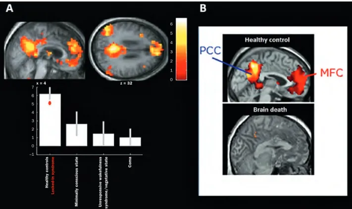
con-sciousness in DOCs patients. These studies have identified a fronto-parietal network encompassing midline (i.e., ante-rior cingulate/mesiofrontal and posteante-rior cingulate/pre-cuneus) and lateral (i.e., prefrontal and posterior parietal) associative cortices that is consistently hypometabolic in unresponsive patients and regains metabolic activity when the patient recovers signs of awareness [6, 46, 47]. More-over, data obtained in sleep (for a review e.g., see [48]) and general anaesthesia (for a review e.g., see [49]) also corrob-orates these findings. A practical example of the FDG-PET imaging studies is the identification of a comparable impair-ment in the fronto-parietal network in anoxic chronic pa-tients with or without clinical fixation, suggesting that this item would not reflect a sign of consciousness [50]. The use of functional magnetic resonance imaging (fMRI) paradigms also permitted to highlight particular brain activation linked to the different states of consciousness. Resting state con-nectivity studies have demonstrated that a resting brain is characterised by coherent fluctuations in the blood-oxygen-level-dependent (BOLD) signal. The midline fronto-parietal or default-mode network (DMN), comprising cortical regions that are known to be more active during rest (encompassing precuneus/posterior cingulate cortex, mesiofrontal/anterior cingulate cortex, and temporoparietal junction areas), has been shown to be informative of cognitive function [51, 52] and to be correlated with bedside behavioural assessment [53]. Furthermore, DMN activity was found to be com-pletely absent in a brain-dead patient (fig. 1) [54].
Figure illustrating the default-mode network encompassing the posterior parietal cortex (PCC) and the midline frontal cortex (MFC). A The default-mode network’s functional connectivity seems to be correlated with the patients’ consciousness level as assessed
by the Coma recovery scale-revised. (Modified from: Vanhaudenhuyse A, Noirhomme Q, Tshibanda LJF, Bruno MA, Boveroux P, Schnakers C, et al. Default network connectivity reflects the level of consciousness in non-communicative braindamaged patients. Brain. 2010;133:161–71.)
B Absence of functional connectivity in the network in a brain-dead patient (modified form [54]). Figure 1 Healthy contr ols Locked-in syndr ome Minimally conscious state Unr esponsive wakefulness syndr ome/vegetative state Coma x = 4 z = 32 7 6 5 4 3 2 1 0 –1 6 5 4 3 2 1 0
With this aim of developing resting automated para- clinical measures that could help in improving DOCs diagno-sis, quantitative EEG is also used for the differentiation between UWS/VS and MCS patients. Although most quan-titative EEG findings can be informative, most of the data only provide information about the brain’s general function-ing. To address this lack in physiological details, a resting EEG study using power spectra and connectivity measures, aimed at exploring the functional differences in conscious and unconscious patients. Taking into account coherence measures (the measure of connectivity between two elec-trode sites), it is possible to gather useful information about the level of integration and connection of the brain’s net-works [55]. Using this technique, recent work showed that, compared to UWS/VS, MCS patients have a better connected network in the theta and alpha bands and that UWS/VS patients may show increased delta power but will at the same time show a decrease in alpha power, as compared to MCS patients [56]. Interestingly, quantitative EEG con-nectivity measures are correlated with the clinical diagnosis obtained with repeated CRS-R assessments at the bedside [56, 57]. To complement these approaches, transcranial mag-netic stimulation (TMS) together with high-density EEG is now employed to evaluate effective connectivity (i.e., the influence one neural system exerts over another) at the bedside. According to theoretical models of consciousness, effective connectivity represents a basic requirement for consciousness and means that multiple, specialised areas of the brain (i.e., the thalamocortical system) must engage in rapid causal interactions [7, 58]. TMS/EEG measures report that, in UWS/VS patients, a simple and local electrical response is obtained after stimulating the brain, indicating a breakdown of effective connectivity like previously observed in unconscious sleeping or anaesthetised subjects [59–61].
In contrast, TMS/EEG in MCS patients will trigger much more complex activations in the brain that will involve sequentially distant cortical ipsilateral and contralateral areas to the site of stimulation, similar to activations re-corded in LIS patients [59].
Detection of consciousness and communication
Specific paradigms using external stimuli are used in order to identify possible residual higher order cognitive processes in DOCs patients. First, neuroimaging passive paradigms will look at brain processes during the presentation of stimula-tion without the active participastimula-tion of the patient. For example, with the use of FDG-PET scan and fMRI, a signifi-cantly different cerebral processing can be identified be-tween conscious and unconscious patients. Indeed, while MCS patients show a widespread cortico-cortical functional activity in associative areas following visual, somatosensory and auditory stimuli, UWS/VS patients only show isolated brain activity in primary sensory cortices [62–66]. Associa-tive areas are therefore thought to be essential for an inte-grated and more elaborated interpretation of the stimulation (i.e., fronto-parietal network and insula) [67, 68]. The iso-lated and low-level brain activation seen in UWS/VS pa-tients suggests the absence of information integration, thus the absence of conscious perception [7].
Although passive paradigms can be informative in terms of brain processing [69], they lack in diagnosis information since they were not specifically designed for detecting relia-ble signs of conscious awareness. Active paradigms might be more demanding in terms of patients’ cognitive abilities but they can reliably gather diagnosis insights with the objectification of command following through specific brain activations. In fact, recent functional neuroimaging studies based on active tasks provided evidence for awareness in patients diagnosed with UWS/VS (and MCS) as they pre-sented with volitional brain activity and thus clear signs of awareness as detected with fMRI [70–73], electroence-phalography (EEG) [74–76] or electromyography (EMG) [77]. In an active motor imagery fMRI paradigm, 2 UWS/VS and 3 MCS patients were able to correctly imagine them-selves as visiting their house and playing tennis as compared to healthy controls [72]. An EEG active paradigm based on the P3 could correctly diagnose a LIS who had previously been considered as being comatose [78]. EMG permitted to objectify preparatory motor responses to command in 1 UWS/VS and 2 MCS non-communicative patients pre-senting with extensive motor deficits with the electrical recordings of muscle activity [77]. Finally, a recent high-density EEG motor imagery task study permitted detec- tion of reliable signs of response to commands in 3 out of 16 UWS/VS patients (fig. 2) [76].
In addition to the detection of signs of consciousness, brain-computer interfaces (BCIs), recordings of brain signals might also allow communication enabling self-expression in non-communicative patients with motor deficits [79, 80]. BCIs have classically been developed for LIS and other pathologies involving severe motor deficits (e.g., amyo-trophic lateral sclerosis) in order to provide a means of inter-action with their environment. In DOCs, BCIs using active
Figure 2 Figure showing a similar brain activation pattern in motor cortices
during performance of a high-density EEG motor imagery task in a patient who received a prior clinical diagnosis of UWS/VS.
UNRESPONSIVE
“VEGETATIVE”
PATIENT
HEALTHY
CONTROL
paradigms are now developed to allow patients to express their consciousness and to communicate. For example, a remarkable case of a behaviourally diagnosed UWS/VS patient who could correctly answer autobiographical yes–no type questions (e.g., “Is your name John?”), by producing specific brain activations through mental imagery tasks (i.e., to say yes, imagine yourself playing tennis; to say no, imaging yourself moving around in your house) in the MRI with the previously reported paradigm [72]. Therefore, this revolutionary finding strikingly demonstrated the possibility of establishing binary communication using patients’ brain responses alone. Recently, an EEG-based BCI aimed for the first time to use an auditory-evoked potential task (P3-based paradigm) for communication purposes in a population with DOCs, LIS and healthy volunteers. The results showed that 20% of the MCS patients were able to show command following but without showing communication with this device, 50% of the LIS patients and 81% of the healthy vol-unteers could reliably use the communication system [81]. These findings highlight the challenges encountered in the development of BCI paradigms. Indeed, the proof of the presence of higher cognitive functions can be obtained with these active paradigms and BCIs, but the absence of brain activation or low performances do not constitute a proof of absence of such high-level information processing. There-fore, it is impossible to know whether DOCs patients tried to perform the task or not. Also, an absence of command following could be explained by the fact that the proposed task and commands might actually require more cognitive resources than expected and thus be too difficult. One solu-tion to this issue, as well as for paradigms, aimed at the detection of signs of consciousness, would be to provide training sessions [82]. Repeated sessions are also recom-mended to rule out the absence of significant performance that could result from the rapid changes in vigilance in DOCs populations.
Therapeutic interventions
Recent surgical and pharmacological trials have shown significant effects of specific treatments on patients’ levels of awareness in DOCs. For instance, deep brain stimulation (DBS) has been proposed as a therapeutic approach in MCS patients. A successful DBS case was reported in 2007 when a 6-year-post-injury patient recovered complex cognitively mediated behavioural patterns after the application of bi-lateral thalamic electrical stimulation (Schiff et al., 2007). This case report showed that DBS could improve arousal level and fluctuations as well as promoting more complex behavioural responsiveness as measured with the CRS-R. Pharmacological trials have been focused mainly on tadine, Zolpidem and Apomorphine in clinical trials. Aman-tadine was initially used in the treatment of Parkinson’s disease, and because of its antiviral properties, it was also employed against influenza. Amantadine is a dopaminergic agonist that has been suggested to improve recovery in DOCs patients. A case report of a MCS patient revealed that, using multiple CRS-R evaluations and FDG-PET cerebral metabolism measurements, motor and cognitive abilities
showed significant improvement after 3 weeks of Amanta-dine treatment [83]. A recent placebo-controlled trial also showed that patients receiving Amantadine showed a signif-icant faster recovery after a 4-week treatment plan [84]. Zolpidem is an imidazopyridine which acts like an agonist on sub-type 1 of the inhibiting receptors of the gamma-Amin-obutyric acid (GABAA). This agent is initially recommended
in the treatment of insomnia and presents sedative, anti-convulsive, anxiolytic and myorelaxant effects. Zolpidem is often described as a “miracle drug” for awakening patients with DOCs. However, the real proportion of Zolpidem is still not well documented. A placebo-controlled trial conducted in 2009 aimed at obtaining an estimate of the frequency of clinically significant responses among patients with DOC. The authors found 1 responder of a total of 15 DOC patients. Behaviourally, the patient went from an UWS/VS to a MCS diagnosis according to the CRS-R assessments pre- and post- treatment [85]. Finally, Apomorphine is a non-selective dopaminergic agonist that was initially indicated to treat Par-kinson’s disease and erectile impotence but has also showed to have positive effects in a few cases of severely brain-injured patients. In a previous clinical prospective study, subcutaneous administration of Apomorphine was used in traumatic UWS/VS and MCS to improve consciousness. According to the clinical assessments, the results showed an outcome improvement in all patients within the first 24 hours and the positive effects were still present after 4 weeks with 50% of the sample completely recovered consciousness. Moreover, improvements in consciousness were sustained for at least 1 year, even after the treatment was discontinued [86].
Ethical issues
DOCs raise a lot of ethical debates. First, one of the most debatable issues about this population is pain perception. The International Association of Pain Specialists (IAPS) defines it as “an unpleasant sensory and emotional experience asso-ciated with real or potential tissue damage” [87]. Thus pain is a first-person experience and classic pain assessments require the verbal feedback of the patients. When it comes to DOCs patients, the question of pain perception is far more complex since they are unable to communicate their feelings and possible pain experiences. Detecting and treating pain represents important medical and ethical considerations, especially in severely brain-injured patients, thus neuro-imaging and behavioural studies can help to address the question. Since pain represents a conscious first person per-ception, nociception is a more appropriate term that should be used regarding DOCs patients. As discussed previously in this review, on a neurofunctional perspective, it appears that MCS patients show a pain-matrix activation that is, although reduced, similar to what is seen in healthy volunteers while UWS/VS patients do not show this higher-order, widespread brain activation. Since the communication between associa-tive brain areas and networks represent one key component of conscious awareness, it has been suggested that uncon-scious patients would not feel pain like the MCS patients and healthy volunteers do. These results obviously have major
consequences on patients’ daily care and management. Despite these findings, according to a recent European sur-vey, still high rates of medical doctors (56%) and para-medical professionals (68%) believe that UWS/VS do feel pain [88]. These attitudes may have major consequences in patients’ care management and especially in cases where UWS/VS patients are withdrawn from life-support treat-ment. In these cases patients may be left without administra-tion of analgesic drugs during their dying process. Moreover, pain could be experienced by patients without demonstrat-ing any behavioural sign of such discomfort. As well as the high rate of misdiagnosis of the altered states of conscious-ness highlighted above in this paper, nociception and pain could also be easily missed in this non-communicative pop-ulation. Therefore, pain prophylaxis and treatment have been proposed for all patients suffering from DOCs [89, 90]. To date, the presence or absence of nociception was inferred via motor responses following noxious stimulation, such as stereotypical responses, flexion withdrawal and localisation responses [89]. In DOCs patients, only a clear localisation to noxious stimulation is considered to be an indicator of conscious perception [16]. In order to accurately non-ver-bally assess nociception in this challenging population, a behavioural scale has been proposed for the first time. The Nociception Coma Scale (NCS) assesses behavioural responses at rest, during daily nursing care and during nociceptive stimulation [91]. Recently, a revised version of the NCS has been proposed (NCS-R). The NCS-R encompasses motor, verbal and facial behaviours, excluding the previous visual subscale that was found to be uninformative of the patient’s level of discomfort since the behaviours included in the subscale were frequently observed in response to non- noxious situations. According to this new version, the need of adequate pain management is recommended at a total cut-off score of 4 (on a maximum of 9) or higher [92].
A second significant ethical challenge concerns the per-ceived quality of life of DOCs patients. Healthy individuals and medical professionals sometimes assume that their quality of life might be so poor that it is not worth living. To address the question, a survey on quality of life has been proposed to be filled in by LIS patients. Although the LIS is not considered to be part of the DOCs, this pathological condition is often misdiagnosed as being such and might represent one of the cruelest physical disabilities. On 65 LIS patients interviewed, 47 self-reported a meaningful quality of life, while a minority of 17 patients rated themselves as being unhappy [93]. Moreover, demand for euthanasia is surprisingly infrequent in chronic LIS patients [94]. Indeed, less than 30% of the chronic LIS patients would report the wish to die or suicidal thoughts [94]. As healthcare prac-tioners, these findings stress the importance of leaving our personal attitudes and beliefs aside when dealing with severely disabled patients. Indeed, contrary to popular creed, it seems that life is worth living it even in cases of severe disabilities. Biased clinicians interpretations of the patients’ conditions might modify medical treatment plans and influence families in inappropriate ways.
Conclusion
We have reviewed the recent advances regarding coma and related disorders. As we have previously highlighted, disentangling between conscious and UWS/VS patients re-presents a major challenge that can generate severe con-sequences. It is to these latter problematic challenges that the ethical and legal end-of-life issues of withholding and withdrawal of life-sustaining treatment are related [95, 96]. The rate of misdiagnosis among the altered states of con-sciousness is still very high; therefore, the use of behavioural scales in parallel with the increasingly powerful neuroimag-ing technologies will help to refine our understandneuroimag-ing and definition of DOCs thus, leading to a more accurate diagno-sis and prognodiagno-sis. Although these technologies still need to be validated in a larger population of patients for finer inter-pretation of the provided information, they have already revealed themselves as promising complementary assess-ment tools. In this sense, multi-centric studies must be supported in order to address the sensitivity and specificity of the neuroimaging or electrophysiological tools. Collabora-tive work also seems essential to gather comparable data for the clinical behavioural assessments and for the potential prognostic value of the para-clinical technologies [97, 98].
The rapidly growing neuroscientific findings on DOCs must be taken into account for patient’s future care needs and to promote adequate policies to keep up with the findings. In fact, new findings in consciousness research has led to the redefinition of clinical criteria for diagnosis and brings to the clinician new knowledge about patient’s recovery and prognosis [44, 45]. Because most of these reported complementary para-clinical procedures remain mainly investigational, clinicians must be aware of the level of evidence supporting the research findings and of the unavoidable ethical and social issues involved. Indeed, we previously discussed patients who were first diagnosed as being un conscious at bedside; but then correctly diagnosed when assessed with neuroimaging and electrophysiological techniques. These cases seem to be increasing as does our understanding of the human brain and its consciousness’s correlates. As a result, clinicians must increasingly answer questions and requests from family members and surrogate decision makers about the new diagnostic and therapeutic procedures. Finally, the future of consciousness research, from a scientific and clinical point of view, should focus on further validation of the clinical techniques and para-digms used. This would be especially advisable in the acute phase, when the patient’s medical condition allows it, in order to tract the patient’s evolution and to provide solid prognosis clues for clinicians and patient’s families.
References
1 James W. The Principles of Psychology. New York: Dover; 1890. 2 Moruzzi G, Magoun HW. Brain stem reticular formation and activation
of the EEG. Electroencephalogr Clin Neurophysiol. 1949;1:455–73. 3 Sherman SM, Guillery RW. The role of the thalamus in the flow of
information to the cortex. Philos Trans R Soc Lond B Biol Sci. 2002;357: 1695–708.
4 Buckwalter JA, Parvizi J, Morecraft RJ, van Hoesen GW. Thalamic projections to the posteromedial cortex in the macaque. J Comp Neurol. 2008;507:1709–33.
5 Laureys S, Faymonville ME, Luxen A, Lamy M, Franck G, et al. Restoration of thalamocortical connectivity after recovery from persistent vegetative state. Lancet. 2000;355:1790–1.
6 Laureys S, Goldman S, Phillips C, Van Bogaert P, Aerts J, et al. Impaired effective cortical connectivity in vegetative state: preliminary investigation using PET. NeuroImage. 1999;9:377–82.
7 Laureys S. The neural correlate of (un)awareness: lessons from the vegetative state. Trends Cogn Sci. 2005;9:556–9.
8 Zorab J. The resuscitation greats. Bjorn Ibsen. Resuscitation. 2003;57:3–9. 9 Zeman A. Consciousness. Brain. 2001;124:1263–89.
10 Posner JB, Saper CB, Plum F. Diagnosis of stupor and coma. New-York: Oxford University Press; 2007.
11 Vanhaudenhuyse A, Demertzi A, Schabus M, Noirhomme Q, Bredart S, et al. Two distinct neuronal networks mediate the awareness of environ - ment and of self. J Cogn Neurosci. 2011;23:570–8.
12 Laureys S, Celesia GG, Cohadon F, Lavrijsen J, Leon-Carrion J, et al. Unresponsive wakefulness syndrome: a new name for the vegetative state or apallic syndrome. BMC Med. 2010;8:68.
13 Jennett B, Plum F. Persistent vegetative state after brain damage. A syndrome in search of a name. Lancet. 1972;1:734–7.
14 Gosseries O, Bruno MA, Chatelle C, Vanhaudenhuyse A, Schnakers C, et al. Disorders of consciousness: what’s in a name? NeuroRehabilitation. 2011;28:3–14.
15 The Multi-Society Task Force Report on PVS. Medical aspects of the persistent vegetative state. N Engl J Med. 1994;330:1499–508. 16 Giacino JT, Ashwal S, Childs N, Cranford R, Jennett B, et al. The minimally
conscious state: definition and diagnostic criteria. Neurology. 2002;58: 349–53.
17 Bruno MA, Vanhaudenhuyse A, Thibaut A, Moonen G, Laureys S. From unresponsive wakefulness to minimally conscious PLUS
and functional locked-in syndromes: recent advances in our understanding of disorders of consciousness. J Neurol. 2011;258:1373–84.
18 Bruno MA, Majerus S, Boly M, Vanhaudenhuyse A, Schnakers C, et al. Functional neuroanatomy underlying the clinical subcategorization of minimally conscious state patients. J Neurol. 2012;259:1087–98. 19 Thibaut A, Bruno MA, Chatelle C, Gosseries O, Vanhaudenhuyse A, et al.
Metabolic activity in external and internal awareness networks in severely brain-damaged patients. J Rehabil Med. 2012;44:487–94.
20 Cairns H, Oldfield RC, Pennybacker JB, Whitteridge D. Akinetic mutism with an epidermoid cyst of the third ventricle. Brain. 1941;64:273–90. 21 Giacino JT. Disorders of consciousness: differential diagnosis and neuro-
pathologic features. Semin Neurol. 1997;17:105–11.
22 Laureys S, Berré J, Goldman S. Cerebral function in coma, vegetative state, minimally conscious state, locked-in syndrome and brain death. In: Yearbook of Intensive Care and Emergency Medicine. Berlin: Springer- Verlag; 2001. p. 386–96.
23 Luaute J, Maucort-Boulch D, Tell L, Quelard F, Sarraf T, et al. Long-term outcomes of chronic minimally conscious and vegetative states. Neurology. 2010;75:246–52.
24 Bruno MA, Ledoux D, Vanhaudenhuyse A, Gosseries O, Thibaut A, et al. Prognosis of patients with altered state of consciousness. In: Coma and Altered States of Consciousness. Paris: Springer-Verlag; 2012. p. 11–23. 25 Bernat JL. Questions remaining about the minimally conscious state.
Neurology. 2002;58:337–8.
26 Plum F, Posner JB. The Diagnosis of Stupor and Coma. Philadelphia, PA: Oxford University Press; 1983.
27 Bruno MA, Schnakers C, Damas F, Pellas F, Lutte I, et al. Locked-in syndrome in children: report of five cases and review of the literature. Pediatr Neurol. 2009;41:237–46.
28 Smart CM, Giacino JT, Cullen T, Moreno DR, Hirsch J, et al. A case of locked-in syndrome complicated by central deafness. Nat Clin Pract Neurol. 2008;4:448–53.
29 Gosseries O, Bruno M, Vanhaudenhuyse A, Laureys S, Schnakers C. Consciousness in the locked-in syndrome. In:The Neurology of Conscious-ness: Cognitive Neuroscience and Neuropathology. Oxford: Elsevier; 2009. p. 191–203.
30 Laureys S, Piret S, Ledoux D. Quantifying consciousness. Lancet Neurol. 2005;4:789–90.
31 Giacino JT, Kalmar K, Whyte J. The JFK Coma Recovery Scale-Revised: measurement characteristics and diagnostic utility. Arch Phys Med Rehabil. 2004;85:2020–9.
32 Schnakers C, Vanhaudenhuyse A, Giacino J, Ventura M, Boly M, et al. Diagnostic accuracy of the vegetative and minimally conscious state: clinical consensus versus standardized neurobehavioral assessment. BMC Neurol. 2009;9:35.
33 Bruno MA, Ledoux D, Lambermont B, Damas F, Schnakers C, et al. Comparison of the Full Outline of UnResponsiveness and Glasgow Liege Scale/Glasgow Coma Scale in an intensive care unit population. Neurocrit Care. 2011;15:447–53.
34 Schnakers C, Giacino J, Kalmar K, Piret S, Lopez E, et al. Does the FOUR score correctly diagnose the vegetative and minimally conscious states? Ann Neurol. 2006;60:744–5.
35 Seel RT, Sherer M, Whyte J, Katz DI, Giacino JT, et al. Assessment scales for disorders of consciousness: evidence-based recommendations for clinical practice and research. Arch Phys Med Rehabil. 2010;91:1795–813.
36 Strens LH, Mazibrada G, Duncan JS, Greenwood R. Misdiagnosing the vegetative state after severe brain injury: the influence of medication. Brain Inj. 2004;18:213–8.
37 Majerus S, Gill-Thwaites H, Andrews K, Laureys S. Behavioral evaluation of consciousness in severe brain damage. Prog Brain Res. 2005;150: 397–413.
38 Majerus S, Bruno MA, Schnakers C, Giacino JT, Laureys S. The problem of aphasia in the assessment of consciousness in brain-damaged patients. Prog Brain Res. 2009;177:49–61.
39 Vanhaudenhuyse A, Giacino J, Schnakers C, Kalmar K, Smart C, et al. Blink to visual threat does not herald consciousness in the vegetative state. Neurology. 2008;71:1374–5.
40 Vanhaudenhuyse A, Schnakers C, Bredart S, Laureys S. Assessment of visual pursuit in post-comatose states: use a mirror. J Neurol Neurosurg Psychiatry. 2008;79:223.
41 Laureys S. Functional neuroimaging in the vegetative state. Neuro Rehabilitation. 2004;19:335–41.
42 Schiff ND. Multimodal neuroimaging approaches to disorders of consciousness. J Head Trauma Rehabil. 2006;21:388–97.
43 Laureys S, Schiff ND. Coma and consciousness: paradigms (re)framed by neuroimaging. NeuroImage. 2012;61:478–91.
44 Fins JJ. Being conscious of their burden: severe brain injury and the two cultures challenge. Ann N Y Acad Sci. 2009;1157:131–47.
45 Fins JJ. The ethics of measuring and modulating consciousness: the imperative of minding time. Prog Brain Res. 2009;177:371–82. 46 Laureys S, Lemaire C, Maquet P, Phillips C, Franck G. Cerebral
metabo-lism during vegetative state and after recovery to consciousness. J Neurol Neurosurg Psychiatry. 1999;67:121.
47 Lull N, Noe E, Lull JJ, Garcia-Panach J, Chirivella J, et al. Voxel-based statistical analysis of thalamic glucose metabolism in traumatic brain injury: relationship with consciousness and cognition. Brain Inj. 2010;24: 1098–107.
48 Maquet P. Understanding non rapid eye movement sleep through neuro- imaging. World J Biol Psychiatry. 2010;11(Suppl 1):9–15.
49 Boveroux P, Bonhomme V, Boly M, Vanhaudenhuyse A, Maquet P, et al. Brain function in physiologically, pharmacologically, and pathologically altered states of consciousness. Int Anesthesiol Clin. 2008;46:131–46. 50 Bruno MA, Vanhaudenhuyse A, Schnakers C, Boly M, Gosseries O, et al. Visual fixation in the vegetative state: an observational case series PET study. BMC Neurol. 2010;10:35.
51 Greicius MD, Krasnow B, Reiss AL, Menon V. Functional connectivity in the resting brain: a network analysis of the default mode hypothesis. Proc Natl Acad Sci U S A. 2003;100:253–8.
52 Mevel K, Grassiot B, Chetelat G, Defer G, Desgranges B, et al. The default mode network: cognitive role and pathological disturbances. Rev Neurol (Paris). 2010;166:859–72.
53 Soddu A, Vanhaudenhuyse A, Demertzi A, Bruno MA, Tshibanda L, et al. Resting state activity in patients with disorders of consciousness. Funct Neurol. 2011;26:37–43.
54 Boly M, Tshibanda L, Vanhaudenhuyse A, Noirhomme Q, Schnakers C, et al. Functional connectivity in the default network during resting state is preserved in a vegetative but not in a brain dead patient. Hum Brain Mapp. 2009;30:2393–400.
55 Chorlian DB, Rangaswamy M, Porjesz B. EEG coherence: topography and frequency structure. Exp Brain Res. 2009;198:59–83.
56 Lehembre R, Marie-Aurelie B, Vanhaudenhuyse A, Chatelle C, Cologan V, et al. Resting-state EEG study of comatose patients: a connectivity and frequency analysis to find differences between vegetative and minimally conscious states. Funct Neurol. 2012;27:41–7.
57 Gosseries O, Schnakers C, Ledoux D, Vanhaudenhuyse A, Bruno MA, et al. Automated EEG entropy measurements in coma, vegetative state/ unresponsive wakefulness syndrome and minimally conscious state. Funct Neurol. 2011;26:25–30.
58 Tononi G, Koch C. The neural correlates of consciousness: an update. Ann N Y Acad Sci. 2008;1124:239–61.
59 Rosanova M, Gosseries O, Casarotto S, Boly M, Casali AG, et al. Recovery of cortical effective connectivity and recovery of consciousness in vegetative patients. Brain. 2012;135:1308–20.
60 Massimini M, Ferrarelli F, Huber R, Esser SK, Singh H, et al. Breakdown of cortical effective connectivity during sleep. Science. 2005;309:2228–32. 61 Ferrarelli F, Massimini M, Sarasso S, Casali A, Riedner BA, et al.
Breakdown in cortical effective connectivity during midazolam-induced loss of consciousness. Proc Natl Acad Sci U S A. 2010;107:2681–6. 62 Boly M, Faymonville ME, Peigneux P, Lambermont B, Damas P, et al.
Auditory processing in severely brain injured patients: differences between the minimally conscious state and the persistent vegetative state. Arch Neurol. 2004;61:233–8.
63 Giacino JT, Hirsch J, Schiff N, Laureys S. Functional neuroimaging applications for assessment and rehabilitation planning in patients with disorders of consciousness. Arch Phys Med Rehabil. 2006;87:67–76. 64 Laureys S, Faymonville ME, Degueldre C, Fiore GD, Damas P, et al.
Auditory processing in the vegetative state. Brain. 2000;123:1589–601. 65 Di HB, Yu SM, Weng XC, Laureys S, Yu D, et al. Cerebral response
to patient’s own name in the vegetative and minimally conscious states. Neurology. 2007;68:895–9.
66 Schiff ND, Rodriguez-Moreno D, Kamal A, Kim KH, Giacino JT, et al. fMRI reveals large-scale network activation in minimally conscious patients. Neurology. 2005;64:514–23.
67 Boly M, Faymonville ME, Schnakers C, Peigneux P, Lambermont B, et al. Perception of pain in the minimally conscious state with PET activation: an observational study. Lancet Neurol. 2008;7:1013–20.
68 Schiff ND. Bringing neuroimaging tools closer to diagnostic use in the severely injured brain. Brain. 2007;130:2482–3.
69 Boly M, Garrido MI, Gosseries O, Bruno MA, Boveroux P, et al. Preserved feedforward but impaired top-down processes in the vegetative state. Science. 2011;332:858–62.
70 Bardin JC, Fins JJ, Katz DI, Hersh J, Heier LA, et al. Dissociations between behavioural and functional magnetic resonance imaging-based evaluations of cognitive function after brain injury. Brain. 2011;134:769–82. 71 Boly M, Coleman MR, Davis MH, Hampshire A, Bor D, et al. When
thoughts become action: an fMRI paradigm to study volitional brain activity in non-communicative brain injured patients. NeuroImage. 2007;36: 979–92.
72 Monti MM, Vanhaudenhuyse A, Coleman MR, Boly M, Pickard JD, et al. Willful modulation of brain activity in disorders of consciousness. N Engl J Med. 2010;362:579–89.
73 Owen AM, Coleman MR, Boly M, Davis MH, Laureys S, et al. Detecting awareness in the vegetative state. Science. 2006;313:1402.
74 Schnakers C, Perrin F, Schabus M, Majerus S, Ledoux D, et al. Voluntary brain processing in disorders of consciousness. Neurology.
2008;71:1614–20.
75 Goldfine AM, Victor JD, Conte MM, Bardin JC, Schiff ND. Determination of awareness in patients with severe brain injury using EEG power spectral analysis. Clin Neurophysiol. 2011;122:2157–68.
76 Cruse D, Chennu S, Chatelle C, Bekinschtein TA, Fernandez-Espejo D, et al. Bedside detection of awareness in the vegetative state: a cohort study. Lancet. 2011;378:2088–94.
77 Bekinschtein TA, Coleman MR, Niklison J, 3rd, Pickard JD, Manes FF. Can electromyography objectively detect voluntary movement in disorders of consciousness? Journal of neurology, neurosurgery, and psychiatry. 2008;79:826–8.
78 Schnakers C, Majerus S, Goldman S, Boly M, Van Eeckhout P, et al. Cognitive function in the locked-in syndrome. J Neurol. 2008;255:323–30. 79 Kubler A, Kotchoubey B. Brain-computer interfaces in the continuum
of consciousness. Curr Opin Neurol. 2007;20:643–9.
80 Laureys S, Boly M. The changing spectrum of coma. Nat Clin Pract Neurol. 2008;4:544–6.
81 Lule D, Noirhomme Q, Kleih SC, Chatelle C, Halder S, et al. Probing command following in patients with disorders of consciousness using a brain-computer interface. Clin Neurophysiol. 2012.
82 Kubler A, Birbaumer N. Brain-computer interfaces and communication in paralysis: extinction of goal directed thinking in completely paralysed patients? Clin Neurophysiol. 2008;119:2658–66.
83 Schnakers C, Hustinx R, Vandewalle G, Majerus S, Moonen G, et al. Measuring the effect of amantadine in chronic anoxic minimally conscious state. J Neurol Neurosurg Psychiatry. 2008;79:225–7.
84 Giacino JT, Whyte J, Bagiella E, Kalmar K, Childs N, et al. Placebo- controlled trial of amantadine for severe traumatic brain injury. N Engl J Med. 2012;366:819–26.
85 Whyte J, Myers R. Incidence of clinically significant responses to zolpidem among patients with disorders of consciousness: a preliminary placebo controlled trial. Am J Phys Med Rehabil. 2009;88:410–8.
86 Fridman EA, Krimchansky BZ, Bonetto M, Galperin T, Gamzu ER, et al. Continuous subcutaneous apomorphine for severe disorders of consciousness after traumatic brain injury. Brain Inj. 2010;24:636–41. 87 IASP. Classification of chronic pain: Description of chronic pain syndromes
and definitions of pain terms. Task force on taxonomy, suppl. 3. Seattle: IASP Press; 1994.
88 Demertzi A, Schnakers C, Ledoux D, Chatelle C, Bruno MA, et al. Different beliefs about pain perception in the vegetative and minimally conscious states: a European survey of medical and paramedical professionals. Prog Brain Res. 2009;177:329–38.
89 Schnakers C, Zasler ND. Pain assessment and management in disorders of consciousness. Curr Opin Neurol. 2007;20:620–6.
90 Schnakers C, Faymonville ME, Laureys S Ethical Implications: Pain, coma, and related disorders. In: Encyclopedia of Consciousness. Oxford: Elsevier; 2009. p. 243–50.
91 Schnakers C, Chatelle C, Vanhaudenhuyse A, Majerus S, Ledoux D, et al. The Nociception Coma Scale: a new tool to assess nociception in disorders of consciousness. Pain. 2010;148:215–9.
92 Chatelle C, Majerus S, Whyte J, Laureys S, Schnakers C. A sensitive scale to assess nociceptive pain in patients with disorders of consciousness. J Neurol Neurosurg Psychiatry. 2012;83(12):1233–7.
93 Bruno MA, Bernheim JL, Ledoux D, Pellas F, Demertzi A, et al. A survey on self-assessed well-being in a cohort of chronic locked-in syndrome patients: happy majority, miserable minority. BMJ Open. 2011;1: e000039.
94 Laureys S, Pellas F, Van Eeckhout P, Ghorbel S, Schnakers C, et al. The locked-in syndrome: what is it like to be conscious but paralyzed and voiceless? Prog Brain Res. 2005;150:495–511.
95 Celesia G. Persistent vegetative state: clinical and ethical issues. 2000;460–2.
96 Jennett B. The vegetative state. Medical facts, ethical and legal dilemmas. Cambridge: Cambridge University Press; 2002.
97 Coleman MR, Davis MH, Rodd JM, Robson T, Ali A, et al. Towards the routine use of brain imaging to aid the clinical diagnosis of disorders of consciousness. Brain. 2009;132:2541–52.
98 Di H, Boly M, Weng X, Ledoux D, Laureys S. Neuroimaging activation studies in the vegetative state: predictors of recovery? Clin Med. 2008;8: 502–7.