1 Neural changes associated with semantic processing in healthy aging despite intact
behavioral performance
Jacinthe Lacombe a,b,c *, Pierre Jolicoeur a,b,c,d, Stephan Grimault a,b,e, Jessica Pineault a,b,c, Sven Joubert a, c
a
Département de psychologie, Université de Montréal, Pavillon Marie-Victorin C.P. 6128, succursale Centre-Ville, Montréal, Québec, Canada H3C 3J7
b
CERNEC, Université de Montréal, Pavillon Marie-Victorin,
Département de psychologie C.P. 6128, succursale Centre-Ville, Montréal, Québec, Canada H3C 3J7
c
Centre de recherche de l’Institut universitaire de gériatrie de Montréal (CRIUGM), 4565 Chemin Queen-Mary, Montréal, Québec, Canada H3W 1W5
d
International Laboratory for Brain, Music and Sound Research (BRAMS), Pavillon 1420 Mont-Royal, FAS – Département de psychologie, C.P. 6128,
succ Centre-ville, Montréal, Québec, Canada H3C 3J7 e
Centre national de la recherche scientifique (CNRS), 3 rue Michel-Ange 75794 Paris cedex 16, France
* Corresponding author: Sven Joubert, Département de psychologie, Université de
Montréal, C.P. 6128, Succursale Centre-Ville, H3C 3J7, Montréal, QC, Canada. Tel : 1-514-343-6940 Fax: 1-514-340-2801. Email address: [email protected]
2
Abstract
Semantic memory recruits an extensive neural network including the left inferior prefrontal cortex (IPC) and the left temporoparietal region, which are involved in semantic control processes, as well as the anterior temporal lobe region (ATL) which is considered to be involved in processing semantic information at a central level. However, little is known about the underlying neuronal integrity of the semantic network in normal aging. Young and older healthy adults carried out a semantic judgment task while their cortical activity was recorded using magnetoencephalography (MEG). Despite equivalent behavioral performance, young adults activated the left IPC to a greater extent than older adults, while the latter group recruited the temporoparietal region bilaterally and the left ATL to a greater extent than younger adults. Results indicate that significant neuronal changes occur in normal aging, mainly in regions underlying semantic control processes, despite an apparent stability in performance at the behavioral level.
Keywords: semantic memory; language; normal aging; MEG; elderly; semantic executive
3
1. Introduction
Several cognitive abilities decline over the course of normal aging, including episodic memory, working memory, and executive functions (Park, et al., 2002). Declining abilities in episodic memory and working memory in older adults have been associated with bilateral recruitment of inferior prefrontal cortex in older adults, while young adults tended to activate the same region but only in one hemisphere, a phenomenon described as hemispheric asymmetry reduction in older adults (HAROLD) (Cabeza, 2002; Rajah & D'Esposito, 2005). Semantic memory, in contrast, which represents general knowledge about the world, remains relatively stable or even improves in normal aging (Burke & Mackay, 1997; Luo & Craik, 2008; Park, et al., 2002; Verhaeghen, 2003). Despite this behavioral evidence indicating preserved semantic abilities in healthy aging, little is known about the integrity of the neural network underlying semantic processing in healthy aging. More specifically, it is not clearly understood whether older individuals show similar or distinct patterns of brain activation relative to younger adults while they perform a semantic task.
Semantic processing recruits a widespread network of brain regions, which is mainly left-lateralized for verbal material (Binder, Desai, Graves, & Conant, 2009; Binney, Embleton, Jefferies, Parker, & Ralph, 2010). Typically, this semantic network includes key regions such as the anterior temporal lobe (ATL) region, which is considered to represent a candidate site for the storage of conceptual representations, and for processing concepts at an amodal and abstract level (Jefferies & Lambon Ralph, 2006; Patterson, Nestor, & Rogers, 2007). The left inferior prefrontal cortex (IPC) and left temporoparietal region, in contrast, appear to be involved in strategic search and control
4 processes required for semantic processing (Binder, et al., 2009; Jefferies, 2013; Jefferies & Lambon Ralph, 2006; Whitney, Kirk, O'Sullivan, Lambon Ralph, & Jefferies, 2011). Semantic control processes modulate relevant information to provide a task-appropriate response regardless of task modalities (Jefferies & Lambon Ralph, 2006). However, these regions of the semantic network are recruited differently depending of the type of stimuli that are processed. For instance, differences of activation in ATL and IPC have been reported between concrete and abstract words in neuroimaging studies, using functional magnetic resonance imaging or positron emission topography (Hoffman, Binney, & Lambon Ralph, 2014; Sabsevitz, Medler, Seidenberg, & Binder, 2005; Wang, Conder, Blitzer, & Shinkareva, 2010). Studies on clinical patients, with lesions in regions of the semantic network also showed differences in performance during semantic processing of abstract and concrete words (Loiselle, et al., 2012). The use of concrete and abstract words is thus of interest to explore age-related changes in semantic processing.
In healthy aging, even though the store of knowledge remains intact, rapid access to conceptual knowledge and the executive components related to the manipulation and retrieval of this knowledge may become less efficient. For instance, diminished semantic control processes may impact language production in tasks requiring lexical semantic retrieval (Wierenga, et al., 2008), such as evidenced by the commonly-encountered tip-of-the-tongue (TOT) phenomenon in older individuals. During picture naming tasks, it has been shown that in order to compensate for effortful retrieval and to maintain a high level of performance, older adults recruit inferior prefrontal regions to a greater extent than younger adults (Galdo-Alvarez, Lindin, & Diaz, 2009; Nielson, et al., 2006; Wierenga, et al., 2008). This indicates that neural changes underlying semantic control
5 processes in tasks exerting demands upon these processes may be taking place despite intact behavioral performance in older adults. It remains important to determine, however, if similar age-related brain changes can be observed during receptive language tasks tapping comprehension such as semantic judgment tasks, which do not exert as much demands on effortful semantic retrieval processes required in naming and other language production tasks.
The aim of this study was thus to compare the patterns of cortical activity, using MEG, in younger and older adults while they performed a semantic judgment task in which they had to determine whether a list of single words were concrete or abstract. Our hypothesis was that differences in cortical activation would be observed between young and older adults in brain regions associated with semantic control processes, including the IPC and the temporoparietal region, but not in the ATL, which has a role in processing conceptual representations at a central level, even though we expected behavioral performance to be similar across age groups.
2. Material and Methods 2.1 Subjects
Eleven healthy older adults aged between 60 and 85 years old and thirteen healthy young adults aged between 18 and 30 years of age participated in this study. The groups did not differ significantly in terms of number of years of education (t(14.787) = 1.194, p = 0.251). Demographic data are presented in Table 1.
INSERT TABLE 1 HERE
All participants were volunteers, right-handed, French native speakers, and were recruited from the local community. Exclusion criteria included neurological or
6 psychiatric conditions, a history of alcohol or drug abuse, or a general anesthesia in the past 6 months. In order to exclude the presence of cognitive impairment in healthy older adults, older participants completed cognitive screening to ensure they had a normal level of cognitive functioning. The mean score at the MMSE (Mini Mental State Examination, Folstein, Folstein, & McHugh, 1975) was 29.7 (s.d.=0.7). In addition, older participants carried out the RL/RI16 test, a test of verbal learning (RL/RI16, Van der Linden, et al., 2004). A participant was excluded if his/her performance at this test was less than 1.5 standard deviation below that of an age-and-education-matched normative group. All participants gave signed informed consent to participate in the study, which had been vetted by the appropriate institutional research ethics committee. Before carrying out the MEG study, participants were screened for MEG artefacts, which included for instance dental work, metal implants or abnormal magnetization of the brain resulting from a previous MRI. In addition to the participants described above, three younger participants and five older participants also participated in this study but were excluded from the study because of large movements of the head during the recording that compromised the accuracy of source localizations or because of excessive loss of trials due to eye blinks during MEG acquisition.
2.2 Materials and Procedure 2.2.1 MEG semantic judgment task
The semantic judgment task consisted of deciding whether each of 460 French words, presented one by one, was abstract or concrete. Half of the words (230) were abstract (A) and the other half were concrete (C). Words were selected from the Omnilex Database (www.omnilex.uottawa.ca). Abstract and concrete words were balanced in
7 terms of number of letters (between 5 and 7 letters; A: M = 6.32, s.d. = 0.77; C: M = 6.30, s.d. = 0.73, p = .803), as well as in terms of written (A: M = 19.63, s.d. = 22.82; C:
M = 19.57, s.d. = 13.10, p =. 969) and spoken (A: M = 13.08, s.d. = 21.58; C: M = 13.34, s.d. = 15.09, p = .881) word frequencies. As expected, the two word categories differed
significantly in terms of imageability (t(456)= 82.8, p < .001), the imageability score being
greater for concrete than for abstract words (A: M = 2.6, s.d, = 0.4; C: M = 5.5, s.d. = 0.3). Abstract and concrete words were then split using a median split on the imageability score of each category of words (abstract/low imageability, abstract/high imageability, concrete/low imageability and concrete/high imageability). In term of imageability ratings, the abstract/low imageability words has the lowest imageability scores (M = 2.4,
s.d, = 0.5), followed by abstract/high imageability words (M = 2.9, s.d, = 0.1),
concrete/low imageability words (M = 5.3, s.d, = 0.2), and finally followed by concrete/high imageability words (M = 5.8, s.d, = 0.2). This procedure was established in order to investigate if a linear effect of imageability on brain activation might be observed. There were no statistical differences between the four word classes, either in terms of written or spoken word frequency.
Words were presented one by one on a computer screen, and each participant had to judge if the word presented was concrete or abstract. A short description of what was meant by abstract and concrete words was provided, as well as a 32-word practice block, before the beginning of the experimental task. Each trial began with a white circle, indicating to the participant that he/she could blink eyes. Once the participant was ready, he/she was instructed to push simultaneously the button under each thumb. This procedure was established to avoid false starts. Once the trial was launched, a white
8 fixation cross appeared for 600 ms ± 100 ms, in the center of the screen. The word then appeared for 500 ms, before returning to the white cross. The subject then had to decide whether the word referred to a concrete or an abstract concept. He/she responded by pushing the button under the index of the left or right hand, and the side of response for concrete and abstract words was counterbalanced across subjects. The fixation cross stayed on the screen until the participant responded. The participant had 2500 ms to answer. Once a response was recorded, a colored circle appeared and provided feedback to the subject. A green circle indicated a correct answer, whereas a red circle appeared when an incorrect response or no response was recorded. Providing feedback generally helps subjects to ensure they have not forgotten the particularities of the task and that they have their fingers on the correct response buttons (when the task involves manual responses). Providing feedback on accuracy also ensures proper calibration in terms of the speed-accuracy trade-off function, in that responding too quickly will lead to errors that are signaled to the subject. When the 2500 ms-delay was over, the white circle appeared, indicating to the subject that he/she could once again blink eyes. Once the participant was ready, he/she simultaneously pushed the button under each thumb and the next trial began. Experimental trials were divided into 10 blocks of 46 words each (half abstract, half concrete). Order of presentation was pseudorandomized.
Stimuli were presented using E-Prime software and were projected on a rear-projection screen, placed about 69 cm from the participant’s eyes. Words were presented in Times New Roman font. Stimuli subtended a horizontal visual angle of about 4.2° (width) and a vertical visual angle of about 0.6° (height) at the center of the field of view.
9 Functional neural activity of the participants carrying out the semantic judgment task was recorded continuously using a whole-head CTF-VSM 275 sensors MEG system. MEG is a technique with excellent temporal resolution and good spatial resolution and, in contrast with fMRI, provides a better signal of neural activity in the anterior temporal lobe region, a key region of the semantic network (Noppeney & Price, 2004; Patterson, et al., 2007; Visser, Jefferies, & Lambon Ralph, 2010). It offers a direct measure of cortical activity, as opposed to fMRI, which provides an indirect measure of cerebral activation inferred from the hemodynamic (blood oxygen level dependent, BOLD) response (Marinkovic, 2004). MEG also offers the possibility to explore a precise cognitive process by focusing on a specific temporal window, in contrast with the brain activity recorded by fMRI which represents a summation of several successive activations related to different cognitive processes (Vartiainen, Liljestrom, Koskinen, Renvall, & Salmelin, 2011).
For six participants in the older group, plastic lenses were installed in front of their eyes to ensure optimal visual acuity of the stimuli during the experimental task and to avoid magnetic perturbations (from personal glasses with metal parts). Bipolar electroocculogram (EOG) (electrodes placed at the left and right canthi for horizontal EOG and above and below the left eye for vertical EOG) as well as unipolar electroencephalogram ECG were also recorded. Head shape and fiducial points (nasion, left and right pre-auricular points) were digitized using a Polhemus Fastrak (Polhemus Inc., Colchester, VT, USA). Pictures of the positions of the coils for each subject were also taken to allow the co-registration process. A cervical collar was used to limit head movement and help subjects relax in the apparatus without neck pain. The sampling rate
10 was 1200 Hz. Two optical response boxes, one in each hand, were used to record participants’ responses during the semantic judgment task.
2.2.2 Structural MR Image acquisition
Following MEG recording, each subject underwent a high-resolution anatomical MRI scan obtained with a 3T Siemens Trio MRI (Siemens, Erlangen, Germany) at the Unité de neuroimagerie fonctionnelle (UNF, Montréal, Canada) (http://www.unf-montreal.ca/siteweb/Home_en.html). These images were acquired using an optimized MPRAGE protocol (TR = 2.3 s, TE = 2.94 ms, TI = 900 ms, flip angle = 9º, FOV = 256×240, voxel 1mm×1mm×1.2mm) using an 8-channel coil. Images were acquired in the horizontal plane.
2.3 Data Analysis
2.3.1 Behavioral Data
Correct responses were averaged for each participant, for each of the four categories of words. The absence of response was recorded as an incorrect response. A repeated-measure ANOVA (group x type of words) was carried out. For global reaction times, an independent-group t-test was carried out.
2.3.2 MEG Data
Third-order gradient noise reduction and low-pass-filter at 40 Hz were used on data (computed with CTF-VSM software). Blinks and eye movements were identified by visual inspection of the horizontal EOG and vertical EOG signals, and trials containing them were excluded from the analyses. Trials with an incorrect response or excessive head movement (exceeding 10 mm from baseline recording) or other external magnetic
11 artefact were also removed. Approximately 23% of the trials had been removed due to blinks and eye movements, head movement or external artefacts in both groups. The MEG recording was segmented from - 200 ms to + 1000 ms relative to stimulus onset with the 200 ms pre-stimulus period used as a baseline period. Event-related magnetic field maps were computed by averaging signals recorded from artefact-free correct trials. We thus obtained four event-related magnetic field maps (one for each of the four categories of stimuli, i.e. abstract/low imageability; abstract/high imageability; concrete/low imageability; concrete/high imageability) for each participant. A visual inspection of the averaged event-related magnetic field temporal curves allowed the selection of a temporal window specific from 330 ms to 430 ms for younger adults, and from 410 to 510 ms for older adults to isolate a component, the N400, associated with semantic processing of meaningful semantic stimuli (Federmeier & Kutas, 2005; Marinkovic, 2004). These curves are shown in figure 1. Previous studies in ERP have shown that the peak latency of this component is generally delayed in normal aging (Federmeier & Kutas, 2005; Giaquinto, Ranghi, & Butler, 2007).
INSERT FIGURE 1 HERE
The event-related fields were used to perform source localization, with the maximum entropy on the mean method (MEM : Amblard, Lapalme, & Lina, 2004; Grova, et al., 2006). This method is a cortically-constrained distributed source-localization approach. The cortical surface (we used the white/gray matter boundary in a 3D surface) was segmented from each anatomical MRI scan using BrainVisa software (http://brainvisa.info/index_f.html). We used a standard co-registration process: like all CTF-VSM MEG systems, three coils that emit signals at three different locations (nasion,
12 left and right pre-ocular) were recorded by the MEG sensors. The positions of the coils relative to the MEG sensors were then calculated. On the processed MRI (3D reconstruction of the subject's head), we manually indicated the position of the three coils with the aid of the pictures taken during the experimental session. The position of the subject's head and the MEG sensors were then co-registered by superimposing the MEG data and MRI image, by repositioning the coil locations from one image to the other. Approximately 4000 sources, orthogonal to the local surface, were distributed over the cortex of each hemisphere of each participant, and these sources were used in distributed source localization analyses, for each participant.
Cortically-constrained source images were computed for each time sample, in each condition. These images were averaged over time, during the selected time window (around the N400), to estimate source activity for each of the four conditions (abstract/low imageability; abstract/high imageability; concrete/low imageability; concrete/high imageability) and each participant. Each cortical-surface average localization map was interpolated in the volume MRI image for each participant and the resulting image was smoothed using a Gaussian filter with an 8-mm FWHM (full width at half maximum). All the images were finally normalized using SPM2 to a common template in MNI-Talairach space (ICBM 152, Montreal Neurological Institute).
We performed analyses using a General Linear Model (AFNI ; Cox, 1996) analysis of the normalized source-localization maps. For statistical analysis on group, we used random field theory (RFT; Worsley, et al., 2002) to determine our statistical thresholds corrected for multiple comparisons. According to RFT, a cluster of voxels with a volume of at least 68 mm3 and a t = 3.87 was significant at p < .001, corrected for multiple
13 comparisons. In order to investigate if there was an interaction between age x concreteness, three regions of interest were specifically selected: the left IPC, the temporoparietal region and the left ATL, all key regions of the semantic network. Abstract words (abstract/high imageability and abstract/low imageability) were combined together, as well as concrete words (concrete/low imageability and concrete/high imageability). A statistical threshold of p < .01 non-corrected was used.
3. Results
3.1 Semantic judgment task
Semantic performance, such as determined by mean accuracy, was strictly equivalent for younger and older adults for all types of words. However, reaction times were longer for older than for younger adults (t(22)=3.22, p < .05). Results are shown in Table 2.
INSERT TABLE 2 HERE
No principal effect on group was found (p = .948). A principal effect of the type of words was observed (F(2,059) = 13.523, p < .001). Concrete/high imageability words were processed more accurately than the other three types of words (concrete/low
imageability; abstract/high imageability and abstract/low imageability). No interactive effect of age x concreteness was found (p = .856). In sum, older and younger adults’ performance was strictly equivalent, in term of accuracy, on the semantic judgment task.
3.2 MEG results
Direct contrasts between the two groups were carried out. Differences in terms of cortical activation between the groups of young and older adults related to the semantic decision task were found in several regions of the semantic network, even though both groups showed identical performance at the task, such as expressed by the accuracy rates
14 of both groups. Localization images emphasizing differences across younger and older adults are illustrated in Figure 2, while specific regions are listed in Table 3. The Talairach coordinates and Brodmann areas were provided by AFNI software (Cox, 1996), and have been validated with the Talairach Client software (Lancaster, et al., 2000).
INSERT TABLE 3 HERE
The temporoparietal region was found to be more activated in older adults relative to young adults. Indeed, the largest cluster of voxels with group difference was found in the right posterior middle temporal gyrus, extending into the inferior parietal lobule and the angular gyrus. An independent cluster in the left posterior middle temporal gyrus was also found to be more activated in the group of older adults. In addition, a locus in the left middle temporal lobe, located in the anterior temporal lobe (ATL), was found to be more activated in the group of older adults. In contrast, a significant locus of activation was found to be more activated in the left IPC, including Brodmann area 47, for younger than older adults. In addition, group differences in activation were also observed in additional brain regions including the fusiform gyrus and the occipital lobes, which were more activated by older adults mostly in the right hemisphere, while the right cingulate gyrus was more activated by younger adults.
INSERT FIGURE 2 HERE
In terms of abstract vs. concrete words, no statistically significant differences in source localization between the different types of words were found. However, an interaction effect age x concreteness was found in the left inferior prefrontal region. As shown in figure 3, a small locus in the left IPC (BA47) was found to be more activated in older adults for abstract words than for concrete words, whereas the younger adults had
15 similar activations in this region for both categories of words. Note that the locus of difference of activation between abstract and concrete words is anterior to the one found in the group comparison. No difference of activation was found in either the temporoparietal region or the left ALT.
INSERT FIGURE 3 HERE
Overall, younger adults recruited the IPC to a greater extent than older adults while they performed a semantic judgment task, whereas older adults recruited the bilateral temporoparietal region and the left ATL significantly more than younger adults. However, older adults recruited a specific sub-region of the IPC to a greater extent when abstract words were examined specifically. Finally, the pattern of cortical activation associated with semantic processing in the group of healthy older adults was found to be overall more widespread than in the younger group, recruiting for instance posterior regions of the right-hemisphere to a greater extent.
4. Discussion
The main goal of this study was to investigate the neural bases of semantic processing in healthy aging. In terms of cognitive performance, younger and older adults performed equally well on the single-word semantic judgment task, as indicated by the accuracy score. Our results are in line with previous findings showing that verbal knowledge, which represents an intrinsic aspect of semantic memory, remains intact in normal aging, in contrast to other memory systems such as episodic and working memory, which decline with age (Park, et al., 2002; Verhaeghen, 2003). However, even though percentage of correct responses was equivalent for both groups, the pattern of brain activity that emerged from the semantic task differed between younger and older
16 participants. Consistent with our hypothesis, significant age-related changes were found within several key regions of the semantic network: the left IPC, the temporoparietal region bilaterally and, more surprisingly, the left ATL (Binder, et al., 2009; Jefferies, 2013; Jefferies & Lambon Ralph, 2006; Patterson, et al., 2007; Whitney, et al., 2011). The left IPC was more activated in younger participants, while the temporoparietal cortices and the left ATL were more activated in older participants during semantic processing. It is noteworthy to mention that the peak of the locus of difference of activation in the left posterior middle temporal gyrus found in our study is posterior to the one reported in previous semantic studies in young adults (Noonan, Jefferies, Visser, & Lambon Ralph, 2013). In addition, patterns of cortical activation in older subjects were more widely distributed across both hemispheres than in younger subjects. Therefore, these results indicate that while semantic processing remains intact during the course of aging, its neurofunctional organization undergoes specific changes.
According to current models of semantic memory (Patterson et al., 2007; Jefferies, 2013), the ATL region represents the primary site of conceptual knowledge, where concepts are assumed to be processed at an abstract and amodal level. The left IPC and the temporoparietal region, in turn, are thought to be involved in the executive aspects of semantic processing, such as the selection, retrieval, and manipulation of semantic information (Jefferies, 2013; Jefferies & Lambon Ralph, 2006; Whitney, et al., 2011). This posterior region includes the posterior middle/superior temporal gyrus as well as the inferior parietal lobe (Jefferies, 2013). Although both the IPC and the temporoparietal cortex play a role in selection of context-appropriate and task-oriented semantic knowledge (including the inhibition of inappropriate semantic candidates or
17 distracters), it is not clear at this time whether these two regions play distinct roles within the semantic control system or whether they act synergistically as part of a connected network of regions (Jefferies, 2013). It has been recently proposed that the IPC may be involved in semantic control processing regardless of the receptive/expressive aspects of the task while the posterior temporal middle temporal gyrus may be engaged only when comprehensive task are performed (Noonan et al., 2013). In the current study, age-related changes were found in both of these cortical areas. More specifically, the left IPC was less activated in older than in younger adults, while the reverse pattern was found in the temporoparietal cortex during a receptive semantic task.
The finding in this study of reduced prefrontal activation in older adults differs from some previous studies which have reported increased activation in this region during a naming task (Galdo-Alvarez, et al., 2009; Nielson, et al., 2006; Wierenga, et al., 2008). According to these authors, increased frontal activation in older adults reflected greater demands on the semantic control system required to maintain a high-level of performance during picture naming, which requires effortful lexical retrieval processes. Because this cognitive process becomes less efficient in normal aging, seniors have to activate the IPC to a greater extent in order to maintain a performance equivalent to that of younger adults. This kind of hyperactivation in a production task that requires controlled-searching strategies seems coherent with the compensation-related utilization of neural circuits hypothesis (CRUNCH) (Reuter-Lorenz & Cappell, 2008). According to this model, inefficient processing leads older adults to recruit a cerebral region to a greater extent to maintain a high level of performance. If the task becomes too difficult, a pattern of hypoactivation is then observed, along with a drop in behavioral performance.
18 Thus, this compensation model is of interest to explain the age-related changes observed when an expressive language task is engaged.
However, receptive language tasks tapping comprehension such as semantic judgment tasks do not exert as much demands on effortful semantic retrieval processes required in naming and other language production tasks. Since picture naming (language production) is inherently different from semantic judgment (language comprehension), such differences in the nature of the tasks may account for the dissimilar patterns of prefrontal activation reported in the current study. Indeed, studies investigating the neural bases of semantic processing in normal aging using receptive tasks have reported mainly a pattern of prefrontal hypoactivation in healthy older adults relative to younger adults (Berlingeri, et al., 2010; Grossman, et al., 2002; Peelle, Troiani, Wingfield, & Grossman, 2010; Tyler, et al., 2010), in contrast with a prefrontal hyperactivation in production tasks. These studies, however, focused mainly on syntactic processing of sentence comprehension, which recruits a number of high-order cognitive processes, including complex syntactic comprehension and working memory, which are also known to decline with age (Burke & Shafto, 2008; Wingfield & Grossman, 2006).
Nonetheless, two studies which have used single-word semantic judgment tasks reported similar results. In a functional MRI study (fMRI), Stebbins et al. (2002) found less activation in left prefrontal cortex in older adults relative to younger adults while they carried out a single-word semantic judgment task. Using magnetoencephalography (MEG), Kemmotsu et al. (2012) compared young to middle-aged adults (mean age of 50 years-old) during single-word semantic judgment task, and reported that middle-aged adults recruited the inferior prefrontal region to a lesser extent than young adults. Thus,
19 even when the impact of working memory load and syntactic demands was reduced by presenting single-words, patterns of prefrontal hypoactivation were still observed in older adults during semantic judgment, even though behavioral performance was equivalent across groups.This pattern of prefrontal hypoactivation differed from the one observed in studies investigating age-related changes in language production, which revealed mainly a pattern of prefrontal hyperactivation.
In the context of a semantic comprehension task where the behavioral performance of elderly participants is preserved, the classic CRUNCH pattern is not observed in the IPC. According to this model, hypoactivation of IPC would be associated with a decrease in the behavioral performance, which was not the case in our study, where a hypoactivation of the IPC was found in elderly despite preserved behavioral performance. Thus, it is possible that optimal performance on effortful naming tasks in older adults may be associated with hyperactivation of the prefrontal cortex, whereas optimal performance on semantic comprehension tasks may be associated with a pattern of hypoactivation within this region. Moreover, Davis et al. (2008) and Huang et al. (2012) postulated that compensatory neural mechanisms in older adults occur not only in prefrontal regions but also in posterior regions, such as the temporoparietal region, which is less affected by normal aging than prefrontal regions (Dennis & Cabeza, 2008). Along this line, age-related differences may be observed in other cerebral region, such as the posterior middle temporal gyrus, that undertakes the same cognitive process than IPC and might play a more critical role in a comprehensive semantic judgment task (Noonan et al., 2013).
20 Indeed, the posterior regions found to be more activated in older adults in our study included the posterior middle temporal gyrus and the inferior parietal lobule. Both of these regions have been reported to play a key role in semantic control processes (Jefferies, 2013; Noonan, et al., 2013; Whitney, Kirk, O'Sullivan, Lambon Ralph, & Jefferies, 2012). Other studies have also reported greater recruitment of temporoparietal regions during semantic processing in normal aging (Grossman, et al., 2002; Nielson, et al., 2006; Peelle, et al., 2010). Grossman et al. (2002) concluded that greater activation in the right posterolateral temporoparietal regions for older adults during a sentence comprehension task may reflect a compensatory mechanism enabling older adults to maintain sentence comprehension at the same level as younger adults. This interpretation therefore seems to fit with the CRUNCH model, mentioned previously (Reuter-Lorenz & Cappell, 2008). However, this over-recruitment was interpreted by Grossman et al. (2002) as a material-specific role of working memory. In regards to the current study, greater activation in the right posterior temporal regions in older participants seems hardly explainable only in terms of working memory demands. We specifically chose a single-word semantic judgment task in order to reduce the working memory load and complex syntactic structure processing, and right temporoparietal regions were still found to be more activated in older adults. In addition, in a recent fMRI study, Peelle et al. (2013) shown that older adults who performed as well as younger adults on a semantic judgment task activated the bilateral temporoparietal region to a greater extent than the younger group, while low-performing older adults in the group showed reduced activation most notable in the left inferior parietal lobule, in contrast with the high-performing older adults. As reviewed by Jefferies (2013), different subregions in the
21 temporoparietal region may play distinct roles in semantic processing. Specific semantic control processes may depend upon the posterior portion of the middle/superior temporal gyrus, while more general executive control mechanisms (not limited to semantic processing, and including working memory) may depend to a greater extent upon the adjacent inferior parietal lobe (Jefferies, 2013; Noonan, Jefferies, Corbett, & Lambon Ralph, 2010; Whitney, et al., 2012). Moreover, increased activation found in the right inferior parietal lobule for older adults may be non-specific and may not be related only to semantic processing. For instance, some authors have postulated that this latter region is part of the so-called “default-mode” network (Fox, et al., 2005; Seghier, Fagan, & Price, 2010; Toro, Fox, & Paus, 2008). Because older adults may be less efficient in “deactivating” the default-mode network during task completion, greater activation may be found for elders in some regions of this network (Grady, 2008; Grady, et al., 2010). In addition, it has been proposed that the semantic control network highly overlaps the more general frontoparietal control network (Noonan et al., 2013), implicated in cognitive control and decision-making process (Ridderinkhof, Ullsperger, Crone, & Nieuwenhuis, 2004; Ridderinkhof, van den Wildenberg, Segalowitz, & Carter, 2004; Vincent, Kahn, Snyder, Raichle, & Buckner, 2008). Thus, it is also possible that age-related changes in executive functioning might partly be associated with the changes in brain activation observed in the present study.
In summary, greater activation in posterior middle temporal cortex in older adults may reflect functional reorganization of semantic control processes. Changes in executive functioning may be the first way by which the brain adapts to aging (Reuter-Lorenz & Cappell, 2008). Because of a less important role of the IPC in a comprehension task, the
22 semantic control network may be prone to a functional reorganization within anatomically-connected posterior regions of this network during aging. Older adults thus activate the posterior regions to a greater extent than younger adults despite equivalent behavioral performance on a semantic judgment task. One hypothesis for this functional reorganization would be that, like attentional processes, the IPC may be more implicated in controlled processes while the posterior regions may be more related to automatic processes (Lezak, Howieson, & Loring, 2004). Considering that automatic processes are more resistant to normal aging (Grieder, et al., 2012; Wlotko, Lee, & Federmeier, 2010) than controlled processes, older adults might rely more on posterior regions to complete a semantic judgment task. Nonetheless, when task demands are too important, for instance when abstract words have to be processed, older adults can recruit the IPC to a greater extent, in order to maintain a high-level performance.
In addition, the group of older adults, who performed as well as the group of younger adults at the behavioural level, relied to a greater extent on right hemisphere structures than the group of young adults. This right-hemispheric over-recruitment has previously been typically associated with numerous cognitive processes that engage prefrontal regions, such as episodic and working memory (Cabeza, 2002). However, some studies have also reported bilateral activation in posterior parietal regions in older adults during various cognitive tasks , such as episodic retrieval, working memory task, visual attention or numerical magnitude judgment task (Cabeza, et al., 2004; Huang, et al., 2012). The current results are thus in line with these studies, and with those published by Ansado et al. (2013) who found more bilateral parietal activation in older adults compared with young adults during a semantic judgment task using fMRI.
23 A more striking finding in the current study was that significantly greater activation was found in the ATL region in older relative to young adults when they carried out the semantic judgment task. To our knowledge, very few studies have found or interpreted differences between young and older healthy adults in this region during a semantic task (Berlingeri, et al., 2010; Peelle, et al., 2010). Consistent with our own findings, Ansado et al. (2013) found a pattern of hyperactivation in the bilateral temporal pole for older adults during a semantic judgment task on visually-presented words using fMRI. They concluded that a neurofunctional reorganization occurred in temporal areas in older adults in order to help them better achieve a semantic task. An alternate interpretation may be that because of the functional role of the ATL in processing semantic information at an abstract and amodal level (Patterson, et al., 2007), an over-recruitment of this region in elders may reflect more “expert use” of this region reflecting a better organization and efficient use of accumulated knowledge, or greater reliance on stored representations. Clearly, more studies are needed to better understand this unexpected pattern of results within the ATL.
4.1. Strengths and limitations
To our knowledge, our study is the first one to use this type of semantic judgment task using MEG in older adults (>65 years), which is important considering that significant changes in MEG signal have been reported to occur after the age of 60 (Gomez, Perez-Macias, Poza, Fernandez, & Hornero, 2013). Second, the use of a semantic judgment task on single-word reduces the contribution of other high-order cognitive processes, such as complex syntactic comprehension and working memory. The results obtained can be more directly related to a specific process, the semantic one in the
24 present case. The use of the MEG also allows the possibility to focus specifically on a precise cognitive process, by focusing on a specific temporal window, and offers a direct measure of the cortical activity. Finally, from a clinical perspective, knowing which types of changes occur in normal aging may help to better distinguish these changes from those that occur in pathological aging. Indeed, while semantic knowledge are well preserved in normal curse of aging, it is affected very early in age-related pathological conditions such as Mild cognitive impairment, Alzheimer’s disease and semantic dementia (Brambati, Peters, Belleville, & Joubert, 2012; Gainotti, Quaranta, Vita, & Marra, 2014; Joubert, et al., 2010; Joubert, et al., 2008; Seidenberg, et al., 2009).
Despite these strengths, there are limitations to the present study which need to be acknowledged. We used a single-word processing task to investigate the neural substrates of semantic processing. This task offers many advantages, as mentioned previously, but also some possible drawbacks. First, in everyday life, language comprehension rarely relies solely on single-words. Many words are used to express a concept, and the context provided by sentences and discourse modulates ongoing semantic processing in complex and dynamic ways. Nevertheless, single-word comprehension surely contributes to comprehension in everyday conversation and in sentence comprehension. Some confounders may have also contributed to the current results. For instance, a general cognitive slowing accompanies normal aging (Salthouse, 1996), and may account for the slowed reaction times observed in the behavioral task for older adults. Moreover, these latter might be more prone to fatigue. However, no difference was found in the performance across the different experimental blocks between subjects. Second, the semantic network relies on numerous brain regions, and the involvement of specific areas
25 depends on the type of material processed. For instance, visual material, such as picture and faces, and verbal material, such as words or sounds, recruit different brain regions. Similarly, the semantic network is globally left-lateralized for visually-presented verbal material whereas auditory stimuli often recruit bilateral structures (Marinkovic, 2004). While we can assume that regions implicated in semantic control system may be amodal, specific subregions within this network may be recruited differently, depending on the type of conceptual knowledge required to be processed, on the nature of the input or on the nature of the task demands (Brambati, Benoit, Monetta, Belleville, & Joubert, 2010). In the same line, we can’t exclude the possibility that some age-related differences of activation might be related to other cognitive processes, such as attentional processes. Third, while the temporal resolution of the MEG is excellent, the spatial resolution provided by this technique is not as precise as the one offered by the fMRI. Finally, the relatively small sample size in the current study should be considered and similar studies with larger groups should be carried out.
4.2. Conclusions
In summary, this study was the first to our knowledge to compare patterns of cortical activation between young and older healthy adults during semantic judgment of single-word using MEG. Age-related changes were observed primarily in regions underlying semantic control processes. Despite identical behavioural performance on the semantic task, older adults activated the bilateral temporoparietal region to a greater extent than young adults, whereas young adults activated the left IPC to a greater extent than older adults. This functional reorganization in normal aging may reflect compensatory mechanisms allowing to maintain a high-level of performance during
26 semantic processing, which may fit with the CRUNCH approach. In addition, greater activation in the ATL region was found for older adults, a result which requires further investigation in order to be better understood.
Overall, this study provides new insights into the functional architecture of the semantic network over the course of healthy aging. It provides evidence in favour of a domain-specific functional reorganization in aging within a functionally and anatomically connected prefrontal-temporal-parietal network underlying conceptual knowledge.
27
Acknowledgements
JL was supported by the FQRNT and ASC. SJ is supported by a FRQ-S chercheur boursier senior award. We are grateful to Nathalie Bouloute and Manon Robert for their technical work during MEG recordings, as well as Ping Hei Lam, computer engineer, for his invaluable assistance.
Disclosure statement
The authors have nothing to disclose and declare they have no actual or potential conflicts of interest that could prejudice or bias the results and outcomes reported in this manuscript.
28
References
Amblard, C., Lapalme, E., & Lina, J. M. (2004). Biomagnetic source detection by maximum entropy and graphical models. IEEE transactions on bio-medical
engineering, 51, 427-442.
Ansado, J., Marsolais, Y., Methqal, I., Alary, F., & Joanette, Y. (2013). The adaptive aging brain: evidence from the preservation of communication abilities with age. The
European journal of neuroscience, 37, 1887-1895.
Berlingeri, M., Bottini, G., Danelli, L., Ferri, F., Traficante, D., Sacheli, L., Colombo, N., Sberna, M., Sterzi, R., Scialfa, G., & Paulesu, E. (2010). With time on our side? Task-dependent compensatory processes in graceful aging. Experimental brain
research. Experimentelle Hirnforschung, 205, 307-324.
Binder, J. R., Desai, R. H., Graves, W. W., & Conant, L. L. (2009). Where is the semantic system? A critical review and meta-analysis of 120 functional neuroimaging studies. Cerebral Cortex, 19, 2767-2796.
Binney, R. J., Embleton, K. V., Jefferies, E., Parker, G. J., & Ralph, M. A. (2010). The ventral and inferolateral aspects of the anterior temporal lobe are crucial in semantic memory: evidence from a novel direct comparison of
distortion-corrected fMRI, rTMS, and semantic dementia. Cerebral Cortex, 20, 2728-2738. Brambati, S. M., Benoit, S., Monetta, L., Belleville, S., & Joubert, S. (2010). The role of
the left anterior temporal lobe in the semantic processing of famous faces.
29 Brambati, S. M., Peters, F., Belleville, S., & Joubert, S. (2012). Lack of semantic priming
effects in famous person recognition in Mild Cognitive Impairment. Cortex; a
journal devoted to the study of the nervous system and behavior, 48, 414-420.
Burke, D. M., & Mackay, D. G. (1997). Memory, language, and ageing. Philosophical
transactions of the Royal Society of London, 352, 1845-1856.
Burke, D. M., & Shafto, M. A. (2008). Language and aging. In F. I. M. Craik & T. A. Salthouse (Eds.), The Handbook of Aging and Cognition. 3rd ed. (3rd ed., pp. 373-443). New York (NY): Psychology Press.
Cabeza, R. (2002). Hemispheric asymmetry reduction in older adults: the HAROLD model. Psychology and aging, 17, 85-100.
Cabeza, R., Daselaar, S. M., Dolcos, F., Prince, S. E., Budde, M., & Nyberg, L. (2004). Task-independent and task-specific age effects on brain activity during working memory, visual attention and episodic retrieval. Cerebral Cortex, 14, 364-375. Cox, R. W. (1996). AFNI: software for analysis and visualization of functional magnetic
resonance neuroimages. Computers and biomedical research, an international
journal, 29, 162-173.
Davis, S. W., Dennis, N. A., Daselaar, S. M., Fleck, M. S., & Cabeza, R. (2008). Que PASA? The posterior-anterior shift in aging. Cerebral Cortex, 18, 1201-1209. Dennis, N. A., & Cabeza, R. (2008). Neuroimaging of healthy cognitive aging. In F. I. M.
Craik & T. A. Salthouse (Eds.), The handbook of aging and cognition. 3rd ed. (pp. 1-54). New York (NY): Psychology Press.
Federmeier, K. D., & Kutas, M. (2005). Aging in context: age-related changes in context use during language comprehension. Psychophysiology, 42, 133-141.
30 Folstein, M. F., Folstein, S. E., & McHugh, P. R. (1975). "Mini-mental state". A practical
method for grading the cognitive state of patients for the clinician. Journal of
psychiatric research, 12, 189-198.
Fox, M. D., Snyder, A. Z., Vincent, J. L., Corbetta, M., Van Essen, D. C., & Raichle, M. E. (2005). The human brain is intrinsically organized into dynamic, anticorrelated functional networks. Proceedings of the National Academy of Sciences of the
United States of America, 102, 9673-9678.
Gainotti, G., Quaranta, D., Vita, M. G., & Marra, C. (2014). Neuropsychological
predictors of conversion from mild cognitive impairment to Alzheimer's disease.
Journal of Alzheimer's disease : JAD, 38, 481-495.
Galdo-Alvarez, S., Lindin, M., & Diaz, F. (2009). Age-related prefrontal over-recruitment in semantic memory retrieval: Evidence from successful face naming and the tip-of-the-tongue state. Biological psychology, 82, 89-96.
Giaquinto, S., Ranghi, F., & Butler, S. (2007). Stability of word comprehension with age: An electrophysiological study. Mechanisms of Ageing and Development, 128, 628-636.
Gomez, C., Perez-Macias, J. M., Poza, J., Fernandez, A., & Hornero, R. (2013). Spectral changes in spontaneous MEG activity across the lifespan. Journal of neural
engineering, 10, 066006.
Grady, C. L. (2008). Cognitive neuroscience of aging. Annals of the New York Academy
of Sciences, 1124, 127-144.
Grady, C. L., Protzner, A. B., Kovacevic, N., Strother, S. C., Afshin-Pour, B., Wojtowicz, M., Anderson, J. A., Churchill, N., & McIntosh, A. R. (2010). A multivariate
31 analysis of age-related differences in default mode and task-positive networks across multiple cognitive domains. Cerebral Cortex, 20, 1432-1447.
Grieder, M., Crinelli, R. M., Koenig, T., Wahlund, L. O., Dierks, T., & Wirth, M. (2012). Electrophysiological and behavioral correlates of stable automatic semantic retrieval in aging. Neuropsychologia, 50, 160-171.
Grossman, M., Cooke, A., DeVita, C., Alsop, D., Detre, J., Chen, W., & Gee, J. (2002). Age-related changes in working memory during sentence comprehension: an fMRI study. NeuroImage, 15, 302-317.
Grova, C., Makni, S., Flandin, G., Ciuciu, P., Gotman, J., & Poline, J. B. (2006). Anatomically informed interpolation of fMRI data on the cortical surface.
NeuroImage, 31, 1475-1486.
Hoffman, P., Binney, R. J., & Lambon Ralph, M. A. (2014). Differing contributions of inferior prefrontal and anterior temporal cortex to concrete and abstract
conceptual knowledge. Cortex; a journal devoted to the study of the nervous
system and behavior, 63C, 250-266.
Huang, C. M., Polk, T. A., Goh, J. O., & Park, D. C. (2012). Both left and right posterior parietal activations contribute to compensatory processes in normal aging.
Neuropsychologia, 50, 55-66.
Jefferies, E. (2013). The neural basis of semantic cognition: converging evidence from neuropsychology, neuroimaging and TMS. Cortex; a journal devoted to the study
of the nervous system and behavior, 49, 611-625.
Jefferies, E., & Lambon Ralph, M. A. (2006). Semantic impairment in stroke aphasia versus semantic dementia: a case-series comparison. Brain, 129, 2132-2147.
32 Joubert, S., Brambati, S. M., Ansado, J., Barbeau, E. J., Felician, O., Didic, M., Lacombe,
J., Goldstein, R., Chayer, C., & Kergoat, M. J. (2010). The cognitive and neural expression of semantic memory impairment in mild cognitive impairment and early Alzheimer's disease. Neuropsychologia, 48, 978-988.
Joubert, S., Felician, O., Barbeau, E. J., Didic, M., Poncet, M., & Ceccaldi, M. (2008). Patterns of semantic memory impairment in Mild Cognitive Impairment.
Behavioural neurology, 19, 35-40.
Kemmotsu, N., Girard, H. M., Kucukboyaci, N. E., McEvoy, L. K., Hagler, D. J., Jr., Dale, A. M., Halgren, E., & McDonald, C. R. (2012). Age-related changes in the neurophysiology of language in adults: relationship to regional cortical thinning and white matter microstructure. The Journal of neuroscience : the official
journal of the Society for Neuroscience, 32, 12204-12213.
Lancaster, J. L., Woldorff, M. G., Parsons, L. M., Liotti, M., Freitas, C. S., Rainey, L., Kochunov, P. V., Nickerson, D., Mikiten, S. A., & Fox, P. T. (2000). Automated Talairach atlas labels for functional brain mapping. Human brain mapping, 10, 120-131.
Lezak, M. D., Howieson, D. B., & Loring, D. W. (2004). Neuropsychological Assessment
(4th edition). New York: Oxford University Press.
Loiselle, M., Rouleau, I., Nguyen, D. K., Dubeau, F., Macoir, J., Whatmough, C., Lepore, F., & Joubert, S. (2012). Comprehension of concrete and abstract words in
patients with selective anterior temporal lobe resection and in patients with selective amygdalo-hippocampectomy. Neuropsychologia, 50, 630-639.
33 Luo, L., & Craik, F. I. (2008). Aging and memory: a cognitive approach. Canadian
journal of psychiatry, 53, 346-353.
Marinkovic, K. (2004). Spatiotemporal dynamics of word processing in the human cortex. The Neuroscientist : a review journal bringing neurobiology, neurology
and psychiatry, 10, 142-152.
Nielson, K. A., Douville, K. L., Seidenberg, M., Woodard, J. L., Miller, S. K., Franczak, M., Antuono, P., & Rao, S. M. (2006). Age-related functional recruitment for famous name recognition: an event-related fMRI study. Neurobiology of aging,
27, 1494-1504.
Noonan, K. A., Jefferies, E., Corbett, F., & Lambon Ralph, M. A. (2010). Elucidating the nature of deregulated semantic cognition in semantic aphasia: evidence for the roles of prefrontal and temporo-parietal cortices. Journal of cognitive
neuroscience, 22, 1597-1613.
Noonan, K. A., Jefferies, E., Visser, M., & Lambon Ralph, M. A. (2013). Going beyond inferior prefrontal involvement in semantic control: evidence for the additional contribution of dorsal angular gyrus and posterior middle temporal cortex.
Journal of cognitive neuroscience, 25, 1824-1850.
Noppeney, U., & Price, C. J. (2004). Retrieval of abstract semantics. NeuroImage, 22, 164-170.
Park, D. C., Lautenschlager, G., Hedden, T., Davidson, N. S., Smith, A. D., & Smith, P. K. (2002). Models of visuospatial and verbal memory across the adult life span.
34 Patterson, K., Nestor, P. J., & Rogers, T. T. (2007). Where do you know what you know?
The representation of semantic knowledge in the human brain. Nature reviews, 8, 976-987.
Peelle, J. E., Chandrasekaran, K., Powers, J., Smith, E. E., & Grossman, M. (2013). Age-related vulnerability in the neural systems supporting semantic processing.
Frontiers in aging neuroscience, 5, 46.
Peelle, J. E., Troiani, V., Wingfield, A., & Grossman, M. (2010). Neural processing during older adults' comprehension of spoken sentences: age differences in resource allocation and connectivity. Cerebral Cortex, 20, 773-782.
Rajah, M. N., & D'Esposito, M. (2005). Region-specific changes in prefrontal function with age: a review of PET and fMRI studies on working and episodic memory.
Brain, 128, 1964-1983.
Reuter-Lorenz, P. A., & Cappell, K. A. (2008). Neurocognitive aging and the
compensation hypothesis. Current Directions in Psychological Science, 17, 177-182.
Ridderinkhof, K. R., Ullsperger, M., Crone, E. A., & Nieuwenhuis, S. (2004). The role of the medial frontal cortex in cognitive control. Science, 306, 443-447.
Ridderinkhof, K. R., van den Wildenberg, W. P., Segalowitz, S. J., & Carter, C. S. (2004). Neurocognitive mechanisms of cognitive control: the role of prefrontal cortex in action selection, response inhibition, performance monitoring, and reward-based learning. Brain and cognition, 56, 129-140.
Sabsevitz, D. S., Medler, D. A., Seidenberg, M., & Binder, J. R. (2005). Modulation of the semantic system by word imageability. NeuroImage, 27, 188-200.
35 Salthouse, T. A. (1996). The processing-speed theory of adult age differences in
cognition. Psychological review, 103, 403-428.
Seghier, M. L., Fagan, E., & Price, C. J. (2010). Functional subdivisions in the left angular gyrus where the semantic system meets and diverges from the default network. The Journal of neuroscience : the official journal of the Society for
Neuroscience, 30, 16809-16817.
Seidenberg, M., Guidotti, L., Nielson, K. A., Woodard, J. L., Durgerian, S., Zhang, Q., Gander, A., Antuono, P., & Rao, S. M. (2009). Semantic knowledge for famous names in mild cognitive impairment. Journal of the International
Neuropsychological Society : JINS, 15, 9-18.
Stebbins, G. T., Carrillo, M. C., Dorfman, J., Dirksen, C., Desmond, J. E., Turner, D. A., Bennett, D. A., Wilson, R. S., Glover, G., & Gabrieli, J. D. (2002). Aging effects on memory encoding in the frontal lobes. Psychology and aging, 17, 44-55. Toro, R., Fox, P. T., & Paus, T. (2008). Functional coactivation map of the human brain.
Cerebral Cortex, 18, 2553-2559.
Tyler, L. K., Shafto, M. A., Randall, B., Wright, P., Marslen-Wilson, W. D., & Stamatakis, E. A. (2010). Preserving syntactic processing across the adult life span: the
modulation of the frontotemporal language system in the context of age-related atrophy. Cerebral Cortex, 20, 352-364.
Van der Linden, M., Coyette, F., Poitrenaud, J., Kalafat, M., Calicis, F., & Wyns, C. (2004). L'épreuve de rappel libre/rappel indicé à 16 items (RL/RI). In M. Van der Linden, S. Adam, A. Agniel, C. Baisset-Mouly, F. Bardet, F. Coyette, B.
36 Juillerat, M. Kalafat, J. Poitrenaud, F. Sellal & C. Thomas-Antérion (Eds.),
L'évaluation des troubles de la mémoire. Présentation de quatre tests de mémoire épisodique (avec leur étalonnage) (pp. 25-47). Marseille (France): Solal.
Vartiainen, J., Liljestrom, M., Koskinen, M., Renvall, H., & Salmelin, R. (2011).
Functional magnetic resonance imaging blood oxygenation level-dependent signal and magnetoencephalography evoked responses yield different neural
functionality in reading. The Journal of neuroscience : the official journal of the
Society for Neuroscience, 31, 1048-1058.
Verhaeghen, P. (2003). Aging and vocabulary scores: a meta-analysis. Psychology and
aging, 18, 332-339.
Vincent, J. L., Kahn, I., Snyder, A. Z., Raichle, M. E., & Buckner, R. L. (2008). Evidence for a frontoparietal control system revealed by intrinsic functional connectivity.
Journal of neurophysiology, 100, 3328-3342.
Visser, M., Jefferies, E., & Lambon Ralph, M. A. (2010). Semantic processing in the anterior temporal lobes: a meta-analysis of the functional neuroimaging literature.
Journal of cognitive neuroscience, 22, 1083-1094.
Wang, J., Conder, J. A., Blitzer, D. N., & Shinkareva, S. V. (2010). Neural representation of abstract and concrete concepts: a meta-analysis of neuroimaging studies.
Human brain mapping, 31, 1459-1468.
Whitney, C., Kirk, M., O'Sullivan, J., Lambon Ralph, M. A., & Jefferies, E. (2011). The neural organization of semantic control: TMS evidence for a distributed network in left inferior frontal and posterior middle temporal gyrus. Cerebral Cortex, 21, 1066-1075.
37 Whitney, C., Kirk, M., O'Sullivan, J., Lambon Ralph, M. A., & Jefferies, E. (2012).
Executive semantic processing is underpinned by a large-scale neural network: revealing the contribution of left prefrontal, posterior temporal, and parietal cortex to controlled retrieval and selection using TMS. Journal of cognitive
neuroscience, 24, 133-147.
Wierenga, C. E., Benjamin, M., Gopinath, K., Perlstein, W. M., Leonard, C. M., Rothi, L. J., Conway, T., Cato, M. A., Briggs, R., & Crosson, B. (2008). Age-related
changes in word retrieval: role of bilateral frontal and subcortical networks.
Neurobiology of aging, 29, 436-451.
Wingfield, A., & Grossman, M. (2006). Language and the aging brain: patterns of neural compensation revealed by functional brain imaging. Journal of neurophysiology,
96, 2830-2839.
Wlotko, E. W., Lee, C. L., & Federmeier, K. D. (2010). Language of the aging brain: Event-related potential studies of comprehension in older adults. Language and
linguistics compass, 4, 623-638.
Worsley, K. J., Liao, C. H., Aston, J., Petre, V., Duncan, G. H., Morales, F., & Evans, A. C. (2002). A general statistical analysis for fMRI data. NeuroImage, 15, 1-15.
38
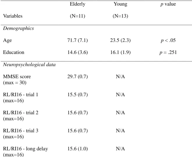
Table 1. Demographic and neuropsychological data
Variables Elderly (N=11) Young (N=13) p value Demographics Age 71.7 (7.1) 23.5 (2.3) p < .05 Education 14.6 (3.6) 16.1 (1.9) p = .251 Neuropsychological data MMSE score (max = 30) 29.7 (0.7) N/A RL/RI16 - trial 1 (max=16) 15.5 (0.7) N/A RL/RI16 - trial 2 (max=16) 15.6 (0.7) N/A RL/RI16 - trial 3 (max=16) 15.6 (0.7) N/A
RL/RI16 - long delay (max=16)
15.6 (1.0) N/A
Table 1. Demographic data for both groups, and neuropsychogical data for elderly. Standard deviations are in parenthesis. MMSE = Mini Mental State Examination; RL/RI16 = Rappel libre/Rappel indicé 16.
39
Table 2. Behavioral performance on the semantic judgment task
Abstract/low imageability Abstract/high imageability Concrete/low imageability Concrete/high imageability Global performance Reaction time Elderly 92.9 (4.0) 92.3 (4.2) 94.2 (3.8) 97.5 (2.1) 94.3 (3.5) 856.6 (97.5) Young 93.6 (6.6) 92.3 (4.1) 93.8 (3.9) 97.5 (2.8) 94.2 (2.7) 734.5 (77.7)
Table 2. Mean performance on the semantic judgment task for both groups, for each type of words. Global performance and global reaction time (in milliseconds) are also provided. The performance is given in percentage, with standard deviation in parenthesis.
40 Table 3. Cortical areas showing difference of activation between young and older adults
Regions (Brodmann’s Areas) x y z Cluster Size t-value Elderly > Young
R. posterior Middle Temporal Gyrus (BA39) 51 -64 19 908 6.85 L. Middle Temporal Gyrus (ATL) (BA21) -53 8 -35 712 6.41
L. Declive -35 -80 -19 492 6.87
R. Middle Occipital Gyrus (BA18) 47 -76 -11 356 6.44 L. posterior Middle Temporal Gyrus (BA19) -49 -76 19 187 5.87
R. Precentral Gyrus (BA6) 57 -6 29 172 7.11
R. Fusiform Gyrus (BA19) 23 -82 -13 140 5.52
L. Precentral Gyrus (BA4) -15 -26 61 124 5.66
L. Inferior Temporal Gyrus (BA20) -53 -18 -35 120 5.44 R. Middle Occipital Gyrus (BA19) 33 -76 21 114 7.90
R. Lingual Gyrus (BA17) 17 -100 -13 112 6.85
R. Precentral Gyrus (BA6) 21 -18 65 82 5.68
Young > Elderly
L. Superior Frontal Gyrus (BA6) -3 28 61 146 6.40
R. Anterior Cingulate (BA24) 5 30 13 106 6.70
L. Insula -31 18 -1 94 6.24
L. Inferior Frontal Gyrus (BA47) -45 18 -11 92 6.13
R. Cingulate Gyrus (BA32) 7 20 27 74 5.64
Peak of the greatest statistical significance (according to the t-test value) of activation differences between younger and older adults. A minimum cluster of 68 voxels and a significance statistical threshold at p<.001 corrected for multiple comparisons have been used. Talairach stereotaxic coordinates are provided.
41
Figure 1. Event-related magnetic time curves for young adults (top) and elderly (bottom).
A visual inspection on the curves allowed the selection of time windows that corresponds to the semantic cognitive process, around the N400.
42
Figure 2. Difference of activation between younger and older adults during semantic
judgment task. Brain regions showing activation differences between younger and older adults. Areas of greater activation for young adults relative to older adults are represented by red colors, while blue colors represent areas of greater activation for older adults relative to young adults. The scale illustrated the value of the t-test (for a direct contrast Young - Old). Images are in neurological orientation (left = left hemisphere). Areas of greater activation for young adults include the left inferior prefrontal cortex (L. IPC), while areas of greater activation for older adults include the bilateral temporoparietal regions (Bilat. TPR) and the left anterior temporal lobe (L. ATL).
43
Figure 3. Interactive effect age*concreteness in the left inferior prefrontal cortex. An
interactive effect age*concreteness was found in the left IPC (BA47). A small locus was found to be more activated in older adults for abstract words than for concrete words, whereas the younger adults had similar activations in this region for both categories of words.