T h e s i s s u b m i t t e d t o f u l f i l l t h e r e q u i r e m e n t s f o r t h e d e g r e e o f P h D i n B i o m e d i c a l a n d P h a r m a c e u t i c a l s c i e n c e s
2 0 1 5 - 2 0 1 6
UNIVERSITY OF LIÈGE
Faculty of Medicine
Laboratory of Medicinal Chemistry Pr. Bernard Pirotte
Center for Interdisciplinary Research on Medicines (CIRM)
Laboratory of Molecular Pharmacology Dr. Julien Hanson GIGA - Molecular Biology of Diseases
DEVELOPMENT OF PHARMACOLOGICAL
TOOLS FOR THE IDENTIFICATION OF G
PROTEIN-COUPLED RECEPTORS LIGANDS
Julie Gilissen
5
Au terme de la rédaction de ce manuscrit, il m’est agréable de remercier les nombreuses personnes qui ont participé à ces travaux de recherches et sans qui la réalisation de ceux-ci n’aurait pas été possible. Ce projet pluridisciplinaire est le fruit de nombreux échanges et collaborations.
Je souhaite tout d’abord remercier mon promoteur, le Docteur Julien Hanson, pour m’avoir transmis l’envie de me spécialiser dans la pharmacologie des récepteurs couplés aux protéines G, de m’avoir initiée aux techniques de la biologie cellulaire et moléculaire ainsi que les techniques associées au criblage de librairies. Je le remercie chaleureusement pour son soutien, sa disponibilité, son implication, son enthousiame, ses précieux conseils et ses grandes qualités scientifiques. Je lui suis reconnaissante de la confiance dont il a fait preuve à mon égard en me laissant mener à bien ce projet au sein du laboratoire de pharmacologie moléculaire.
J’exprime ma gratitude envers mon co-promoteur, le Professeur Bernard Pirotte qui m’a permis de mener à bien cette thèse de doctorat. Je le remercie de m’avoir acceuillie au sein du laboratoire de chimie pharmaceutique, d’avoir suivi ce projet et d’avoir pris le temps de relire mes écrits. Je le remercie chaleureusement pour sa confiance, sa gentillesse, son aide et ses nombreux conseils.
J’exprime toute ma reconnaissance à l’ensemble de mon jury de thèse, le Professeur André Luxen, président de mon comité, ainsi qu’au Professeur Vincent Seutin et au Docteur Jacques Piette pour leurs conseils avisés et le temps qu’ils ont consacré à l’évaluation de mon travail. Je voudrais également remercier le Docteur Laurent Provins de chez UCB Biopharma et le Professeur Graeme Milligan de l’université de Glasgow, membres extérieurs du jury, pour leur aimable participation.
Merci à tous les scientifiques travaillant aux laboratoires de chimie pharmaceutique et de pharmacologie moléculaire avec qui j’ai eu plaisir à travailler. Je remercie particulièrement Pierre Geubelle pour son aide précieuse et sa rigeur scientifique. Pierre, ce travail n’aurait pas été aussi fructueux sans ta collaboration sur le projet SUCNR1, merci pour tout. Je remercie vivement Céline Piron, notre technicienne pour son enthousiasme, son implication et ses compétences scientifiques. Ton aide en culture cellulaire et biologie moléculaire m’a permis d’avancer plus rapidement et de générer de nombreux résultats. Je remercie Nadine Dupuis pour nos nombreuses conversations enrichissantes ainsi que pour le développement du test utilisé pour la mesure du recrutement des arrestines. Enfin, je remercie vivement Monsieur Anouar Derj et mademoiselle Céline Laschet pour m’avoir fait bénéfier de leur expérience sur les techniques de western blot. Je vous remercie également pour votre temps et nos nombreux échanges.
6
Je souhaite remercier le Docteur Sébastien Dilly pour sa collaboration et son aide indispensable pour la partie concernant la modélisation moléculaire de ce projet.
Je tiens à exprimer ma reconnaissance au Docteur François Jouret pour ses nombreux conseils pour la partie in vivo de ce projet ainsi que d’avoir pris le temps de relire mes écrits. Je remercie également Madame Laurence Poma pour avoir réalisé les expériences sur les rats au sein du laboratoire de chirurgie expérimentale (GIGA-Cardiovascular Sciences).
Je tiens à exprimer ma profonde gratitude envers la Fondation Léon Fredericq (FLF), le Fonds National de la Recherche Scientifique (F.N.R.S) ainsi que l’Université de Liège pour m’avoir accordé les moyens financiers indispensables à la réalisation de ces travaux.
Un grand merci à tous mes collègues et amis pour leur soutien et les moments que nous avons partagés : Nadine, Anouar, Céline L, Céline P, Pierre, Catherine, Monique, Céline D, Gaël, Aurélie et Philippe.
Je terminerai ces remerciements en ayant une pensée particulière pour mes proches, ma famille et mes amis pour leurs encouragements, leur patience et leur écoute. Je tiens également à remercier Daniel pour sa présence, son soutien et son aide tout au long de la réalisation de ce travail.
7
Abstract
G protein-coupled receptors (GPCRs) represent the protein family most successfully targeted for treating human diseases. They couple to G proteins to mobilize second messenger pathways that lead to cellular responses and ultimately to physiological changes. However many are poorly characterized with few ligands reported or remain completely orphans. Therefore, there is a growing need for screening-compatible and sensitive assays in order to identify new ligands.
The present project aims at developing pharmacological tools to characterize the pharmacology and physiology of GPCRs. Our approach rely on i) development of receptor models and assays for the identification of ligands, ii) screening of chemical and virtual small molecules libraries and iii) analysis of structure-activity relationships study of active molecules. The project has been divided in two parts.
To set-up assays for the evaluation of GPCRs activation, we selected the understudied succinate receptor 1 (SUCNR1) that is proposed to affect cellular metabolism and pathophysiology of diseases in multiple organs. Nevertheless the receptor has never been validated as a drug target because very few ligands have been described. So, developing pharmacological tools for SUCNR1 remains of great interest in therapeutic drug discovery.
First, we have started by examining SUCNR1 signaling pathways in HEK293 cells. Our investigations have highlighted the efficient coupling to Gαi and thus the negative
modulation of intracellular cAMP levels. Consequently we have implemented an assay sensitive to cAMP variations to identify ligands able to induce SUCNR1 activation. However, an important drawback to track agonists for Gαi-coupled receptors is the
mandatory stimulation of cAMP levels. Inducers such as forskolin must be used and are sources of variations and errors. In order to avoid these artifacts we have set-up and validated a cAMP-inducer free method based on the GloSensor biosensor. This real time assay was amenable to high-throughput screening for the detection of Gαi-coupled
receptors agonists. The strategy monitoring basal cAMP levels compared to the stimulated cAMP levels allowed to decrease recording time and artifcats from forskolin use, leading to the identification of fewer false positives and unidentified false negatives.
8
Although both methods found agonists in the chemical library screened, no active new scaffolds on SUCNR1 were discovered.
We infer that this method could facilitate the study and screening of Gαi-coupled
receptors for active ligands.
Secondly, given the interesting potential of SUCNR1 for promising therapeutic advances, we have carried out the study of the receptor interaction with its natural ligand, succinate. We have optimized the previous three-dimensional model for SUCNR1 binding pocket by means of more detailed structure-activity relationships study of succinate related molecules. The study of structure-activity relationships performed by Pierre Geubelle, in parallel to this work, allowed the deduction of the structural elements required to be active on SUCNR1. Thus we have defined a pharmacophore for activity on the receptor and subsequently evaluated various cycloalkanes. With our cAMP assay, Pierre Geubelle has highlighted the (1R, 2S)-1,2-cyclopropanedicarboxylate to be able to activate SUCNR1. We confirmed the activity of this compound on SUCNR1 capacity to recruit arrestin 3 and determined the pharmacological properties of this new ligand as SUCNR1 agonist, in vitro and in vivo. To confirm our in vitro results, we have also assessed the hypertensive properties of this cyclic analogue. Intravenous addition at the dose of 0.1 mg.kg-1 in rats has been demonstrated to increase blood pressure in the same range as
succinate.
Consequently we have demonstrated that (1R, 2S)-1,2-cyclopropanedicarboxylate could be regarded as an original synthetic full agonist for SUCNR1. In addition, the pharmacophore for SUCNR1 should help to generate synthetic compounds characterized by an increased potency and/or efficacy compared to succinate.
9
Résumé
Les récepteurs couplés aux protéines G (RCPGs) constituent la plus grande famille de récepteurs membranaires intervenant dans la transduction des signaux extracellulaires. Via la régulation de messagers intracellulaires, ils sont responsables du contrôle de nombreuses fonctions physiologiques et sont la cible de nombreux médicaments commercialisés. Cependant, de nombreux RCPGs sont encore peu caractérisés avec peu de ligands décrits ou encore même orphelins. De ce fait, ils font l’objet de nombreuses études ayant pour but de déterminer leur(s) rôle(s) physiologique(s), mais également d’identifier leurs ligands afin de déterminer leur intérêt en tant que cibles pour la découverte de nouveaux médicaments. C’est pourquoi il y a un besoin croissant de disposer de tests pharmacologiques sensibles et compatibles avec le criblage de librairies afin d’identifier de nouveaux ligands.
Ce projet a pour but de développer des outils pharmacologiques pour caractériser la pharmacologie et la physiologie de tels récepteurs. Notre approche est basée sur i) le développement de modèles cellulaires et de tests pharmacologiques pour identifier des ligands, ii) le criblage de librairies chimiques et virtuelles, et iii) l’analyse des relations structure-activité des molécules actives. Le projet peut être divisé en deux parties.
Afin de développer des tests pharmacologiques et ainsi évaluer l’activation de RCPGs, nous avons sélectionné le récepteur au succinate, SUCNR1. Ce récepteur a été décrit comme affectant le métabolisme cellulaire ainsi que de nombreuses pathologies dans plusieurs organes. Il n’a toutefois jamais été validé comme cible thérapeutique. Ceci est dû au fait que peu de ligands ont été décrits. Le développement d’outils pharmacologiques pour SUCNR1 constitue donc un intérêt majeur en recherche thérapeutique.
D’abord, l’investigation des voies de signalisation de SUCNR1 surexprimé dans des cellules HEK293, nous a permis d’identifier le récepteur comme étant efficacement couplé à la protéine Gαi et donc capable de moduler négativement les taux d’AMPc
intracellulaire. Par conséquent, nous avons mis au point un test pharmacologique capable de mesurer les variations d’AMPc pour identifier des ligands qui induisent l’activation de
10
SUCNR1. Toutefois, l’identification d’agonistes de récepteurs couplés à Gαi nécessite la
stimulation préalable des taux d’AMPc par un activateur tel que la forskoline. Leur utilisation est un grand inconvénient puisqu’ils génèrent de nombreuses variations et erreurs. Dans le but de s’affranchir de cette source d’erreurs, nous avons mis au point et validé une méthode qui rend possible la mesure des taux d’AMPc de base sans nécessiter l’utilisation d’un activateur. Le test pharmacologique est basé sur un biosenseur appelé GloSensor qui permet une mesure en temps réel des taux d’AMPc et est facilement adaptable au criblage à haut débit pour détecter des agonistes pour les récepteurs couplés à Gαi. Cette stratégie a dès lors permis de diminuer le temps de lecture ainsi que
le nombre d’artéfacts liés à l’utilisation de la forskoline. En effet, la méthode sans activateur des taux d’AMPc a conduit à l’identification de moins de faux positifs mais a aussi permis de révéler des faux négatifs. Bien que les deux méthodes aient mené à l’identification d’agonistes, aucunes nouvelles structures actives sur SUCNR1 n’ont pu être découvertes.
L’intérêt de cette méthode reside dans le fait qu’elle facilitera l’étude et le criblage de récepteurs couplés à Gαi pour l’identification de ligands.
Ensuite, nous avons étudié l’interaction du récepteur avec son ligand naturel, le succinate. Nous avons optimisé le modèle tridimensionnel de la poche de liaison de SUCNR1 existant grâce à des informations obtenues lors de l’étude des relations structure-activité d’analogues du succinate menée par Pierre Geubelle. Son travail a permis de déduire les éléments structuraux importants pour l’activité sur SUCNR1. Dès lors nous avons pu en déduire un pharmacophore et nous avons investigué l’activité de cycloalcanes. Avec le système GloSensor, Pierre Geubelle a identifié un nouvel agoniste de SUCNR1, le (1R, 2S)-1,2-cyclopropanedicarboxylate. Son activité a été confirmée sur une deuxième voie de signalisation. En effet, il induit le recrutement de l’arrestine 3 via l’activation de SUCNR1. Nous avons ensuite investigué son activité in vivo et mesuré son effet hypertenseur. Le composé injecté par intraveineuse à la dose de 0.1 mg.kg-1 augmente
la pression artérielle chez le rat dans le même ordre de grandeur que le succinate. Par conséquent, nous avons identifié un agoniste plein synthétique et original pour SUCNR1. De plus, le pharmacophore devrait aider à générer de nouveaux ligands pour SUCNR1, plus puissants et/ ou efficaces par rapport au succinate.
11
Publications
Forskolin-free cAMP assay for Gi-coupled receptors. Julie Gilissen, Pierre Geubelle, Nadine
Dupuis, Bernard Pirotte, and Julien Hanson. Biochemical Pharmacology, 2015.
Insight into SUCNR1 (GPR91) structure and function. Julie, Gilissen; François, Jouret; Bernard Pirotte and Julien Hanson. Pharmacology & Therapeutics, 2016.
Manuscript in preparation
Discovery of the first synthetic agonist for SUCNR1. Pierre Geubelle, Julie Gilissen, Sébastien Dilly, Nadine Dupuis, Laurence Poma, Eric Goffin, François Jouret, Bernard Pirotte and Julien Hanson.
13
Abbreviations list
[Ca2+]i intracellular free calcium
3D three-dimensional
7TM seven transmembrane domains AA amino acid
AB antibody
AC adenylate cyclase
ALPHA amplified luminescent proximity homogeneous assay
AMP adenosine monophosphate ATP adenosine triphosphate BP blood pressure
BRET bioluminescence resonance energy transfer
CAM constitutively active mutant cAMP cyclic adenosine
3´,5´-monophosphate CCD charge-coupled device
CDS cellular dielectric spectroscopy CFP cyan fluorescent protein
CHO cells chinese hamster ovary cells CNG cyclic nucleotide-gated channel CRE cAMP-response element
CREB cAMP response element-binding protein
DAG diacylglycerol
DMSO dimethylsulphoxide
DOS diversity-oriented synthesis EC50 effective concentration
ECL extracellular loop EFC enzyme fragment complementation Emax maximum efficacy
ERKs extracellular signal-regulated kinases
FACS fluorescence-activated cell sorting
FBS fragment-based screening FLIPR fluorimetric imaging plate reader FLuc firefly luciferase
14
FP fluorescence polarization FRET fluorescence resonance energy
transfer FSK forskolin
GDIs guanine nucleotide dissociation inhibitors
GEFs guanine nucleotide exchange factors
GFP green fluorescent protein GPCRs G protein-coupled receptors GRKs G protein-coupled receptor
kinases
HCS high-content screening HEK293 cells human embryonic kidney cells HRP horseradish peroxidase HTRF homogeneous time-resolved fluorescence HTS high-throughput screening IBMX 1-methyl-3-(2-methylpropyl)-7H-purine-2,6-dione ICL intracellular loop IP inositol phosphate
IP3 inositol-1,4,5-triphosphate
JNKs c-Jun N-terminal kinases KO knockout
LTB4 leukotriene B4
LOPAC library of pharmacologically active compounds
MAPKs mitogen-activated protein kinases
MDCK madin darby canine kidney NMR nuclear magnetic resonance PCR polymerase chain reaction PDE phosphodiesterase
p-ERK1/2 phosphorylated ERK1/2 PI3Ks phosphatidylinositol 3-kinases PIP2
phosphatidylinositol-4,5-bisphosphate PKA protein kinase A PKC protein kinases C PLC-β phospholipase C-β PTX pertussis toxin RNA Ribonucleic acid
15
RGS regulators of G protein signaling RhoGAPs GTPase-activating proteins SA succinic acid
SAR structure-activity relationships SDH succinate dehydrogenase siRNA small interfering RNA
SOSA selective optimization of side activities
SPA scintillation proximity assays SREB Super conserved receptor
expressed in brain
TR-FRET time-resolved fluorescence resonance energy transfer VPR Volume pressure recording WT wild type
17
Table of contents
I. INTRODUCTION ... 23
I.1.PHARMACOLOGY OF G PROTEIN-COUPLED RECEPTORS 25
I.1.1. GPCRs general description ... 25
I.1.2 Classification of GPCRs ... 26
I.1.3. Structure of GPCRs ... 27
I.1.4. Signal transduction and regulation ... 30
I.1.4.1. Heterotrimeric G proteins ... 30
I.1.4.2. G protein-coupled receptor kinases (GRKs) ... 34
I.1.4.3. Arrestins ... 35
I.1.4.3.1. G protein-dependent mechanism ... 35
I.1.4.3.2. G protein-independent mechanism ... 39
I.1.4.4. Other mechanisms for the modulation of GPCRs signaling ... 41
I.1.5. GPCRs Ligands ... 43
I.1.5.1. Nature of ligands ... 43
I.1.5.2. Affinity and efficacy of ligands ... 43
I.1.5.3. Classification of ligands ... 45
I.1.5.4. Constitutive activity... 47
I.1.5.5. Biased ligands... 48
I.2.SUCNR1 51
I.2.1. "Deorphanization" and characterization of SUCNR1 ... 51
I.2.2. Implications in (patho)physiology ... 55
I.2.3. Ligands and binding pocket ... 56
18
I.3.METHODS FOR THE IDENTIFICATION OF LIGANDS FOR GPCRS 61
I.3.1. Interest of ligands ... 61
I.3.2. Computational approach ... 63
I.3.3. Pharmacological approach... 64
I.3.3.1. Pharmacological screenings... 65
I.3.3.1.1. Biological extract preparations ... 65
I.3.3.1.2. Libraries ... 65
I.3.3.1.3. Cell-based assays ... 68
I.3.3.1.3.1. General receptor activity measurement ... 68
I.3.3.1.3.2. G protein activity measurement ... 70
I.3.3.1.3.2.1. Ca2+ measurement ... 71
I.3.3.1.3.2.2. cAMP measurement ... 73
I.3.3.1.3.2.2.1. Competition assays for cAMP detection ... 74
I.3.3.1.3.2.2.2. Biosensors ... 79
I.3.3.1.3.3. Challenges of the quantification of Gαi activation ... 87
I.3.3.1.4. Critical parameters for screening... 89
I.3.3.2. Medicinal chemistry ... 94
II. AIM OF THE WORK ... 97
III. RESULTS AND DISCUSSION ... 103
III.1.CHARACTERIZATION OF SUCNR1 SIGNALING PATHWAYS AND DEVELOPMENT OF CELL-BASED ASSAYS 105
III.1.1. Gαi signaling pathway ... 105
III.1.1.1. The biosensor ... 105
III.1.1.2.Gαi signaling pathway activation measure ... 106
III.1.2. Gαq signaling pathway ... 110
19
III.1.2.2. Gαq signaling pathway assessment ... 112
III.1.3. Arrestins recruitment ... 113
III.1.3.1. The biosensor ... 113
III.1.3.2. Ability of SUCNR1 to recruit arrestins ... 114
III.1.4. Determination of ERK phosphorylation ... 116
III.1.5. Gα12/13 signaling pathway ... 118
III.2.SCREENING OF SUCNR1 WITH A FSK-FREE CAMP ASSAY 121
III.2.1. Real time analysis of cAMP levels modulation mediated by ... 122
SUCNR1 activation ... 122
III.2.2. Optimization of a screening protocol ... 127
III.2.3. Screening of the Sigma LOPAC1280™ library ... 129
III.2.3.1. Primary screening ... 129
III.2.3.2. Secondary screening of the selected hits ... 135
III.3.CHARACTERIZATION OF SUCCINATE BINDING SITE 141
III.3.1. In silico screening of a virtual library ... 141
III.3.1.1. Three-dimensional model of SUCNR1 ... 141
III.3.1.2. Virtual screening of ZINC database ... 143
III.3.2. Optimization of the existing receptor model ... 145
III.3.2.1. Structural elements ... 145
III.3.2.1.1. Screening of analogues ... 145
III.3.2.1.2. Pharmacophore ... 147
III.3.2.2. Binding pocket ... 149
III.3.3. Characterization of cycloalkanes activity on SUCNR1 ... 150
III.3.3.1. In vitro assays ... 150
20
III.3.4. Other ligands ... 153
IV. CONCLUSIONS AND PERSPECTIVES ... 157
V. MATERIAL AND METHODS ... 167
V.1.CHARACTERIZATION OF SUCNR1 SIGNALING PATHWAYS AND DEVELOPMENT OF CELL-BASED ASSAYS 171
V.1.1. Protocols for transfections ... 171
V.1.1.1. Transfection using lipofectamine... 171
V.1.1.2. Transfection using calcium phosphate ... 171
V.1.1.3. Transfection using Xtreme gene ... 171
V.1.2. Generation of stable cell lines ... 172
V.1.2.1. cAMP assay ... 172
V.1.2.2. Intracellular calcium mobilization assay ... 173
V.1.2.3. Arrestin complementation assay ... 173
V.1.2.4. Flow cytometry analysis ... 174
V.1.2.4.1. Non-permeabilized cells ... 174
V.1.2.4.2. Permeabilized cells ... 174
V.1.3. Immunofluorescence staining and confocal microscopy... 175
V.1.4. Cell-based assays and second messengers measurement ... 175
V.1.4.1. cAMP Assay ... 175
V.1.4.2. Intracellular calcium mobilization assay ... 176
V.1.4.3. Arrestin complementation assay ... 176
V.1.4.4. Determination of ERK phosphorylation ... 176
V.1.4.5. Determination of Rho activation ... 177
V.2.SCREENING OF SUCNR1 WITH A FSK-FREE CAMP ASSAY 179
V.2.1. Calculation of Z’ factor ... 179
21
V.2.3. Data analysis and statistical procedure ... 179
V.3.CHARACTERIZATION OF SUCCINATE BINDING SITE 181
V.3.1. In silico models ... 181
V.3.1.1. Receptor model ... 181
V.3.1.2. Docking of the ZINC database ... 181
V.3.1.3. Pharmacophore model ... 181
V.3.2. SUCNR1 mutants ... 182
V.3.2.1. Site-Directed Mutagenesis ... 182
V.3.2.2. Transient transfection of mutants ... 182
V.3.3. In vivo experiments ... 183
VI. BIBLIOGRAPHY... 185
VII. APPENDIX ... 211
VII.1.SCREENING RESULTS 213
VII.1.1 Primary screening ... 213
VII.1.2. Cherry pick for secondary screening ... 238
23
25
I.1. Pharmacology of G protein-coupled receptors
I.1.1. GPCRs general description
G protein-coupled receptors (GPCRs) belong to the largest known membrane receptors family in mammals (Fredriksson et al., 2003; Civelli et al., 2013), exhibiting seven transmembrane domains (7TM) as their characteristic feature (Strader et al., 1995). They couple to G proteins to mobilize second messenger pathways that lead to cellular responses and ultimately to physiological changes.
Since the pioneering works of Langley and Ehrlich at the beginning of the twentieth century, GPCRs became one of the most important pharmaceutical research areas (Langley, 1901; Limbird, 1996). They play an essential role in regulating various physiological functions (pain, neurotransmission, muscle contraction, insulin secretion, …) and many are implicated in human diseases, making them the most druggable gene family (Overington et al., 2006). Approximately 82 GPCRs are the direct or indirect target of 30-50% of drugs currently on the market or in clinical trials for cancer, neurodegeneration, diabetes, pain, allergy, viral infection, … (Wilson et al., 1998; Wittenberger et al., 2001; Civelli et al., 2013). In addition, some of them are expressed in therapeutically relevant tissues and may represent a source of therapeutic targets with similar potential for drug discovery as seen with known GPCRs (Wilson et al., 1998).
The completion of human genome sequence allowed the identification of 800 genes encoding GPCRs (Alexander et al., 2015). Most of these genes encode for chemosensory receptors that mediate the perception of sensory stimuli such as odors, light, sweet and bitter taste substances via specialized heterotrimeric G proteins modulating the activity of primary sensory cells (Wettschureck and Offermanns, 2005). 356 are coding for "nonsensory" GPCRs in which most are under-interrogated or poorly characterized with few physiologically relevant ligands reported, 61 are awaiting further input to be considered as deorphanized and still 60 remain completely uncharacterized (Alexander et al., 2015). Although their DNA sequences look like known GPCRs, they are initially unmatched to their activating ligands and their signaling mechanisms are unknown. They
Introduction
26
are designated as "orphan" GPCRs and are of considerable interest in enriched understanding of physiological responses. GPCRs represent an useful and important target class for therapeutic drug discovery and biochemical study (Davenport and Harmar, 2013).
The study of GPCRs is currently an intensive and exciting field of research, the 2012 Nobel Prize in chemistry was awarded jointly to Robert J. Lefkowitz and Brian K. Kobilka for studies of G-protein-coupled receptors" (NobelPrize.org, 2012). Their finding is of high interest as structure and mechanism characterization of GPCRs may help for the design of ligands and thus new drugs (Wilson et al., 1998; Jacoby et al., 2006).
I.1.2 Classification of GPCRs
GPCRs are classified into different families sharing characteristic highly conserved residues according to phylogenetic analyses and sequence homology (Fredriksson et al., 2003) as well as the various types of ligands they bind to (Foord et al., 2005).
The most recent sequence analysis proposes the clustering of human GPCRs into five groups named "GRAFS", on the basis of its five main classes : Glutamate, Rhodopsin, Adhesion, Frizzled, and Secretin (Fredriksson et al., 2003). The Rhodopsin family has the largest number of GPCRs with 719 members (284 "nonsensory" including 87 orphans (however endogenous ligand has been proposed in at least one publication for 54 of them) and 435 olfactory mostly orphans) and is divided into four α (amines, lipids, …), β (peptides), γ (opioids, chemokines,…) and δ (glycoproteins, purines) groups.
However some orphan receptors, such as Super Conserved Receptor Expressed in Brain (SREB) show closer homology to each other than to known class A GPCRs, suggesting that they may represent new families of receptors with distinct ligands. These sub-families are distributed throughout the GPCRs superfamily tree, suggesting that they will have a diverse range of functions (Wilson et al., 1998; Alexander et al., 2015).
27
Glutamate, frizzled and secretin families contain 12, 11 and 15 "deorphanized" GPCRs, respectively. Although the 26 adhesion members are orphans, endogenous ligand has been described in at least one publication for 6 of them (Alexander et al., 2015).
I.1.3. Structure of GPCRs
GPCRs share a common molecular architecture of 7TM helices connected by three extracellular loops (ECL1-3) and three intracellular loops (ICL1-3) which are involved in heterotrimeric G protein-coupling (Figure I-1) (Strader et al., 1995; Wettschureck and Offermanns, 2005).
Figure I-1 : 3D general structure of GPCRs. Blue : Amino acids (AAs) conserved between GPCRs; Red : AAs conserved between GPCRs of the Rhodopsin family (Kenakin et al., 2010).
The characteristic 7TM structure was for the first time identified for rhodopsin, in 1975. However the sequence homology between β2-adrenoceptor and bovine rhodopsin was
only revealed in the 1980s with the cloning of the adrenergic receptor. A structure-function relationship was established between the two receptors and the β2
-adrenoceptor advances are considered as a historic breakthrough that catalyzed the molecular GPCRs research (Jacoby et al., 2006). Besides, the study of the rhodopsin
Introduction
28
signaling mechanism highlighted its linkage to the G proteins. These significant information made rhodopsin the ideal model for other GPCRs investigations. However the speculation of a large family of such receptors with the 7TM arrangement as a fold characteristic was only confirmed in the following years by the successful cloning of other GPCRs (Jacoby et al., 2006).
Although GPCRs have been studied for almost a century, the first high-resolution structure (2.8 Å) of the rhodopsin was only available in 2000 (Palczewski, 2000). It allowed pharmacologists to understand GPCRs mechanisms and predict the overall folding of the 7TM (Audet and Bouvier, 2012). Methodological difficulties associated with the crystallization of transmembrane proteins resulted in a delay of many years in generating crystal structures of other GPCRs. But recent technological advances in engineering including the production and purification of membrane proteins, crystal formation and development of microfocus X-ray synchrotron technologies that deliver a microscale beam to a crystal, greatly contribute to the explosion of elucidated GPCRs structures. Notably in 2007, Brian K. Kobilka and co-workers published the first crystallographic structure of the active conformation of the β2-adrenoceptor (Figure I-2) (Rasmussen et
al., 2011).
Figure I-2 : High resolution (3.2 Å) crystal structure of β2-adrenoceptor (green) bound to an agonist (BI-167107), interacting with the three sub-units of a Gs protein (Rasmussen et al., 2011).
29
Between 2007 and 2015, 47 high-resolution structures representing 13 distinct GPCRs have been solved. 20 were cocrystallized with antagonists, 16 with inverse agonists whereas 11 were cocrystallized with agonists (Audet and Bouvier, 2012; Zhang et al., 2015b). The structural and functional models that arise from them have changed our view of the way ligands bind to GPCRs and helped to understand conformational dynamics of GPCRs. In addition, GPCRs cocrystallization with G protein or G protein-mimetics provided insights into the activation processes and conformational rearrangement of the G protein (Audet and Bouvier, 2012). These studies resulted in the current paradigm that conformational changes leading to receptor activation require ligand binding and concomitant stabilization by G protein. Agonist binding promotes the folding of an active conformation and lock the receptor in a more stable state. As a consequence the receptor interacts with a G protein and triggers reliable signaling pathways (Figure I-2) (Audet and Bouvier, 2012; Zhang et al., 2015b).
However GPCRs are highly dynamic proteins that adopt different conformational states and go through various transitions in their interactions with ligands and intracellular signaling. Although these crystal structures provide valuable information, they represent static frozen conformations of a single GPCR state (Millar and Newton, 2010). Therefore new biophysical techniques to monitor GPCR dynamic structural changes in relation to ligand activation are emerging (Millar and Newton, 2010). These ones include site-directed spin labelling of rhodopsin, substitution of putative interacting AA of a receptor with a labeled cysteine and tryptophan or even combination of two-dimensional nuclear magnetic resonance (NMR) spectroscopy and labelling lysine or methionine residues with
13C (Millar and Newton, 2010). Recent structural dynamics study of the β2-adrenoceptor
cytoplasmic domain revealed that unliganded or inverse-agonist-bound receptors exist predominantly in two inactive conformations that exchange within hundreds of microseconds. Stimulation with an agonist increases conformational heterogeneity (inactive, intermediate, and active states) and requires interaction with a G protein or an intracellular G protein mimetic to favor the active conformation (Manglik et al., 2015).
Introduction
30
I.1.4. Signal transduction and regulation
Each component of the transmembrane signaling system, the receptor, the G proteins as well as the effectors can be regulated independently by additional proteins, soluble mediators, or even at transcriptional level. Upon agonist stimulation, the receptors undergo conformational changes that promote their binding to intracellular partners. Currently, three main families of proteins interacting with GPCRs have been extensively studied : heterotrimeric G proteins, G protein-coupled receptor kinases (GRKs), and arrestins. The complex organization of GPCRs provides a huge variety of signaling pathways that allow distinct cell types to respond adequately to extracellular signals (Wettschureck and Offermanns, 2005).
I.1.4.1. Heterotrimeric G proteins
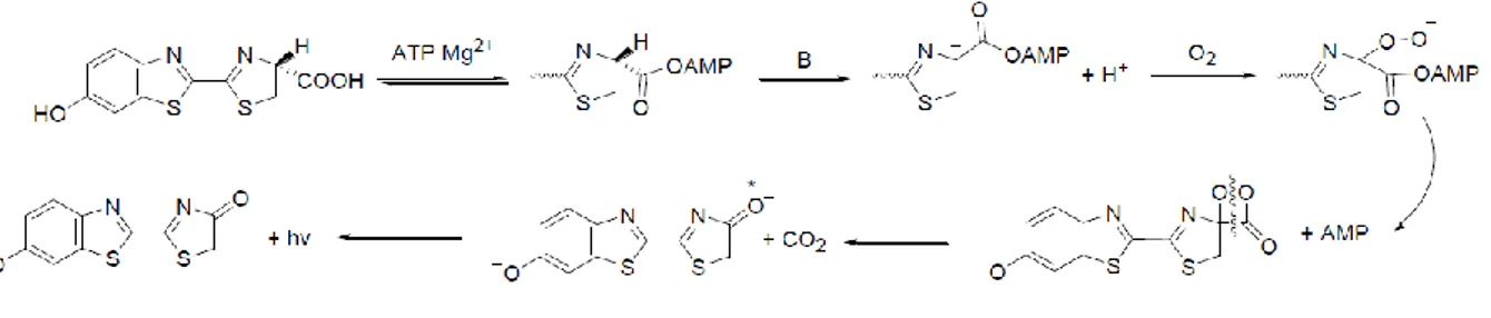
GPCRs respond to a wide range of extracellular stimuli by activating intracellular signal transduction pathways that lead to second messengers changes and/or entry of ions at the plasma membrane. These elements of the signaling cascade including cyclic adenosine 3´,5´-monophosphate (cAMP), adenylate cyclase (AC), intracellular free calcium ([Ca2+]i), inositol phosphate (IP), phospholipases, kinases and ion channels, emerged as
important effector systems during 1960s and 1970s, even before knowing the molecular nature of the receptors (Jacoby et al., 2006).
A ligand activating GPCR is able to recruit one or more heterotrimeric G proteins that undergo an activation-inactivation cycle to dynamically couple activated receptors to effectors.
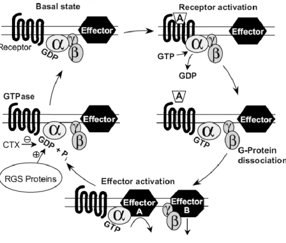
In the basal state, the heterotrimer composed of associated -complex and the GDP-bound -subunit can be recognized by an appropriate activated receptor which couple to. This coupling promotes the exchange of GDP for GTP on the -subunit. The GTP-bound -subunit dissociates from the activated receptor as well as from the -complex. Consequently these free subunits are able to modulate the activity of effectors and thus mediate cellular responses. Signaling is ended by the intrinsic GTPase activity of the G protein -subunit which hydrolysis the bound GTP to GDP, resulting in the re-association
31
of the GDP-bound -subunit and the -complex (Figure I-3). The heterotrimeric G protein re-formed can then enter a new cycle if activated receptors are present.
Thirty regulators of G protein signaling (RGS proteins) are known to contribute to the deactivation of G protein-mediated signaling by increasing GTPase activity of G protein
-subunit (Wettschureck and Offermanns, 2005; Jacoby et al., 2006). Additionally to their role in the modulation of G protein-mediated signaling kinetics, they also influence the regulation of protein localization, the intracellular trafficking as well as the receptor selectivity (Magalhaes et al., 2012).
Introduction
32
A GPCR can activate four main families of G proteins which differ in the signaling
pathways they couple to (Figure I-4) (Wettschureck and Offermanns, 2005) :
- Gs : this G protein subunit regulates positively cAMP through AC modulation. The AC
enzyme catalyzes the conversion of adenosine triphosphate (ATP) to cAMP and inorganic pyrophosphate. In addition, AC can also be regulated by βγ complex, [Ca2+]
i, and protein
kinase C (PKC).
- Gi/o : Go is mainly present in the nervous system and modulates the opening of
voltage-gated Ca2+ channels. Gi family (Gi1,2,3) activation induces AC shut down that
leads to a downregulation of cAMP levels. Gi are also able to activate a variety of
phospholipases and phosphodiesterases (PDE), and promote the opening of several ion channels.
- Gq : This family comprises five members, Gq, G11, G14, G15 and G16 that regulate
the activity of phospholipase C-β (PLC-β) isoforms, resulting in the intramembrane hydrolysis of phosphatidylinositol-4,5-bisphosphate (PIP2) to inositol-1,4,5-triphosphate
(IP3) and diacylglycerol (DAG) production. Subsequently, DAG increases the activity of
protein kinases including PKC which regulate several processes inside the cell, and IP3
triggers the release of [Ca2+]i from endoplasmic reticulum to cytoplasm.
In addition, G15 and G16 are well-known to be able to link numerous Gs-, Gi- and Gq
-GPCRs to PLC-β activation.
- G12/13 : This last family triggers the activity of Rho GTPase (RhoA, RhoB and RhoC)
mediated by GTPase-activating proteins (RhoGAPs), guanine nucleotide exchange factors (GEFs) and guanine nucleotide dissociation inhibitors (GDIs). Active Rho subsequently activates downstream effectors such as ROCK, which phosphorylates multiple cellular substrates (i.e. Rhotekin).
These G12/13 proteins are known to be involved in the formation of actomyosin-based
structures and the modulation of their contractility. They also interact with numerous proteins such as cadherins, causing the release of -catenin and induce many signaling pathways leading to various effectors. Therefore these proteins are related to cell proliferation, migration, growth and cell division (Siehler, 2007).
33
Figure I-4 : Signaling pathways of G proteins (Thomsen et al., 2005).
The second messengers modulated by G proteins, in turn, trigger activation of various signaling cascades, for instance phosphorylation events that regulate enzymes and transcription factors. A well-described example of kinases activated by an important number of GPCRs are the mitogen-activated protein kinase (MAPK) superfamily. This large group of enzymes comprises extracellular signal-regulated kinases (ERKs), c-Jun N-terminal kinases (JNKs), ERK3/4, ERK5, and p38 MAPKs. They are key components of intracellular signaling pathways that control cell proliferation, inflammation, apoptosis, ... Activation of MAPKs frequently results in their rapid translocation to the nucleus, where they phosphorylate and regulate the functional activity of various transcription factors by acting on their promoter (Gutkind, 2000; Qi and Elion, 2005). Other examples of GPCR-mediated signaling events also include phosphorylation of cytosolic factors through protein kinases A (PKA) and PKC, and also nuclear transcription factors (Brivanlou and Darnell, 2002). PKA and PKC are phosphotransferases, activated in some cell types by response to GPCR-stimulated increases in intracellular second messengers (cAMP, Ca2+
and DAG). The kinases can mediate the phosphorylation of downstream target proteins as well as the receptor itself (Ferguson, 2001).
Introduction
34
dimers, for their part, are combinations of five known isoforms of the subunit and thirteen known isoforms of the subunit. Although specific function of individual
dimers is not fully explored, each individual isoform can associate with a set of effectors and regulators. Therefore, dimers themselves regulate the activity of many signaling molecules including ion channels, phospholipases, phosphoinositide kinases like phosphatidylinositol 3-kinases (PI3Ks), particular isoforms of AC and ERKs pathways (Wettschureck and Offermanns, 2005; Jacoby et al., 2006).
I.1.4.2. G protein-coupled receptor kinases (GRKs)
Seven GRKs (GRK1 - GRK7) exist and are divided into three subfamilies : GRK 1 and 7 localized to the retina; GRK 2 and 3 which interact with the βγ complex via their pleckstrin homology domain; and membrane-associated GRK 4, 5, and 6. GRKs 2, 3, 5, and 6 are widely distributed in mammalian tissues (Pitcher et al., 1998). They are composed of three functional domains, an amino-terminal RGS homology domain, a central catalytic domain and a carboxyl-terminal membrane targeting domain (Ferguson, 2001).
Agonist-activated receptor (or activated conformation in case of constitutive activity) is able to recruit GRKs that translocate to the receptor and thus catalyze the phosphorylation of serine and threonine residues within either the third intracellular loop (such as M2 receptor and A2A receptor) (Nakata et al., 1994) or carboxyl-terminal tail
domains (such as rhodopsin and β2-adrenoceptor) (Bouvier et al., 1988). They play a
central role in the regulation of GPCRs, G protein signaling uncoupling (desensitization) and the endocytosis of GPCRs to endosomes to allow GPCR dephosphorylation and resensitization (Magalhaes et al., 2012). However receptor phosphorylation alone is insufficient to mediate the desensitization of many GPCRs which often requires arrestins binding (Magalhaes et al., 2012).
Besides their role as regulators of GPCR signaling at the level of the receptor, GRKs also regulate the activity of the G protein activity through their RGS domain (Ferguson, 2001). In addition, they are able to influence GPCR signaling via G protein-independent mechanisms. But still so, there are numerous factors, including the expression,
activation-35
deactivation, kinases and phosphatases that can regulate the phosphorylation of receptors. Indeed a receptor can be phosphorylated by different kinases at distinct sites and it is proposed that characteristic fingerprints of receptor phosphorylation exist for different cell types. This may translates to a "bar code" that directs the signaling outcome of the receptor and a different phenotype in cells (Millar and Newton, 2010).
I.1.4.3. Arrestins
I.1.4.3.1. G protein-dependent mechanism
The arrestin family consists of four isoforms (arrestin 1- arrestin 4), arrestin 1 and arrestin 4 (previously known as visual arrestin or v-arrestin and cone arrestin, respectively) are specifically localized to the visual system whereas arrestin 2 (initially termed β-adrenoceptor arrestin-1 or β-arrestin 1) and arrestin 3 (β-arrestin 2) are ubiquitously expressed (Pitcher et al., 1998). Arrestins have been widely investigated and are of important interest in physiology. For example, arrestin 2 knockout (KO) mice have altered cardiac responsiveness to β2-adrenoceptor stimulation (Conner et al., 1997) and arrestin
3 KO mice show enhanced morphine analgesia (Bohn et al., 1999) whereas both arrestins KO caused neonatal lethality in mice because of respiratory distress (Zhang et al., 2010). Crystal structures of arrestins 1 and 2 in the "basal state" indicate an intact polar core at the junction of N and C domains, which are essentially composed of antiparallel β sheets, with the C tail in close proximity to the junction (Figure I-5, inactive arrestin) (Granzin et al., 1998; Han et al., 2001). Interaction with the phosphorylated tail of activated receptor promotes the disruption of the polar core. This leads to the activation of conformational changes in arrestin through the release of the C tail that results in the increase of the accessibility of both clathrin and AP2-binding domains (Figure I-5, active arrestin) (Gurevich and Gurevich, 2003; Xiao et al., 2004).
Introduction
36
Figure I-5 : Model of inactive and active conformations of arrestin 3 (Lefkowitz et al., 2006).
Originally arrestins were discovered as molecules that bind to activated rhodopsin or β2-
adrenoceptor (Benovic et al., 1987; Lohse et al., 1990) contributing to desensitization by physically impairing further G protein binding or even inactivation by promoting internalization of the receptors. Next, many other GPCRs were reported to recruit arrestins and multiple endocytic mechanisms characterized by different kinetics emerged.
The activation of the receptor and its phosphorylation stabilizes a conformation state (higher arrestin binding affinity) that promotes the recruitment of arrestin. Thus the receptor-arrestin interaction competes with G protein interaction that results in the waning of G protein-dependent signal. Preventing primary signaling leads to a universal desensitization mechanism of GPCRs that may vary from attenuation of agonist potency (Von Zastrow and Kobilka, 1994) to termination of signaling (Magalhaes et al., 2012). This desensitization allows the protection against both acute and chronic receptor overstimulation (Ferguson, 2001; Kohout and Lefkowitz, 2003). Additionally PKA and PKC may also contribute to desensitization as they are able to phosphorylate both agonist-activated receptors and inactive receptors (Hausdorff et al., 1989).
Arrestins are also able to act as endocytic adaptors that link receptors to the clathrin-coated pits (Von Zastrow and Kobilka, 1994) and mediate further signaling (Kohout and
37
Lefkowitz, 2003; Shenoy and Lefkowitz, 2003). Arrestin can associate transiently with phosphorylated receptors, bring them to clathrin-coated pits, and then dissociate as the receptors internalize. These receptors including β2-adrenoceptor, belong to class A
(Oakley et al., 2000; Magalhaes et al., 2012) and are generally resensitized by dephosphorylation in acidic endosomal compartment (Krueger et al., 1997) to rapidly recycle back to the membrane (Figure I-6) (Oakley et al., 2000; Magalhaes et al., 2012). By contrast, arrestin dissociates very slowly from class B receptors and the complex may reside for extended periods in endosomal vesicles before being degraded by lysosomes (i.e. ETB and NTS receptors, PAR1 (Hermans et al., 1997; Trejo and Coughlin, 1999;
Bremnes et al., 2000)) or slowly recycled (i.e. NK1 receptor (Grady et al., 1995)) back to the cell surface (Figure I-6) (Oakley et al., 2000; Magalhaes et al., 2012).
Introduction
38
As a consequence GPCRs are involved in regulating receptor resensitization as well as desensitization. However kinetics of receptors endocytosis, which depend on GPCR subtype and cell line where it is expressed, are very different. Indeed receptor desensitization takes seconds to minutes whereas receptor recycling and resensitization is less efficient with a lower rate at a minimum of several minutes (Ferguson, 2001). In addition, arrestins are important for the regulation of the endocytic machinery, as they interact with protein complexes implicated in the regulation of clathrin-mediated endocytosis (Magalhaes et al., 2012).
Although arrestins represent the major mechanism of receptor internalization, several GPCRs do not require arrestins to internalize such as the secretin receptor (Walker et al., 1999), 5-HT2A (Gray et al., 2001, 2003), PAR1 (Paing et al., 2002), IP (Smyth et al., 2000)
or M2 (Vögler et al., 1998) receptors. As an alternative, they can remain at the cell surface
or internalize through a dynamin- and clathrin-dependent pathway, independent of arrestins (Zhang et al., 1996). But also, ligand-induced internalization can be dependent on GRK expression levels. For instance, BLT1 internalization has been shown after
leukotriene B4 (LTB4) stimulation in cell type that express high levels of endogenous GRK 2 or when it is overexpressed. This arrestins-independent internalization has been demonstrated to be blocked by coexpressing dominant-negative GRK 2 K220R (Chen et al., 2004). In addition, the agonist activation and the cell line in which the receptor is expressed are two important factors that regulate internalization. For example, etorphine, but not morphine, stimulates µ receptor phosphorylation and internalization in HEK293 cells whereas morphine requires GRK2 overexpression to induce phosphorylation (Zhang et al., 1998).
39
I.1.4.3.2. G protein-independent mechanism
In addition to their central role in GPCRs desensitization and internalization, a new paradigm shows GRKs and arrestins as being signal transducers themselves. Indeed, GPCRs are able to signal in a G protein-independent manner through arrestins that serve as adaptor and scaffold proteins, bringing molecules involved in signal transduction within spatial and thus functional proximity of each other (Lefkowitz and Shenoy, 2005). These proteins are described to dynamically assemble a wide range of multiprotein complexes that mediate receptor signaling, trafficking and degradation by modulating novel effectors through nonclassical pathways (Lefkowitz and Shenoy, 2005).
Arrestins scaffolding of intracellular signaling molecules was first demonstrated for the nonreceptor tyrosine kinase Src, facilitating activation of MAPK including ERK1/2 (Luttrell et al., 1999). Later, arrestins were identified to be involved in the recruitment of other nonreceptor tyrosine kinases (Lefkowitz and Shenoy, 2005) that results in the phosphorylation of various transcription factors (Morrison and Davis, 2003) and cytosolic substrates (Ge et al., 2003) through the activation of MAPK pathway signaling (Raf/MEK/ERK) (Azzi et al., 2003). Arrestin 3 for example, is described as a scaffold protein that aligns individual components of MAPK pathways in appropriate orientation that activates ERK1/2 as well as JNK3 (Ahn et al., 2004; Rajagopal et al., 2005).
In addition, GRKs play an important role in regulating ERK1/2 arrestin-mediated-signaling (Kim et al., 2005; Shenoy et al., 2006). More precisely GRK 5 and 6 isoforms favor ERK1/2 whereas GRK 2 and 3 behave as signaling inhibitors for AT1 receptor (Kim et al., 2005).
In vitro the contribution of arrestins-mediated signaling pathways can be assessed with small interfering RNA (siRNA), specific inhibitors of PKC (Ahn et al., 2004) or negative mutants of arrestins (Azzi et al., 2003).
Although arrestins- and GRKs-dependent signaling pathways are largely demonstrated in vitro, it remains essential to establish the physiologic and pathophysiologic roles of such mechanisms in vivo. For instance, AT1 receptor in the cardiovascular system was
studied by Zhai et al. by using transgenic mice unable to couple to G protein signaling, they highlighted physiologic consequences (ventricular dilatation, hypertrophy and
Introduction
40
myocardial apoptosis) that can be correlated with arrestin-mediated ERK activation (arrestin-Src-ERK signaling) (Rajagopal et al., 2005). GRKs and arrestins KO mice are also valuable tools to explore cellular responses mediated by G protein-independent signaling pathways in vivo. For example these models allow understanding the diversity of the effectiveness of β-blockers such as metoprolol. In addition to β-blocking action, metoprolol has been reported to signal through GRK 5 and arrestin 2-dependent pathway. This results in an expression of fibrotic genes increase that is responsible for cardiac fibrosis in cardiomyocytes and lead to cardiac dysfunction (Nakaya et al., 2012).
It seems likely that these G protein-independent mechanisms will lead to distinct physiological consequences due to their kinetics features. For example the initial phase of Gαq signaling is rapid and transient (inferior to 5 minutes) whereas arrestins signal in
a slower and persistent manner (superior to 20 minutes) (Ahn et al., 2004; Kim et al., 2005; Lefkowitz et al., 2006). Another difference is the G protein-dependent distribution of the activated MAPK including ERK1/2, which accumulates in the nucleus where it phosphorylates and activates various transcription factors. In contrast ERK1/2 arrestin-mediated is confined to a cytoplasmic compartment where it presumably phosphorylates a distinct set of effectors (Kim et al., 2005).
As a consequence of arrestins acting as scaffolds and signal transducers, the diversity of signaling possibilities for a single receptor are significantly increased. In addition, it suggests that arrestins are involved in many physiological and pathophysiological cellular processes including chemotaxis, metastasis, apoptosis and behavior (Lefkowitz et al., 2006).
41
I.1.4.4. Other mechanisms for the modulation of GPCRs signaling
The preponderance of GPCRs and intracellular molecules is dynamically regulated at many levels from their biosynthesis including gene transcription, translation, and post-translation through their trafficking to the cell membrane. Many intracellular proteins including chaperones facilitate GPCRs translocation to the plasma membrane and thus their cell-surface expression (Millar and Newton, 2010). Once at the cell surface, GPCRs may interact with a wide variety of accessory proteins that modulate ligand affinity and selectivity, G protein-coupling and signaling, cytoskeletal and extracellular matrix interactions, as well as receptor desensitization and internalization (Millar and Newton, 2010; Magalhaes et al., 2012). These proteins may induce GPCRs phosphorylation, acetylation, palmitoylation, ubiquitination, and myristoylation that also modify receptor functional properties (Millar and Newton, 2010). Interactions with these proteins also allow the formation of novel signal transduction complexes that alter cellular functions.
In addition, GPCRs may undergo homo- or hetero-oligomerization to induce transactivation of other receptors or signal modification (Millar and Newton, 2010). They are designated as "homomeric/heteromeric receptors" when inactive monomers become active in binding or signaling as oligomers. For example GABAB1 when expressed alone
is able to bind ligands but is non-functional. Co-expression of GABAB1 and GABAB2 leads
to the formation of a heteromeric functional complex where GABAB2 mediates G
protein-coupled signaling (Ahmad et al., 2015).
In contrast receptors that are intrinsically active as monomers but have new activities as oligomers are called "receptor homomers/heteromers" (Ferré et al., 2010). Oligomerization of GPCRs can affect receptor function either by influencing their signaling and diversifying the pharmacological responses or through "transactivation" in which oligomerization of two defective receptors is able to restore receptor functionality. For example, monomers D1 and D2 signal through Gαs and Gαi, respectively, whereas
D1/D2 heteromers trigger Gαq signaling pathway, the more used in the brain, suggesting
Introduction
42
The discovery that some GPCRs appear to function in complexes with other signal transduction and scaffolding proteins highlighted numerous additional GPCR activities and offer many novel therapeutic possibilities (Jacoby et al., 2006). For example the understanding of side effects of a drug, rimonabant, a CB1 receptor antagonist used as
a medication to aid smoking cessation is associated with appetite suppression because of CB1 receptor dimerization with appetite-stimulating OX1 receptor, also antagonized
43
I.1.5. GPCRs Ligands
I.1.5.1. Nature of ligands
The endogenous ligands for GPCRs have tremendous variations including ions, small molecules (organic odorants, amines, metabolites, ...), peptides, proteins, lipids, nucleotides, and photons. Analyses of physical properties of crystallized GPCR-ligand complexes revealed ligand properties important for crystallization propensity (an appropriate ligand improves receptor stability) such as thermal stability upon ligand binding, molecular weight between 200 and 500 kDa, easy access to the binding pocket, high affinity, sufficient solubility to remain in solution at the concentrations required to provide complete occupancy of the ligand-binding site and hydrogen bond-forming capacity (Zhang et al., 2015b).
I.1.5.2. Affinity and efficacy of ligands
To characterize a ligand-receptor pair, two fundamental and distinct parameters need to be determined : its propensity to bind to the receptor and its ability to produce a response once bound.
The capacity of a ligand to bind a receptor through electrostatic, hydrogen or Van der Walls interactions determines its affinity for one state of the receptor, resulting in the increase of the population of receptors in that conformation (Wermuth, 2008).
Ligand affinity for a protein is characterized by a dissociation constant KD (equivalent to
the ratio of the rate that the ligand leaves the surface of the protein (koff) and the rate
it approaches the protein surface (kon)), usually determined with saturation experiment
with a radioactively labeled ligand (Wermuth, 2008; Kenakin, 2014). Competition-binding assays are performed to determine IC50 (of unlabeled ligand), which represents the ligand
concentration that decreases half the radioactivity (Figure I-7). Classically labeled ligands are used to saturate all the binding sites of the receptor and evaluate the ability of an unlabeled ligand to displace it (Wermuth, 2008; Kenakin, 2014).
Introduction
44
Ki and IC50 are used to compare affinity for receptors, a lower IC50 means a higher affinity
for the receptor (Wermuth, 2008). The relation between these parameters is based on Cheng-Prusoff equation : Ki = IC50/(1+([L]/KD)) (Cheng and Prusoff, 1973).
Figure I-7 : Theorical curve of affinity of unlabeled ligand that displaces labeled bound ligand (Wermuth, 2008).
As a GPCR is a dynamic protein existing at equilibrium between numerous active and inactive states, classically the efficacy of a ligand represents its ability to displace this equilibrium. However the definition of the efficacy can be expanded to a wide variety of GPCRs behaviors (G proteins interaction, desensitization, internalization or oligomerization) as the property of a ligand to cause the receptor to change its behavior toward the host cell. The efficacy of a ligand is influenced by the coupling efficiency of the receptor pathway of interest and the receptor concentration (Kenakin, 2002; Wermuth, 2008). The comparison of efficacy of different ligands is based on their medium effective concentration, EC50 which corresponds to the ligand concentration required to
get half the maximum efficacy (Emax ; maximal response capable of being produced in a
given system) (Figure I-8). A lower EC50 means a better efficacy at lower concentrations
45
IC50 is used to define the concentration of a blocker that reduces by 50% the maximum
efficacy of the system (Wermuth, 2008).
Figure I-8 : Theorical concentration-response curves of different ligands (Wermuth, 2008).
I.1.5.3. Classification of ligands
GPCRs are subject to constant local folding and unfolding reactions occurring in different regions that expose crucial regions. Therefore a receptor can adopt numerous micro-conformations, some of them are related to active states capable of producing a pharmacological effect. A ligand interacts with a GPCR binding site, which can promote receptor folding and stabilization (Kenakin, 2002; Congreve et al., 2011).
GPCR ligands are divided into two main categories, agonists and antagonists that promote and block receptor activation, respectively.
An agonist is a ligand that preferentially binds to and stabilizes active conformations of the GPCR that results in locking the receptor in a stable state and in a biological response increase. Full agonists stimulate the maximum capacity of the receptor (an endogenous ligand is classically considered as a full agonist) whereas a partial agonist does not reach
Introduction
46
the maximum response capacity. The designation of full versus partial agonist is system-dependent, and a full agonist for one tissue or measurement might be a partial agonist in another (Jacoby et al., 2006; Congreve et al., 2011).
Antagonists can be subdivided into inverse agonists (See I.1.5.4.) and neutral antagonists, these ones block receptor activation by preventing the binding of agonists or inverse agonists to the receptor. In competitive antagonism, the binding of the agonist and antagonist is mutually exclusive, either because the agonist and antagonist compete for the same binding site or combine with spatially adjacent and overlapping binding sites (synoptic interaction) or occupy different binding sites in such way that simultaneous binding is impossible (Jacoby et al., 2006).
A non-endogenous agonist may combine either with the same site as the natural agonist (primary or orthosteric site) or with a topographically site distinct from the orthosteric one (allosteric or allotropic site), such that the receptor is able to accommodate two ligands simultaneously. An allosteric modulator is a ligand that enhances (PAM, positive allosteric modulator) or inhibits (NAM, negative allosteric modulator) the action of an orthosteric agonist or antagonist by simultaneously combining with an allosteric site on the receptor (Jacoby et al., 2006; Congreve et al., 2011). The effect of the allosteric modulator is dependent of the orthosteric ligand (Lazareno et al., 2000) but it is also able to directly activate the receptor in absence of orthosteric agonist (Langmead and Christopoulos, 2006).
47
I.1.5.4. Constitutive activity
Constitutive or intrinsic activity of a GPCR is defined by its ability to adopt constitutively or spontaneously an active conformation allowing the interaction with G proteins or regulatory proteins and subsequently triggering functional responses in the absence of ligand (Kenakin et al., 2010). This GPCR characteristic was introduced in 1989 with the description of antagonists able to inhibit basal GTPase activity with negative intrinsic activity at an endogenously expressed δ receptor (Costa and Herz, 1989). This kind of ligands that are able to specifically block constitutive activity, are nowadays commonly called inverse agonists. Further convincing data obtained with artificially generated constitutively active mutants (CAM) confirmed the concept of receptors constitutive activity (Smit et al., 2007). This GPCR ability is induced either by selective mutations in the sequence or by expression at high levels in the cell (Dunlop and Eglen, 2004). Many studies have shown that single point mutations of conserved motifs result in a loss of an intramolecular interaction that results in a constitutive activity (Parnot et al., 2002). For example, Hase et al. showed the importance of the DRY motif for Gαi activation of
the GPR20 receptor because the mutant R148A is not able to inhibit prostaglandin E2-induced cAMP formation (Hase et al., 2008). In addition, N-terminus domain might maintain the constitutive activity by acting as a tethered intramolecular ligand in MC4
receptor as well as deletion or specific mutations in the N-terminus impairs GPR61 constitutive activity (Toyooka et al., 2009).
However constitutive activity might reflect the presence of an endogenous ligand that either is difficult to remove or which is produced by the cell. For example the constitutive activity of FFA1 receptor is actually due to a permanent occupation of the receptor binding site by its endogenous FFA ligand (Ahmad et al., 2015). In addition, an inverse agonist might be responsible for the constitutive activity by stabilizing an inactive conformational state of the receptor, leading to a reduced signaling background (Toyooka et al., 2009). Finally an inverse agonist might improve GPCR expression at membrane by reducing internalization, enhancing membrane trafficking, and assisting in receptor folding (Zhang et al., 2015b).
Introduction
48
I.1.5.5. Biased ligands
Although GPCRs have been described for a long time as being able to couple to multiple G proteins and activate various signaling pathways, some ligands were identified as possessing different efficacies toward separate pathways. In recent years such ligands have been called biased ligands.
A biased ligand favors one response over another (G protein, arrestin or another direct signaling partner of the GPCR) compared with the endogenous ligand, which is considered to be neutral (Rajagopal et al., 2010b). Indeed, an agonist toward a specific signaling pathway can act, through the same receptor, as an antagonist on a different pathway in the same cell (Galandrin et al., 2007). For example, ICI118551 and propranolol ligands for β2-adrenoceptor are inverse agonists of the Gαs pathway whereas they have
partial agonist efficacy on the arrestin-dependent MAPK pathway (Azzi et al., 2003). The biased ligands concept is mechanistically explained by their capacity to selectively stabilize different receptor conformations that differ in their propensities to activate the various signaling pathways (Galandrin et al., 2007; Millar and Newton, 2010; Magalhaes et al., 2012).
This phenomenon might provide new insights in specific ligands that will restrict off-target effects of new developed drugs (Lefkowitz and Shenoy, 2005; Millar and Newton, 2010). In addition selectively blocking GPCR desensitization may improve a long-term agonist treatment or may even avoid the need to use receptor agonists (Ferguson, 2001). All currently known angiotensin receptor blockers and β-blockers act as antagonists for both signaling (McMurray and Pfeffer, 2005) but biased antagonists would provide great benefits in cardiovascular diseases by blocking the deleterious effects of chronic G protein signaling and simultaneously engage cytoprotective and anti-apoptotic arrestin signaling pathways (Lefkowitz et al., 2006).
However selective ligands might complicate the choice of an assay for a screening campaign which should measure the appropriate intracellular signal involved in desired phenotypic response of a cell for a disease state or pathophysiology (Millar and Newton, 2010). In addition, potential therapeutic compounds identified by high-throughput screening (HTS) based on a single signaling pathway will require deeper pharmacological investigations (Galandrin et al., 2007).
51
I.2. SUCNR1
I.2.1. "Deorphanization" and characterization of SUCNR1
Succinate receptor 1 or SUCNR1 (Figure I-9) (Davenport et al., 2013) was first spotted in a megacaryocytic cell line in 1995 and called "P2U2", a name coined for its homology
with the purinergic receptor P2Y2, known as P2U at that time (Gonzalez et al., 2004).
SUCNR1 gene was later re-discovered as GPR91 in 2001 on human chromosome 3q24-3q25 using an expressed sequence tag data mining strategy (Wittenberger et al., 2001).
Figure I-9 : Snake plot of SUCNR1. Blue : AAs conserved within rhodopsin family; Red : disulphide bridges; Green : V6.42 may exclude the formation of an ionic lock between E6.32 and the DRY motif; Yellow
: glycosylation sites; Brown : phosphorylation site; Purple : AAs involved in the interaction SUCNR1-succinate (Gilissen et al., 2016).
Introduction
52
It encodes a protein of 330 AAs which shares a high degree of homology between human and mouse (68%) with exception of the C-terminus, which is 12 AAs shorter in rodents (Wittenberger et al., 2001; Ariza et al., 2012).
SUCNR1 belongs to the δ group of rhodopsin-like GPCRs family (Fredriksson et al., 2003) and was initially viewed as a purinergic receptor due to its high sequence homology with P2Y receptors (29% with P2Y1 (Wittenberger et al., 2001)). P2Y family was described as a
local gene amplification and genes encoding for these receptors were classified according to their chromosomal localization in two subgroups, "a" constituted of genes present on chromosome 3q24 and "b" are genes clustered on one hand to chromosome 11q13.5 and on the other hand on chromosome 3q24-25.1. These two subgroups are composed of nucleotide-receptors whereas "n" represent the related non-nucleotide receptors (Figure I-10) (Wittenberger et al., 2002).
Although it was predicted to bind purinergic ligands (Joost and Methner, 2002; Wittenberger et al., 2002; Fredriksson et al., 2003), SUCNR1 has been paired by He et al. with a molecule not even remotely similar to purines, i.e. succinate (He et al., 2004). Interestingly GPR99 (homology of 33%), the closest homologue of SUCNR1 (Wittenberger et al., 2002) also has a citric acid cycle intermediate as a natural ligand, α-ketoglutarate (Wittenberger et al., 2002; He et al., 2004).