Université de Montréal
Inhibition of the lactic acid transporters MCT1 and MCT4
as an underlying mechanism for drug-induced myopathy
par Yat Hei (Henry) Leung
Faculté de pharmacie
Thèse présentée
en vue de l’obtention du grade de doctorat en Sciences Pharmaceutiques
option Pharmacologie
Décembre, 2017
Résumé
Les myopathies induites par les médicaments représentent un effet secondaire sérieux causé par plusieurs médicaments. Ces symptômes musculaires varient de myalgies légères avec ou sans élévation de créatine kinase, faiblesse musculaire, myosite, jusqu’à de rares rhabdomyolyses potentiellement mortelles. Bien que les myalgies légères soient tolérables, les myopathies chroniques affectent la qualité de vie des patients, requérant souvent la cessation d’une thérapie efficace.
Le mécanisme sous-jacent à ces myotoxicités causées par les médicaments est connu pour certains composés, mais demeure obscur pour plusieurs (ex. statines). Les statines constituent une thérapie efficace pour la diminution du cholestérol, mais elles sont reconnues pour causer ces effets secondaires. De nombreux facteurs augmentant les concentrations plasmatiques de statines (ex. doses élevées, interactions médicamenteuses, polymorphismes génétiques) semblent être liés à une fréquence de myotoxicité plus élevée. Conséquemment, le métabolisme et le transport des médicaments, contrôlant l’absorption globale, la distribution et l’élimination, peuvent devenir importants. Cependant, ces facteurs peuvent seulement expliquer partiellement les désordres musculaires observés.
Même si plusieurs mécanismes sont proposés pour les myotoxicités induites par les statines, le mécanisme exact responsable de cet effet est controversé, puisque les études rapportent des résultats contradictoires. Puisque l’exercice semble exacerber les douleurs musculaires chez les patients prenant des statines, l’hypothèse derrière ce projet est que le transport de l’acide L-lactique par les transporteurs de monocarboxylates serait impliqué dans le développement des myotoxicités. Puisque l’acide L-lactique est l’un des sous-produits
majeurs résultant de l’activité physique, son élimination efficace des cellules musculaires est essentielle. L’administration de médicaments inhibant compétitivement ces transporteurs pourrait mener à une perturbation de l’homéostasie de l’acide L-lactique et à des désordres musculaires.
L’objectif du premier volet de cette étude était d’évaluer le potentiel d’inhibition des médicaments acides sur le transport d’acide L-lactique en utilisant les lignées cellulaires cancéreuses Hs578T et MDA-MB-231 exprimant sélectivement MCT1 ou MCT4, respectivement. Ces lignées cellulaires ont permis la caractérisation des transporteurs avec la détermination de leurs paramètres cinétiques et d’inhibition. Le but principal du deuxième volet de cette étude était de confirmer le potentiel d’inhibition de l’atorvastatine, simvastatine, rosuvastatine et loratadine sur le transport d’acide L-lactique dans un cadre plus physiologique en utilisant des cellules musculaires squelettiques primaires humaines (SkMC). L’objectif global de ce projet de doctorat était de mieux comprendre les mécanismes derrière certaines myopathies induites par les médicaments, plus spécifiquement celles induites par les statines et la loratadine, en étudiant les transporteurs de monocarboxylates impliqués dans le transport d’acide L-lactique et l’homéostasie du pH dans le muscle.
La loratadine et l’atorvastatine ont démontré le meilleur potentiel d’inhibition de l’efflux d’acide L-lactique dans les lignées cellulaires, une observation confirmée dans les SkMC. Cette inhibition pourrait causer une accumulation intracellulaire d’acide L-lactique menant à une acidification et à des désordres musculaires.
De futures études dans des modèles in vivo sont requises pour confirmer l’impact physiologique de nos résultats dans un cadre clinique. Ces données permettraient une
meilleure compréhension des myopathies induites par les statines et la loratadine et permettront ainsi de prévenir leur occurrence en optimisant les stratégies thérapeutiques.
Mots-clés : Acide lactique, transporteurs de monocarboxylates, statines, transporteurs de médicaments, myopathie induite par les médicaments, muscle, effets indésirables, loratadine, cholestérol.
Abstract
Drug-induced myopathy is a serious side effect caused by various widely-administered medications. These muscle-related symptoms range from mild myalgia with or without creatine kinase increase, muscle weakness, myositis, to rare life-threatening rhabdomyolysis. While mild myalgias can be tolerable, chronic myopathies can affect the patients' quality of life, frequently requiring the cessation of an effective drug.
The underlying mechanism of these drug-induced myotoxicities is known for some drugs but remains unclear for most (e.g. statins). Statins constitute an effective cholesterol-lowering therapy, but they are known to cause these adverse drug reactions. Various factors
increasing statin plasma levels (e.g. high doses, drug-drug interactions, genetic polymorphisms) seem to be linked with a higher occurrence of myotoxicity. Consequently, systemic drug metabolism and transport, controlling overall absorption, distribution and elimination, can become important. However, these factors only partly explain the observed muscular disorders.
Although there are several proposed mechanisms for statin-induced myotoxicity, the exact mechanism responsible for this effect is still debated with studies reporting conflicting results. Since exercise seems to exacerbate muscle pain in patients under statin treatment, the premise of this project is that L-lactic acid transport via the monocarboxylate transporters is involved in the development of drug-induced myopathy. Since lactic acid is one of the major byproducts resulting from physical activity, its efficient removal from the muscle cells is essential. Therefore, the administration of drugs competitively inhibiting those transporters may potentially lead to perturbation of L-lactic acid homeostasis and muscular disorders.
The aim of the first part of this study was to assess the inhibitory potential of acidic drugs on L-lactic acid transport using breast cancer cell lines Hs578T and MDA-MB-231, which selectively express MCT1 or MCT4, respectively. These cell lines allowed transporter characterization with the determination of their kinetic parameters and inhibition. The main objective of the second part of this study was to confirm the inhibitory potentials of atorvastatin, simvastatin, rosuvastatin and loratadine on L-lactic acid transport in a more physiological setting using primary human skeletal muscle cells (SkMC). The overall goal of this doctoral project was to better understand the mechanisms behind certain drug-induced myopathies, more specifically those induced by statins and loratadine, by studying monocarboxylate transporters involved in lactic acid transport and pH homeostasis in the muscle.
Loratadine and atorvastatin demonstrated the greatest potency for inhibition of L-lactic acid efflux first in cancer cell lines, an observation confirmed in SkMC. This inhibition may cause an accumulation of intracellular L-lactic acid leading to acidification and muscular disorders.
Further studies with in vivo models are required to confirm the physiological impact of our findings in a clinical setting. These data will help understand statin- and loratadine-induced myopathy and prevent its occurrence by optimizing treatment strategies.
Keywords: Lactic acid, monocarboxylate transporters, statins, drug transporters, drug-induced myopathy, muscle, adverse events, loratadine, cholesterol.
Table of contents
Résumé ... i
Abstract...iv
Table of contents ...vi
List of tables ...ix
List of figures ... x
List of abbreviations ...xi
Acknowledgements ... xiv
Chapter 1 – Introduction ... 1
1. Introduction ... 2
1.1 Adverse drug reaction ... 3
1.1.1 Drug-induced myopathy ... 4
1.2 Drugs that can induce myopathy ... 5
1.2.1 Statins... 6
1.2.1.1 Cardiovascular disease: risk factors, hypercholesterolemia and treatments ... 6
1.2.1.2 Statin characteristics ... 10
1.2.3 Other drugs that can induce myopathy ... 17
1.2.3.1 Loratadine ... 20
1.3 Mechanisms of drug-induced myotoxicities ... 22
1.3.1 Depletion of cholesterol, isoprenoids and coenzyme Q10... 23
1.3.2 Disturbed calcium metabolism ... 24
1.3.3 Autoimmune ... 24
1.3.4 Drug-drug interactions ... 25
1.3.5 Genetic polymorphisms ... 26
1.3.6 Inhibition of lactic acid efflux by MCTs and drug-induced myopathy ... 26
1.4 Drug transporters ... 30 1.4.1 ABC transporters ... 31 1.4.1.1 ABCB1 ... 32 1.4.1.2 ABCC ... 33 1.4.1.3 ABCG2 ... 33 1.4.2 SLC transporters ... 34 1.4.2.1 SLCO transporters ... 34 1.4.2.1.1 SLCO1B1...35 1.4.2.1.2 SLCO1B3...36 1.4.2.1.3 SLCO2B1...36 1.4.2.2 SLC22 transporters ... 37 1.4.2.3 Monocarboxylate transporters ... 37 1.4.2.3.1 MCT1...41 1.4.2.3.2 MCT4...44 1.4.2.3.3 Other MCT...45 1.4.3 Statin transporters ... 46
1.4.3.1 Intestinal and hepatic transporters ... 46
1.4.3.2 Muscular transporters ... 47
1.5 Rationale, hypothesis and objectives ... 48
Chapter 2- Articles ... 51
2.1 Article 1: Effects of a series of acidic drugs on L-lactic acid transport by the monocarboxylate transporters MCT1 and MCT4 ... 52
2.1.1 Introduction ... 52
2.1.2 Objectives ... 53
2.1.3 Article ... 54
2.1.4 Discussion ... 87
2.1.5 Author contributions ... 88
2.2 Article 2: Statins and loratadine-induced muscle pain in human skeletal muscle cells ... 89
2.2.2 Objectives ... 90
2.2.3 Article ... 91
2.2.4 Discussion ... 127
2.2.5 Author contributions ... 128
Chapter 3 - Discussion and Conclusion ... 129
3.1 General discussion ... 130
3.2 Conclusion ... 139
List of tables
Table I. The pharmacodynamic properties of statins ... 13
Table II. The physicochemical and pharmacological properties of statins ... 14
Table III. Potential myopathy-inducing drugs ... 19
Table IV. Examples of H1-antihistamine from different generations ... 21
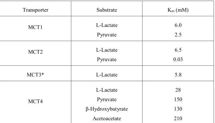
Table V. MCT1/4 substrates and inhibitors ... 40
Table VI. Kinetic parameters of human MCT ... 41
Table VII. MCT1 and MCT4 Km values from different studies... 133
List of figures
Figure 1. Bioabsorption pathway of cholesterol from diet ... 8
Figure 2. Cholesterol homeostasis ... 9
Figure 3. Biosynthesis pathway of cholesterol and its pharmaceutical inhibitors ... 12
Figure 4. The structures of the statins that have been evaluated ... 15
Figure 5. Loratadine structure ... 20
Figure 6. Glycolysis pathway. ... 29
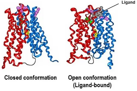
Figure 7. Protein structure of MCT1 under two conformational states ... 39
List of abbreviations
ABC: ATP-binding cassette transporters ACE: Angiotensin-converting enzyme
ACEi: Angiotensin-converting enzyme inhibitors ADR: Adverse drug reaction
AP1/2: Activator protein 1/2
ARB: Angiotensin-II receptor blockers ATP: Adenosine triphosphate
BCRP: Breast cancer resistance protein CLint: Intrinsic clearance
CNT: Concentrative nucleoside transporter CVD: Cardiovascular disease
CK: Creatine phosphokinase CYP450: Cytochrome P450 DNA: Deoxyribonucleic acid
ENT: Equilibrative nucleoside transporter GI: Gastrointestinal
GWAS: Genome-wide association study HDL: High-density lipoprotein
HDL-C: High-density lipoprotein cholesterol HIF-1α: Hypoxia-inducible factor 1α
HMG-CoA: 3-hydroxy-3-methylglutaryl coenzyme A HMGCR: HMG-CoA reductase
HPLC: High performance liquid chromatography IGF-I: Insulin-like growth factor I
Ki: Inhibition constant
Km: Michaelis-Menten constant
LDL: Low-density lipoprotein
logD: Log10(coefficient of distribution)
logP: Log10(coefficient of partition)
MCT: Monocarboxylate transporter MDR: Multidrug resistance protein mRNA: Messenger ribonucleic acid
MRP: Multidrug resistance-associated protein
NFκB: Nuclear factor kappa-light-chain-enhancer of activated B cells NSAID: Non-steroidal anti-inflammatory drug
OAT: Organic anion transporter
OATP: Organic anion transporter polypeptide OCT: Organic cation transporter
OCTN: Organic cation/carnitine transporter PEPT: Peptide transporter
P-gp: P-glycoprotein
RCT: Randomized controlled trial RR: Relative risk
S.D.: Standard deviation SkMC: Skeletal muscle cells SLC: Solute carrier
SMCT: Sodium-coupled monocarboxylate transporter SNP: Single nucleotide polymorphism
SP1: Stimulating protein 1 TC: Total cholesterol TG: Triglycerides
ULN: Upper limit of normal USF: Upstream stimulatory factors UV: Ultraviolet
To my family and friends, Whose support got me through the daily struggles
Acknowledgements
I would like to thank Dr Jacques Turgeon for accepting me in his lab for my masters' degree. At that point, I just wanted to get higher education to get a better quality of life. It was Dr Turgeon who convinced me that I can achieve more and be the best in whatever field of study I decided to pursue in. Thank you for giving me the opportunity to fast-track my project to a doctorate degree. To follow that, I would like to thank my co-director Dr Véronique Michaud for taking over the supervision of my project and supporting me, when Dr Turgeon had less time due to his administrative duty. Thank you to both of my directors for believing in me and having given me the opportunity to grow as an independent researcher.
I would like to thank my committee members for watching over my development as a scientist and giving me their advice and opinions on my project. I would like to thank the Faculty of Pharmacy for the financial and administrative support they provided throughout my studies.
Thank you to my evaluation committee for taking the time to read and give their comments on my thesis.
Thank you to the FRQS for their financial support during the majority of my studies, so that I can focus all my energy on my research without worrying too much about my finances.
Throughout my years in the lab, many people have not only helped me for my experiments, but also making my time there an overall good experience. The different lab members that I would like to acknowledge are, in order of appearance: Catherine Armstrong, François Bélanger, Jennifer Lu, Fleur Gaudette, Jade Huguet, Alexia Grangeon, Liliam Moya, Marie-Ève Papillon, Sophie Gravel, Nour Ghazal, Roxanne Pelletier, Sarah Maximos, Michel Chamoun, Charles Homsi, Valérie Clermont, and various summer students. I would also like to thank the other students of the 8th floor of CRCHUM who brought me some good laughs over the past few years.
Finally, I thank my family and friends for being there throughout this journey and supporting my career choice.
1. Introduction
Muscle pain and weakness, defined as myopathy, can be associated with drug administration. Drug-induced myopathy is common with widely-used medications.1 However,
there was an increase of these drug-associated toxicities since the introduction of HMG-CoA reductase inhibitors (statins) for lipid management in patients suffering from cardiovascular disease.2
Statin treatment is highly prescribed for its effectiveness in decreasing low-density lipoprotein cholesterol (LDL-C) plasma levels.3 Statins are well tolerated but the muscle
symptoms associated with their use can limit adhesion to treatment or even lead to drug discontinuation.4 The definitive mechanism of statin-induced muscle disorders is still not
known. High doses and the presence of drug-drug interactions or genetic polymorphisms increasing statin plasma levels seem to be linked with a higher occurrence of myotoxicity.5, 6
Consequently, systemic drug metabolism and transport, controlling overall absorption, distribution and elimination, can become important.7 However, these factors only partly
explain the observed muscular disorders, since only 10-15% of patients under statin treatment are affected by myotoxicity.
Therefore, it has been postulated that the tissue-specific local (muscle) drug absorption facilitated by membrane drug transporters is determinant for the occurrence of muscular events. Relevant drug transporters affecting systemic concentrations, and particularly those that can affect local concentrations, will be discussed below.
Exercise seems to exacerbate muscle pain in patients under statin treatment who are physically active.8, 9 Since lactic acid is one of the major byproducts resulting from physical
activity, its efficient removal from the muscle cells is essential.10 Thus, in this doctoral project,
we will attempt to better understand the mechanisms behind certain drug-induced myopathies, more specifically those induced by statins and loratadine (over-the-counter antihistamine commonly used to relieve allergies) by studying monocarboxylate transporters (MCT) involved in lactic acid transport and pH homeostasis in the muscle. Our work will be focused on the interactions between different drugs and these MCT in different in vitro models.
1.1 Adverse drug reaction
Adverse drug reactions (ADR) are a substantial public health concern. In the 2014 report from the Center for Disease Control and Prevention, it was found that unintentional injuries were the 4th cause of death in the United States.11 These included unintentional motor
transport-related injuries, unintentional poisonings, firearms and falls. The ADR-related death rate has been increasing over the years, and in 2011, it has actually surpassed the motor vehicle traffic-related injuries.11
Results from a meta-analysis showed that adverse reactions (including non-serious, serious, and fatal reactions) related to a medication significantly affect around 15.1% of the hospitalized patients.12 Since it is estimated that 6.5% of hospital admissions are related to
ADR, it is important to comprehend the underlying mechanism of these adverse events.13
Many factors can contribute to this situation, such as the aging population, which is subject to comorbidities, polypharmarcy to treat these conditions, drug-drug interactions, and the interindividual genetic variability modulating the pharmacokinetics and pharmacodynamics of drugs inside the organism.14, 15
The subpopulation that is most affected by these adverse effects are the 10 million Canadians and the 107 million Americans suffering from cardiovascular diseases (CVD).16
These pathologies are often associated with a great number of comorbidities which require the administration of a multitude of medications.17 Treatments for CVD comprise various
therapeutic approaches, including healthy diet and exercise, but they rely mainly on drugs that can effectively manage dyslipidemia.4 The treatment of cardiovascular diseases and their
comorbidities is associated with a significant interindividual variability, where some patients are resistant to the treatments, while others are over-sensitive to some drugs. Therefore, the management of therapeutics can become complex and would require various dose adjustments, increasing or decreasing depending on the medications. In 2001, there was raised awareness on the potential toxicity associated with the use of lipid-lowering agents, since cerivastatin was withdrawn from the market.4 Cerivastatin was found to be associated with
severe muscle toxicity and rhabdomyolysis causing 52 deaths.4 Nevertheless, these cases were
caused by higher doses or having it used concomitantly with gemfibrozil (same drug-metabolizing enzymes; CYP2C8), increasing plasma levels of cerivastatin.18
1.1.1 Drug-induced myopathy
Myopathy accounts for approximately 20% of common reasons for general practice consultations.19, 20 Muscle pain is not uncommon with widely-used medications.21 Since many
drugs can cause musculoskeletal symptoms, patients presenting with these drug-induced myopathies should always be classified by differential diagnosis in order to separate them from the rest of the musculoskeletal disorders.20 These muscle injuries can be produced in
several different ways, such as direct toxicity (main cause), which is often dose-dependent, or indirect muscle-damaging effects such as electrolyte disturbances, excessive energy
requirements, inadequate delivery of energy or oxygen, or via immunological reaction.20
Drug-induced myotoxicities can be classified by the absence or presence of muscle pain, including asymptomatic creatine phosphokinase (CK) elevation, mild to severe myalgias, cramps, exercise intolerance, muscle weakness, severe myositis, and rhabdomyolysis.21
While mild myalgias are relatively tolerable, chronic myopathies can affect quality of life. Therefore, an early recognition of these ADR is really important for patients as most of these effects are partially or completely reversible with a dose adjustment or drug substitution.22 However, many of these drug-induced myopathies are observed in the context
of a drug-drug interaction, which can complicate their diagnosis since they are not always related to one single agent.22 Moreover, milder symptoms such as myalgia and muscular
fatigue are not frequently reported by physicians, complicating the estimation of the actual incidence of these adverse events.6
Drug-induced myotoxicities are rare forms of ADR. The mechanisms by which they are caused are still relatively unknown. Some classes of drugs are more frequently associated with these adverse drug events, such as antifungal agents, antimalarials, antiviral agents, cardiovascular agents, corticosteroids, immunosuppressants and lipid-lowering agents (statins and fibrates).1, 23, 24 In this doctoral thesis, we will be focusing mainly on the effects of statin,
loratadine and other acidic drugs administration, to determine if myopathies observed with those drugs are generated through the same mechanisms.
1.2 Drugs that can induce myopathy
As previously stated, there are many drug classes that can cause myotoxicity, but this project will be focused on statin-induced myopathy.
1.2.1 Statins
1.2.1.1 Cardiovascular disease: risk factors, hypercholesterolemia and
treatments
Coronary heart disease is the leading cause of death in the world.11 About 610 000
Americans die of heart disease every year, which represents approximately 1 in 4 deaths. On top of having the highest mortality rate, CVD represents a major economic burden for the health care system, with an approximate $207 billion yearly expense (services, medication, loss of productivity).11, 25
High blood pressure, smoking and high levels of LDL-C constitute the three main risk factors for the development of CVD.26, 27 In fact, about 33.5 million adults (16.2%) have high
serum cholesterol levels (>240 mg/dL) in the United States.28 It was also reported by the
American Heart Association that less than half of the patients who should take lipid-lowering treatments actually adhere to their therapy.28 Other contributing risk factors would be diabetes,
obesity, poor diet, physical inactivity and excessive alcohol intake.25 Even though CVD
encompass a wide spectrum of diseases, this project will address mainly drugs used to manage LDL-C levels.
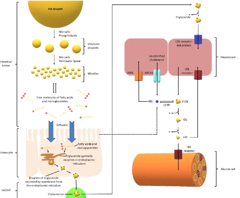
Cholesterol is either absorbed from the diet or synthesized endogenously in small quantities.29The average human consumes around 70-100 g of lipids every day, most of which
are triglycerides. Pancreatic lipase is implicated in fat digestion in the small intestine and acts on the surface of lipid droplets.29 Fat absorption is enhanced by its emulsification, which is
obtained via mechanical disaggregation and emulsifying agents, such as phospholipids and biliary salts (amphipathic molecules derived from cholesterol).29 Fatty acids and
monoglycerides are found in micelles and are absorbed by enterocytes.29 However, those two
compounds are used in cells to form triglycerides, maintaining a concentration gradient. Chylomicrons are formed inside of the cell and are composed of triglycerides, phospholipids, cholesterol and liposoluble vitamins.29 Chylomicrons and very low-density lipoprotein
(VLDL; produced from the liver) deliver triglycerides to cells throughout the body by having these triglycerides stripped by lipoprotein lipase. After the loss of these particles, VLDL becomes the denser LDL. LDL transports cholesterol to cells, which require cholesterol to function, by binding it with a specific LDL receptor. The high-density lipoprotein (HDL) precursor (synthesized in the liver and small intestine) collects excess cholesterol to form mature HDL and is brought back to the liver (reverse cholesterol transport).29
Figure 1. Bioabsorption pathway of cholesterol from diet
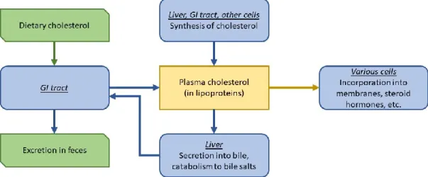
Cholesterol is an important precursor in many biological structures, such as plasma membranes, biliary salts and steroid hormones.29 However, high concentrations of cholesterol
cause atherosclerosis, which leads to several CVD, such as myocardial infarction and cerebral vascular accidents.29 Most cells use plasma cholesterol for their biological functions, whereas
only hepatic and intestinal cells release cholesterol into the blood stream. Cholesterol is secreted in the bile by the liver, where part of it is reabsorbed as dietary cholesterol, and the other part is eliminated in the feces.29 The liver also metabolizes a substantial amount of
cholesterol into biliary salts, which are components of the bile helping with cholesterol absorption. Cholesterol levels are dictated by the liver, where high cholesterol concentrations inhibit the enzyme HMG-CoA reductase responsible for its own synthesis in a retrocontrolled manner. This inhibition is highly variable between individuals.29
Figure 2. Cholesterol homeostasis
Figure adapted from Vander’s Human Physiology- The Mechanisms of Body Function (2008)29
Recently, there is a novel drug class that targets the proprotein convertase subtilisin/kexin type 9 (PCSK9).31 When PCSK9-bound LDL receptor binds to LDL, this
complex is ingested and degraded. Therefore, PCSK9 inhibition can block its ability to cause LDL receptor degradation and lower blood LDL-particle concentrations. However, most cholesterol lowering drugs target either biliary absorption (ezetimibe) or the hepatic cholesterol synthesis enzyme HMG-CoA reductase (statins).29
A meta-analysis regrouping 27 statin trials in patients at low risk of major vascular events found that the risk of these events decreased by 11 per 1000 over 5 years following every 1.0 mmol/L reduction in LDL-C levels.32 This reduction of risk justifies the use of
statins in lipid management. It was reported that patients using statins see their risk of heart attack decrease from 20 to 50%.32
An updated guideline from the US Preventive Services Task Force published in 2016 recommends the use of statins in patients from 40 to 75 years old who present at least one CVD risk factor without history of a previous cardiovascular event. Patients eligible for primary prevention statin therapy have a calculated 10-year CVD event of at least 10%, which is based on age, sex, race, cholesterol levels, systolic blood pressure, antihypertension treatment, diabetes and smoking status.33 In the case of secondary prevention, patients already
experienced a cardiovascular event and statins are prescribed to lower the risk of subsequent events.34, 35
1.2.1.2 Statin characteristics
Statins, 3-hydroxy-3-methylglutaryl coenzyme A (HMG-CoA) reductase inhibitors, are the first line therapeutics for cardiovascular protection with well-demonstrated advantages.4
Statins and other lipid-lowering drugs form the most prescribed class of drugs in the United States (255.4 million prescriptions in 2010) due to their capacity to lower the cardiovascular mortality and morbidity.36 In 2012, there were 38 million statin prescriptions in Canada
alone.37 The efficacy of statins is largely explained by their ability to diminish the plasma
levels of LDL by inhibiting competitively the HMG-CoA reductase while increasing the level of HDL, and sequentially lowering the incidence of clinical cardiovascular endpoints.38 LDL
transports cholesterol to cells, whereas HDL extracts excess cholesterol from tissues.29 The
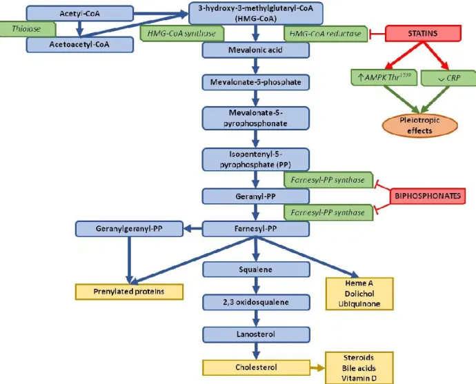
biosynthesis of cholesterol and where statins act (rate limiting step of the pathway) are presented in Figure 3. Statins also have other pleiotropic effects, like anti-inflammatory effects which can help treat the comorbidities associated with cardiovascular diseases, such as atherosclerosis and coronary heart disease.39
Figure 3. Biosynthesis pathway of cholesterol and its pharmaceutical inhibitors Figure adapted from Du Souich et al. (2017)40 and Mabuchi et al. (2005)41
Statins can inhibit HMG-CoA reductase by competing with HMG-CoA for the binding site. Crystallization studies have found that statins have the 3-hydroxy-3-methylglutaryl group in common, which is the part of the molecule interacting with the enzyme.42
Each statin has different physicochemical, pharmacokinetic and pharmacodynamic properties, as shown in Tables I and II. Cerivastatin and simvastatin are among the most lipophilic statins, while rosuvastatin and pravastatin represent the most hydrophilic. It was
found in the literature that there are fewer side effects associated with hydrophilic statins compared to lipophilic ones.23, 43 The risk/benefit profile of each statin is determined by these
differences in their characteristics.
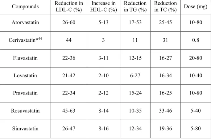
Table I. The pharmacodynamic properties of statins Compounds Reduction in LDL-C (%) Increase in HDL-C (%) Reduction in TG (%) Reduction in TC (%) Dose (mg) Atorvastatin 26-60 5-13 17-53 25-45 10-80 Cerivastatin*44 44 3 11 31 0.8 Fluvastatin 22-36 3-11 12-15 16-27 20-80 Lovastatin 21-42 2-10 6-27 16-34 10-40 Pravastatin 22-34 2-12 15-24 16-25 10-80 Rosuvastatin 45-63 8-14 10-35 33-46 5-40 Simvastatin 26-47 8-16 12-34 19-36 5-80
Table adapted from Vaughan et al., 200438
* Pharmacodynamic properties of cerivastatin were found in a different source since it has been removed from the market. (Stein et al., 1999)44
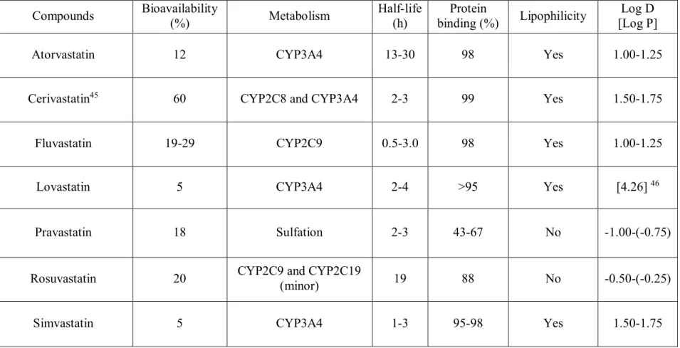
Table II. The physicochemical and pharmacological properties of statins
Compounds Bioavailability (%) Metabolism Half-life (h) binding (%) Protein Lipophilicity [Log P] Log D
Atorvastatin 12 CYP3A4 13-30 98 Yes 1.00-1.25
Cerivastatin45 60 CYP2C8 and CYP3A4 2-3 99 Yes 1.50-1.75
Fluvastatin 19-29 CYP2C9 0.5-3.0 98 Yes 1.00-1.25
Lovastatin 5 CYP3A4 2-4 >95 Yes [4.26] 46
Pravastatin 18 Sulfation 2-3 43-67 No -1.00-(-0.75)
Rosuvastatin 20 CYP2C9 and CYP2C19 (minor) 19 88 No -0.50-(-0.25)
Simvastatin 5 CYP3A4 1-3 95-98 Yes 1.50-1.75
* Physicochemical and pharmacological properties of cerivastatin were found in a different source45 since it has been removed from
The oral bioavailability of statins differs largely from one statin to the other due to their distribution coefficient (logD), their metabolism and their transporters.3 The chemical
structures of the different tested statins are presented in the figure below (Figure 4) Simvastatin and lovastatin have a low bioavailability of 5%, while it is elevated to 12-20% for atorvastatin, pravastatin and rosuvastatin, and 51% for pitavastatin.49, 50 The CYP450
superfamily is responsible for the metabolism of most statins. CYP3A4 is the isoenzyme implicated in the metabolism of simvastatin, lovastatin, atorvastatin, and cerivastatin (which is also metabolized by CYP2C8). Fluvastatin is mostly metabolized by CYP2C9. Even though pravastatin is metabolized by CYP3A4, pravastatin and rosuvastatin are mainly eliminated in the bile and via renal secretion.38, 47, 48
Statins are generally well tolerated by patients, but their chronic use can sometimes be associated with two major side effects: asymptomatic elevation of hepatic enzymes or musculoskeletal disorders.4, 51 Statin-associated myopathies present a large clinical spectrum
of disorders. The symptoms are considered self-limiting, since they generally resolve upon cessation of the drug.52 The mechanisms underlying statin-induced muscle disorders are still
not well understood. Various hypotheses have been proposed to explain these adverse drug events including isoprenoid depletion, ubiquinone synthesis inhibition, sarcolemmal cholesterol alteration, calcium metabolism modification, apoptosis activation and immune reaction.53, 54
Statin-induced muscle symptoms can be classified based on CK levels and the presence of symptomatic myopathy. Myopathies are defined as muscle pain and weakness, which can be further classified as the following: 1) asymptomatic elevation of CK, 2) mild myalgia with or without CK elevation, 3) myalgia with CK mild elevation < 5 times upper limit of normal (ULN), 4) myositis with moderate CK elevation of between 5 and 10 times ULN, and 5) rhabdomyolysis with severe CK increase ˃ 10 times ULN.4, 55 The occurrence of severe
myopathy (including myositis) and rhabdomyolysis induced by statins is rare (0.1-0.5% and 0.04-0.2%, respectively).56 However, statins are more frequently (10-15%) associated with
mild muscle symptoms including myalgia with or without creatine kinase elevation, cramps and muscle weakness.49 Apart from high statin doses, the presence of drug-drug interactions or
genetic polymorphisms increasing plasma levels seem to correlate with a higher frequency of muscular disorders.
Considering the relatively high frequency of clinically observed statin-induced myopathies, their occurrence in randomized controlled trials (RCTs) is relatively low (as low
as <0.1%, <1% and 1.5-3%), and comparable to placebo.1, 23, 55, 57 The observed discrepancy
can partly be explained by the fact that subjects chosen in RCTs are generally not at high risk for these ADR, while in clinic, patients can be subjected to polypharmacy and other predisposing factors (e.g. age, exercice level, pathology, sex).23 This discrepancy can also be
attributed to the definition of muscular disorder in RCTs vs clinical practice. Moreover, many patients are advised by their medical practitioner to be vigilant about any muscle pain that can possibly emerge during statin treament.55 This can lead to a nocebo effect since patients may
expect negative effects from their therapy, making them believe that statins can be toxic and therefore associating any myalgia to the statin.55 Sometimes, this effect can even persist
beyond the original regimen, affecting a statin rechallenge with a different statin and even with non-statin hypolipidemiants.55
Overall, statins play an important role in cholesterol lowering, which can lead to a better prevention and management of CVD.4 Although their long-term use can cause a
spectrum of muscular ADR, statin therapy discontinuation may result in an increased risk of cardiovascular events.4 Since the mechanism of statin-induced myopathy is still not well
understood, it is imperative for scientists to attempt to determine how these myotoxicities are caused so that patients can have the optimal treatment.
1.2.3 Other drugs that can induce myopathy
Other than statins, drugs that could be used concomitantly during lipid-lowering treatment, such as antihypertensives and angiotensin-converting enzyme (ACE) inhibitors, were analyzed to determine if they can exert an additional or synergistic effect. Analgesics
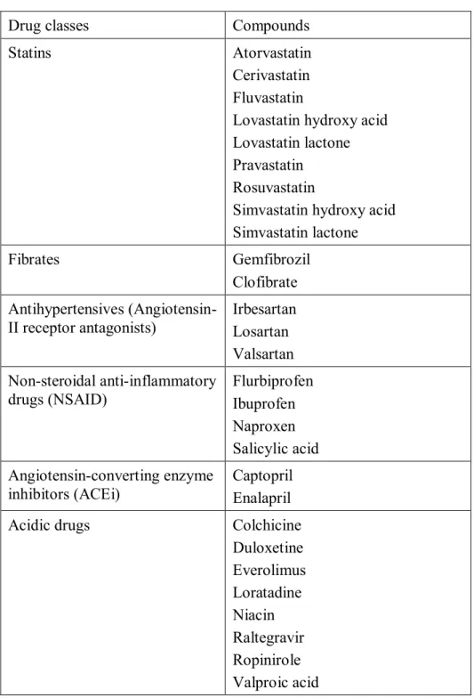
that could be administered to relieve muscle pain after the onset of drug-induced myopathies, specifically non-steroidal anti-inflammatory drugs (NSAID), were assessed to know if their use could exacerbate the pre-existing condition. Other acidic drugs known to cause myopathies were also studied, to determine if they cause muscle pain through the same mechanism as some statins. The characterized possible myopathy-inducing drugs relevant for this study are listed in Table III.
Table III. Potential myopathy-inducing drugs
Drug classes Compounds
Statins Atorvastatin
Cerivastatin Fluvastatin
Lovastatin hydroxy acid Lovastatin lactone Pravastatin
Rosuvastatin
Simvastatin hydroxy acid Simvastatin lactone Fibrates Gemfibrozil Clofibrate Antihypertensives (Angiotensin-II receptor antagonists) Irbesartan Losartan Valsartan Non-steroidal anti-inflammatory drugs (NSAID) Flurbiprofen Ibuprofen Naproxen Salicylic acid Angiotensin-converting enzyme inhibitors (ACEi) Captopril Enalapril
Acidic drugs Colchicine
Duloxetine Everolimus Loratadine Niacin Raltegravir Ropinirole Valproic acid
1.2.3.1 Loratadine
Loratadine is a tricyclic H1-antihistamine commonly
used to relieve allergy symptoms, such as allergic rhinitis and chronic urticaria.58, 59 Loratadine is part of the second generation
(non-sedating) antihistamine class characterized by their selective inverse agonist action on the peripheral histamine H1
-receptors.58 Similar to other second-generation antihistamines,
such as cetirizine, ebastine and terfenadine, loratadine competitively blocks the histamine receptor site instead of preventing histamine release.58 Figure 5 represents the structure
of loratadine.
Unlike their predecessors, the second-generation antihistamines do not present the major ADR of sedation. The first-generation antihistamines can cause these side effects due to their lack of selectivity for H1-receptors (action on acetylcholine receptors, α-adrenergic
receptors and 5-HT receptors) and their capacity to cross the brood-brain barrier.58, 60 These
second-generation antihistamines have a higher selectivity for the peripheral H1-receptors than
the ones in the central nervous system.61 This selectivity is largely attributed to their polarity,
with most of them being zwitterions (molecules that are both positively and negatively charged).62 They are therefore unable to cross the blood-brain barrier, making them less
susceptible to antihistamine-related sedation.58, 61 Third-generation antihistamines are mainly
active metabolites and enantiomers of second-generation antihistamines.61 They do not present
with any notable advantage, with the exception of fexofenadine, the active metabolite of Figure 5. Loratadine structure
terfenadine, which has a lower risk for cardiac arrhythmia than the parent molecule.61 A partial
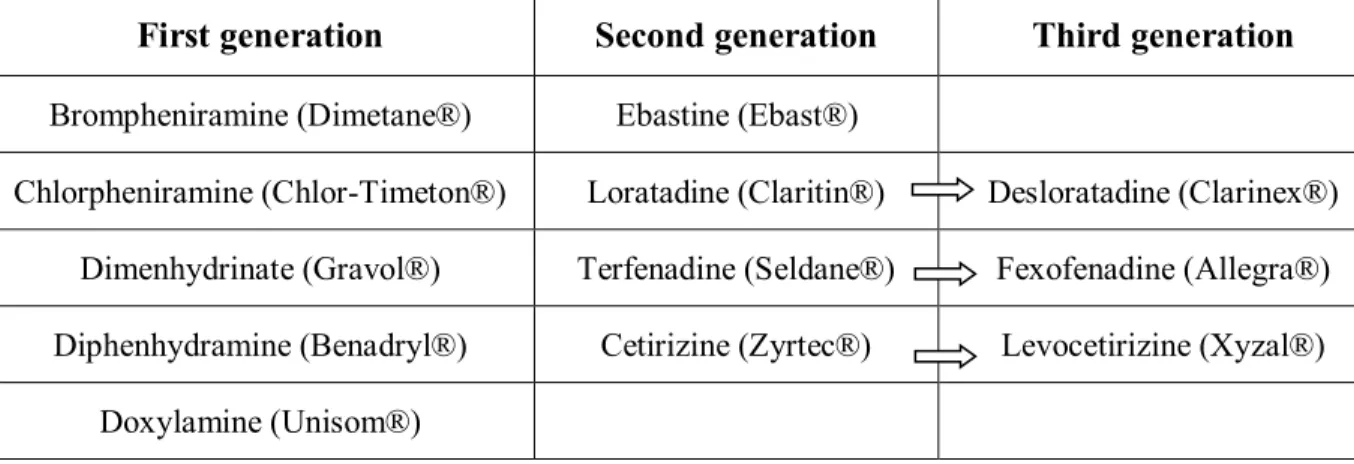
list of H1-antihistamines is presented in Table IV.
Table IV. Examples of H1-antihistamines from different generations (Brand names)61, 63
First generation Second generation Third generation
Brompheniramine (Dimetane®) Ebastine (Ebast®)
Chlorpheniramine (Chlor-Timeton®) Loratadine (Claritin®) Desloratadine (Clarinex®) Dimenhydrinate (Gravol®) Terfenadine (Seldane®) Fexofenadine (Allegra®) Diphenhydramine (Benadryl®) Cetirizine (Zyrtec®) Levocetirizine (Xyzal®)
Doxylamine (Unisom®)
Loratadine allergy treatment can also be associated with muscle pain and other adverse effects, such as headache, dry mouth, fatigue, and gastrointestinal problems.64-66 During
clinical trials involving loratadine and its metabolite desloratadine, the frequency of muscular adverse events was low (~2%) and not significantly different from placebo.67, 68 However, the
occurrence of these events is not representative of clinical reality where patients are not in such controlled environments.69 Loratadine was also found to be more likely to synergistically
increase the risk of myopathy when paired with simvastatin (relative risk (RR) = 1.69), alprazolam (RR = 1.86), duloxetine (RR = 1.94), and ropinirole (RR = 3.21).22 The
mechanism by which loratadine causes these muscle side effects is also still unknown. Moreover, since loratadine is a widely used over-the-counter antihistamine, the frequency of the adverse events related to its use is not well reported and can lead to an underestimation of myopathies reported for this drug.
1.3 Mechanisms of drug-induced myotoxicities
Drug-induced myotoxicities, as mentioned previously, are associated with many widely-used drugs. Over the years, many mechanisms have been attributed to these adverse effects, such as alteration in cellular membrane cholesterol, mitochondrial impairments, increase of lysosomal activity, injuries to electrolyte homeostasis, alteration in protein synthesis and degradation, inhibition of myogenesis, oxidative stress, cell apoptosis and immune reactions.6, 70, 71
Corticosteroids constitute the most common class of drugs to cause muscle toxicity by inhibiting protein synthesis.24 This results from the lowered expression of insulin-like growth
factor I (IGF-I), which has antiapoptotic effects.24 It can also be exacerbated by increased cytoplasmic protease activity (proteolysis) which leads to myofibrillar destruction.24 In addition, steroids can lower glutamine synthase and glycogen phosphorylase activities, leading to muscular atrophy.24
Mitochondrial dysfunction is also associated with myalgia. The antiviral zidovudine (nucleoside reverse transcriptase inhibitor of γ-DNA polymerase) can interfere with the replication of mitochondrial DNA, whereas the immunosuppressant cyclosporin A blocks the mitochondrial permeability transition pore, leading to lowered cellular energy production.2, 24
Lysosomal activity can also play a pathogenic role in the antimalarial chloroquine-induced myopathy. An accumulation of inflammatory cells in the lysosomal system can be generated by chloroquine and hydroxychloroquine, leading to the alkalinisation of lysosomes as well as the alteration of protein glycosylation and membrane lipid metabolism.2
Other drugs, such as colchicine, can produce myotoxicities via lesions of the microtubular system by binding firmly to tubulin molecules.2 D-penicillamine can cause
inflammatory myopathy with genesis of polymyositis and dermatomyositis.2 NSAID
analgesics can incite muscle necrosis and rhabdomyolysis, but they are rare adverse events.2
Despite the various mechanisms identified in drug-induced myopathy, the specific mechanisms related to statin-induced muscle pain are still unknown.50 Different hypotheses
have been proposed and will be discussed below.
1.3.1 Depletion of cholesterol, isoprenoids and coenzyme Q10
One of the first hypotheses proposed for statin-induced myopathy is that the reduction of cholesterol content in skeletal muscle cells can make their membrane unstable.5, 52
Cholesterol is an essential component of the membrane structure and function, so its decrease during statin treatment can influence the membrane fluidity, changing in turn the membrane excitability.5, 72 However, this mechanism has been proven not plausible since the specific in
vitro inhibition of squalene synthase (downstream steps in cholesterol synthesis) did not precipitate to the observed myotoxicity.5, 52
This finding, combined with the ones that have shown that the addition of mevalonate or mevalonic acid (intermediate products of cholesterol synthesis) during statin treatment in rat muscle cells or mice can revert myotoxicity, suggest that the mechanism behind the statin-induced myopathy seems to involve the depletion of either isoprenoids or coenzyme Q10 (ubiquinone).5, 40 The most important isoprenoids in the cholesterol synthesis pathway are
farnesyl pyrophosphate and geranylgeranyl pyrophosphate, which activate the regulatory guanosine 5’-triphosphate-binding proteins, promoting cell maintenance and reducing apoptosis (cell death) by a process known as protein prenylation.5, 52 Statins can inhibit this
process, which increases cytosolic calcium, resulting in apoptosis following caspase-3 activation.52
The reduction in CoQ10 is another possible mechanism for statin-induced myopathy. Statins can inhibit the production of CoQ10, since it is one of the end products of the cholesterol synthesis pathway.52 CoQ10 participates in the mitochondrial electron transport
during oxidative phosphorylation. Therefore, a diminution of CoQ10 can impair the mitochondrial function in the respiratory chain. However, the effects of CoQ10 depletion on statin-induced myotoxicity remain controversial, since a supplement of CoQ10 during statin therapy did not result in a consistent improvement of symptoms in each study.5
1.3.2 Disturbed calcium metabolism
The impaired calcium metabolism has also been considered to be a possible mechanism for the observed muscular symptoms during statin treatment. In vitro studies on rat tissue have shown that statins activate the release of calcium from the sarcoplasmic reticulum by enhancing the expression of calcium channels ryanodine receptors 3 (RYR3).5, 40 It has also
been demonstrated that statins can increase calcium accumulation by activating mitochondrial depolarization through the permeability transition pore (mPTP) and the sodium-calcium exchanger (NCE).5, 40 This increase in intracellular calcium levels (disturbance in calcium
homeostasis) can lead to muscle contraction and cramps.52
1.3.3 Autoimmune
In recent years, other than the previously described self-limited statin-induced myopathies, there have been reports regarding the development of autoimmune myopathies during statin treatment.73 The symptoms of these statin-associated autoimmune myopathies
persist or worsen even after statin discontinuation.73 These immune-mediated necrotizing
myopathies are rare, and are frequently associated with the production of anti-HMGCR autoantibody.74 Therefore, patients suffering from these conditions can be identified using
anti-HMGCR screening and treated with immunosuppressants.75
1.3.4 Drug-drug interactions
Patients with cardiovascular diseases are generally on long-term statin treatment and most of them are elderly with concomitant morbidities (heart disease, hypertension and diabetes).14, 76In general, these patients need a therapeutic arsenal including many drugs. This
clinical reality increases the risk of drug-drug interactions, which in turn increases the occurrence of undesirable events and drug toxicity. The myopathy incidence rate is low with statin monotherapy; however, there is a significant increase in polymedicated patients.47, 56
There is some data suggesting that drug-drug interactions that can affect the pharmacokinetics of statins, inducing a significant increase in statin plasma levels (CYP2C8, CYP2C9 and CYP3A4 inhibitors), are associated with a higher risk of musculoskeletal disorders such as myopathies and rhabdomyolysis.47 However, this relation can only explain the higher potential
of drug-related morbidity associated with statins. For example, the reported incidence rate for lovastatin monotherapy is 0.15%, but this is increased to 2%, 5% and even 28% when patients receive it in concomitance with niacin, niacin plus cyclosporine, and cyclosporine plus gemfibrozil, respectively.47 A drug interaction prediction study by Duke et al. found that the
combination of loratadine and simvastatin increased the risk for myopathy (RR = 1.69).22, 77
Loratadine has also been reported to cause muscle pain.64 It is therefore possible that this
1.3.5 Genetic polymorphisms
Genetic polymorphisms in statin transporters can affect drug disposition and increase drug plasma levels, which lead to the observed ADR.49 An important example of this is that
the membrane expression of the SLCO1B1 V174A (SLCO1B1 521T>C) variant is reduced compared to the wild-type SLCO1B1 in human liver samples.78 SLCO1B1 is a drug
transporter expressed exclusively on the sinusoidal membrane of hepatocytes and it is implicated in the uptake of drugs from the blood into the liver.7 In clinic, subjects homozygous
for SLCO1B1 521CC have an increased exposure to pravastatin 3.21-fold higher than subjects with the SLCO1B1 521TT genotype.79 However, the higher plasma levels can only partly
explain the potential muscular toxicity.80 Moreover, a genome-wide association study
(GWAS) revealed an association between the SLCO1B1 521T>C polymorphism and the incidence of myopathy with simvastatin administration, with an odds ratio (OR) of 4.5 per copy of the C allele.81 Drug transporters and their effects on statin pharmacokinetics will be
discussed further in this thesis.
1.3.6 Inhibition of lactic acid efflux by MCT and drug-induced
myopathy
Other than systemic or local drug levels, statin-induced myotoxicities can be precipitated through their effects on other drug transporters.
In recent studies, it was found that some statins (mostly lipophilic) can be transported by the proton-linked lactic acid transporters, MCT.7, 82-84 This suggests that statins could
inhibit lactic acid transport in a competitive manner.84 Some published data obtained in
accumulation of lactic acid and lead to muscle cramps and cell apoptosis (via activation of caspases).54 We will further investigate this hypothesis in this thesis.
1.3.6.1 Muscle and lactic acid
The muscle is the largest organ in the human body, representing 45% of body weight, and is particularly prone to adverse drug reactions since it is highly vascularized, hence increasing its exposition to circulating drugs.85 Indeed, it receives a large fraction of the blood
supply and it is highly metabolically active. Skeletal muscle is the major producer of lactic acid in the organism through glycolysis, but it can also use lactic acid as a source of energy.85
Briefly, muscle is defined by two distinct muscle fibers: type I and type II fibers. Type I fibers are highly oxidative and are considered slow twitch, whereas type II are highly glycolytic and are referred to as fast twitch.86 The stereoselective transport of lactic acid through the muscle
membrane is catalysed by the proton-linked monocarboxylate transporters.87 The rapid
transport of lactic acid through the membrane is crucial to maintain the intracellular pH homeostasis.88
During the first 5 to 10 minutes of moderate physical activity, ATP is produced via phosphorylative oxidation using glycogen catabolism as a primary fuel source.29 When the
effort or length of the physical activity is too high, glycolysis can start playing a major role and the anaerobic pathway becomes predominant as oxygen supplies are depleted.29This leads
to an increased production of lactic acid. Following physical activity, muscle supplies of creatine phosphate, glycogen and oxygen are depleted. Oxygen is needed to metabolize lactic acid and restore normal lactic acid concentrations.29
Muscular fatigue can be induced by three mechanisms: impaired calcium metabolism (increased expression of RYR3, mPTP and NCE), lowered glucose replenishing (decreased glucose transporter GLUT4 expression) and increased lactic acid levels (inhibition of MCT).40
Consequently, a lowered pH and higher lactic acid concentrations following exercise can alter protein conformation and activity in the muscle, such as actin and myosin, as well as proteins regulating calcium relargage.5, 40, 89 Lactic acid intracellular accumulation will be discussed
more in detail.
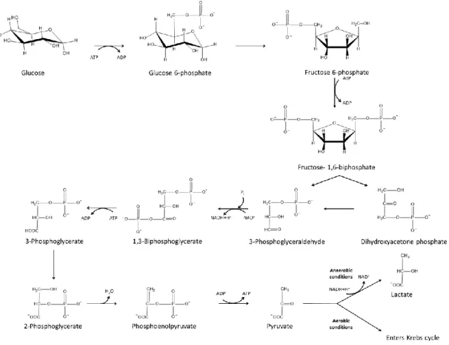
During glycolysis, pyruvate is formed.29 At the end of this pathway, pyruvate can be
processed in two different ways, depending on the oxygen supply. If the metabolism is in aerobic conditions, pyruvate enters the Krebs cycle, but in anaerobic conditions, pyruvate is converted to lactate. Lactate is the ionized form of lactic acid. The production of lactate through the glycolysis pathway is detailed in the figure below (Figure 6). Glucose metabolism in mammalian cells results in lactic acid production. Indeed, one glucose molecule breaks down to two lactic acid molecules and protons. Lactic acid production leads to a decrease in intracellular pH. MCT can transport lactic acid out of the cells (efflux), which increases the intracellular pH.90
Figure 6. Glycolysis pathway
Figure adapted from Vander's Human Physiology- The Mechanisms of Body Function29
It has been reported that around 1,500 mM of lactic acid enters blood circulation every day from various tissues, such as muscle, skin, brain, red blood cells and intestine.91 Lactic
acid concentrations range from 0.5 to 2 mM at rest, but they can go as high as 10 to 15 mM during intensive exercise.92 Lactic acid is generally considered as a byproduct of glycolysis.
However, there is evidence suggesting that lactate can actually be used as an energy source in the neurons and cardiomyocytes.93
GPR81 is part of a GPCR subfamily recognized for binding hydroxy-carboxylic acids. This receptor binds lactate with an affinity (EC50) of 1.5 to 3 mM and is expressed
and brain.91, 94 Several studies have supported the hypothesis that the signaling mechanism of
GPR81 is mediated by its coupling to Gi- type G proteins, which results in an adenylyl cyclase
inhibition. GPR81 activation by lactate is associated with lipolysis inhibition.91, 92, 94 A study
in cancer cells by Roland et al. showed that GPR81 activation increased the mRNA expression of the monocarboxylate transporters MCT1 and MCT4, critical for lactic acid transport.95
Lactic acid transport by monocarboxylate transporters will be discussed further in this thesis. During statin treatment, intense physical exercise can cause an increase of muscle intracellular lactic acid accumulation. It has been proposed that an inhibition of transport of major monocarboxylates related to exercise, such as L-lactate, can play a role in these drug-induced muscle disorders.96-98
1.4 Drug transporters
Transporters are transmembrane proteins located across physiological membranes. They mediate the uptake or efflux of numerous compounds, such as nutrients, endogenous substrates and xenobiotics at the cellular level.7 Their role is to facilitate the transport of these
compounds in and out of the cells, to either provide necessary elements or protect the organism against dietary and environmental toxins.7 Transporters are mainly classified into
two superfamilies, based on their transport mechanism: ATP-Binding Cassette (ABC) transporters and Solute Carrier (SLC) transporters.7 ABC transporters are primary active
transporters and mainly mediate efflux, whereas SLC transporters are mostly bidirectional, depending mainly on electrochemical gradient of the substrates, which classifies them as facilitated transporters.99 However, some SLC can use an ion gradient, such as sodium or
potential difference; these are classified as secondary active transporters.100Efflux transporters
pump the compounds out of the cells, while influx transporters bring them inside the cells. Membrane-bound transporters are found either on the apical or on the basolateral membrane, which determines their function.7
It was previously thought that drug pharmacokinetics and pharmacodynamics were mostly dependent on plasma concentrations.7 However, it is now known that some
transporters, which are identified as drug transporters, play an important role in drug absorption, distribution in specific organs and disposition.7 These drug transporters are further
classified into 13 different families depending on their function. The ABC superfamily comprises MDR, MRP and BCRP, whereas the SLC superfamily includes organic cation transporter (OCT), organic cation/carnitine transporter (OCTN), organic anion transporter (OAT), organic anion transporter polypeptide (OATP; SLCO), peptide transporter (PEPT), concentrative nucleoside transporter (CNT), equilibrative nucleoside transporter (ENT), bile acid transporter, sodium-coupled monocarboxylate transporter (SMCT) and MCT.
There are a multitude of drug transporters, but only the most relevant ones for this project will be discussed in detail.
1.4.1 ABC transporters
The ABC transporters superfamily, as their name suggests, require ATP hydrolysis to translocate their substrates across the membranes against their electrochemical gradient. ABC transporters share a common structure, generally including two nucleotide-binding domains and two transmembrane domains as the core unit. The nucleotide-binding domains are
required for ATP hydrolysis, whereas the transmembrane domains bind to the substrate and allow translocation across the membrane.99
The 49 ABC transporters genes are classified into 7 families (ABCA-ABCG), according to their sequence similarity.99 Three of those families are better characterized:
multidrug resistance protein (MDR; ABCB), multidrug resistance-associated protein (MRP; ABCC) and the breast cancer resistance protein (BCRP; ABCG).7 The MDR family regroups
11 genes (ABCB), the MRP family contains 13 genes (ABCC), and there are only 5 members in the ABCG family, with BCRP (ABCG2) being the most studied.99
1.4.1.1 ABCB1
ABCB1 (P-gp; MDR1) is one of the most studied efflux transporters. It is expressed in multiple organs, such as the liver, the kidney and the gastrointestinal (GI) tract.101 This
transporter’s localization and wide range of substrates make it determinant in the bioavailability of many drug classes: anticancer drugs, HIV protease inhibitors, analgesics, immunosuppressive agents, antibiotics, histamine H2-receptor antagonists, etc.7 In the liver,
ABCB1 mediates the flux of drugs, such as statins, towards the bile and facilitates drug elimination.101 There are around 30 characterized SNP for ABCB1. Two common ABCB1
polymorphisms (2677G>T/A and 3435C>T) yield a lower ABCB1 expression and activity and may influence statin-associated myopathy.7 However, regardless of ABCB1 genetic
polymorphisms, only a weak association has been found between reduced ABCB1 transport activity and statin-induced myotoxicity. A stronger association was found between ABCB1 3435C>T carriers and drug-induced muscle symptoms when patients were treated with statins for more than 5 months.40
1.4.1.2 ABCC
The ABCC family mediates drug efflux and can be found in multiple tissues. ABCC1 and ABCC5 have a ubiquitous expression, with low ABCC1 levels in the liver, whereas ABCC4 is expressed in the muscle. ABCC1 can mediate the transport of a wide range of substrates, with a preference for organic anions, while ABCC4 and ABCC5 transport predominantly nucleotide analogues.102 It was reported that these ABCC isoforms can mediate
the transport of atorvastatin and rosuvastatin in heterologous expression systems.80
ABCC2 plays an important part in statin efflux from the liver towards the bile and ABCC2 inhibition may result in significantly higher drug exposure. In a study to determine whether ABCC2 polymorphisms could be associated with statin-induced myopathy, three common polymorphisms were characterized: ABCC2 -24C>T, ABCC2 1249G>A and ABCC2 3972C>T. They have found that these variants generate a higher drug exposure and could possibly contribute to statin-induced adverse events.103
1.4.1.3 ABCG2
ABCG2 is a main efflux transporter involved in statin elimination in the liver. Similar to P-gp, it mediates drug transport towards the bile canalicular duct. Therefore, a decrease in transport activity for ABCG2 may also lead to increased drug exposure. The ABCG2 421C>A polymorphism (frequency of 7.4-11.1% in Caucasians and 27-35% in Asians) results in decreased ABCG2 activity and higher drug exposure for fluvastatin, simvastatin, atorvastatin and rosuvastatin.40
ABCG2 is also expressed in the muscular cell membrane where it mediates the efflux of statins and other drugs. It has been proposed that decreased efflux transport via ABCG2
might result in statin accumulation in the muscle cell.40 As previously mentioned, ABCG2
mediates the efflux of drugs at the hepatic level towards the bile canalicular duct and the intestine. Impaired ABCG2-mediated transport would therefore lead to higher drug plasma concentrations, and accumulation into the myocyte.40 However, because several patients
present with polymorphisms in ABCG2 and do not experience myopathy during statin treatment, it has been suggested that more than one SNP or predisposing muscle conditions are present in the same individual.40
1.4.2 SLC transporters
Most drugs transporters are part of the SLC transporters superfamily functioning either according to a concentration gradient of its substrate or by an electrochemical gradient created by other transporters.100 The 395 SLC transporters are divided into 52 gene families
(SLC1-SLC52). SLC transporters are classified in the same family if they share 20% of their sequence.104 Transporters found in the SLC superfamily can have multiple transport
mechanisms at the cell membrane, such as coupled transport, exchange and passive transport.104
1.4.2.1 SLCO transporters
SLCO transporters, coded by the SLC21 gene family, are also known as the organic anion-transporting polypeptides (OATP) family. Similar to the ABC transporters, the SLCO genes are classified by sequence similarity.105 Indeed, SLCO transporters sharing over 40% of
their sequence belong to the same family, whereas those sharing over 60% of their sequence are classified in the same subfamily.105
The SLCO transporters are characterized by 12 transmembrane domains and generally perform bidirectional transport, depending on the solute gradient.106 A few members of the
SLCO family are especially important in the liver, such as OATP1B1, OATP1B3 and OATP2B1 on the basolateral membrane of the hepatocytes.107-109
Various endogenous substrates have been found to be transported by the SLCO transporters, as well as drug classes including statins, angiotensin II receptor antagonists and ACE inhibitors.110, 111
1.4.2.1.1 SLCO1B1
SLCO1B1 is expressed in the liver and transports a wide variety of substrates, but the most relevant to this project are the statins cerivastatin, atorvastatin, rosuvastatin and pitavastatin.7, 112-115 However, SLCO1B1 activity can be inhibited by structurally diverse
compounds and commonly used drugs, such as ketoconazole, clarithromycin, erythromycin, verapamil and warfarin among others.116
Statin-induced myopathy has been associated with a decrease in SLCO1B1 function. Various genetic polymorphisms result in a reduced SLCO1B1-mediated transport activity, such as the missense variant 521T>C (SLCO1B1*5), found in 1% of Caucasians.40, 117 The
SLCO1B1*1b (388A>G) variant is observed at a frequency of 40% in Caucasians, 75% in African-American and 60% in Asians. The SLCO1B1*1b allele is in linkage disequilibrium (frequently associated with each other) with the SLCO1B1*5 allele, resulting in the SLCO1B1*15 variant. Both SLCO1B1*5 and SLCO1B1*15 result in decreased SLCO1B1-mediated transport activity.7 This leads to a reduced influx of statins and other drugs (i.e.
plasma concentrations.7 It has been reported that SLCO1B1 521CC homozygotes have higher
AUC for lovastatin acid (3.4-fold), pravastatin (1.9-fold), simvastatin (3.2-fold), pitavastatin (3.2-fold), atorvastatin (3.1-fold) and rosuvastatin (1.8-fold).40, 118 The SLCO1B1 521T>C
genotype has also been associated with statin intolerance defined by an increase of serum creatine kinase or a modification in statin regimen, with an odds ratio (OR) of 2.05.40
Moreover, a GWAS correlated the prevalence of statin-induced myopathy during simvastatin treatment with the SLCO1B1 521T>C allele, with an OR of 4.5 per copy of the allele and an OR of 16.9 for CC homozygotes.40 This mutation in SLCO1B1 could account for up to 60% of
myopathies associated with simvastatin therapy.1, 6 In summary, it has been reported that
SLCO1B1 genetic polymorphisms can result in increased drug exposure, which could increase the risk of experiencing statin-induced myotoxicity.119
1.4.2.1.2 SLCO1B3
SLCO1B3 has been reported to be expressed in liver, small intestine and placenta.7
SLCO1B3 is very similar to SLCO1B1 (80% sequence similarity) and has a comparable spectrum of substrates. As for SLCO1B1, decreased SLCO1B3-mediated transport could also affect statin concentrations. The two most common SLCO1B3 variants are the SLCO1B3 334T>G and SLCO1B3 699G>A, which result in a lower SLCO1B3 activity, possibly increasing the risk of drug-induced myopathy via higher drug plasma concentrations.120
1.4.2.1.3 SLCO2B1
SLCO2B1 is expressed in a variety of organs, such as liver, kidney, intestine, brain and placenta.7 This transporter facilitates the oral absorption of its substrates at the apical