The logopenic/phonological variant of
primary progressive aphasia
M.L. Gorno-Tempini,
MD, PhD
S.M. Brambati, PhD
V. Ginex, BSc
J. Ogar, MS
N.F. Dronkers, PhD
A. Marcone, MD
D. Perani, MD
V. Garibotto, MD
S.F. Cappa, MD
B.L. Miller, MD
ABSTRACTObjective:
Primary progressive aphasia (PPA) is characterized by isolated decline in language functions. Semantic dementia and progressive nonfluent aphasia are accepted PPA variants. A “logopenic” variant (LPA) has also been proposed, but its cognitive and anatomic profile is less defined. The aim of this study was to establish the cognitive and anatomic features of LPA.Methods:
Six previously unreported LPA cases underwent extensive neuropsychological evalua-tion and an experimental study of phonological loop funcevalua-tions, including auditory and visual span tasks with digits, letters, and words. For each patient, a voxel-wise, automated analysis of MRI or SPECT data were conducted using SPM2.Results:
In LPA, speech rate was slow, with long word-finding pauses. Grammar and articulation were preserved, although phonological paraphasias could be present. Repetition and comprehen-sion were impaired for sentences but preserved for single words, and naming was moderately affected. Investigation of phonological loop functions showed that patients were severely im-paired in digit, letter, and word span tasks. Performance did not improve with pointing, was influ-enced by word length, and did not show the normal phonological similarity effect. Atrophy or decreased blood flow was consistently found in the posterior portion of the left superior and middle temporal gyri and inferior parietal lobule.Conclusions:
Logopenic progressive aphasia (LPA) is a distinctive variant of primary progressive aphasia. Cognitive and neuroimaging data indicate that a deficit in phonological loop functions may be the core mechanism underlying the LPA clinical syndrome. Recent studies suggest that Alzheimer disease may be the most common pathology underlying the LPA clinical syndrome.Neurology®2008;71:1227–1234
GLOSSARY
AD⫽ Alzheimer disease; BA ⫽ Brodmann area; CDR ⫽ Clinical Dementia Rating; CVLT-MS ⫽ California Verbal Learning Test–Mental Status Edition; ECD⫽ ethyl cysteinate dimer; FWHM ⫽ full-width at half-maximum; GM ⫽ gray matter; LPA ⫽ logopenic progressive aphasia; MMSE⫽ Mini-Mental State Examination; PNFA ⫽ progressive nonfluent aphasia; PPA ⫽ primary progressive aphasia; Rey-O⫽ Rey–Osterrieth; SemD ⫽ semantic dementia; VBM ⫽ voxel-based morphometry;
WAB⫽ Western Aphasia Battery; WAIS-III ⫽ Wechsler Adult Intelligence Scale, Third Edition.
Since Mesulam’s original description of primary progressive aphasia (PPA) in 1982,
1it has
become clear that progressive isolated language disorders due to neurodegeneration are
clini-cally heterogeneous. They are caused by patterns of localized cerebral dysfunction that do not
necessarily match those affected “typically” by vascular strokes. Thus, the present-day
distinc-tion between progressive nonfluent aphasia (PNFA) and semantic dementia (SemD) may
re-flect an oversimplification of the clinical presentations of progressive aphasia.
2In a recent
study,
3the term “logopenic”
4was revived to label PPA patients who did not show the pattern
Supplemental data atwww.neurology.org Editorial, page 1218
Address correspondence and reprint requests to Dr. Maria Luisa Gorno-Tempini, UCSF Memory and Aging Center, 350 Parnassus Ave., Suite 506, San Francisco, CA 94143 [email protected]
e-Pub ahead of print on July 16, 2008, at www.neurology.org.
From the Memory Aging Center (M.L.G.-T., S.M.B., J.O., B.L.M.), University of California, San Francisco, Department of Neurology, San Francisco; Vita-Salute San Raffaele University (S.M.B., V.G., A.M., D.P., V.G., S.F.C.), Department of Nuclear Medicine, IRCCS San Raffaele, Department of Clinical Neurosciences, San Raffaele Turro, National Institute of Neuroscience, Milan, Italy; University of California (J.O., N.F.D.), Davis, CA, and San Diego, CA.
Supported by the National Institute of Neurological Disorders and Stroke (R01 NS050915), the State of California (DHS 04-35516), the National Institute on Aging (P50 AG03006, P01 AG019724), the Alzheimer’s Disease Research Center of California (03-75271 DHS/ADP/ARCC), and the Italian Ministry for University and Scientific Research (Interlink Grant to S.F.C. and M.L.G.-T.).
of language dysfunction typical of PNFA and
SemD, respectively. Logopenic progressive
aphasia (LPA) was characterized by slow
speech, sentence repetition, and
comprehen-sion deficits, and relative sparing of motor
speech, grammar, and single-word
compre-hension.
3Significant atrophy was found in
the left posterior temporoparietal region,
rather than in the left frontoinsular or anterior
temporal areas typically affected in PNFA and
SemD, respectively. In agreement with the
lo-cation of atrophy, it was hypothesized that a
deficit in auditory verbal short-term memory
could be the core mechanism in LPA.
3,5Short-term memory is a limited-capacity
sys-tem allowing the sys-temporary storage and
manipulation of information.
6Baddeley
7pro-posed a multicomponent model of verbal
working memory comprising the central
exec-utive and the phonological loop. The
phono-logical loop is a component of short-term
memory that includes a “store,” in which
phonological memory traces are held over a
period of few seconds, and an articulatory
“re-hearsal” process that refreshes them.
Despite the initial observation,
3studies
inves-tigating the cognitive and anatomic mechanism
in LPA are lacking, and even its existence is
un-der debate. Here, we performed a detailed
neu-roimaging and cognitive study of six new cases
of LPA, including an experimental assessment of
phonological loop functions. We hypothesized
that all patients would show phonological loop
deficits and anatomic involvement of the left
posterior temporoparietal region. Confirmation
of this hypothesis would establish LPA as a
rec-ognizable, separate clinical variant of PPA with
specific neuroimaging correlates.
METHODSSubjects. Six LPA patients (table 1) at the
Univer-sity of California, San Francisco Memory and Aging Center (n⫽ 4) or at the Neurology Unit, San Raffaele Turro, Milan (n⫽ 2), participated in the study. Patients were selected from a pool of LPA cases on the basis of their ability to participate in numerous testing sessions and the availability of a brain scan. They were all new cases, not included in our previous article.3Patients were
evalu-ated by a team of experienced clinicians and underwent a baseline neurologic, neuropsychological, and language evaluation. LPA was diagnosed when patients did not meet criteria for SemD and PNFA because of the presence of slow, well-articulated and grammatic speech (thus excluding PNFA) and good single-word comprehen-sion (thus excluding SemD).3Imaging findings were not used for
diagnosis. Informed consent was obtained, and the study was ap-proved by each local ethics committee.
All patients had similar clinical histories with approximately 3 years of word-finding difficulties starting in their 50s. At the time of examination, three patients had mild calculation, writing, and ver-bal memory difficulties. None had visuospatial deficits. Anxiety, de-pression, and irritability were common. Two patients showed mild slowing of rapidly alternating movements in the right hand. Al-though extensive follow-up is still not available in most patients, clinical evolution was generally slow, with relative sparing of func-tional status 5 years after onset.
Cognitive assessment. Neuropsychological assessment. All
patients underwent a comparable neuropsychological assessment, using language-specific tests of verbal long-term memory, verbal and visuospatial short-term memory, visuospatial abilities, and exec-utive functions (table e-1 on the Neurology®Web site at www.
neurology.org).
General language evaluation. Language and cognitive
functions were assessed in each patient’s native language with comparable measures. We tested fluency, single-word and sen-tence repetition, word–picture matching, sensen-tence-to-picture matching, and confrontation naming using the Western Aphasia Battery,8 the Boston Naming test,9 and portions of Curtiss
Yamada Comprehension Language Evaluation–Receptive10
(cases 1– 4), and with the Battery for the Analysis of the Aphasic Deficit11(cases 5 and 6). The Pyramid and Palm Trees Test
pictures were used in all patients.12In the four US patients, the
Motor Speech Evaluation was also administered.13
Experimental phonological loop assessment. The
short-term memory model proposed by Baddeley7accounts for several
effects that characterize the normal functioning of the phonolog-ical loop:
1. The phonological similarity effect: Sequences of items that sound similar (e.g., the letters B and T) are harder to remem-ber compared with dissimilar sounding examples (e.g., the letters W and Z). The absence of this effect indicates a phono-logical store deficit.
2. The word length effect: Sequences of long words are harder to remember than sequences of shorter words because longer words take more time to rehearse and their trace fades more quickly.
The following testing procedures were applied to all patients to test for these effects.
Digit span tasks. Digit span tasks were administered under
au-ditory/verbal, visual/verbal, and auditory/pointing conditions.14For
each condition, patients were presented with a sequence of digits at a rate of one digit per second and were asked to recall the exact se-quence immediately after presentation. Ten trials of each sese-quence length (i.e., one digit, two digits, etc.) were tested. Each correctly recalled trial was scored as 0.1, so the perfect score for any given sequence length would be 1. Span level was established when the patient recalled at least six trials at a given length.
For the auditory/verbal condition, stimuli were presented auditorily, and patients responded verbally. For the visual/verbal condition, each digit was presented sequentially in black font on a white background, and patients were required to reproduce the sequence verbally. For the auditory/pointing condition, stimuli were presented auditorily, and patients responded by pointing to the correct items within an array of single digits. The pointing condition excluded an articulation disorder.
Letter span tasks: Phonological similarity effect. Span levels for
tested. The same procedures as for the digit span were adopted but tested only in the auditory/verbal and visual/verbal condi-tions. Two sets of letters, phonologically similar (e.g., B, C, D,
G, P, and T for both languages) and phonologically dissimilar
(e.g., F, K, Q, R, X, W, and Z), were used in both languages as previously described.14Ten sequences of letters, increasing in
length and including letters from one set alone, were presented at a rate of one per second.
A group of 11 age- and education-matched normal con-trols (six Italian, five US) showed a significant effect of pho-nological similarity when tested with this material (mean phonologically similar: 6.00, mean phonologically dissimilar: 6.55; t test, p⬍ 0.05).
Word span tasks: Word length effect. The auditory/verbal and
visual/verbal conditions were tested. Two sets of two- or four-syllable words, matched by frequency, were used for each lan-guage.14,15For each set, 10 word sequences of increasing length
were generated. No word was presented twice.
The same group of 11 controls showed a significant effect of word length (mean long word: 6.09, mean short word: 7.18; t test, p⬍ 0.01).
Patients with a specific deficit in the phonological store are expected to show normal immediate recall of individual items,
defective span in both auditory/verbal and visual/verbal condi-tions with no improvement with pointing, and absence of pho-nological similarity. The word length effect should be preserved, unless the rehearsal is also affected.
Neuroimaging study. Voxel-based morphometry analysis of MRI data (cases 1– 4). T1-weighted MRI scans were
ac-quired using a 1.5-tesla Magnetom VISION system (Siemens Inc., Iselin, NJ) as previously described for cases 1– 4.3
Voxel-based morphometry (VBM) analysis was implemented in the SPM2 software package (The Wellcome Department of Imaging Neuroscience, London, UK) using standard procedures.16
Im-ages were normalized to an age-matched template, modulated, and spatially smoothed with a 12-mm full-width at half-maximum (FWHM) isotropic gaussian kernel.
Group analyses of gray matter (GM) and white matter (WM) images were used to compare patients with a control group of 40 age- and sex-matched healthy controls. Single-subject GM analyses were used to compare each patient with controls. Age, total intracranial volume, and sex were con-founding variables. In the group analysis, significance was set at p⬍ 0.05 corrected for multiple comparisons (family-wise error corrected). Because of the risk of false negatives in
Table 1 Demographic, functional, and cognitive assessment for each patient (cases 1– 4 from the United States, cases 5 and 6 from Italy)
Case 1 Case 2 Case 3 Case 4 Case 5 Case 6 Age, y/sex 56/F 60/F 56/F 63/M 57/F 61/F
Education, y 14 16 17 12 13 8
CDR total (0–3) 0.5 0.5 0.5 0.5 0.5 0.5
MMSE 22 25 17 22 16 28
Copy modified Rey-O figure (maxⴝ 17) 15 17 16 13 — —
Copy Rey-O complex figure (maxⴝ 36) — — — — 12 32
WAIS-III no. digits forward 4 4 4 4 2 4
WAIS-III spatial span 4 7 4 7 — —
Corsi span — — — — 2 5
CVLT 10-minute Delayed Free Recall 5 7 4 5
Rey word list 15-minute Delayed Free Recall 0 14
WAIS-III no. digits backward 2 4 3 3 — 3
Trail Making 120 120 120 74 — 103
Spontaneous speech WAB (maxⴝ 20) 19 19 17 14 13 16
Motor Speech Evaluation (0–7 severity scale)
Apraxia of speech 0 0 0 0 — —
Dysarthria 0 0 0 0 — —
Phonemic fluency 7 18 5 7 3 9
Semantic fluency 13 16 8 8 2 11
Confrontation naming, % correct 73 80 93 80 80 93
Word repetition, % correct 100 100 100 100 70 93
Sentence repetition, % correct 68 75 76 76 50 63
Single-word comprehension, % correct 95 100 98 98 90 100
Sentence comprehension, % correct 66 68 47 60 56 73
Pyramid and Palm Trees pictures, % correct 100 96 92 100 90 96
Raw scores are reported for general cognitive assessment and percentage correct for language measures.
CDR⫽ Clinical Dementia Rating; MMSE ⫽ Mini-Mental State Examination; Rey-O ⫽ Rey–Osterrieth; WAIS-III ⫽ Wechsler Adult Intelligence Scale, Third Edition; CVLT⫽ California Verbal Learning Test; WAB ⫽ Western Aphasia Battery.
single-subject and WM VBM analyses,17we report results of
these analyses at p⬍ 0.001, uncorrected for multiple compar-isons.
99mTc-ethyl cysteinate dimer SPECT analysis (cases 5 and 6). 99mTc-ethyl cysteinate dimer SPECT imaging was
used to detect regional hypoperfusion in the two Italian patients compared with 15 age-matched normal controls, using previ-ously described procedures.18
Image preprocessing (spatial normalization to a standard Montreal Neurological Institute SPECT template and 12-mm FWHM smoothing) and statistical analysis were implemented in SPM99 using standard validated procedures.19Age and total
global distribution of the tracer’s uptake were entered as con-founding variables. Single-subject analyses were performed by comparing each patient with the control group, with significance set at p⬍ 0.001 uncorrected.
RESULTS Cognitive assessment. Neuropsychological assessment.
Mini-Mental State Examination scores
were abnormal in all patients, whereas the Clinical
Dementia Rating was in the mild impairment
range (0.5 in each patient) (table 1). The profile of
neuropsychological and language impairment was
compatible with an LPA diagnosis, as previously
reported.
3Visuospatial abilities and spatial
work-ing memory span were preserved with the
excep-tion of case 5, in whom they were mildly impaired.
Conversely, most tests with a verbal component,
Table 2 Digit, letter (similar and dissimilar), and word (short and long) span results
Verbal response Pointing Digits Digits Sequence length,
no. of items
per sequence Auditory Visual Auditory
1 1.0 (0) 1.0 (0) 1.0 (0)
2 0.9 (0.2) 0.9 (0.2) 0.9 (0.1)
3 0.7 (0.3) 0.6 (0.3) 0.7 (0.3)
Immediate verbal recall Immediate verbal recall Letters similar Letters dissimilar Auditory Visual Auditory Visual
2 0.9 (0.2) 0.8 (0.3) 0.9 (0.2) 0.8 (0.3)
3 0.6 (0.2) 0.6 (0.2) 0.6 (0.3) 0.5 (0.1)
Short words Long words
Auditory Visual Auditory Visual
1 1.0 (0) 1.0 (0) 1.0 (0.1) 1.0 (0.1)
2 0.8 (0.2) 0.9 (0.2) 0.1 (0.1) 0.6 (0.4)
3 0.6 (0.4) 0.6 (0.3) — —
Data are mean (SD) of correctly recalled trials (0.1 for each trial, 1.0 maximum).
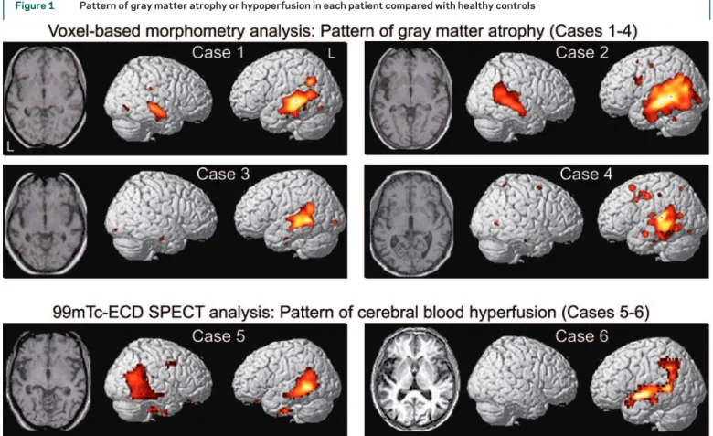
Figure 1 Pattern of gray matter atrophy or hypoperfusion in each patient compared with healthy controls
Representative axial sections of each original structural MRI are displayed in the left panel. Voxel-based morphometry and SPECT results are thresholded at p⬍ 0.005 uncorrected for multiple comparisons and superimposed on the three-dimensional rendering of the Montreal Neurological Institute standard brain. ECD⫽ ethyl cysteinate dimer.
such as the verbal short- and long-term memory
tests, revealed deficits. Case 6 was the exception,
showing normal performance in long-term verbal
memory.
Language evaluation.
Spontaneous speech
produc-tion was characterized by slow, hesitant speech with
word-finding pauses (see e-audio, table 2). Speech in
response to a picture was characterized by decreased
rate and occasional phonemic paraphasias and
word-finding difficulty. Sentences were simple, but
gram-matically well formed and without omission of
grammatic morphemes. Motor speech abilities were
within normal limits, and no apraxia of speech or
dysarthria was noted.
Comprehension was spared at the single-word
level, and Pyramid and Palm Trees Pictures
perfor-mance was mildly impaired in the two more
ad-vanced cases (cases 3 and 5), indicating that semantic
memory was relatively preserved (table 1).
Con-versely, patients showed word-finding pauses in
speech production and poor confrontation naming
ability. Errors consisted mostly of complete anomia
(no response) or phonemic paraphasias, suggesting a
mixed mechanism of paraphasic and word-selection
anomia.
20Comprehension at the sentence level was
impaired, but there was no evidence of a structural
complexity effect.
A specific pattern of repetition difficulties was
found in all patients. Single-word repetition was
largely preserved, whereas sentence repetition was
se-verely impaired, especially for low-probability
sen-tences. The pattern of errors in sentence repetition
suggested that patients were using a semantic rather
than a phonological route.
21For example, when
asked to repeat “It looks as if nobody is around,” one
patient responded, “It looks like nobody is there.”
Phonological loop assessment. Digit span.
Patients
per-formed normally on the immediate recall (repetition) of
individual and pairs of digits but were severely impaired
in sequences of more than three digits (table 2).
Perfor-mance was unrelated to the modality of presentation
(auditory or visual) or output (verbal or pointing).
Letter span: Phonological similarity effect.
Span was three in
both conditions (table 2). In contrast to normal
sub-jects, phonologically dissimilar letters provided no
ben-efit, either with auditory or with visual presentation.
Word span: Word length effect.
Span was three for short
words, but patients could repeat only one long word
(table 2). This effect of word length was particularly
evident in the auditory modality.
Neuroimaging study. Analysis of gray matter atrophy or hypoperfusion.
A consistent pattern of GM atrophy
(cases 1– 4) or hypoperfusion (cases 5 and 6) was found
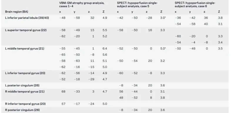
Table 3 Results of the VBM gray matter group analysis (p < 0.05 FWE corrected for multiple comparisons) and SPECT single-subject analysis (p < 0.001 uncorrected)
VBM: GM atrophy group analysis, cases 1– 4
SPECT: hypoperfusion single-subject analysis, case 5
SPECT: hypoperfusion single-subject analysis, case 6
Brain region (BA) x y z Z x y z Z x y z Z
L inferior parietal lobule (39/40) ⫺48 ⫺58 32 4.9 ⫺42 ⫺50 ⫺28 3.0* ⫺36 ⫺42 36 3.8
⫺54 ⫺58 40 3.1
L superior temporal gyrus (22) ⫺58 ⫺49 15 5.5 ⫺58 ⫺50 16 3.3
⫺62 ⫺20 1 5.2 ⫺60 ⫺20 0 3.3
⫺54 ⫺4 ⫺8 3.4
L middle temporal gyrus (21) ⫺55 ⫺45 1 6.4 ⫺52 ⫺50 0 5.0† ⫺50 ⫺48 0 3.5
⫺65 ⫺50 ⫺8 5.6
⫺58 ⫺63 11 5.1 ⫺50 ⫺54 20 3.2
⫺62 ⫺16 ⫺15 5.0
L inferior temporal gyrus (20) ⫺62 ⫺56 ⫺14 4.9 ⫺60 ⫺52 ⫺8 3.3
⫺52 ⫺18 ⫺29 4.7
L posterior cingulum (26) ⫺8 ⫺34 20 3.6
R middle temporal gyrus (21) 68 ⫺33 3 4.7 56 ⫺44 0 3.1
48 ⫺52 8 3.8
R inferior temporal gyrus (20) 57 ⫺17 ⫺24 5.0
R posterior cingulum (26) ⫺8 ⫺34 20 3.6
* p⫽ 0.002 uncorrected for multiple comparisons.
† p⬍ 0.05 family-wise error (FWE) corrected for multiple comparisons in the SPECT single-subject analysis. VBM⫽voxel-based morphometry; GM ⫽ gray matter; BA ⫽ Brodmann area.
in the posterior portion of the left superior and middle
temporal gyri and in the inferior parietal lobule (cases
1– 4 and 6) (figure 1 and table 3). In three cases,
dam-age spread from the posterior temporal areas to the
infe-rior and/or more anteinfe-rior temporal regions. The
corresponding contralateral right posterior temporal
re-gion was involved in cases 1, 2, and 5. Case 5 alone showed
hypoperfusion bilaterally in the posterior cingulum.
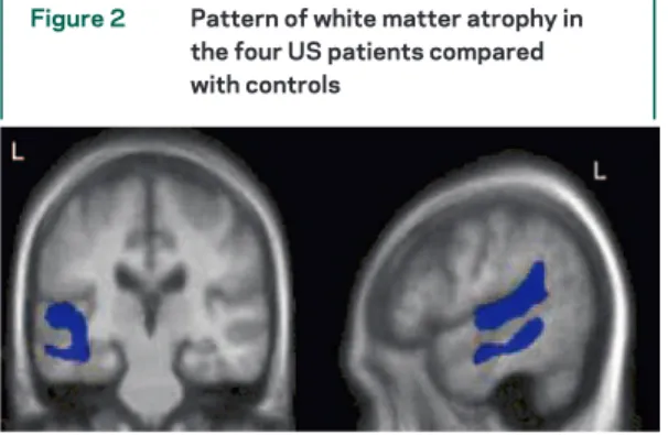
White matter atrophy.
WM loss was observed in
correspondence to GM atrophy in the posterior
por-tion of left middle temporal and bilateral posterior
superior temporal gyri (figure 2).
DISCUSSION
We defined the neuropsychological
and imaging features of the logopenic variant of PPA
that in our experience represents 30% of all PPA
cases. Our results suggested that the core cognitive
deficit in LPA was a phonological loop disorder.
Consistently, the imaging investigation showed
in-volvement of GM and WM in the left posterior
tem-poral and inferior parietal regions.
LPA is characterized by a decreased rate of
spon-taneous language production with frequent halts due
to word-finding pauses. Phonemic paraphasias are
common, but motor speech and grammar are spared.
This pattern of language production is different from
the fast output typical of early SemD patients, who
usually fill word-finding pauses with circumlocutions
and filler words. It is also distinct from the
produc-tion deficit typical of PNFA, in which articulaproduc-tion
deficits and agrammatism predominate.
22LPA
pa-tients, therefore, show a pattern of “intermediate”
fluency distinct from the fluent SemDs and the
non-fluent PNFAs, raising the issue of how to label their
language production. Fluency is a composite
mea-sure, defined by multiple features of spontaneous
language production. The concept was introduced
by Goodglass et al.
23to describe language production
in vascular aphasia. Within this framework, a patient
with would be described as “nonfluent” if he or she
exhibited slow, effortful production, with motor
speech impairment, defective prosody, and omission
of grammatic morphemes. Conversely, fluent aphasic
production would be characterized by normal motor
speech and production rate, spared grammar, and the
presence of phonological and lexical errors. The
fea-tures of LPA patients’ spontaneous production do
not easily fit within this dichotomy, because motor
speech is unaffected and grammar is preserved, yet
speech production is slowed and halting. LPA
pa-tients score as “fluent” on formal aphasia tests, which
give more weight to articulation and grammar, but
may be labeled as “nonfluent” by the clinician who is
impressed by their slow and hesitant production at
the bedside (see e-audio for example of spontaneous
speech in case 2 two years after initial evaluation
re-ported in table 1). We suggest that the label
“nonflu-ent” should be reserved for patients who show the
motor speech and grammar deficits typical of PNFA.
The term logopenic (from the Greek: “lack of words”)
could instead be used to describe the slow language
output typical of LPA. Given the phonological
na-ture of this deficit, the term phonological variant
could also be applied.
The disorder of language production in LPA affects
sentence repetition severely, yet spares the repetition of
single words. A phonological loop disorder had been
proposed to underlie this symptom.
3The performance
of our patients on the experimental phonological loop
battery seems to confirm this hypothesis. LPA patients
have markedly reduced digit span but perform normally
on single-digit repetition, indicating that their deficit
cannot be attributed to defective speech perception.
The abolition of the phonological similarity effect
sug-gests that the store component of the phonological loop
system is impaired.
5The articulatory rehearsal process
was less affected, as evinced by the lack of improvement
when the response was made by pointing, and by the
partially preserved word length effect, especially in the
visual modality.
The phonological loop sustains comprehension by
maintaining online incoming verbal information, thus
allowing syntactic interpretation of word strings.
7A
ca-pacity reduction in the system has been shown to
inter-fere with the ability to process sentences.
24Consistently,
LPA patients showed sentence comprehension deficits,
regardless of syntactic complexity, whereas single-word
comprehension was spared.
LPA most closely resembles vascular conduction
aphasia, a syndrome characterized by fluent speech and
Figure 2 Pattern of white matter atrophy in the four US patients compared with controls
Voxel-based morphometry group analysis results are su-perimposed on the template image. Results are thresholded at p⬍ 0.005 uncorrected for multiple comparisons. Peaks of white matter (WM) atrophy were located in the posterior portion of the left superior (most significant WM peaks: x⫽ ⫺53, y ⫽ ⫺40, z ⫽ 16, Z ⫽ 4.2; x ⫽ ⫺49, y ⫽ ⫺21, z ⫽ ⫺1, Z ⫽ 3.9) and middle (x ⫽ ⫺55, y ⫽ ⫺40, z ⫽ ⫺9, Z ⫽ 3.8; x⫽ ⫺55, y ⫽ ⫺40, z ⫽ ⫺9, Z ⫽ 3.6) temporal gyri, and in the right posterior superior temporal gyrus (x⫽ 56, y ⫽ ⫺33, z⫽ 17, Z ⫽ 3.6).
defective repetition and comprehension of sentences.
25Cases similar to LPA have been reported by Hillis et
al.
26as “progressive conduction aphasia” and also by
other authors under other PPA-related labels.
27,28Con-duction aphasia has been hypothesized to be caused a
lesion in the arcuate fasciculus,
29although damage to
posterior GM temporoparietal regions has been
associ-ated with persistent repetition disorders.
30,31Consis-tently, our patients with LPA showed GM and WM
atrophy within the same region.
Neuroimaging results in LPA showed atrophy or
hypoperfusion in left posterior middle and superior
temporal and inferior parietal regions, a pattern
dif-ferent from SemD and PNFA. This finding
con-firmed, at the single-subject level, across languages
and across imaging modalities, a previous group
study of 10 different LPA patients.
3Both patient
se-ries showed similar location of damage,
demonstrat-ing that the LPA syndrome is associated with a
typical and consistent pattern of anatomic
impair-ment in the left posterior temporoparietal cortices.
The previous association of these regions with the
phonological store
5,31,32strengthens our findings of a
defective functioning of this component of auditory
short-term memory in LPA.
Although the term PPA refers to a clinical
syn-drome and not to a specific pathologic substrate, it
was generally accepted that most cases would show
non-Alzheimer, frontotemporal lobar
degenera-tion–type pathology at autopsy. However, recent
articles have demonstrated that focal, atypical
dis-tribution of Alzheimer disease (AD) pathology is
responsible for 20% to 30% of cases with various
forms of PPA.
2,33-35Retrospective PET and MRI
studies
36,37demonstrated in PPA patients with AD
pathology a pattern of temporoparietal
involve-ment similar to LPA. Furthermore, all US patients
described in this article had cortical amyloid
bind-ing on PET scans usbind-ing the Pittsburgh compound
B tracer.
38Taken together, all these findings
sug-gest that AD could be the most frequent cause of
LPA, whereas it may be less frequently responsible
for the other PPA syndromes.
We have shown that LPA is a distinctive clinical
variant of PPA, associated with a phonological loop
disorder and with anatomic damage to the left
poste-rior temporoparietal region. Future studies will
es-tablish whether AD is the most common pathology
underlying LPA.
ACKNOWLEDGMENT
The authors thank Drs. Juliana Baldo and Serena Amici for their helpful comments.
Received September 26, 2007. Accepted in final form May 2, 2008.
REFERENCES
1. Mesulam MM. Slowly progressive aphasia without gener-alized dementia. Ann Neurol 1982;11:592–598. 2. Kertesz A, Munoz DG. Primary progressive aphasia: a
re-view of the neurobiology of a common presentation of Pick complex. Am J Alzheimers Dis Other Demen 2002; 17:30–36.
3. Gorno-Tempini ML, Dronkers NF, Rankin KP, et al. Cognition and anatomy in three variants of primary pro-gressive aphasia. Ann Neurol 2004;55:335–346. 4. Weintraub S, Rubin NP, Mesulam M-M. Primary
pro-gressive aphasia: longitudinal course, neuropsychological profile, and language features. Arch Neurol 1990;47: 1329–1335.
5. Vallar G, DiBetta AM, Silveri MC. The phonological short-term store-rehearsal system: patterns of impairment and neural correlates. Neuropsychologia 1997;35:795– 812.
6. Baddeley AD, Hitch GJ. Working memory. In: Bower G, ed. Recent Advances in Learning and Motivation. London: Academic Press, 1974:47–90.
7. Baddeley A. Cognitive psychology and human memory. Trends Neurosci 1988;11:176–181.
8. Kertesz A. Western Aphasia Battery. London, Ontario, Canada: University of Western Ontario Press, 1980. 9. Goodglass H, Kaplan E. Boston Diagnostic Aphasia
Ex-amination (BDAE). Philadelphia: Lea & Febiger; distrib-uted by Psychological Assessment Resources, Odessa, FL, 1983.
10. Curtiss S, Yamada J. Curtiss-Yamada Comprehensive Lan-guage Evaluation. Unpublished test. 1988.
11. Miceli G, Laudanna A, Burani C. Batteria per l’Analisi dei Deficit Afasici. Roma: Universita’ Cattolica, 1994. 12. Howard D, Patterson K. Pyramids and Palm Trees: A Test
of Semantic Access from Pictures and Words. Suffolk: Thames Valley Test Company, 1992.
13. Wertz RT, LaPointe LL, Rosenbek JC. Apraxia of Speech: The Disorder and Its Management. New York: Grune and Stratton, 1984.
14. Vallar G, Baddeley AD. Fractionation of working mem-ory: neuropsychological evidence for a phonological short-term store. J Verbal Learning Verbal Behav 1984;23:151– 161.
15. Baddeley AD, Thomson N, Buchanan M. Word length and the structure of short term memory. J Verbal Learning Verbal Behav 1975;14:575–589.
16. Good CD, Scahill RI, Fox NC, et al. Automatic differenti-ation of anatomical patterns in the human brain: valida-tion with studies of degenerative dementias. Neuroimage 2002;17:29–46.
17. Buchel C, Raedler T, Sommer M, Sach M, Weiller C, Koch MA. White matter asymmetry in the human brain: a diffu-sion tensor MRI study. Cereb Cortex 2004;14:945–951. 18. Borroni B, Anchisi D, Paghera B, et al. Combined
99mTc-ECD SPECT and neuropsychological studies in MCI for the assessment of conversion to AD. Neurobiol Aging 2006;27:24–31.
19. Signorini M, Paulesu E, Friston K, et al. Rapid assessment of regional cerebral metabolic abnormalities in single sub-jects with quantitative and nonquantitative [18F]FDG PET: a clinical validation of statistical parametric map-ping. Neuroimage 1999;9:63–80.
20. Benson DF. Aphasia, Alexia and Agraphia. New York: Churchill Livingstone, 1979.
21. Howard D, Franklin S. Missing the Meaning? A Cognitive Neuropsychological Study of the Processing of Words by an Aphasic Patient. Cambridge: MIT Press, 1988. 22. Grossman M, Mickanin J, Onishi K, et al. Progressive
non-fluent aphasia: language, cognitive and PET measures contrasted with probable Alzheimer’s disease. J Cogn Neu-rosci 1996;8:135–154.
23. Goodglass H, Quadfasel FA, Timberlake WH. Phrase length and type and severity of aphasia. Cortex 1964;1:133–153. 24. Vallar G, Papagno C. Neuropsychological impairment of
verbal short-term memory. In: Baddeley AD, Kopelman MD, Wilson BA. The Handbook of Memory Disorders, 2nd ed. New York: Wiley, 2002:249 –270.
25. Caramazza A, Brendt RS, Basili AG, Koller JJ. Syntactic processing deficits in aphasia. Cortex 1981;17: 333–348.
26. Hillis AE, Selnes OA, Gordon B. Primary progressive con-duction aphasia: a cognitive analysis of two cases. Brain Lang 1999;69:478–481.
27. Mendez MF, Clark DG, Shapira JS, Cummings JL. Speech and language in progressive nonfluent aphasia compared with early Alzheimer’s disease. Neurology 2003; 61:1108–1113.
28. Galton CJ, Patterson K, Xuereb JH, Hodges JR. Atypical and typical presentations of Alzheimer’s disease: a clinical, neuropsychological, neuroimaging and pathological study of 13 cases. Brain 2000;123(pt 3):484–498.
29. Benson DF, Sheremata WA, Bouchard R, Segarra JM, Price D, Geschwind N. Conduction aphasia: a
clinicopathological study. Arch Neurol 1973;28: 339–346.
30. Selnes OA, Knopman DS, Niccum N, Rubens AB. The critical role of Wernicke’s area in sentence repetition. Ann Neurol 1985;17:549–557.
31. Baldo JV, Dronkers NF. The role of inferior parietal and inferior frontal cortex in working memory. Neuropsychol-ogy 2006;20:529–538.
32. Paulesu E, Frith CD, Frackowiak RS. The neural corre-lates of the verbal component of working memory. Nature 1993;362:342–345.
33. Knibb JA, Xuereb JH, Patterson K, Hodges JR. Clinical and pathological characterization of progressive aphasia. Ann Neurol 2006;59:156–165.
34. Davies RR, Hodges JR, Kril JJ, Patterson K, Halliday GM, Xuereb JH. The pathological basis of semantic dementia. Brain 2005;128(pt 9):1984–1995.
35. Alladi S, Xuereb J, Bak T, et al. Focal cortical presentations of Alzheimer’s disease. Brain 2007;130(pt 10):2636–2645. 36. Nestor PJ, Balan K, Cheow HK, et al. Nuclear imaging can predict pathologic diagnosis in progressive nonfluent aphasia. Neurology 2007;68:238–239.
37. Josephs KA, Whitwell JL, Duffy JR, et al. Progressive aphasia secondary to Alzheimer disease vs FTLD pathol-ogy. Neurology 2008;70:25–34.
38. Rabinovici G, Furst A, Miller BL, Jagust WJ, Gorno-Tempini ML. [11C] PIB PET in three variants of primary progressive aphasia. Neurology 2007;68 (suppl 1):A60.