*Dedicated to Prof. D. Seebach on the occasion of his 60th birthday. **To whom correspondence should be addressed.
KLUWER/ESCOM
© 1997 Kluwer Academic Publishers. Printed in The Netherlands.
J-CAMD 410
Fine specificity of antigen binding to two class I major histocompatibility
proteins (B*2705 and B*2703) differing in a single amino acid residue*
Didier Rognan
a,**, Stefan Krebs
a, Oliver Kuonen
a, José R. Lamas
b,
José A. López de Castro
band Gerd Folkers
aaDepartment of Pharmacy, Swiss Federal Institute of Technology, Winterthurerstrasse 190, CH-8057 Zürich, Switzerland bCentro de Biologia Molecular Severo Ochoa, Universidad Autonoma de Madrid, Facultad de Ciencas, E-28049 Madrid, Spain
Received 10 March 1997 Accepted 26 May 1997
Keywords: MHC; HLA-B27; Drug design; Molecular dynamics simulations
Summary
Starting from the X-ray structure of a class I major histocompatibility complex (MHC)-encoded protein (HLA-B*2705), a naturally presented self-nonapeptide and two synthetic analogues were simulated in the binding groove of two human leukocyte antigen (HLA) alleles (B*2703 and B*2705) differing in a single amino acid residue. After 200 ps molecular dynamics simulations of the solvated HLA–peptide pairs, some molecular properties of the complexes (distances between ligand and protein center of masses, atomic fluctuations, buried versus accessible surface areas, hydrogen-bond frequencies) allow a clear discrimination of potent from weak MHC binders. The binding specificity of the three nonapep-tides for the two HLA alleles could be explained by the disruption of one hydrogen-bonding network in the binding pocket of the HLA-B*2705 protein where the single mutation occurs. Rearrangements of interactions in the B pocket, which binds the side chain of peptidic residue 2, and a weakening of interactions involving the C-terminal end of the peptide also took place. In addition, extension of the peptide backbone using aβ-Ala analogue did not abolish binding to any of the two HLA-B27 subtypes, but increased the selectivity for B*2703, as expected from the larger peptide binding groove in this subtype. A better understanding of the atomic details involved in peptide selection by closely related HLA alleles is of crucial importance for unraveling the molecular features linking particular HLA alleles to autoimmune diseases, and for the identification of antigenic peptides triggering such pathologies.
Introduction
Major histocompatibility complex (MHC)-encoded class I proteins play a major role in the immune surveil-lance of intracellular pathogens by presenting antigenic peptides to cytotoxic T-lymphocytes (CTLs) at the surface of infected cells [1]. In the last decade, tremendous re-search efforts have been made to delineate the molecular aspects of antigen presentation to CTLs. Three major breakthroughs in this field were the description of the first class I MHC crystal structure [2], the identification of allele-specific motifs for naturally bound peptides [3], and the prominent role played by MHC-encoded TAP heterodimers for antigen transport [4]. Starting from these three major observations, experimental data on antigen
presentation to MHC proteins are presently accumulating at an incredible pace. The huge amount of molecular details have made human leukocyte antigen (HLA) pro-teins very interesting drug design targets for two reasons. First, it is likely that the three-dimensional (3D) structures of all class I and class II alleles are very similar, and that the MHC binding mode is rather conserved for most of the presented peptides, within each HLA class [5,6]. Reliable 3D pictures of MHC–peptide complexes are now accessible and can be interpreted with respect to biological data [7– 10]. Second, the expression of certain MHC alleles is asso-ciated with either resistance or susceptibility to human immunological diseases like ankylosing spondylitis [11], diabetes [12], rheumatoid arthritis [13] or malaria [14].
to a pathology is that of HLA-B27 to inflammatory dis-eases of the joints called spondyloarthropathies [15,16]. For example, more than 95% of patients suffering from ankylosing spondylitis are HLA-B27 positive, while this MHC type is only expressed in 7%of the healthy popula-tion [11]. Interestingly, among the 11 HLA-B27 alleles (HLA-B*2701 to HLA-B*2711) reported to date, at least two are not associated with spondyloarthropathies: HLA-B*2706 [17,18] and HLA-B*2709 [19]. All subtypes differ among each other only at a few residues, mostly located in the peptide binding groove.
Deciphering the molecular parameters responsible for the binding of antigenic peptides to the different HLA-B27 subtypes is an absolute prerequisite for better under-standing their differential association with susceptibility to spondyloarthropathy, and to identify the sequence of potential arthritogenic peptides [15] that may trigger the disease. Sequencing of peptides naturally presented by the different alleles [20–26] shows that Arg at position 2 (P2) is a conserved motif for peptides binding to all subtypes. Gln2
is an additional motif among B*2701-bound pep-tides [25], but is a suboptimal residue (not found in vivo) for other subtypes [27–30]. Two other anchoring positions could be disclosed from the frequency of occurrence of amino acids in peptides naturally bound to B*2705. Hy-drophobic amino acids are preferred at P3, and hydro-phobic as well as positively charged residues can be found at P9. Other positions are more variable and probably indicate a less important binding role. These observations are compatible with the crystal structure of one allele (HLA-B*2705) in complex with a peptide pool [31,32]. As for all other class I alleles crystallized to date, bound ligands are mainly nonapeptides strongly hydrogen-bonded in a sequence-independent manner at their N-and C-termini to both ends of the MHC binding groove. The central part of the peptide (from P4 to P8) bulges out of the binding groove (Fig. 1) [32]. Some peptide side chains (P2, P3 and P9) are responsible for the allele speci-ficity of the recognition process, by binding to comple-mentary pockets of the MHC. Notably, the conserved Arg2 of B27-bound peptides is perfectly centered in a polar subsite (pocket B) composed of MHC polymorphic residues (Tyr7 , His9 , Thr24 , Glu45 , Cys67 ). HLA-B*2705 is the only B27 subtype that has been crystallized up to now. Furthermore, although peptide binding motifs have been described for various HLA-B27 subtypes, the exact peptide repertoire selected by each allele is still under investigation, and the exact contribution of individual amino acids at the nine peptide positions to the various subtypes remains largely unknown.
HLA-B*2703 contains a single point mutation (Tyr59→His) in a subsite (pocket A) responsible for the binding of the peptide N-terminal residue. HLA-B*2703 probably selects a subset of peptides presented by the
residues (Lys, Arg and His) at P1 have been proposed to be one of the main characteristics of this subset common to both subtypes as the replacement of Ser1
by Arg re-stored the binding of a B*2705-restricted viral epitope to HLA-B*2703 [35]. In addition, basic residues were pre-dominant among self-peptides naturally bound to B*2703 [23]. Additional interactions given by basic side chains could compensate for the weaker interaction of the pep-tide N-terminus to B*2703 pocket A. However, this is not a stringent requirement because amino acids other than Arg/Lys at P1 (Ala for example) are compatible with a good binding to this allele [28].
To rationalize these data, molecular dynamics (MD) simulations of both alleles in complex with three different peptides were undertaken to delineate similarities and dif-ferences in MHC binding, which may explain specificity variations.
Materials and Methods
Coordinates setup
Starting coordinates were taken from the crystal struc-ture of HLA-B*2705, solved at 2.1 Å resolution [32] and deposited in the Brookhaven Protein Databank [36] with the entry 1HSA. In order to save computational time, only the antigen-bindingα1–α2 domains were taken into account in the study. This approximation was previously shown not to alter the accuracy of MD simulations [9,37,38] because only limited interactions exist between theα1-α2 part and the other two domains (α3 andβ2m) that do not significantly contact antigenic peptides in the binding groove. This observation has been validated by the crystal structure of one class I MHC protein lacking the membrane-proximal α3 domain, for which a con-served 3D fold of theα1–α2 antigen-binding domain was reported [39]. The C-terminal residue of theα2 domain (Thr182) was protected by an N-methyl group to avoid unrealistic electrostatic interactions. The HLA-B*2703 subtype was obtained from the HLA-B*2705 crystal structure by mutating Tyr59
into His, without changing the direction of the side chain. All MHC-bound nonamers were built from the peptide (ARAAAAAAA) modeled in the original crystal structure [32] by substituting the cor-responding residue for alanine without altering the direc-tion of the side chains. Six crystal water molecules were explicitly taken into account, as they are located in the peptide binding cleft and bridge the binding of the pep-tide to the protein X-ray structure. Polar hydrogen atoms were then added and the complexes were centered in a 7.5 Å thick shell of TIP3P water molecules [40] without posi-tional constraint on solvent atoms. Any water atom closer than 1.75 Å to any solute atom was discarded, so that approximately 1500 water molecules were added to each MHC–peptide binary complex.
Parametrization of the N-terminalb-alanine monomer The N-terminal β-alanine was parametrized for AMBER 4.0 [41] using a previously described procedure [42]. Briefly, atomic coordinates for β-alanine were ob-tained from the SYBYL biopolymer dictionary [43] and optimized by semiempirical quantum mechanics (MOPAC 6.0) using the PM3 Hamiltonian [44]. Potential-derived atomic charges were then computed on this geometry by a single-point SCF calculation at the MNDO level [45]. As new atom types were not defined, nonexisting force constants have been assigned according to available AMBER values for closely related bond, angle and di-hedral types. They were incorporated into the parm91 parameter set.
Molecular mechanics and dynamics simulations
All computations were performed on a CRAY J90 using the AMBER 4.0 program [41] and the united-atom representation of the parm91 parameter set. Since explicit water molecules were taken into account, a dielectric constant of 1 was used for all calculations. To avoid splitting dipoles, nonbonded interactions were calculated within a residue-based cutoff of 10 Å. The solvent atoms were first relaxed by 1000 steps of steepest descent energy minimization, the solute being held fixed. The solvated complex was then fully minimized by 1000 steps of steep-est descent, followed by a conjugate gradient minimiza-tion procedure until the rms gradient of the potential energy was less than 0.25 kcal mol−1
Å−1
. The minimized coordinates were thereafter used as a starting point for an MD simulation at constant temperature. Initial velocities were taken from a Maxwellian distribution at 50 K and an integration step of 2 fs was used. The system was progressively heated from 50 to 297 K during the first picosecond, the temperature being held at 297 K for the rest of the simulation by coupling the system to a heat bath [46] using a temperature coupling constant of 0.05 ps. All bond lengths were constrained to their equilibrium values using the SHAKE algorithm [47] with a bond length tolerance of 2.5×10⁻⁴Å. Coordinates, energies and velocities were collected and saved every 250 steps (0.5 ps) for 200 ps. The analyses of MD trajectories were achieved using in-house routines and the CARNAL module of AMBER [41].
Calculation of electrostatic interaction energies
Electrostatic free energies were computed by solving the linear form of the Poisson–Boltzmann equation using the finite-difference method [48,49] of the DelPhi program [50,51]. Peptides, MHC proteins and MHC–peptide com-plexes were centered in three-dimensional boxes with resolutions of 2.0, 1.20 and 1.10 grid points per Å, re-spectively. For each calculation, 90%of the box was filled with the corresponding molecule. Atomic radii and charges were taken from the AMBER 4.0 united-atom
parameter set [41]. Inner and outer dielectrics were as-signed values of 2.0 and 1.0 (vacuum) or 80 (water envi-ronment). An ionic strength of 0.145 M and an ion exclu-sion radius (Stern layer) of 2.0 Å were used according to previously reported solvent calculations [51]. A probe radius of 1.8 Å was utilized for computing the surface at which the electrostatic potential was extrapolated. Peptide synthesis
Peptides 1 and 3 were synthesized as previously de-scribed [28]. Peptides 2 and 4 were synthesized by auto-mated, multiple solid-phase peptide synthesis with a robot system (Syro, MultiSynTech, Bochum, Germany) using an Fmoc/tBu strategy. For side-chain protection, Tyr(tert-butyl), Ser(tert-Tyr(tert-butyl), Thr(tert-Tyr(tert-butyl), Glu(tert-Tyr(tert-butyl), Gln(trityl), Arg(2,2,3,5,5-pentamethyl-chromansulfonyl) and Lys(tert-butyloxy-carbonyl) were used. The N-ter-minal residues were obtained using single couplings with diisopropylcarbodiimide/1-hydroxy-benzotriazole activa-tion, 10-fold excess and a coupling time of 1 h on the 2-chlorotritylchloride resin. The peptides were cleaved with trifluoroacetic acid/thioanisole/thiocresol (20:1:1) within 3 h, collected by centrifugation and lyophilized from water.
They were then purified by reversed-phase HPLC (Merck-Hitachi, Darmstadt, Germany) on a nucleosil 5
µM/C18column (125×3 mm) at a flow rate of 600µl/min. The absorbance was measured at 220 nm. The solvent system used consisted of 0.1% trifluoroacetic acid in water (A) and 0.1%trifluoroacetic acid in acetonitrile (B). A linear gradient from 10 to 60% B in 30 min was ap-plied. The peptides were purified to homogeneity by a second HPLC on a versapack 10µm C18column (300× 7.8 mm) at a flow rate of 2 ml/min with the same buffer system, and a linear gradient from 0 to 40%B in 35 min followed by a 40–60%B linear gradient for 20 min. Fur-thermore, the peptides were analyzed by ion spray mass spectrometry on a triple-quadrupole mass spectrometer API III with a mass range of m/z = 10–2400 equipped with an ion spray interface (Sciex, Thornhill, ON, Canada), and quantified by amino acid analysis using a 6300 amino acid analyser (Beckman, Palo Alto, CA, U.S.A.).
Peptide binding assay
The quantitative assay used has been described previ-ously [29]. Briefly, RMA-S transfectants expressing B*2705 or B*2703 were used. These are murine cells with impaired TAP-mediated peptide transport and low sur-face expression of (empty) class I MHC molecules, which can be induced at 26 °C [52] and stabilized at the cell surface through the binding of exogenously added pep-tides. These cells were incubated at 26°C for 24 h. After this, they were incubated for 1 h at 26°C with 10−4–10−9 M peptides, transferred to 37°C and collected for flow
microcytometry (FMC) analysis with the ME1 mAb P1 P2 P3 P4 P5 P6 P7 P8 P9 A B C D E F
Fig. 1. Orientation of a canonical nonapeptide (ball-and-stick model) in the binding groove of HLA-B27 (cyan ribbons) [32]. Peptide positions are labeled at the Cαatoms from 1 (P1) to 9 (P9). MHC specificity pockets (A to F) are displayed according to the usual nomenclature [72]. The figure has been obtained with the MOLSCRIPT [73] and Raster3D [74] programs.
TABLE 1
RELATIVE BINDING OF THREE NONAPEPTIDES TO TWO HLA-B27 SUBTYPES
Peptide Sequence Relative bindinga
B*2703 B*2705
1 RRYQKSTELb 1 (2×10⁻⁶) 1 (2×10⁻⁶)
2 ARYQKSTEL 1.5 2
3 RQYQKSTEL >>100 10
aData are expressed as the molar excess of peptide analogue, relative
to the wild-type peptide 1, at which HLA-B27 fluorescence (meas-ured by FMC analysis with an anti-B27 monoclonal antibody) on RMA-S cells was half the maximum obtained with peptide 1. The molar concentration of peptide 1 at 50% maximum fluorescence (C50) is given in parentheses.
bHuman histone H3 peptide: a self-peptide, naturally bound to
HLA-B*2705 [20] and to B*2703 [23]. Dominant anchor residues are dis-played in boldface.
(IgG1, specific for HLA-B27, B7 and B22) [53] after 4 h for B*2705, or after 2 h for B*2703. The determinant recognized by ME1 is not affected by bound peptides or by HLA-B27 polymorphism (data not shown). The bind-ing of a given peptide was measured as its C50. This is its molar concentration at 50%of the fluorescence obtained with that peptide at 10−4
M. Peptides with C50 ≤ 5 µM were considered to bind with high affinity, as these were the values obtained for most of the natural B27-bound ligands. C50values between 5 and 50µM were considered to reflect intermediate affinity. C50≥50µM indicated low affinity. Peptides having C50>100 µM were assumed not to bind. The binding of peptide analogues was measured as the concentration of the peptide analogue required to obtain the fluorescence value at the C50of the unchanged peptide. This was designated as EC50. Relative binding was expressed as the ratio between the EC50of the pep-tide analogue and the C50of the corresponding unchanged peptide.
Results and Discussion
A naturally bound nonapeptide (1) from the human histone H3 protein was taken as reference for its equal binding to both subtypes (Table 1). The Ala1analogue (2) was chosen for its unexpected high affinity for both sub-types [28,35]. Finally, the Gln2
analogue 3 of the human
histone peptide was also studied, in order to investigate the influence of pocket B–P2 interactions that may also participate in the peptide discrimination [28].
Dynamical properties of MHC–peptide complexes Monitoring instantaneous rms deviations (rmsd’s) of protein atoms from the starting structure is usually per-formed to ascertain the reliability of MD simulations [54]. For both alleles complexed to peptides 1–3, similar rmsd values were observed (about 1.7 Å for backbone atoms;
data not shown), indicating that protein distortion upon 0 50 100 150 200 7.0 7.5 8.0 8.5 9.0 9.5 10.0 1a, 1b 2a, 2b 3a, 3b
Dist
ance, Å
Time, ps
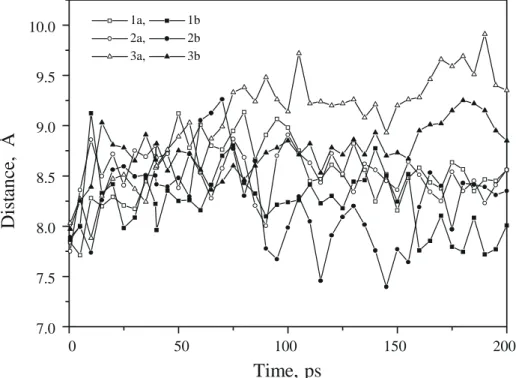
Fig. 2. Time course of the distance (in Å) between the center of mass of peptides 1–3 and that of the host HLA-B27 subtype (a: B*2703; b: B*2705).
ligand binding cannot account for the different binding affinities of these peptides. Stable rmsd values (1–1.5 Å) were also observed for bound peptides. Interestingly, they remain in the same range as that observed for different peptides co-crystallized with the same MHC host protein [55,56]. One exception concerns the weakest binder (pep-tide 3 to HLA-B*2703), for which larger distortions (up to 2.5 Å) were observed and still increasing after 200 ps simulation.
To follow a possible dissociation of peptides 1–3 from the two HLA-B27 alleles, the distance between ligand and protein center of mass (cmass) was monitored. For bind-ing peptides (1, 2), whatever the B27 subtype, this inter-molecular distance increases during the warm-up phase from 8 to 8.5 Å and remains constant for the rest of the simulation (Fig. 2). The weak binder (peptide 3) exhibits a slight but continuous increase of the intermolecular distance, suggesting a partial dissociation of the ligand from the binding groove. This phenomenon was signifi-cantly enhanced for the less stable complex (3a: peptide
3 in complex with HLA-B*2703). However, localization
of the dissociating peptide amino acids is not possible with this analysis. For example, TcR-binding residues may bulge even more out of the peptide binding site and induce similar shifts in the intermolecular distance be-tween center of masses.
Therefore, this analysis was extended by computing the cmass of bound peptide substructures (MHC anchors: P1, P2, P3, P9; TcR anchors: P4, P5, P6, P7, P8) for each MD conformation and the distance relating it to the
protein cmass (Table 2). An examination of d2 and d3 intermolecular distances for the weak binder 3 clearly shows that MHC-anchoring amino acids only are pro-gressively expelled from the binding groove. The dissocia-tion is more significant when compound 3 is complexed to HLA-B*2703, which relates well with the observed binding data (Table 1). The TcR-binding region (P4–P8) is similarly bulging out (at least quantitatively) of the binding cleft for all the six studied complexes (d3 dis-tance, Table 2). An even more precise analysis has been done by evaluating the inter-cmass distance between individual MHC anchor residues and their complemen-tary pocket (A, B, D and F; d4–d7 distances, Table 2). Surprisingly, the repulsion noticed for peptide 3 in com-plex with both alleles seems to be located at the P9–pocket F interaction level (d7=7.3 Å), far from where protein and peptide single-point mutations occur. Interest-ingly, the critical d5 distance, illustrating the strength of the most important interaction between the invariant Arg2 and pocket B, is higher (5.1±0.4 Å) for the less stable complex (3a) than for other pairs (Table 2). The different d4 distances reported here, especially for peptides 1 and
3 and peptide 2, are related to the size of the
correspon-ding P1 side chain (Arg versus Ala). It indicates that the Arg1
side chain is pointing away from its binding subsite (pocket A) and therefore induces higher d4 inter-cmass distances. However, for a given P1 side chain, higher d4 distances are always observed for HLA-B*2703. This indicates that the Tyr59→
His mutation found in the latter allele is detrimental for a stable and strong binding of P1 side chains to pocket A of the protein. The highest
stan-dard deviations are found for the P3–pocket D
interac-TABLE 2
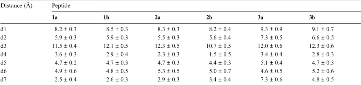
DISTANCE BETWEEN PROTEIN AND PEPTIDE CENTER OF MASSES Distance (Å) Peptide 1a 1b 2a 2b 3a 3b d1 08.2±0.3 08.5±0.3 08.3±0.3 08.2±0.4 09.3±0.9 09.1±0.7 d2 05.9±0.3 05.9±0.3 05.5±0.3 05.6±0.4 07.3±0.5 06.6±0.5 d3 11.5±0.4 12.1±0.5 12.3±0.5 10.7±0.5 12.0±0.6 12.3±0.6 d4 03.6±0.3 02.9±0.4 02.3±0.3 01.5±0.5 03.4±0.4 02.8±0.3 d5 04.7±0.2 04.7±0.3 04.7±0.3 04.4±0.3 05.1±0.4 04.7±0.3 d6 04.9±0.6 04.8±0.5 05.3±0.5 05.0±0.7 04.6±0.5 05.2±0.6 d7 02.5±0.4 02.6±0.3 02.9±0.3 03.4±0.4 07.3±0.6 04.8±0.5
d1: protein–peptide; d2: protein–MHC anchors (P1–P3, P9); d3: protein–TcR anchors (P4–P8); d4: pocket A–P1; d5: pocket B–P2; d6: pocket D–P3; d7: pocket F–P9. P1 P2 P3 P4 P5 P6 P7 P8 P9 0.6 0.8 1.0 1.2 1.4 1.6 1.8 1a, 1b 2a, 2b 3a, 3b
rm
sf
,
Å
Pn
Fig. 3. Rms atomic fluctuations of peptides 1–3 when bound to an HLA-B27 protein (a: B*2703; b: B*2705). Pn represents the peptide position (from 1 to 9).
tion (d6, Table 2). The 0.6 Å variations, observed in such nonbonded distances, correspond to the alternative estab-lishment of strong and weak hydrophobic contacts, which may be explained by the topology of the binding groove. Pocket D is a hydrophobic subsite open to the central part of the binding cleft, and partially filled by hydro-phobic side chains [20]. The significantly higher variance of the d6 distance (Table 2) suggests that this interaction is the most flexible one and that different local conforma-tions at P3 are compatible with a good occupancy of pocket D. Retrospectively, it explains why the P3–pocket D interaction may be important for an optimal peptide binding to HLA-B*2705. Bulky hydrophobic nonnatural side chains (α- andβ-naphthylalanine, cyclohexylalanine) have been shown to significantly enhance binding to the
HLA-B*2705 protein [38,57]. This positive effect probably results from two correlated components: (i) the existence of additional contacts to pocket D, a pure enthalpic ef-fect; and (ii) a reduced flexibility of the MHC-bound P3 side chain, a favorable entropic effect.
Atomic fluctuations
Atomic fluctuations of MHC-bound peptides were com-puted from mean conformations, time-averaged over the last 50 ps (Fig. 3). As expected, MHC anchors are much less flexible than the TcR-binding middle part. If atomic mobility of the bound ligand is considered, it is not poss-ible to depict real differences in the binding of peptides 1 and 2 to the two subtypes. However, the C-terminal an-chor residue clearly tends to be more flexible when the corresponding ligand does not strongly bind to the B27
subtype. The highest flexibilities are observed for the P1 P2 P3 P4 P5 P6 P7 P8 P9 0.0 0.5 1.0 1.5 2.0 1a, 1b 2a, 2b 3a, 3b
Acces
sible
/
Buried
Pn
Fig. 4. Ratio of accessible to buried surface area for relaxed MD time-averaged conformations. Surface areas were calculated with the MS program [75] using a 1.4 Å probe radius. High ratios were truncated to a value of 2.0. Pn represents the peptide position (from 1 to 9).
1a 1b 2a 2b 3a 3b 0 5 10 15 20 25 30
H-bonds
Number
Complex
>50% 25-50%Fig. 5. MHC–peptide H-bonding frequency for peptides 1–3 in complex with HLA-B27 subtype (a:B*2703; b: B*2705). H-bonds have been geometrically defined by an acceptor (A) to donor (D) distance less than 3.25 Å and a D-H..A angle greater than 120°. Interactions were statistically monitored throughout the simulations for a total of 400 conformations per MHC–peptide complex. Two categories of H-bonds were defined: strong ones with frequencies higher than 50%and medium ones with occurrences between 25 and 50%.
weakest binder (peptide 3 to B*2703), especially at the important anchoring positions (P2, P3 and P9). It is logi-cal to find that the mutation at P2 (for peptide 3)
weakens the interactions to MHC pocket B that has been designed to accommodate an arginine side chain [32]. However, the P2 amino acid is more flexible when the corresponding peptide is complexed to HLA-B*2703
(compare 3a and 3b, Fig. 3). Again, the most striking
Tyr59
Tyr59
His59
His59
Glu45
Glu45
Glu63
Glu63
Tyr171
Tyr171
Trp167
Trp167
Met5
Met5
Tyr159
Tyr159
Tyr99
Tyr99
His9
His9
Thr24
Thr24
Tyr7
Tyr7
P1
P1
P2
P2
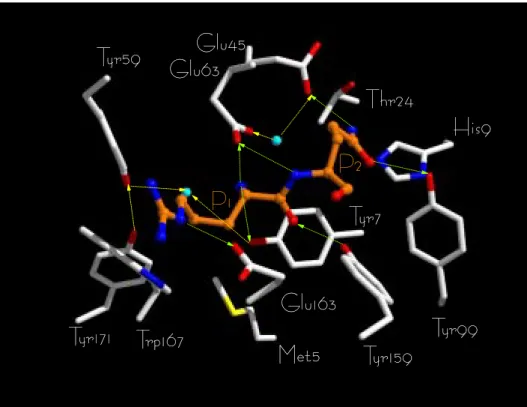
Fig. 6. Crystal structure of HLA-B*2705 [32]. The view is focused on MHC pocket A (Met5, Tyr7, Tyr59, Glu63, Tyr159, Trp167, Tyr171) and pocket
B (His9, Thr24, Glu45, Tyr99) side chains interacting via H-bonds (direct H-bonds: green broken lines; water-mediated H-bonds: yellow broken lines)
with the P1–P2 positions of a bound peptide. The following color coding has been used: carbon, white (protein) or orange (peptide); nitrogen, blue; oxygen, red; sulfur, yellow. Bound water molecules are shown as cyan balls. Arrows indicate the direction of the H-bonds (from the donor to the acceptor). HLA-B*2703 was obtained by mutating Tyr59into His (SYBYL Biopolymer module) [43]. The side-chainχ2dihedral was just
modified here in order to bring the Nεatom as close as possible to the bound water molecule. However, whatever the rotamer chosen, a direct H-bond to a water molecule is not possible. The interatomic distance between the peptide N-terminus and Tyr59(OH) or His59(Nε2) atoms is 4.31
and 5.35 Å, respectively. Figures 6–8 have been obtained by using the rendering program Raster 3D [74].
differences are not observed at the variable amino acids of the peptides (P1, P2) but at the C-terminal anchor (P9), a feature already noticed for other MHC–peptide complexes [9]. One may hypothesize that the N-terminal tripeptide (P1-P2-P3) determines the stability of peptide– MHC interactions over the whole length of the binding groove by controlling the conformational space accessible to the bulging middle part and, consequently, the binding capacity of the following C-terminus. However, atomic flexibilities of the P1–P3 and P4–P8 parts are not interre-lated. It is also possible that these differences are related to the short time scales (200 ps) used for simulating the complexes and that much longer simulations are needed to see significant molecular differences at positions P1 and P2 of the peptide ligand. Nanosecond MD simula-tions of macromolecules are nowadays feasible [58,59], but still remain unrealistic as a structure–activity relation-ship tool for comparing a series of ligands in their pro-tein-bound state.
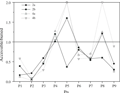
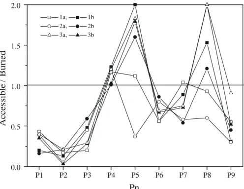
Accessible versus buried surface areas
Whether peptide flexibility correlates with dissociation from the binding cleft was addressed by looking at
access-ible and buried surface areas of each HLA-bound peptide residue (Fig. 4). Only position 9 of peptide 3 in complex with B*2703 was much more accessible than the others. Otherwise, the main anchor P2 was similarly buried what-ever the peptide and the host HLA protein. This means that a partial dissociation was only observed for one position (P9) in one complex (3a) and that the previously reported higher flexibility of Gln2
for the same peptide– MHC complex did not correspond with a release of the P2 side chain from pocket B. It may however influence the quality and frequency of hydrogen bonds between the Gln2
side chain and pocket B of both B27 alleles. The influence of secondary anchor positions is more difficult to ascertain. P3 is similarly buried for all MHC–peptide complexes. P6 (Ser) and P7 (Thr) positions are probably accessory anchor positions that marginally bind, in some conformations, to the central part of the peptide binding groove (pockets C/E). This observation is not incompat-ible with the high atomic fluctuations of P6-P7 amino acids (Fig. 3), as their side chains are directed towards the binding groove but without reaching its floor. P4 (Gln), P5 (Lys) and P8 (Glu) residues are potential candidates for TcR recognition because of their concomitant atomic flexibility and surface accessibility.
Qualitative and quantitative analysis of peptide–MHC
H-Glu45
Glu45
Glu63
Glu63
Tyr171
Tyr171
Trp167
Trp167
Met5
Met5
Tyr159
Tyr159
Tyr99
Tyr99
His9
His9
Thr24
Thr24
Tyr7
Tyr7
P1
P1
P2
P2
His59
His59
Fig. 7. MD model of complex 3a, focused on MHC pockets A–B (see the legend to Fig. 6). A mean conformation was averaged from the last 100 conformers and submitted to 500 steps of steepest descent, followed by 1500 steps of conjugate-gradient energy relaxation.
Glu45
Glu45
Glu63
Glu63
Tyr171
Tyr171 Trp167
Trp167
Met5
Met5
Tyr159
Tyr159
Tyr99
Tyr99
His9
His9
Thr24
Thr24
Tyr7
Tyr7
P1
P1
P2
P2
Tyr59
Tyr59
Glu163
Glu163
Fig. 8. MD model of complex 3b, focused on MHC pockets A–B (see the legend to Fig. 6). The mean conformation was obtained as described in Fig. 7.
bonds
Reporting the number of MHC–peptide H-bonds as well as their frequencies during the simulation allows a
clear distinction between peptides 1 and 2 and the Gln2 analogue (peptide 3, Fig. 5). The stability of protein– peptide H-bonds was assessed by computing the frequency of occurrence of the interaction throughout the MD
tra-jectory (400 conformations). A frequency higher than 50% MHC–PEPTIDE H-BONDS WITH A FREQUENCY HIGHER THAN 50% Pn atom HLA-B27 1a 1b 2a 2b 3a 3b P1 N Tyr7(OH) 1 1 1 His59(NE2) 1 1 1 Glu45(OE2) 3 Glu63(OE1) 1 1 1 Glu63(OE2) 1 1 Glu163(OE2) 3 NE Glu163(OE2) 1 × × 1 NH2 Glu63(OE1) × × 3 Glu63(OE2) × × 3 O Tyr159(OH) 1 1 1 1 P2 N Glu63(OE1) 1 1 Glu63(OE2) 1 1 NE Glu45(OE2) 3 × × Glu63(OE1) 3 × ×
OE1 Tyr99(OH) × × × × 3
NE2 His9(NE2) × × × × 3
Glu45(OE2) × × × × 3 NH1 His9(NE2) 1 1 1 × × Thr24(OG1) 3 × × NH2 Thr24(OG1) 1 1 1 × × Glu45(OE1) 1 1 1 × × Glu45(OE2) 1 1 1 × × Glu63(OE1) 3 × × P3 N Tyr99(OH) 1 1 1 1 P6 OG Ala69(O) 1 Thr73(OG1) 1 P8 O Trp147(NE1) 1 1 1
OE1 Lys146(OE1) 1
P9 N Asp77(OD1) 1 1 1 1 1
OXT Tyr84(OH) 1
Thr143(OG1) 1 1 1
Lys146(NZ) 1 1 1
Empty boxes indicate interactions that are common to at least two complexes, whereas filled boxes represent unique MHC–peptide hy-drogen bonds. The absence of a specific side chain is featured by a cross.
was chosen to characterize strong H-bonds. Medium in-teractions were assigned a frequency between 25 and 50%. About 25 H-bonds have been identified for peptides 1 and 2 in complex with B*2703 and B*2705 while 50%less could be found for the Gln2
analogue with the two sub-types (Fig. 5). The distribution of strong and medium H-bonds correlates well with the binding potency of the peptide. A similar number of strong H-bonds were found
lar binding efficiencies of peptides 1 and 2 to both sub-types. On the other hand, a reduced number of medium and/or strong H-bonds (peptide 3 in complex with the two alleles) correlates with the decreased binding of this peptide. The weakest binding potency (peptide 3 to B*2703) could effectively be qualitatively and quantitat-ively related to the distribution of intermolecular H-bonds. Not only the number but also the quality of the MHC–ligand interactions correlates well with the binding potency.
To accurately localize the interactions that may explain peptide specificity variations, all H-bonds with frequencies higher than 50% were identified for the six complexes (Table 3). The first noticeable difference between the two HLA-B27 alleles is the H-bonding network between the MHC residues involved in binding to the peptide P1 position. In HLA-B*2705, two amino acid side chains are H-bonded to the peptide N-terminus (Tyr7
/Tyr171 in the crystal structure, Tyr7/Glu63in the MD models) (Table 4, Figs. 6–8). Both side chains are fixed by a subtle water-relayed H-bond network involving proximal MHC side chains (Tyr59, Glu45, Tyr171). Tyr59 is directly bound to Tyr171
, and indirectly to Tyr7 , Glu45
and Glu63
. The single point mutation occurring for HLA-B*2703 (Tyr59→
His) perturbs this network. The bound water molecule disap-pears and the peptide N-terminus binds to His59and no more to Tyr7
(Table 3, Fig. 7). The consequence on the H-bond balance is a loss of one direct MHC–MHC inter-action (His59
cannot interact with Tyr171
) and five water-mediated interactions for complex 3a (Fig. 7). The result-ing conformational change may be well accommodated as far as P2 is strongly bound to pocket B (His9
, Thr24 , Glu45
) and the resulting H-bonds are strong enough to maintain the peptide in the binding groove (P2=Arg). If P2 is not an arginine (peptide 3), the resulting interaction to pocket B (Thr24
and Glu45
notably) is much weaker and the conformational rearrangement at P1 is important (see the three new H-bonds for the P1 position in com-plex 3a, Fig. 7). The Arg to Gln change at the P2 posi-tion of the bound peptide is better tolerated by HLA-B*2705 (complex 3b, Fig. 8) as the Tyr59side chain is still able to fix the position of Tyr7
. During the MD simula-tion, the N-terminal Cα-N bond of the bound peptide has rotated to gain a new H-bond to Glu63
. However, it is still bound to Tyr7 as in the reference structure. Import-antly, the Gln2
side chain is bound to Glu45
and Tyr99 , thus providing additional interactions to pocket B when compared to complex 3a (Fig. 8). The quality of the interaction between Gln2
and pocket B is, however, much inferior to that observed for peptide analogues 1 and 2 bearing an optimal Arg residue (Table 4), thus explaining the reduced binding affinity of peptide 3 for HLA-B*2705.
Arg over Ala at P1 could be quantified by the gain of P1 P2 P3 P4 P5 P6 P7 P8 P9 0.0 0.5 1.0 1.5 2.0 2a 2b 4a 4b
Acces
sible/
buried
Pn
Fig. 9. Accessible versus buried surface areas of peptides 2 and 4 in complex with B*2703 (a) and B*2705 (b) alleles (see the legend to Fig. 4).
2a 2b 4a 4b 0 5 10 15 20 25 30
H-Bonds
Complex
> 50% 25- 50%Fig. 10. Intermolecular hydrogen bonds for peptides 2 and 4 in complex with B*2703 (a) and B*2705 (b) alleles (see the legend to Fig. 5). two water accessible salt bridges to Glu63
/Glu163
(Table 3). However, this does not correspond to a higher binding affinity of the Arg analogue when compared to the Ala1 peptide. An Ala side chain is much easier to desolvate and optimally interacts with conserved apolar residues of pocket A (Met5
, Trp167
), thus explaining a rather similar binding affinity of peptides 1 and 2 to both subtypes.
However, the present model cannot fully explain recent data, indicating that basic residues are overrepresented at the P1 position of B*2703-bound natural ligands [23]. From a purely statistical point of view, various rotamers of basic P1 side chains could develop a salt bridge with at least three negatively charged amino acids located at the rim of pocket A (Glu58
, Glu63 , Glu163
), and thus stabilize the MHC–peptide complex.
MHC–peptide interaction energies
MHC–PEPTIDE INTERACTION ENERGIES CALCULATED FROM THE LINEAR POISSON–BOLTZMANN EQUATION AND AMBER FORCE-FIELD CALCULATIONS
Peptide ∆G0 coul a ∆G0 reac b ∆G0 elec c ∆H elec d ∆H vdw e ∆G tot f ∆H tot g 1a −499 487 −12 −87 −90 −−102 −−177 1b −542 498 −44 −79 −97 −−141 −−176 2a −359 340 −19 −72 −77 0−−96 −−149 2b −389 335 −54 −75 −80 −−134 −−155 3a −306 334 −28 −44 −68 0−−40 −−112 3b −371 380 −09 −62 −67 0−−58 −−129 a∆G0
coul: Coulombic component of MHC–peptide electrostatic interaction energy (charge–charge, charge–dipole, dipole–dipole interactions).∆G 0 coul = ∆G0 coul(P–L)− ∆G 0 coul(P)− ∆G 0
coul(L)[50], where P–L describes the protein–ligand complex, P the protein and L the ligand. b∆G0
reac: corrected self-reaction field component of MHC–peptide electrostatic interaction energies (energy required to transfer a molecule from
a continuum dielectric (vacuum) to another (water).∆G0
reac= ∆G0reac(P–L)− ∆G0reac(P)− ∆G0reac(L). As the contribution of the protein–ligand complexes
(∆G0
reac(P–L)) and of the isolated protein (∆G0reac(P)) could be omitted from the calculation without affecting the reliability of the results [61], this
component corresponds here to the free energy of peptide desolvation (−∆G0 reac(L)). c ∆G0
elec: total electrostatic interaction energy (∆G 0 coul+ ∆G
0 reac). d∆H
elec: AMBER electrostatic interaction energy (ε =4rij). e ∆H
vdw: AMBER van der Waals interaction energy.∆Hvdw=A/r
12−B/r6, where r is the distance between atom pairs and A and B are
atom-type-dependent parameters.
f ∆G
tot: total interaction energy (∆G0elec+ ∆Hvdw). gDH
tot: total AMBER interaction enthalpy (∆Helec+ ∆Hvdw).
TABLE 5
INFLUENCE OF A P1 β-AMINO ACID ON THE HLA-B27
SUBTYPE SELECTIVITY OF A MODIFIED HLA-B27
LIGAND Peptide Sequence EC50(µM) a number B*2703 B*2705 (P1-RYQKSTEL) 2 P1=Ala 3.0 04.0 4 P1=Balb 7.5 20
aConcentration of the peptide (in µM) at which HLA-B27
fluor-escence (measured by FMC analysis with an anti-B27 monoclonal antibody) on RMA-S cells was half the maximum obtained with the wild-type peptide (peptide 1, Table 1).
bBal:β-alanine (H
2N-CH2-CH2-CO). Interaction energies were extrapolated for all six
ener-gy-minimized time-averaged conformations (Table 4) by summing up the van der Waals nonbonded interaction energy (calculated with the AMBER 4.0 force field) and the electrostatic component (calculated by solving the linear form of the Poisson–Boltzmann equation), as re-cently described [60]. As both ligands and protein struc-tures are very similar for all complexes, distortion ener-gies as well as translational/rotational entropy losses upon binding were neglected here. Moreover, the self-reaction field energy component of the electrostatic interaction energy was limited to the contribution of the isolated peptide (free energy of desolvation) and calculated from the bound-peptide coordinates extracted from the MHC– peptide binary complexes. It has recently been shown that neglecting the protein contribution to the self-reaction field energy is indeed possible and does not alter the reliability of the obtained results [61].
Our computational protocol is able to properly rank the binding of the three peptides 1–3 to both MHC al-leles. Peptide 3 clearly interacts much weaker than the other two peptides 1 and 2, whatever the MHC allele. The weakest interaction energy was observed for binding of 3 to B*2703, and is thus in agreement with binding data (Table 1). Force-field interaction enthalpies (calcu-lated by summing up both AMBER van der Waals and electrostatic components, using a dielectric permittivity of 4rij) were much less related to the observed binding data, as peptide 2 was always disfavoured with regard to pep-tide 1 (Table 4). Notably, taking into account the peppep-tide desolvation energy by the continuum eletrostatics method permits to compensate for the weakest Coulombic interac-tions provided by peptide 2 (P1=Ala) to both alleles, with
respect to that observed for the Arg1
analogue. It may be noticed that the free electrostatic interaction energies (∆G0
elec, Table 4) computed by the continuum electrostatics method were also in rather good qualitative agreement with the binding data reported in Table 1. Hence, the three peptides are highly polar and interact mainly via H-bonds and salt bridges.
For an even more realistic ranking of highly polar li-gands than those presented here, free energy perturbation [62] is probably the method of choice. Unfortunately, the enormous amount of CPU time that would be necessary for this computation precludes its systematic use in fast screening of a set of congeneric molecules. Synthesis and in vitro binding assays in this case provided a faster and experimentally determined answer.
MD simulation of MHC–peptide complexes could relate observed binding potencies and allele specificity to simple molecular criteria (inter-cmass distances, atomic fluctuations, accessible surface areas, distribution and
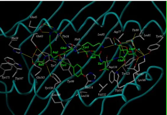
location of intermolecular H-bonds). A single point muta-Bal1 Arg2 Tyr3 Gln4 Lys5 Ser6 Thr7 Glu8 Leu9 Tyr84 Asp77 Thr80 Leu81 Leu95 Lys146 Trp147 Val152 Thr143 Tyr123 Asp116 His114 Leu156 Leu160 Tyr159 Glu163 Trp167 Tyr171 His59 Glu63 Glu45 Thr24 His9 Tyr99 Tyr7
Fig. 11. Energy-minimized time-averaged MD model of complex 4a. The MHC protein backbone is displayed as a solid cyan tube, with peptide-inter-acting side chains. The bound peptide 4 is represented by sticks. The following color coding has been used: carbon, white (protein) or green (peptide); nitrogen, blue; oxygen, red; sulfur, yellow. Bound water molecules are shown as cyan balls. Yellow broken lines indicate MHC–peptide H-bonds.
Bal1 Arg2 Tyr3 Gln4 Lys5 Ser6 Thr7 Glu8 Leu9 Thr80 Leu81 Asp77 Leu95 Trp147 Lys146 Thr143 Ty123 Asp116 His114 Val152 Leu156 Leu160 Tyr159 Tyr99 Tyr7 His9 Thr24 Glu163 Trp167 Ty171 Tyr59 Glu63 Glu45 Tyr84
Fig. 12. Energy-minimized time-averaged MD model of complex 4b (see the legend to Fig. 11). tion in the HLA binding groove is sufficient to break an
H-bond network in the vicinity of the peptide N-terminus. As previously suggested on the basis of peptide binding
analyses [28], this minor change strengthens even more the binding role of the dominant anchor P2 side chain (Arg) for one allele (HLA-B*2703) and explains why changing P2 to Gln has more detrimental effects in
pep-where compensatory stabilization of the MHC–peptide complex is still possible by H-bonded MHC side chains. Interestingly, the most spectacular consequence of pro-tein/peptide variability affects an area far away (10 Å) from the location of the point mutations. It concerns the stability of the interaction between the peptidic C-ter-minal residue and its complementary pocket F, which has recently been shown to play a decisive role in linking particular B27 alleles to spondyloarthropathies [24,26]. Protein-based design
Relating the structure of B27 subtypes to the sequence of their naturally bound peptides is a crucial step in identifying potential immunodominant epitopes that may discriminate alleles and confer susceptibility or resistance to autoimmune diseases. One striking feature concerns the single point mutation (Tyr59His) distinguishing HLA-B*2705 from HLA-B*2703, which is unique among HLA proteins. It is believed that B*2703 selects a subset of the peptides presented by HLA-B*2705 [34]. Recent studies have identified some peptides that are naturally presented by both subtypes, and at least one natural B*2705 ligand (the undecamer RRYLENGKETL) is not presented by B*2703 [23,63]. The only difference between both alleles concerns the position 59 located in pocket A which inter-acts with the N-terminal amino acid of the bound peptide (Fig. 6). As pocket A is slightly wider for HLA-B*2703, extending the peptide backbone towards His59
by replacing the natural P1 residue by aβ-amino acid should theoreti-cally allow a better discrimination of both alleles. This structural change should be much better accommodated by HLA-B*2703 (H-bond between His59
and the N-terminus of the P1β-amino acid) than by HLA-B*2705, for which a steric clash with the Tyr59side chain may be expected.
Starting from the self-peptide 1 (RRYQKSTEL) nat-urally presented by HLA-B*2705 [20] and B*2703 [23], Ala and Bal (β-alanine) were substituted for the natural Arg at P1 (Table 5). The Ala1
peptide analogue was here taken as a reference for its strong binding to both sub-types. The two ligands were synthesized and tested for their binding to B*2703 and B*2705. As expected from the topology of the binding cleft, only the Bal analogue could discriminate between the two subtypes, with a bet-ter binding to HLA-B*2703 (Table 5).
To rationalize the experimental binding data, the non-natural ligand 4 was simulated a posteriori, in complex with both alleles, using exactly the same conditions as those employed for simulating the natural MHC–peptide complexes (see the section Computational procedures). Using two of the previously described molecular parame-ters (accessible versus buried surface area of the bound ligand, intermolecular H-bonds) as quality control of the complex stability, peptide 4 was indeed found to be much better accommodated by B*2703 than by B*2705 (Figs.
peptide to B*2705 could also be related to a partial disso-ciation of the C-terminus from its complementary pocket F (Fig. 9), far away from the peptide mutation site. The present data, in agreement with previous MD simulations of different MHC–peptide complexes [9], suggest that the expulsion of the C-terminus from pocket F could be the very first event in the dissociation of weak binding pep-tides from class I MHC binding grooves. The modified P1 position is, however, significantly more buried when the host protein is the B*2703 allele (Fig. 9). A qualitative and quantitative analysis of intermolecular hydrogen bonds also supports the reported binding data. A total of 15 H-bonds could be depicted for complex 4b (peptide 4 in complex with B*2705), whereas 26 interactions have been found for complex 4a (peptide 4 in complex with B*2703, Fig. 10). However, this analysis was unable to explain the reduced affinity of theβ-Ala compound for B*2703, when compared to that of the natural Ala ana-logue 2 (Table 4). The slight differences seen in the epi-tope stabilization assay are certainly too subtle for the short MD runs reported here. They probably result from: (i) the absence of a side chain at position P1 of ligand 4; and (ii) a weaker binding contribution of the bulging P4–P8 part, for which higher atomic fluctuations (data not shown) and less nonbonded contacts (see the high solvent accessibility of the P5 and P8 residues for ligand
4, Fig. 9) have been noticed. Energy-minimized
time-averaged conformations of both complexes (Figs. 11 and 12) clearly depict significant differences in the MHC pocket A (Tyr59, Trp167, Tyr171), which deviates dramati-cally from the starting crystal coordinates for B*2705 only (rmsd values from all pocket A atoms of 1.5 and 2.5 Å for B*2703 and B*2705, respectively). The major con-formational alterations upon Bal1 binding were observed for the Tyr59
-Trp167 -Tyr171
triad (rmsd values of 1.7 and 2.7 Å for B*2703 and B*2705, respectively)sl. As pre-dicted, the Tyr59 side chain was shifted away from the peptide N-terminus and is now interacting via a water molecule with the β-alanine terminal ammonium (Fig. 12). In contrast, the β-amino acid can directly interact with the larger pocket A of B*2703 through three H-bonds to His59
(mutated position), Glu63
and Glu163 (Fig. 11). Another significant difference in the binding of pep-tide 4 to both alleles concerns the C-terminal amino acid, which has nearly lost, upon binding to B*2705, all H-bonds to the polar side chains of pocket F (Tyr84
, Thr143 , Lys146
; compare Figs. 11 and 12). The incorporation of a
β-amino acid at P1 has modified the above described H-bond network between MHC side chains and the peptide N-terminus. The bound water molecule located in pocket A of HLA-B*2705 (recall Fig. 6) has either disappeared (B*2703, Fig. 11) or has been shifted towards the extreme left end of the binding groove (B*2705, Fig. 12).
incor-poration of a β-amino acid in the peptide sequence does not abrogate binding to HLA-B27 subtypes. Peptide 4 is one of the very first ligands for which the backbone modification of an anchor residue does not abolish class I MHC binding. Up to now, only a retro-inverso (NH-CO instead of (NH-CO-NH) and a reduced peptide bond (CH2-NH instead of CO-NH) pseudopeptide analogue of an HLA-A2-binding peptide have been proposed as suc-cessful P1 modifications [64,65]. However, aβ-amino acid at P1 presents the advantage to preserve the backbone direction of the peptide ligand and the H-bonding capac-ity of the first peptide bond (to Glu63and Tyr159), so that less 3D conformational changes of the MHC binding cleft are necessary to accommodate the modified ligand. The recently described X-ray structure of an MHC–peptide– TcR ternary complex [66] suggests that the latter feature may be of particular importance for a proper recognition of the MHC–ligand pair by a TcR. Furthermore, it opens the door to the incorporation of β-amino acids at other anchor positions, notably P2, P3 and P9. Potential TcR-binding amino acids have already been replaced by vari-ous organic spacers without affecting the binding of the corresponding ligand to class I MHC proteins [38,67,68]. The present design study demonstrates that substituting aβ-amino acid for a natural residue is a further solution for designing high-affinity MHC ligands with improved stability and pharmacokinetic properties. This is an abso-lute prerequisite for the therapeutic use of MHC ligands either as MHC blockers [69] or as T-cell receptor antag-onists [70].
Conclusions
MD simulations have been used in the present study as a tool for explaining peculiar structure–activity relation-ships at the level of the protein–ligand interaction com-plexes. The current study is not aimed at quantitatively ranking MHC ligands and predicting their binding affin-ities. For that purpose, free energy calculations using much longer equilibration and conformational sampling would be necessary. More simply, dynamical properties of the modeled complexes can be qualitatively well related to known binding data. Notably, monitoring protein– ligand intramolecular distances, the atomic mobility of the bound ligands, the ratio of accessible versus buried surface areas, the history and the quality of peptide– protein H-bonds allow a clear discrimination of high-affinity from weak-binding peptides. This computational approach, based on the qualitative analysis of short MD trajectories, has already been used to succesfully predict the bound conformation of a natural HLA-A2-restricted epitope [37] prior to X-ray structure determination [55], to identify T-cell epitopes from the primary structure of potentially interesting proteins [9] and to design high-affinity nonnatural ligands [38,71]. Herewith, we propose
its application to the rationalization of peptide specificity for closely related HLA alleles and the design of non-natural ligands with increased specificity for one HLA-B27 subtype. Identifying the molecular rules, fine tuning peptide selection by HLA alleles is a crucial step for better understanding the peptide–HLA interactions that may confer either susceptibility or resistance to immuno-logical diseases associated with particular HLA alleles.
Acknowledgments
D.R. wishes to thank the computational center of the ETH Zürich for generous allocation of computer time on the CRAY J90 and PARAGON machines. This work was supported by the Schweizerischer Nationalfonds zur Förderung der wissenschaftlichen Forschung (Project No. 31-45504.95) and by Grant SAF 94-0891 from the Plan Nacional de I+D to J.A.L.C. J.R.L. is a fellow of the Basque Government.
References
1 Heemels, M.T. and Ploegh, H.L., Annu. Rev. Biochem., 64 (1995) 643.
2 Bjorkman, P.J., Saper, M.A., Samraoui, B., Bennet, W.S., Strominger, J.L. and Wiley, D.C., Nature, 329 (1987) 506. 3 Falk, K., Rötzschke, O., Stevanovic, S., Jung, G. and
Rammen-see, H.-G., Nature, 351 (1991) 290.
4 Spies, T., Bresnahan, M., Bahram, S., Arnold, D., Blank, G., Mellins, E., Pious, D. and DeMars, R., Nature, 348 (1990) 744. 5 Stern, L.J. and Wiley, D.C., Structure, 2 (1994) 245.
6 Madden, D.R., Annu. Rev. Immunol., 13 (1995) 587.
7 Corr, M., Boyd, L.F., Frankel, S.R., Kozlowski, S., Padlan, E.A. and Margulies, D.H., J. Exp. Med., 176 (1992) 1681.
8 Huczo, E.L., Bodnar, W.M., Benjamin, D., Sakaguchi, K., Zhu, N.Z., Shabanowitz, J., Henderson, R.A., Appella, E., Hunt, D.F. and Engelhard, D., J. Immunol., 151 (1993) 2572.
9 Rognan, D., Scapozza, L., Folkers, G. and Daser, A., Biochemis-try, 33 (1994) 11476.
10 Chelvanayagam, G., Jakobsen, I.B., Gao, X. and Easteal, S., Protein Eng., 9 (1996) 1151.
11 Brewerton, D.A., Hart, F.D., Nicholls, A. and Sturrock, R.D., Lancet 2, (1973) 994.
12 Todd, J.A., Bell, J.I. and McDevitt, H.O., Nature, 329 (1987) 599. 13 Wordsworth, B.P., Lanchbury, J.S., Sakkas, L.I., Welsh, K.I., Panayi, G.S. and Bell, J.I., Proc. Natl. Acad. Sci. USA, 86 (1989) 10049.
14 Hill, A.V.S., Elvin, J., Willis, A.C., Aidoo, M., Allsopp, C.E.M., Gotch, F.M., Gao, X.M., Takiguchi, M., Greenwood, B.M., Townsend, A.R.M., McMichael, A.J. and Whittle, H.C., Nature, 360 (1992) 434.
15 Benjamin, R. and Parham, P., Immunol. Today, 11 (1990) 137. 16 Kingsley, G. and Sieper, J., Immunol. Today, 14 (1993) 387. 17 López-Larrea, C., Sujirachato, K., Mehra, N.K., Chiewsilp, P.,
Isarangkura, D., Kanga, O., Dominguez, O., Coto, E., Peña, M., Setién, F. and Gonzales-Roces, S., Tissue Antigens, 45 (1995) 169. 18 Nasution, A.R., Mardjuadi, A., Kunmartini, S., Suryadhana, N.G., Setyohadi, B., Sudarsono, D., Lardy, N.M. and Feltkamp, T.E.W., J. Rheumatol., 24 (1997) 1111.
Zuccarelli, A., Bitti, P.P., Tosi, R. and Sorrentino, R., Eur. J. Immunol., 25 (1995) 3199.
20 Jardetzky, T.S., Lane, W.S., Robinson, R.A., Madden, D.R. and Wiley, D.C., Nature, 353 (1991) 326.
21 Rojo, S., Garcia, F., Villadangos, J.A. and López de Castro, J.A., J. Exp. Med., 177 (1993) 613.
22 Rötzchke, O., Falk, K., Stevanovic, S., Gnau, V., Jung, G. and Rammensee, H.G., Immunogenetics, 39 (1994) 74.
23 Boisgérault, F., Tieng, V., Stolzenberg, M.C., Dulphy, N., Khalil, I., Tamouza, R., Charron, D. and Toubert, A., J. Clin. Invest., 98 (1996) 2764.
24 Garcia, F., Marina, A. and López de Castro, J.A., Tissue Antigens, 49 (1997) 215.
25 Garcia, F., Galocha, B., Villadangos, J.A., Lamas, J.R., Albar, J.P., Marina, A. and López de Castro, J.A., Tissue Antigens, 49 (1997) 580.
26 Fiorillo, M.T., Meadows, L., D’Amato, M., Shabanowitz, J., Hunt, D.F., Apella, E. and Sorrentino, R., Eur. J. Immunol., 27 (1997) 368.
27 Parker, K.C., Biddison, W.E. and Coligan, J.E., Biochemistry, 33 (1994) 7736.
28 Villadangos, J.A., Galocha, B., Garcia, F., Albar, J.P. and López de Castro, J.A., Eur. J. Immunol., 25 (1995) 2370.
29 Galocha, B., Lamas, J.R., Villadangos, J.A., Albar, J.P. and López de Castro, J.A., Tissue Antigens, 48 (1996) 509.
30 Raghavan, M., Lebron, J., Johnson, J. and Bjorkman, P., Protein Sci., 5 (1996) 2080.
31 Madden, D.R., Gorga, J.C., Strominger, J.L. and Wiley, D.C., Nature, 353 (1991) 321.
32 Madden, D.R., Gorga, J.C., Strominger, J.L. and Wiley, D.C., Cell, 70 (1992) 1035.
33 López, D., García-Hoyo, R. and López de Castro, J.A., J. Immunol., 152 (1994) 5557.
34 Villadangos, J.A., Galocha, B., García-Hoyo, R., López, D., Garcia, F. and López de Castro, J.A., Eur. J. Immunol., 24 (1994) 2548. 35 Colbert, R.A., Rowland-Jones, S.L., McMichael, A.J. and
Frelinger, J.A., Immunity, 1 (1994) 121.
36 Bernstein, F.C., Koetzle, T.F., Williams, G.J.B., Meyer Jr., E.F., Brice, M.D., Rodgers, J.M., Kennard, O., Shimanouchi, T. and Tasumi, M., J. Mol. Biol., 112 (1977) 535.
37 Rognan, D., Zimmermann, N., Jung, G. and Folkers, G., Eur. J. Biochem., 208 (1992) 101.
38 Rognan, D., Scapozza, L., Folkers, G. and Daser, A., Proc. Natl. Acad. Sci. USA, 92 (1995) 753.
39 Collins, E.J., Garboczi, D.N., Karpusas, M.N. and Wiley, D.C., Proc. Natl. Acad. Sci. USA, 92 (1995) 1218.
40 Jorgensen, W.L., Chandrasekhar, J., Madura, J.D., Impey, R.W. and Klein, M.L., J. Chem. Phys., 70 (1983) 926.
41 Pearlman, D.A., Case, D.A., Caldwell, J.C., Seibel, G.L., Singh, U.C., Weiner, P. and Kollman, P.A., AMBER 4.0, University of California, San Francisco, CA, U.S.A., 1992.
42 Besler, B.H., Merz, K.M. and Kollman, P.A., J. Comput. Chem., 11 (1990) 431.
43 SYBYL, release 6.2, TRIPOS Associates Inc., St. Louis, MO, U.S.A.
44 Stewart, J.J.P., J. Comput.-Aided Mol. Design, 4 (1990) 1. 45 Dewar, M.J.S. and Thiel, W., J. Am. Chem. Soc., 99 (1977) 4899. 46 Ryckaert, J.P., Cicotti, G. and Berendsen, H.J.C., J. Comput.
Phys., 23 (1977) 327.
A. and Haak, J.R., J. Chem. Phys., 81 (1984) 3684.
48 Klapper, I., Hagstrom, R., Fine, R., Sharp, K. and Honig, B., Proteins Struct. Funct. Genet., (1986) 47.
49 Warwicker, J. and Watson, H.C., J. Mol. Biol., 157 (1982) 671. 50 Gilson, M.K. and Honig, B., Proteins Struct. Funct. Genet., 4
(1988) 7.
51 Gilson, M.K., Sharp, K. and Honig, B., J. Comput. Chem., 9 (1988) 327.
52 Ljunggren, H.G., Stam, N.J., Ohlen, C., Neefjes, J.J., Hoglund, P., Heemels, M.T., Bastin, J., Schumacher, T.N., Townsend, A., Karre, K. and Ploegh, H.L., Nature, 46 (1990) 476.
53 Ellis, S.A., Taylor, C. and McMichael, A., Hum. Immunol., 5 (1982) 49.
54 van Gunsteren, W.F. and Berendsen, H.J.C., Angew. Chem., Int. Ed. Engl., 29 (1990) 992.
55 Madden, D.R., Garboczi, D.N. and Wiley, D.C., Cell, 75 (1993) 693.
56 Reid, S.W., McAdam, S., Smith, K.J., Klenerman, P., O’Callaghan, C.A., Harlos, K., Jakobsen, B.K., McMichael, A.J., Bell, J.I., Stuart, D.I. and Jones, E.Y., J. Exp. Med., 184 (1996) 2279.
57 Rovero, P., Vigano, S., Pegorado, S., Revoltella, R., Riganelli, D., Fruci, D., Greco, G., Butler, R. and Tanigaki, N., J. Pept. Sci., 1 (1995) 266.
58 Brunne, R.M., Berndt, K.D., Güntert, P., Wüthrich, K. and van Gunsteren, W.F., Proteins Struct. Funct. Genet., 23 (1995) 49. 59 Fox, T. and Kollman, P.A., Proteins Struct. Funct. Genet., 25
(1996) 315.
60 Gallego, J., Ortiz, A.R., de Pascual-Teresa, B. and Gago, F., J. Comput.-Aided Mol. Design, 11 (1997) 114.
61 Taylor, N.R. and von Itzstein, M., J. Comput.-Aided Mol. Design, 10 (1996) 233.
62 Bash, P.A., Singh, U.C., Langridge, R. and Kollman, P.A., Science, 236 (1987) 564.
63 Garcia, F., Marina, A., Albar, J.P. and López de Castro, J.A., Tissue Antigens, 49 (1997) 23.
64 Guichard, G., Calbo, S., Muller, S., Kourilsky, P., Briand, J.-P. and Abastado, J.-P., J. Biol. Chem., 270 (1995) 26057.
65 Guichard, G., Connan, F., Graff, R., Ostankovitch, M., Muller, S., Guillet, J.-G., Choppin, F. and Briand, J.-P., J. Med. Chem., 39 (1996) 2030.
66 Garboczi, D.N., Ghosh, P., Utz, U., Fan, Q.R., Biddison, W.E. and Wiley, D.C., Nature, 384 (1996) 134.
67 Weiss, G.A., Collins, E.J., Garboczi, D.N., Wiley, D.C. and Schreiber, S.L., Chem. Biol., 2 (1995) 401.
68 Bouvier, M. and Wiley, D.C., Proc. Natl. Acad. Sci. USA, 93 (1996) 4583.
69 Adorini, L., Muller, S., Cardinaux, F., Lehmann, P.V., Falcioni, F. and Nagy, Z.A., Nature, 334 (1988) 623.
70 De Magistris, M.T., Alexander, J., Coggeshall, M., Altman, A., Gaeta, F.C.A., Grey, H.M. and Sette, A., Cell, 68 (1992) 625. 71 Rognan, D., Habilitationsschrift, ETH Zürich, Zürich,
Switzer-land, 1997.
72 Saper, M.A., Bjorkman, P.J. and Wiley, D.C., J. Mol. Biol., 219 (1991) 277.
74 Kraulis, P.J., J. Appl. Crystallogr., 24 (1991) 846.
75 Merritt, E.A. and Murphy, M.E.P., Acta Crystallogr., D50 (1994) 869.
![Fig. 1. Orientation of a canonical nonapeptide (ball-and-stick model) in the binding groove of HLA-B27 (cyan ribbons) [32]](https://thumb-eu.123doks.com/thumbv2/123doknet/14864834.636910/4.892.153.743.125.527/orientation-canonical-nonapeptide-stick-model-binding-groove-ribbons.webp)
![Fig. 6. Crystal structure of HLA-B*2705 [32]. The view is focused on MHC pocket A (Met 5 , Tyr 7 , Tyr 59 , Glu 63 , Tyr 159 , Trp 167 , Tyr 171 ) and pocket B (His 9 , Thr 24 , Glu 45 , Tyr 99 ) side chains interacting via H-bonds (direct H-bonds: green b](https://thumb-eu.123doks.com/thumbv2/123doknet/14864834.636910/8.892.185.712.125.526/crystal-structure-focused-pocket-pocket-chains-interacting-direct.webp)