Detection of specific collagen types in
normal and keratoconus corneas
David A. Newsome, Jean-Michel Foidart, John R. Hassell, Jay H. Krachmer, Merlyn M. Rodrigues, and Stephen I. Katz
Keratoconus is a corneal disease of unknown cause that involves a progressive thinning and. scarring of the corneal connective tissue. We examined normal human and keratoconus cor-neas, including one healed penetrating keratoplasty specimen. Organ cell cultures of normal and keratoconus corneal specimens were labeled with radioactive proline and analyzed, by CM-cellulose chromatography and slab gel electrophoresis to determine collagen biosynthesis. Collagen types I and HI were synthesized in similar amounts by normal and keratoconus stromacytes in culture. Specifically purified antibodies were used to determine the distribution of collagen types in tissue sections by immunofluorescence. The distribution of collagen types I, III, and IV in keratoconus was also similar to that in normal corneas, except that scarred regions in keratoconus and at the host-graft: juncture were largely type HI. Immunofluorescent reaction of the anti—type IV collagen antibodies with Bowman's layer, in particular, and Descemet's membrane in keratoconus specimens indicated extensive destruction. Basement membrane destruction may play an important role in this disease.
Key words: keratoconus, corneal collagens, collagen types, immunofluorescence, normal and abnormal corneal extracellular matrix
Ke.
;ratoconus is a slowly progressive, seri-ous corneal disorder of uncertain heritability and unknown cause. The clinical hallmark of the disease is the irregular, cone-shaped pro-trusion of the central cornea which results from weakening and thinning of the stromal elements. Although the disease has been re-ported to exhibit familial patterns and anau-From the Section on Retinal and Ocular Connective Tis-sue Diseases (D. N., J. H.) and Ocular Pathology (M. R.), Clinical Branch, National Eye Institute, the Laboratory of Developmental Biology and Anomalies, National Institute of Dental Research (J-M. F.), Der-matology Branch, National Cancer Institute (S. K.), National Institutes of Health, Department of Health, Education and Welfare, Bethesda, Md., and the De-partment of Ophthalmology, University of Iowa (J. K.). J-M. F. is a Fellow of the FNRS in Belgium. Submitted for publication July 5, 1979.
Reprint requests: Dr. David Newsome, Building 10, Room 10 D-17, National Institutes of Health, Be-thesda, Md. 20205.
tosomal recessive mode of inheritance has been postulated,1 most cases appear to be sporadic. In other clinical reports, kera-toconus has been linked to a heritable disor-der of connective tissue, Ehlers-Danlos syn-drome.2"4
Extensive biomicroscopic and pathological studies of keratoconus have so far failed to demonstrate conclusively whether the pri-mary site of the disease is the corneal stroma or the epithelium and its basement mem-brane.5 Ectasia of the stroma likely involves either primary or secondary alterations in its major structural macromolecules or in their interactions with other matrix elements. Col-lagen, the most abundant stromal element (about 71% of dry weight) has naturally received much attention. Histopathological studies have documented stromal scarring and degradative changes in the stromal lamel-lae but have been of little help in elucidating pathogenetic mechanisms.
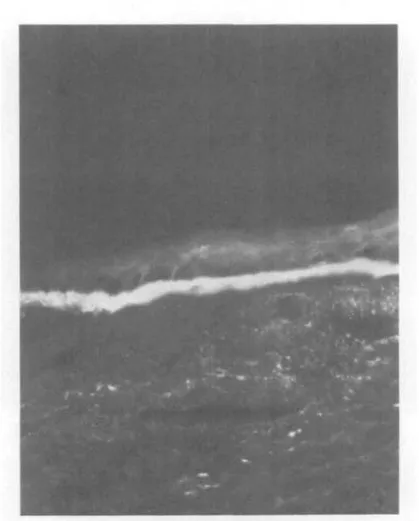
Fig. 1. Fluorescence micrograph of a frozen section of normal cornea reacted with anti-type IV collagen antibodies demonstrates a wide, discrete area of fluorescence in the epithelial basement membrane area. (x250.)
In the present study we have used spe-cifically purified antibodies to characterize the distribution of collagen types in normal human corneal and anterior scleral tissue as well as in keratoplasty specimens from pa-tients with well-documented keratoconus, including one postmortem specimen from a patient who had successful perforating kera-toplasty for keratoconus. We also charac-terized biochemically the collagens synthe-sized by normal and keratoconus stromal cells and normal scleral cells. Our data dem-onstrate the cornea-specific distribution of collagen types as well as some alterations in collagenous components in keratoconus speci-mens.
Materials and methods
Corneal specimens. Five normal and five
kera-toconus corneas were studied. Normal tissue was
obtained at autopsy from individuals aged 7, 22, 28, 35, and 56 years. Corneoscleral specimens were embedded in OCT embedding medium (Ames) and then frozen in liquid nitrogen. Frozen sections (4 jum thick) were transferred to albumin-coated glass slides to serve as substrate for a modified direct immunofluorescent staining reaction. Keratoconus specimens from patients in the age range 28 to 49 years, all with typical clinical criteria for the dis-ease as documented by one of us (J. K.), were placed into cold M-K corneal storage medium at the time of surgery or autopsy and embedded, frozen, and sectioned within 20 hr by the methods described above. The keratoconus specimens had variable amounts of obvious scarring; all had irregu-lar thinning and Fleisher pigmented rings.
Corneal stromal cell cultures were established as previously described6 from explants of pure stroma prepared under a dissecting stereomicroscope. Cell cultures were similarly started from cleanly dissected explants of sclera and conjunctival
740 Newsome et al. Invest. Ophthalmal. Vis. Sci.June 1981
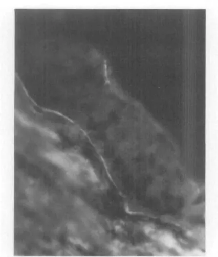
Fig. 2. Fluorescence micrograph of a frozen section of keratoconus cornea reacted with a n t i -type IV collagen antibodies demonstrates a marked reduction in and discontinuity of the amount ol fluorescence in the epithelial basement membrane area as compared with that seen in the normal specimen in Fig. 1. (X500.)
stroma. Primary outgrowths of cells as well as cells in the third serial passage forming confluent layers were used. Cells were maintained in polystyrene culture dishes (Falcon) in Eagle's minimal essen-tial medium with 5% fetal calf serum, 100 u/m] penicillin, and 50 /ng/ml streptomycin, and ex-posed to 37°, 100% humidity, and a 95% air-5% CO2 atmosphere. Confluent cultures were ra-diolabeled by 24 hr incubation in serum-free Dulbecco-Vogt medium (NIH) with 100 ju.Ci/ml
3
H-proline (NET-323; New England Nuclear, Bos-ton), 100 jti.g/ml vitamin C, and 50 yu-g/ml /3-aininoproprionitrile fumarate as a lathyrogen. Medium and cells together with carrier collagen from skins of lathyritic rats were extracted in 0.5M acetic acid containing 1.0 mg/ml pepsin. The colla-gen was purified by NaCl precipitations (25%; two precipitations) in the presence of carrier collagen from skins of lathyritic rats and eluted from a car-boxymethyl (CM)-cellulose chromatography
col-umn (1.6 by 5 cm) as described elsewhere, which permitted an average (n = 4) recovery of 74% of applied counts.7"9 Peak fractions in the effluent were pooled, lyophilized, and electrophoresed in the presence of 0.05M urea on sodium dodecyl sulfate (SDS)-polyacrylamide slab gels.10 Purified types I and III collagen standards were included with each electrophoresis. Reduced samples were obtained by dividing the original 250 /xl samples into two and adding 5 ju.1 of/3-mercaptoethanol to one set of the samples. After electrophoresis the gels were stained with Coomassie blue and were impregnated with 2,5-diphenyloxazole (PPO) in dimethylsulfoxide. Autofluororadiograms on Ko-dak X-Omat film were prepared by exposing the film at —74° for periods up to 7 days.
Preparation of antigens. Types I and 111 colla-gens were isolated from fetal calf skin,8 type II from a rat chondrosarcoma," and type IV from a murine sarcoma.12 Type I procollagen was
ex-tracted from the skin of dermatosporactic calves and was a gift from Dr. C. M. Lapiere. Type III procollagen was purified from fetal calfskin.8 The identity and purity of the collagens was deter-mined by amino acid analysis after hydrolysis in 6N HC1 at 110° for 18 hr; by CM-cellulose and
diethylaminoethyl-cellulose elution profiles, and by SDS-polyacrylamide slab gel electrophoresis with and without /3-mercaptoethanol reduction and with and without cyanogen bromide diges-tion. Human fibronectin was purified from fresh human plasma by affinity chromatography on a gelatin column. The material eluted with 1M po-tassium bromide was electrophoresed after reduc-tion of the disulfide bonds with 20 mM dithio-treitol. The 220,000 dalton chain of fibronectin was eluted from slices of the acrylamide gels and used to immunize rabbits.
Preparation of antibodies to collagens. Rabbits or guinea pigs were immunized by repeated sub-cutaneous or foot pad injections of 0.5 to 5.0 mg of the antigen in Freund's complete adjuvant. Antibodies were isolated and purified by cross-immunoabsorption and checked for cross-reac-tivity to collagen and proteoglycans by indirect immunofluorescence, micropassive hemaggluti-nation assay,13 and radioimmunoassay. Antibodies to collagens were purified from the antisera by affinity chromatography and cross-immunoab-sorption.H Serum from immunized animals was passed sequentially over columns of types II and III collagen and types IV and V collagens which had been covalently bound to Sepharose 4B. The unbound material was then passed over a type I collagen column which was extensively rinsed with phosphate-buffered saline (PBS). Antibodies were eluted from the column with 0.5N acetic acid-0.5M NaCl. This eluate was neutralized with 3M Tris, dialyzed against PBS, and concentrated by filtration on a UM20 Amicon filter. Antibodies to the other antigens were purified according to a similar protocol.
Testing of antibodies to collagens. The spe-cificity of the antibodies was determined by radio-immunoassay (RIA), by immunofluorescence la-beling of target tissues, and by enzyme-linked immunoabsorbent assay (ELISA) as reported pre-viously.15 For RIA, types I and III collagens were labeled with iodine-125 by the chloramine-T method16 (sp. act. 1 to 2 fxCi/ixg). The RIA was
performed according to the method of Rohde et al.17 In quantitative inhibition tests preincubation
of the purified antibody with increasing amounts of the autologous collagen inhibited the binding of the antibody with the labeled antigen, whereas no
Table I. Distribution of ocular connective
tissue proteins in normal and keratoconus specimens as detected by immunofluorescence
Site Proteins detected Cornea] epithelium
Cornea! epithelial basement membrane
Cornea! stroma* Descemet's membrane Corneal scars
Conjunctival blood vessels Sclera No reaction for collagens/fibronectin Type IV collagen Type 1 collagen; fibronectin Type IV collagen; fibronectin Type III collagen; some type I Type IV collagen Type III collagen; some type I
*Type III collagen has been extracted from corneal stroma and characterized biochemically but was not reliably detected by immunofluorescence in our studies.
significant inhibition was observed when the anti-body was preincubated with the heterologous col-lagen. Indirect immunofluorescence studies were performed on fresh-frozen sections of human skin, liver, tendon, and spleen. They established that the purified antibodies reacted only with those structures known to contain these molecules. Fi-nally, indirect immunofluorescence blocking and competition studies confirmed the specificity of the anti-type I and anticollagen antibodies, since a preincubation with the specific antigen blocked the binding of antibodies to known high-affinity substrates. For example, a preincubation with type I collagen blocked the binding of anti-type I collagen antibodies to human skin but did not in-terfere with the binding of anti-type III collagen antibody.
Preparation of antibodies to fibronectin. Specific antibody to fibronectin was purified by affinity chromatography.H Serum from im-munized rabbits was passed over a column of fibronectin which had been covalently bound to Sepharose 4B. The unbound material was then washed away with PBS. Antibody to fibronectin was then eluted from the column with 0.5M acetic acid, pH 2.9, neutralized with 3M Tris, dialyzed against PBS, and concentrated on a UM20 Amicon filter.
Testing of antibody to fibronectin. The spec-ificity of the antibody was determined by RIA, by ELISA,15 by radial immunodiflusion according to Ouchterlony, and by immunoelectrophoresis. For RIA, labeled fibronectin was purified from the cul-ture medium of human fibroblasts grown with
742 Newsome et al. Invest. Ophtlialmol. Vis. Sci.June 1981
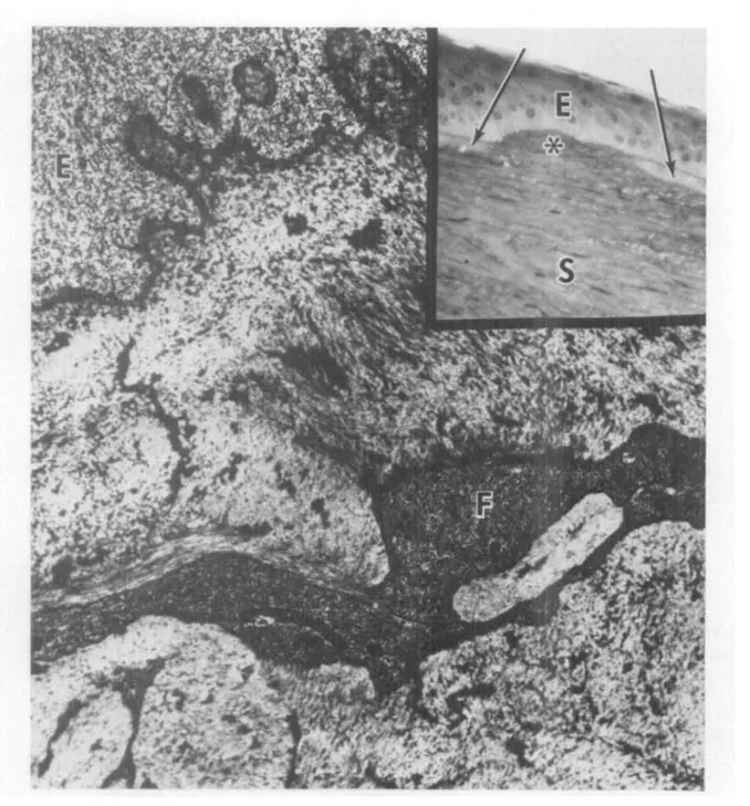
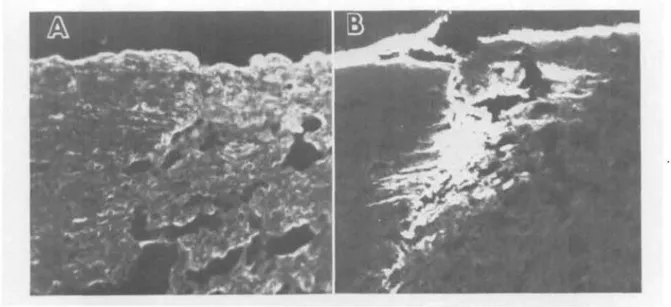
Fig, 3. Inset of the superficial area of a scarred keratoconus cornea shows distruption (asterisk) of Bowman's layer (arrows) and superficial stromal fibrosis. E, epithelium; S, stroma. (To-luidine blue; X350.) The transmission electron micrograph demonstrates irregular subepithe-lial connective tissue excrescences in the basal epithelium (E). Bowman's layer is replaced by irregular connective tissue. F, fibroblast. (x27,000.)
[U-3H]leucine.l8 Fibronectin was also labeled with l2oI by the chloraniine-T method.16 Preincu-bation of the antibody with other antigens, includ-ing types I and III collagens, laminin, and type IV collagen, did not inhibit the binding of the anti-bodv to labeled fibronectin. Bv radial
immu-nodiffusion and immunoelectrophoresis a single line of precipitation was obtained with an an-tifibronectin antibody and purified fibronectin or normal human serum.
Immunofluorescence reaction. Modified direct immunofluorescent staining was performed on
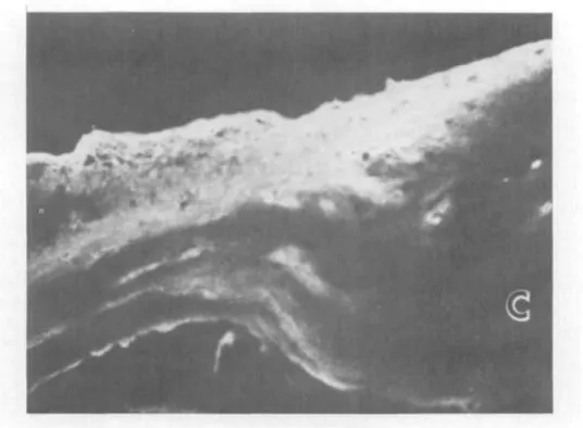
Fig. 4. These sections of a healed perforating keratoplasty specimen from a patient with keratoconus were reacted with anti-type I (A) and anti-type III (B) antibodies. Note the strong fluorescence of the scar with anti-type III and the similarity of staining of the graft and host stromas with both antibodies. (X300.)
sections of cornea and corneoscleral tissue with antibodies and control sera in two dilutions: 10 and 20 /Ag/ml PBS. After a 30 min incubation the sec-tions were washed in PBS and then exposed to fluorescein-conjugated goat-anti-rabbit immuno-globulin for 30 min, washed again in PBS, and mounted with glycerine. Specimens were exam-ined with a Leitz ultraviolet microscope equipped with a camera attachment and a BG 12 filter. Ex-posures were made with an automatic exposure meter on Kodak Ektachrome 200 film.
Results
With the use of the techniques described herein it was not generally possible to assess immunofluorescent reactivity quantitatively. We were able to localize the heterogeneous population of human corneal collagens to specific sites, This distribution is summarized in Table I.
Corneal epithelium. The epithelial layer
it-self did not react with any antibodies. This finding was similar in normal and keratoco-nus corneas. The zone just deep to the epi-thelium showed a strong reaction with an-tibodies to type IV collagen {Fig. 1). In the keratoconus specimens with more advanced stromal thinning, the,reaction with anti-type IV in the epithelial basement membrane
area was strikingly decreased and irregular (Fig- 2).
Bowman's layer. Bowman's layer reacted
similarly with antibodies to types I and III collagen as did the anterior stroma. How-ever, the positivity for anti-type I was noticeably reduced as compared with more posterior layers of the stroma. Bowman's layer reacted more strongly with antibody to fibronectin than did the stroma. Except for the presence of type Ill-containing scars that included part of the Bowman's area in some keratoconus specimens, there was no differ-ence between the types of detectable colla-gens in the normal and keratoconus material. The histological appearance of a typical scar can be seen in Fig. 3.
Corneal stroma. In both normal and
kera-toconus tissue the stroma was strongly posi-tive for type I. Procollagen type I reaction was consistently weaker in all specimens of cornea. In some of the heavily scarred keratoconus specimens there was a patchy increase in detectable procollagen type I in the subepithelial anterior stroma.
Type III collagen was not clearly detected in the normal corneal stroma by fluorescence methods. Consistent reactivity in normal
tis-744 Newsome et al. Invest. Ophthalmol. Vis. Sci.June 1981
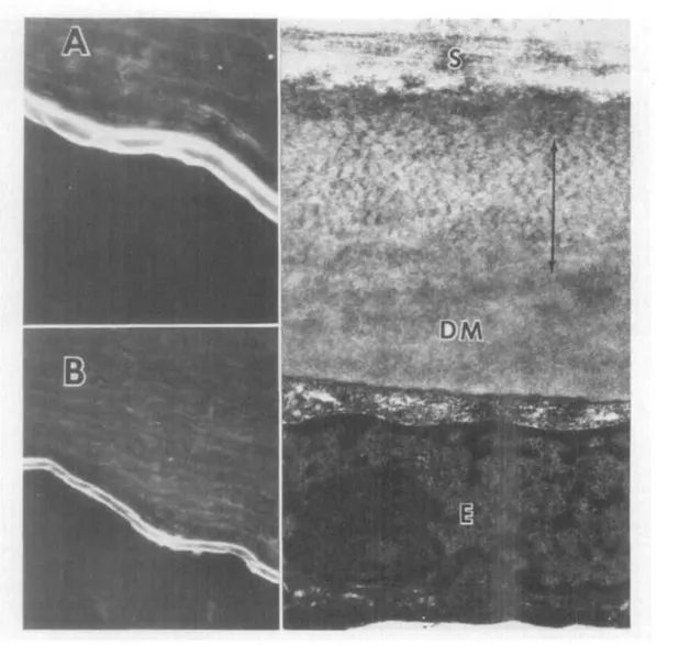
Fig. 5. Fluorescence micrographs (A, X450; B, X300) of a section of normal cornea reacted with antifibronectin antibody exhibits fluorescent anterior and posterior portions of Des-cemet's membrane identical to that pattern seen with anti-type IV antibodies. Right panel, Transmission electron micrograph of normal tissue. The fluorescence pattern was seen in both normal and undisrupted keratoconus Descemet's membrane and may correspond to the nor-mal 100 nm wide-spaced collagen arrow zone (immiinoHuorescent dark interleaf) and anterior and posterior granular basement membrane-like zones of material. S, stroma; DM, posterior Descemet's membrane; E, endothelial cell. (x30,000,) Fibronectin is also detectable in stroma itself in the fluorescence micrographs.
sues was detected only on the edges of stromal lamellae. No differences were visible between the reaction for type III and procol-lagen type III in normal and keratoconus un-scarred stroma.
In the specimen of a successful graft of normal donor into a keratoconus corneal bed, both areas showed the same reaction with antibodies to types I and III. In the grafted
specimen the healed junction between donor keratoconus recipient reacted strongly with antibodies to type III and procollagen type III. The zone of scarring extended into both the host and donor tissues (Fig. 4). Type III collagen could be clearly detected also in the more thinned and scarred keratoconus spec-imens as patches corresponding to stromal scars.
Fig. 6. Fluorescence micrograph of a normal sclerocorneal section reacted with anti—type III antibodies reveals strong fluorescence in Tenon's and the sclera at the liinbal transition zone. Note the relative lack of reaction in the clear cornea (C). This strong reaction was evident in all areas of sclera studied in normals and keratoconus. (X450.)
Both normal and keratoconus corneal stro-mas exhibited high levels of detectable fibronectin. There was a particularly concen-trated positive reaction at the level of the stroma adjacent to Descemet's membrane and in the epithelial and Descemet's basement membrane areas themselves (Fig. 5).
Descemet's membrane. Descemet's mem-brane reacted strongly with antibodies to type IV collagen, exhibiting a striking lami-nar pattern with bright fluorescence on the stromal and endothelial faces and a dark, ap-parently unstained interleaf (Fig. 5). When measured on a fluorescence micrograph, the bright leaves each were about 1 /am wide at 4x, and the dark interleaf 2 /xm. These rela-tive widths were comparable to the three anatomical zones discernible ultrastructural-ly: the anterior zone of granular basement membrane-like material at the stromal inter-faces, the middle zone of wide-spaced colla-gen, and the posterior zone of basement membrane-like material (Fig. 5). In kerato-conus specimens, the staining pattern of
Descemet's membrane was often irregular, and the laminar pattern less defined. This finding appeared to be associated with the frequent folds in Descemet's membrane near the thinned stroma.
Normal Descemet's membrane had de-finitely detectable fibronectin by the immu-nofluorescent stain. The distinctive laminar pattern was present, with more intense stain-ing on the stromal as compared with the endo-thelial surface. The interruption of the pattern of fibronectin reaction in most of the keratoconus specimens was in contrast to the picture in the normal tissues (Fig. 5).
Sclera. Normal human sclera had a pre-ponderance of detectable type III collagen over type I collagen. This pattern, opposite to that seen in the corneal stroma, was most apparent at the scleral spur region, where the two tissues are juxtaposed (Fig. 6). Procolla-gen type I reaction was consistently weaker in all scleral specimens than that of type I itself.
Conjunctiva. Normal human conjunctival stroma reacted strongly with antibodies to
746 Newsome et at. Invest. Ophthalmol. Vis. Sci.June 1981 40,000 30,000 20,000 10,000
-B
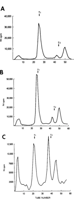
50,000 r 40,000 30,000 • 20,000 • 10,000 • 12,500 10.000 7500 2500 -20 30 40 TUBE NUMBERFig. 7. CM-cellulose chromatograms demonstrate the elution profiles of the radioactively labeled col-lagens synthesized by cultured corneal stroma-cytes from normal (A) and keratoconus (B) corneas and sclera (C). Arrows indicate the elution posi-tions of the carrier rat skin type I collagen peaks. Note the large peak containing the type III colla-gen in C and the similarities of the profiles in A and B.
type I collagen. Walls of blood vessels in the conjunctiva had clearly detectable type IV collagen and fibronectin. The conjunctival epithelium itself did not react with any an-tibodies.
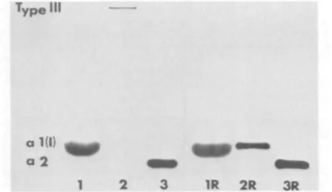
Identification of collagen synthesized in vitro. Normal and keratoconus corneal stromal cells synthesized both types I and III collagen in vitro. The presence of these two collagens was confirmed by the elution posi-tion from CM-cellulose columns (Fig. 7) and SDS slab gel electrophoresis patterns. SDS gel profiles of the migration patterns of pooled fractions from the three collagenous peaks seen on CM-cellulose chromatography showed co-migration of peak 1 collagen chains with purified alpha 1 (I) collagen and peak 3 with authentic alpha 2 collagen chains (Fig. 8). Peak 2 materials migrated largely with the standard type III and exhibited disappearance of the trimeric form when electrophoresed under reducing conditions (Fig. 8).
The ratio of type I to type III collagen as determined from the CM-cellulose profiles was 10:1 for normal corneal tissue and 9:1 for keratoconus tissues (average of five spec-imens). The preponderance of type III colla-gen in the sclera was demonstrated by a ratio of 2.1:1, type I to type III (average of five specimens). An example of this large amount of type III collagen synthesized by scleral fibroblasts in culture can be seen in Fig. 7. The material from peak 2 of the chromato-gram of the scleral collagens again showed reduction with beta-mercaptoethanol when subjected to SDS gel electrophoresis. Discussion
Corneal transparency depends upon the orderly deposition of collagen and glycocon-jugates in the stroma.19' 20 Maintenance of
transparency must depend, at least in part, on the controlled turnover of this extracel-lular matrix. Collagens, the most plentiful group of structural macromolecules in the corneal stroma, are present largely as fibrillar proteins that interact with the stroma-specific proteoglycans to produce a normal optically useful stroma. Thus modifications of the
Type Id
ai(l)
a2
1
1R 2R
3R
Fig. 8. Fluororadioautograph of an SDS-polyacrylamide slab gel of the collagen chains eluted in the three peaks of the CM-cellulose chromatogram demonstrates the presence of the types I and III collagen synthesized by cultured keratoconus stromaeytes. 1, Pooled fractions from the a-1 peak region; 2, pooled from the interpeak; 3, pooled from the a-2 peak. Note the disappearance of the type III trimer after reduction with b- mercaptoethanol (2R). The posi-tion of the radiolabeled type III collagen was identical to that of coelectrophoresed authentic type III, as was that of the labeled type I. These patterns were similar between normal and keratoconus cell synthetic products; the scleral samples had more prominent type III bands on the gels.
types of collagens synthesized or defects in posttranslational processing of the molecule could lead to corneal disease.
Recent advances in collagen characteriza-tion have revealed that there are at least four genetically distinct types which are now re-ferred to as types I, II, III, and IV.21 The tissue-specific distribution of collagen types appears to play an important role in mor-phogenesis, particularly in the cornea,22'23< 27 and in association with the sensory retina.24'25 Corneal stromal matrix stability may de-pend in part upon the proportion of the types of collagen present. We have shown that there are two major collagen types synthe-sized in vitro by normal and keratoconus cor-nea cells, with a similar proportion of types I and III in both. This mixture of types I and III is similar to the collagens that accumulate in vivo and have been extracted and iden-tified.26 It is possible that an alteration in the proportion of these major collagen compo-nents could destabilize the connective tissue
matrix, as in Ehlers-Danlos type IV, where the synthesis of type III collagen is low to absent.27 Maumenee28 reported that two keratoconus corneal specimens contained in-creased type III collagen as detected by im-munofluoesence. In our specimens type III collagen was not reliably detected in unin-volved corneal stroma. We did demonstrate collections of type III collagen in keratoconus corneal scars and at the site of host-graft wound healing in a perforating keratoplasty specimen. Type III collagen has previously been reported in scars in nonocular tissues, even those such as tendon which do not nor-mally have type III.29
The presence of type III collagen in normal cornea, including human, has been contro-versial,30' 3l and type V collagen preliminarily identified.32 Type III had been identified in bovine material,33 and type V in lapine.32 Newsome et al.26 have recently submitted evidence from collagenase sensitivity, cyano-gen bromide peptides, trypsin sensitivity,
748 Newsome et al. Invest. Ophthahnol. Vis. Sci.June 1981
and slab gel electrophoresis with and without reduction identifying types III and V in nor-mal human cornea. These data support the observations of others noted above. Differ-ences with other reports30 may be due at least partially to technique, since type III is heav-ily crosslinked and difficult to extract from native tissues. It is more difficult to explain the conflict of the present data from cul-tures,31 since extraction is not a problem in vitro. We used different media with a short labeling period and early passage cells, in contrast to Stoesser et al.34 In the present study preliminary results with anti-type V antibodies revealed a positive reaction pat-tern identical to that for type IV in both nor-mal and keratoconus corneas. We have not emphasized these results, since our anti-bodies were not, at the time of this work, rigorously proved to be completely specific. Subsequent purification has, however, con-firmed the type V specificity (J-M. F., un-published data).
Our results emphasize some similarities and indicate certain differences between the developing chick cornea and human tissue. Type I collagen was the major detectable col-lagen in our study of the human corneal stroma, a finding corresponding to what has been reported for chick.35 Type III collagen is a small component of the human cornea but is reportedly absent in the chick. The synthesis of both types I and III by a uniform cell population has not been previously re-ported for corneal stromacytes but is known to occur with smooth muscle cells36 and fibroblasts.29 We could not reliably detect type III collagen in all normal stromas, perhaps due to the limits of the immu-nofluorescent reaction. Factors such as mask-ing of type III by other matrix components may also have influenced its detectability.
Type II collagen is in the chick a promi-nent compopromi-nent of Bowman's layer, the sub-epithelial stroma, and Descemet's mem-brane.35 The presence of type II-bearing primary corneal stroma, thought to be critical to the morphogenesis of the adult stroma in the chick, has not been confirmed in human tissue (for review, see ref. 37). We could not demonstrate type II in the Bowman's or
sub-epithelial areas of either the normal or keratoconus specimens.
The laminar staining pattern of Descemet's membrane may indicate a regional distribu-tion of type IV collagen and of the noncol-lagenous fibronectin within the membrane. Such a specific distribution could explain, at least in part, the distinct ultrastructure of this membrane. Jakus38 and later Hay and Revel39 described the approximately 100 nm period-icity of the anterior third of Descemet's membrane, presumably reflecting the pack-ing of membrane subunits in this area. Such orderly packing could be in response to the presence here of type IV fibrils or aggragates. The interface between the stroma and Descemet's membrane is also distinguished ultrastructurally. Patchy, basement mem-brane-like material has been reported here by transmission electron microscopy. The strong reaction in this region with an-tifibronectin suggests that this material con-sists, at least in part, of fibronectin. The pre-cise localization of fibronectin within the basement membrane-like material at the stromal-Descemet's interface must await ul-trastructural studies.
Type IV collagen was also prominently de-tected at the interface of the endothelium with Descemet's membrane. This observation offers further evidence that this cell layer, so important to the maintenance of proper cor-neal hydration, synthesizes basement mem-brane.40' 41 The presence of fibronectin here in posterior Descemet's membrane is consistent with its detection in a wide variety of human basement membranes.42 This protein is also commonly associated with and synthesized by fibroblasts (for review, see ref. 43). It is possi-ble that the stromacytes synthesize the fibronectin that accumulates in Descemet's membrane. However, the corneal endothe-lium is embryologically derived from the neural crest, as are the stromacytes proper,44 and thus may share common attachment pro-teins. This observation contrasts with the ap-pearance of the epithelial basement mem-brane area which had no detectable fibronec-tin. And, of course, the epithelium is not a neural crest derivative.
the sclera may in part explain its anatomical differences from the type I-rich corneal stroma. The proportion of collagen types syn-thesized may affect fibril aggregation and uni-formity. In the sclera the matrix fibrils are randomly arrayed in a feltwork with no signs of the orderly lamellae so characteristic of the corneal stroma. Scleral fibril diameters are much more heterogenous, and the fibrils much larger than those of the cornea. Lapier et al.45 have shown that type III collagen influences the size and type of bundles formed by type I collagen in vitro. Such an interaction may condition the collagen bundle organization in various connective tissues.
It is significant that the collagen profiles obtained from cultured corneal, scleral, and conjunctival stromacytes differed among themselves, and that these differences corre-sponded to differences in immunofluorescent reactivity. For example, the sclera reacted strongly with anti-type III and synthesized the largest proportion of type III collagen in vitro. The persistence of these differences in patterns of synthesis of collagens, each type of which is a distinct gene product, is in con-trast to the rapid decay of keratan sulfate pro-teoglycan synthesis by in vitro corneal stroma, even in organ culture.46
The present work has extended our knowl-edge of the site-specific distribution of the genetically distinct collagen types in normal human and keratoconus corneas and normal sclera. It appears that a normal complement of corneal stromal collagens is present in normal proportions in keratoconus. Thus, if keratoconus is a true collagen disease (i.e., has a defect in collagen on the molecular level), the defect probably occurs as a result of an error in assembly or in the control of turnover, for example, by the overproduction of collagenolytic enzymes. Further investi-gations of these possibilities are in progress.
REFERENCES
1. Hammerstein W: Zur Genetik des Keratokonus. Al-brecht Von Graefes Arch Klin Exp Ophthalmol 190:293, 1974.
2. McKusick VA: Heritable Disorders of Connective Tissue, ed. 4. St. Louis, 1972, The C. V. Mosby Co. 3. Robertson I: Keratoconus and the Ehlers-Danlos
syndrome. A new aspect of keratoconus. Med J Aust 1:571, 1975.
4. Kuming BS and Joffe L: Ehlers-Danlos syndrome associated with keratoconus. A case report. S Afr Med J 52:403, 1977.
5. Pollack FM: Contributions of electronmicroscopy to the study of corneal pathology. Surv Ophthalmol 20:375, 1976.
6. Newsome DA, Takasugi M, Kenyon KR, Stark WF, and Opelz G: Human corneal cells in vitro: mor-phology and histocompatibility (HL-A) antigens of pure cell populations. INVEST OPHTHALMOL 13:23, 1974.
7. Chung E and Miller EJ: Collagen polymorphism: characterization of molecules with the composition [ a l (III)]3 human tissues. Science 183:1200, 1974.
8. Epstein EH Jr: [ a l (III)]3 human skin collagen:
re-lease by pepsin digestion and preponderance in fetal life. J Biol Chem 249:3225, 1974.
9. Trelstad RL: Human aorta collagens: evidence for three distinct species. Biochem Biophys Res Com-mun 57:717, 1974.
10. Lammli UK: Cleavage of structural protein during the assembly of the head of bacteriophage T4. Na-ture 227:680, 1970.
11. Smith BD, Martin EJ, Miller EJ, Dorfman A, and Swarm R: Nature of the collagen synthesized by a transplanted chondrosarcoma. Arch Biochem Bio-phys 166:181, 1975.
12. Orkin RW, Gehron P, McGoodwin EB, Martin GR, Valentine T, and Swarm R: A murine tumor produc-ing a matrix of basement membrane. J Exp Med 145:204, 1977.
13. Andreopoulos NA, Mestecky J, Wright GP, and Mil-ler EJ: Characterization of antibodies to the native human collagens and to their component a chains in the sera and joint fluids of patients with rheumatoid arthritis. Immunochemistry 13:709, 1976. 14. Nowack H, Gay S, Wick G, Becker U, and Timpl R:
Preparation and use in immunohistology of an-tibodies specific for types I and III collagen and pro-collagen. J Immunol Methods 12:117, 1977. 15. Rennard SI, Berg R, Martin GR, Foidart J-M, and
Gehron Robey P: Enzyme-linked immunoassay (ELISA) for connective tissue components. Anal Biochem (in press).
16. McConahey PJ and Dixon FJ: A method of trace iodination of proteins for immunological studies. Int Arch Allergy 29:185, 1966.
17. Rohde H, Nowack H, Becker U, and Timpl R: Ra-dioimmunoassay for the aminoterminopeptide of procollagen p a l (I) chain. J Immunol Methods 11:135, 1976.
18. Engvall E and Rouslahti E: Binding of a soluble form of fibroblast surface protein fibronectin to collagen. Int J Cancer 20:1, 1977.
19. Trelstad RL and Coulombre AJ: Morphogenesis of the collagenous stroma in the chick cornea. J Cell Biol 50:840, 1971.
20. Maurice DM: The structure and transparency of the cornea. J Physiol 136:263, 1957.
750 Newsome et al. Invest. Ophthalmol. Vis. Sci.June 1981
21. Martin GR, Beyers PH, and Piez KA: Procollagen. Adv Enzymol 42:167, 1975.
22. Dodson JW and Hay ED: Secretion of collagenous stroma by isolated epithelium grown in vitro. Exp Cell Res 65:215, 1971.
23. Hay ED: Origin and role of collagen in the embryo. Am Zoologist 13:1085, 1973.
24. Newsome DA, Linsenmeyer RF, and Trelstad RL: Vitreous body collagen. Evidence for dual origin from the neural retina and hyalocytes. J Cell Biol 71:59, 1976.
25. Smith GN, Linsenmeyer TF, and Newsome DA: Synthesis of type II collagen in vitro by embryonic chick neural retina tissue. Proc Natl Acad Sci USA 73:4420, 1976.
26. Newsome DA, Gross J, and Hassell JR: Human corneal stroma contains three distinct collagens. (Manuscript submitted.)
27. Pope FM, Martin GR, Lichtenstein JR, Penttinen R, Gerson G, Rowe DR, and McKusick BA: Patients with Ehlers-Danlos syndrome type IV lack type III collagen. Proc Natl Acad Sci USA 72:1314, 1975. 28. Maumenee IH: The cornea in connective tissue
dis-ease. Ophthalmology 85:1014, 1978.
29. Epstein EH Jr and Munderloh NH: Isolation and characterization of CNBr peptides of human [ a l (III)]3
collagen and tissue distribution o f [ a l ( I ) ]2a 2 a n d [ a l
(III)]3 collagens. J Biol Chem 250:9304, 1975.
30. Freeman IL: Collagen polymorphism in mature rabbit cornea. INVEST OPHTHALMOL VIS SCI 17:171, 1978.
31. Church RL: Procollagen and collagen produced by normal bovine corneal stroma fibroblasts in cell
cul-ture. INVEST OPHTHALMOL VIS SCI 19:192, 1980.
32. Yue B, Baum JL, and Smith BD: Collagen synthesis by cultures of stroma] cells from normal human and keratoconus corneas. Biochem Biophys Res Com-mun 86:465, 1979.
33. Schmut O: The identification of type III collagen in calf and bovine cornea and sclera. Exp Eye Res 25:505, 1977.
34. Stoesser TR, Church RL, and Brown SI: Partial characterization of human collagen and procollagen
secreted by human corneal stromal fibroblasts in cell
culture. INVEST OPHTHALMOL Vis SCI 17:264, 1978.
35. von der Mark K, von der Mark H, Timpl R, and Trelstad RL: Immunofluorescent localization of col-lagen types I, II, and III in the embryonic chick eye. Dev Biol 59:75, 1977.
36. Layman DL, Epstein EH Jr, Dodson RF, and Titus JL: Biosynthesis of type I and III collagens by cul-tured smooth muscle cells human aorta. Proc Natl Acad Sci USA 74:671, 1977.
37. Hay ED: Development of the vertebrate cornea. Int Rev Cytol (in press).
38. Jakus MA: Studies on the cornea. II. The fine struc-ture of Descemet's membrane. J Biophys Biochem Cytol 2(Suppl):243, 1956.
39. Hay ED and Revel JP: Fine Structure of the Devel-oping Avian Cornea. New York, 1969, S. Karger. 40. Perlman M, Baum JL, and Kay GI: Fine structure
and collagen synthesis activity of monolayer cultures of rabbit corneal endothelium. J Cell Biol 63:306, 1974.
41. Kefalides NW, Cameron JD, Tomichek EA, and Yanoff M: Biosynthesis of basement membrane col-lagen by rabbit corneal endothelium in vitro. J Biol Chem 251:730, 1976.
42. Stenman S and Vahen A: Distribution of a major connective tissue protein, fibronectin, in normal human tissues. J Exp Med 147:1054, 1977. 43. Yamada KM and Oldern K: Fibronectin-adhesive
glycoproteins of cell surface and blood. Nature 275:179, 1978.
44. Johnston MC, Nodin DM, Hazleton RD, Cou-lombre JL, and CouCou-lombre AJ: Origins of avian ocu-lar and periocuocu-lar tissues. Exp Eye Res (in press). 45. Lapier CM, Nusgens B, and Pierard GE:
Interac-tion between collagen type I and type III in condi-tioning bundles organization. Connect Tissue Res 5:21, 1977.
46. Klintworth GK and Smith CF: A comparative study of extracellular glycosaminoglycans synthesized by rabbit corneal fibroblasts in organ and confluent cul-tures. Lab Invest 35:258, 1976.