Cytosine deaminase suicide gene therapy for
peritoneal carcinomatosis
Mohamed Bentires-Alj, Anne-Ce´cile Hellin, Chantal Lechanteur, Fre´de´ric Princen,
Miguel Lopez, Georges Fillet, Jacques Gielen, Marie-Paule Merville, and Vincent Bours
Laboratory of Medical Chemistry/Medical Oncology, University of Lie`ge, Lie`ge, Belgium.
Gene therapy is a novel therapeutic approach that might soon improve the prognosis of some cancers. We investigated the feasibility of cytosine deaminase (CD) suicide gene therapy in a model of peritoneal carcinomatosis. DHD/K12 colorectal adenocarcinoma cells transfectedin vitro with the CD gene were highly sensitive to 5-fluorocytosine (5-FC), and a bystander effect could also be observed. Treating CD⫹cells with 5-FC resulted in apoptosis as detected by terminal deoxynucleotidyltransferase-mediated deoxyuridine triphosphate nick-end labeling. In vitro, several human cell lines derived from ovarian or colorectal carcinomas, as well as the rat glioblastoma 9 L cell line, responded to CD/5-FC and showed a very strong bystander effect. 5-FC treatment of peritoneal carcinomatosis generated in syngeneic BDIX rats by CD-expressing DHD/K12 cells led to a complete and prolonged response and to prolonged survival. Our study thus demonstrated the efficacy of CD suicide gene therapy for the treatment of peritoneal carcinomatosis. Cancer Gene Therapy (2000) 7, 20 –26
Key words: Cytosine deaminase; gene therapy; peritoneal carcinomatosis; colorectal carcinoma.
G
ene therapy is a novel approach that might lead to
improved treatments of some types of cancer.
Al-though a number of clinical trials are currently being
performed to evaluate the safety and the efficacy of such
an approach in different settings, more experimental
work is required to define the best models for gene
therapy. Peritoneal carcinomatosis develops in an
ana-tomical cavity and cannot be eradicated by conventional
anticancer therapies. Therefore, it constitutes a suitable
model to study the delivery of therapeutic genes and
their preclinical and clinical efficiencies. We have
re-ported previously that a herpes simplex virus
(HSV)-thymidine kinase (tk)-based suicide gene therapy could
improve the survival of rats with peritoneal
carcinoma-tosis induced by colorectal adenocarcinoma cells.
1,2Other investigators have also delivered therapeutic
genes into anatomical cavities in which cancer had
developed.
3– 6.
Suicide gene therapy is based on the introduction in
target cells of a gene coding for an enzyme that
trans-forms a prodrug into a cytotoxic compound.
7,8The gene
for HSV-1 TK has been the subject of many
investiga-tions in vitro and in vivo.
9 –14The Escherichia coli gene
coding for the cytosine deaminase (CD) enzyme is
another suicide gene. CD transforms 5-fluorocytosine
(5-FC) into cytotoxic 5-fluorouracil (5-FU).
15,16This
gene has already been tested in vitro as well as in several
animal models, most of them being based on colorectal
carcinoma cells,
17–25and is now being considered for
phase I clinical trials.
26The present report investigated the feasibility of a
CD-based suicide gene therapy in a model of peritoneal
carcinomatosis induced by colorectal carcinoma cells in
syngeneic rats. It demonstrates that 5-FC treatment can
eradicate the peritoneal carcinomatosis generated by
colorectal carcinoma cells stably transfected with the CD
gene. It also demonstrates that 5-FC induces apoptosis
in CD
⫹cells, and that several cell lines, including
ovarian and colorectal carcinoma cell lines, are sensitive
in vitro to the CD/5-FC cytotoxic effect.
MATERIALS AND METHODS
Plasmids
The pRSV-CD plasmid carrying the E. coli K12 CD gene (kindly provided by Dr. J. Gebert, Section of Molecular Diagnostics and Therapy, Chirurgische Universita¨ts-klinik, Heidelberg, Germany) has been described previously.16The pCMV-CD plasmid was constructed by inserting a 1.3-kb
HindIII/BamHI CD fragment from pRSV-CD into the
pcDNA3 vector (Invitrogen, San Diego, Calif) at the HindIII/
BamHI sites. The pCMV-CD20 (cluster of differentiation 20
antigens) plasmid was kindly provided by Dr. Jim Koh (Labo-ratory of Molecular Oncology, Massachusetts General Hospi-tal, Harvard Medical School, Charlestown, Mass).
Cell culture and transfections
The DHD/K12 colorectal cancer cell line was derived from a transplantable colon adenocarcinoma induced by
1,2-dimeth-September 11, 1998; February 27, 1999.
Address correspondence and reprint requests to Dr. Vincent Bours, Medical Oncology, CHU B35, Sart-Tilman, Universite´ de Lie`ge, 4000 Lie`ge, Belgium. E-mail address: vbours@ulg.ac.be
ylhydrazine in syngeneic BDIX rats.27 DHD/K12 cells were maintained in Dulbecco’s modified Eagle’s medium (Life Technologies, Gaithersburg, Md) supplemented with 5% fetal calf sera (FCS) (Life Technologies), 1% L-glutamine (200
mM), 1% N-2-hydroxyethylpiperazine-N⬘-2-ethanesulfonic acid (1 M), 1%L-arginine (0.55 mM), penicillin (100 IU/mL),
and streptomycin (100g/mL).
The 9 L glioblastoma cells and the MDA-MB-435 breast cancer cells were a gift from C. Grignet-Debrus (Laboratory of Fundamental Virology and Immunology, University of Lie`ge). 9 L cells were cultured in RPMI 1640 (Life Technologies) supplemented with 10% FCS, 1% sodium pyruvate (100 mM), 1% nonessential amino acids, penicillin (100 IU/mL), and streptomycin (100g/mL). MDA-MB-435 cells were grown in RPMI 1640 supplemented with 10g/mL insulin, 10% FCS, penicillin (100 IU/mL), and streptomycin (100g/mL).
The OVCAR-3 ovarian epithelial cancer cell line was main-tained in RPMI 1640 supplemented with 10% FCS, 1%
L-glutamine (200 mM), penicillin (100 IU/mL), and
streptomy-cin (100g/mL). HCT116 human colon carcinoma cells were grown in McCoy’s 5A modified medium supplemented with 10% FCS, 1%L-glutamine (200 mM), penicillin (100 IU/mL),
and streptomycin (100g/mL).
For stable transfections, plasmids were linearized (XbaI for pRSV-CD and BglII for pCMV-CD) and transfected into DHD/K12 cells with the transfection reagent N-(1-[2,3-dio-leoyloxy]propyl)-N,N,N-trimethylammonium methylsulfate (DOTAP) as recommended by the manufacturer (Boehringer Mannheim, Mannheim, Germany). After selection for geneti-cin resistance (500 g/mL of G418, active concentration, Boehringer Mannheim), 30 clones were isolated and ana-lyzed for integration of the CD gene by polymerase chain reaction and for expression of the CD enzyme by immuno-blot (anti-CD polyclonal rabbit antibody, a generous gift of Dr. Haack, Chirurgische Universita¨tsklinik, Heidelberg, Germany).
The transient transfections were also performed with the DOTAP method. Cells were cotransfected with pCMV-CD and pCMV-CD20 to estimate the transfection rate. We mea-sured immunofluorescence with a fluorescence-activated cell sorter (FACS) using a monoclonal anti-CD20 antibody conju-gated to phycoerythrin (Becton Dickinson, San Jose, Calif).
In vitro
cytotoxicity test
Stably transfected cells or untransfected cells were seeded at a concentration of 1500 cells/well on 96-well, flat-bottom micro-plates in medium supplemented with 15% FCS. After 24 hours, cells were cultivated in 5-FC- (Sigma, St. Louis, Mo) or 5-FU-containing medium that was replaced every other day. After 6 days (5-FU) or 8 days (5-FC) of incubation with the drug, cell viability was measured by a colorimetric assay based on the cleavage of the tetrazolium salt WST-1 by mitochon-drial dehydrogenases in viable cells (Boehringer Mannheim). For transiently transfected cells, the medium was removed the day after the DOTAP transfection and replaced by 5-FC-(2 mM) containing medium, which was replaced 48 hours later. Cell viability was measured after 96 hours of treatment by trypan blue exclusion and compared with control transfected cells grown in the absence of 5-FC.
The bystander effect was measured by coculturing different proportions of DHD/K12-CMV-CD (CD⫹) and DHD/K12 (CD⫺) cells. After seeding and treating the cells as mentioned above, their viability was measured by the WST-1 test.
Treatment of animals
At day 0, 10-week-old male BDIX rats were inoculated intra-peritoneally (i.p.) with 106DHD/K12-CMV-CD cells in 2 mL of serum-free medium. At day 14, the animals were treated with 5-FC (Ancotil, Roche, France) at 500 mg/kg/day or with normal saline buffer injected i.p. 5 days a week for 3 weeks. Two animals were sacrificed at 4 days after the last treatment, and the tumor evolution was evaluated by direct abdominal examination. Kaplan-Meier curves were established for eight animals and compared with the log-rank test.
Detection of apoptotic cells
DHD/K12 and DHD/K12-CMV-CD cells were cultured in medium with or without 5-FC (4 mM) for 48 hours. Cells were washed two times in phosphate-buffered saline, fixed in para-formaldehyde, permeabilized in methanol, and labeled by fluorescein-deoxyuridine triphosphate in the presence of ter-minal deoxynucleotidyltransferase-mediated deoxyuridine triphosphate nick-end labeling (TUNEL) as recommended by the manufacturer (In Situ Cell Death Detection Kit, Boeh-ringer Mannheim).
RESULTS
CD confers 5-FC sensitivity to DHD/K12 cells
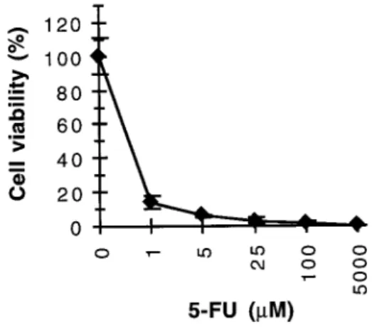
The sensitivity of DHD/K12 colorectal carcinoma cells
to 5-FU was evaluated by incubating the cells for 6 days
in the presence of increasing 5-FU concentrations and
testing cell viability with the WST-1 test. 5-FU killed
DHD/K12 cells in a dose-dependent manner (Fig 1),
thus indicating that these cells could be considered for
CD gene therapy.
Next, we stably transfected DHD/K12 cells with
ex-pression vectors containing a viral eukaryotic promoter
(Rous sarcoma virus or cytomegalovirus (CMV))
up-stream of the CD gene. Two clones were selected that
had integrated the CD gene and expressed the CD
enzyme, as demonstrated by polymerase chain reaction
and immunoblots, respectively (data not shown). These
Figure 1. Cytotoxic effect of 5-FU on DHD/K12 cells in vitro.
DHD/K12 cells were incubated for 6 days in the presence of increasing 5-FU concentrations as indicated. Cell viability was then measured with the WST-1 test.
clones, DHD/K12-RSV-CD and DHD/K12-CMV-CD,
were incubated for 8 days in the presence of increasing
concentrations of 5-FC. Measures of cell viability
dem-onstrated that 5-FC concentrations ranging from 400
M to 2 mM killed a large majority of the
CD-trans-fected cells, whereas the viability of parental DHD/K12
cells was not affected (Fig 2A). Moreover,
DHD/K12-CMV-CD clones were more sensitive to the prodrug
than the DHD/K12-RSV-CD clones (Fig 2A), probably
as a consequence of a better promoter activity.
To test whether a bystander effect could be observed
in vitro, various proportions of DHD/K12-CMV-CD and
untransfected DHD/K12 cells were cocultured and
chal-lenged for 8 days with two different 5-FC concentrations,
which killed 100% of CD-expressing cells but did not
affect parental cells (Fig 2B). Under these experimental
conditions, the presence of 20% of DHD/K12-CMV-CD
cells was sufficient to induce a cytotoxic effect that killed
79% and 92% of the cells after incubation with 1 mM
and 2 mM of 5-FC, respectively. These data also
con-firmed that the survival of untransfected DHD/K12 cells
was unaffected by the 5-FC treatment.
Induction of apoptosis by 5-FC in DHD/K12-CD cells
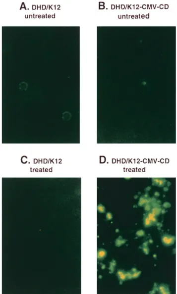
We investigated whether the treatment of
DHD/K12-CMV-CD cells with 5-FC induced cell death through
apoptosis. Untransfected DHD/K12 and
DHD/K12-CMV-CD cells were incubated for 48 hours with or
without 5-FC (4 mM), fixed, and analyzed for apoptosis
by TUNEL. Under these conditions, we did not observe
any significant apoptosis in untransfected DHD/K12
cells treated with such a high 5-FC concentration or left
untreated, or in the DHD/K12-CMV-CD cells incubated
with the medium alone (Fig 3, A–C). However, the
DHD/K12-CMV-CD cells displayed a significant
apo-Figure 2. Cytotoxic effect of 5-FC on DHD/K12 CD⫹cells in vitro. A: stably transfected DHD/K12-RSV-CD and DHD/K12-CMV-CD cells as well as untransfected DHD/K12 cells were incubated in vitro for 8 days in the presence of increasing 5-FC concentrations as indicated. Cell viability was then measured with the WST-1 test. B:
In vitro bystander effect. DHD/K12-CMV-CD cells and untransfected
cells were cocultured in various proportions and incubated for 8 days in the presence of 5-FC (1 or 2 mM). The proportions of CD⫹
cells were 0%, 5%, 20%, 50%, and 100%, respectively. Figure 3. Induction of apoptosis by 5-FC in DHD/K12-CMV-CD
cells. Untransfected DHD/K12 cells (A,C) or DHD/K12-CMV-CD cells (B,D) were left untreated or were treated with 5-FC (4 mM) for 48 hours. Treated cells were fixed, and apoptosis was evaluated by TUNEL.
ptosis after 5-FC treatment, as shown by the high
number of fragmented, fluorescent nuclei (Fig 3D).
In vitro
cytotoxicity and bystander effect with the CD
suicide gene in adenocarcinoma cell lines
Peritoneal carcinomatosis most frequently arises from
digestive or ovarian carcinomas, and many of these
tumors respond poorly to chemotherapy. Therefore, we
investigated whether other cell lines derived from
hu-man colon (HCT116), ovarian (OVCAR-3), or breast
(MDA-MB-435) carcinomas were also sensitive to the
CD gene/5-FC cytotoxic effect. The 9 L rat glioblastoma
cell line, known for its response to the HSV-tk suicide
gene and its high bystander effect, was also included in
this assay. The cell lines mentioned above were
tran-siently transfected with the CD suicide gene and
subse-quently treated with 5-FC for 48 or 96 hours. The
efficacy of the transient transfections was assessed by
cotransfection of an expression vector coding for the
B-cell-specific CD20 surface antigen and FACS counting
of CD20
⫹cells. Transfection efficiencies were low; only
0.7% of HCT116 cells, 0.7% of MDA-MB-435 cells, 3%
of 9 L cells, and 10.5% of OVCAR-3 cells were positive
for CD20 expression (Table 1). Despite this low
trans-fection rate, significant cytotoxicity was observed in
three of the four cell lines after transient transfection of
the CD gene and 5-FC treatment for 48 hours (data not
shown) or 96 hours (Table 1). Indeed, when compared
with control cells transfected with the CD20 antigen
expression vector alone or with an empty pcDNA3
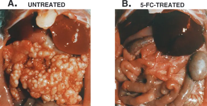
Figure 4. 5-FC treatment of rats injected with DHD/K12-CMV-CD cells. The figure shows the peritoneal cavity at day 36 of a rat injected with
DHD/K12-CMV-CD cells and treated with normal saline buffer (A) and of a rat injected with DHD/K12-CMV-CD cells and treated with 5-FC (500 mg/kg/day) for 3 weeks starting at day 14 (B). This experiment was performed on groups of two rats, and a representative picture is shown.
Table 1. In Vitro CD-Induced Cytotoxicity and Bystander Effect
Transfection (5-FC) Transfected cells (%) Cell viability (%) pcDNA3 (⫹) CD20 (⫹) CD20⫹ CD (⫺) CD20⫹ CD (⫹) HCT116 0.7 100 ⬎100 90.2 7.8 MDA-MB-435 0.7 100 78.9 93.7 67.2 9 L 3 100 81.8 ⬎100 5.1 OVCAR-3 10.5 100 ⬎100 84.6 5.5
* HCT116, MDA-MB-435, 9 L, and OVCAR-3 cells were transiently transfected with expression vectors coding for the CD20 antigen, the CD enzyme, or a combination of both, or with the pcDNA3 empty expression vector. Transfection efficiencies were determined by FACS analysis of the percentages of CD20⫹ cells after transfection of the CD20 expression vector. These percentages of transfected cells were reproducible and were confirmed by two independent experiments. Transfected cells were then treated with 2 mM of 5-FC (⫹) for 96 hours or left untreated (⫺), and cell viability was measured with trypan blue. Cell viabilities were expressed as percentages of living cells compared with cells transfected with the control empty vector and treated with 5-FC. Data are representative of two independent experiments.
vector, CD-transfected HCT116, 9 L, or OVCAR-3 lines
showed only 7.8%, 5.1%, and 5.5% of surviving cells,
respectively, after treatment with 5-FC (Table 1).
How-ever, this cytotoxic effect was much less important in the
MDA-MB-435 cell line, as 67% of cells were still alive
after CD transfection and 5-FC treatment.
Conse-quently, these data indicated that the CD gene is
effi-cient in vitro against several cell lines other than DHD/
K12, including colon and ovarian carcinoma cell lines,
and that a significant bystander effect can be observed in
vitro.
In vivo
response of DHD/K12-CD cells to 5-FC
To demonstrate the in vivo feasibility of a CD-based
suicide gene therapy, we used our model of peritoneal
carcinomatosis induced by DHD/K12 cells in syngeneic
BDIX rats. As described previously, injection of 10
6DHD/K12 cells in the peritoneal cavity of these rats led
to the development of macroscopic peritoneal tumor
nodes within 10 days; all of the animals died of extensive
peritoneal carcinomatosis before day 70.
1,2In an initial experiment, groups of two rats were
injected either with untransfected DHD/K12 cells or
with DHD/K12-CMV-CD cells. These animals were
treated for 3 weeks (days 14–18, 21–25, and 28–32) with
peritoneal injections of 5-FC (500 mg/kg/day) or normal
saline buffer alone. The animals were sacrificed at day
36, and the reduction in tumor volume was assessed by
direct abdominal examination. At day 36, all of the
animals injected with untransfected DHD/K12 cells and
treated with 5-FC or with saline buffer showed an
extensive peritoneal dissemination (data not shown).
Similarly, the two rats injected with
DHD/K12-CMV-CD cells and treated for 3 weeks with saline buffer
displayed a large number of tumor nodes disseminated
in their peritoneal cavity (Fig 4A). However, animals
injected with DHD/K12-CMV-CD and treated with
5-FC displayed a complete regression of the peritoneal
carcinomatosis (Fig 4B).
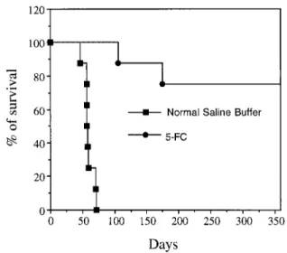
To demonstrate whether this apparent excellent
tu-mor response to CD/5-FC treatment translated into
survival advantages, animals were injected with 10
6DHD/K12-CMV-CD cells and treated for 3 weeks from
day 14 with 5-FC at 500 mg/kg/day; next, survival curves
were established. Animals injected with
DHD/K12-CMV-CD cells and treated with normal saline buffer
alone died between days 45 and 71 (Fig 5), as did
animals that had not been treated or animals injected
with parental DHD/K12 cells and treated with 5-FC
(data not shown). However, rats injected with DHD/
K12-CMV-CD cells and treated with 5-FC for 3 weeks
showed a significantly improved survival, as six of eight
animals were still alive at day 360 (Fig 5, P
⬍ .001).
DISCUSSION
We had previously reported the efficacy of HSV-tk
suicide gene therapy for peritoneal carcinomatosis.
1,2The present report indicates that such a treatment could
also be envisaged with the E. coli CD gene.
Interestingly, when animals are injected with cells
transfected in vitro, the results obtained with the CD
gene are better than those obtained with the tk gene.
Indeed, after a peritoneal injection of TK-positive
DHD/K12 cells and treatment with ganciclovir, the
majority of the animals relapsed and died before day
140,
1whereas in our model, the CD/5-FC approach
apparently eradicated the tumor, or at least led to a
prolonged remission in six of eight animals. These
results confirm previous studies, which indicated a better
cytotoxic effect of CD over TK in vitro or in vivo.
22,28This superiority might be explained by a better bystander
effect in vivo. The expression of a stably transfected gene
might be very heterogenous, and some cells often loose
this expression. We had shown previously that, after
injection of TK-positive cells and treatment with
ganci-clovir, relapsing tumors expressed very low levels of the
tk gene.
1A strong bystander effect would allow a better
eradication of tumor cells that have lost the transgene
expression, and thus a more favorable outcome of the
animals. However, the superiority of the CD gene over tk
in our model might also simply be explained by vector
differences. The tk gene had been inserted in a retroviral
vector, and its expression was driven by the simian virus
40 promoter, whereas the CD gene was cloned in a CMV
expression vector before stable transfection. The CMV
promoter is probably stronger than the simian virus 40
promoter, and therefore allowed a better expression of
the suicide gene. Moreover, our in vitro data with the
Rous sarcoma virus and CMV promoters indicated that
a better promoter led to a stronger bystander effect (data
Figure 5. Survival of rats injected with DHD/K12-CMV-CD cells and
treated with 5-FC. Two groups of eight rats were injected i.p. with 106DHD/K12-CMV-CD cells and treated with saline buffer or with
5-FC (500 mg/kg/day) for 3 weeks starting at day 14. Survival curves for each group are shown. Six rats from the 5-FC-treated groups were still alive at day 360.
not shown). These differences indicated that a careful
choice of the promoter is critical for a better efficiency of
in vivo gene therapy.
The CD gene allowed a very strong in vitro bystander
effect with several cell lines, including ovarian and
colorectal carcinoma cell lines and the well-studied
glioblastoma 9 L cell line. These results confirmed that
the CD gene could be a powerful tool for in vivo cancer
gene therapy. The bystander effect generated by the
CD/5-FC system is generally thought to be related to a
diffusion of 5-FU from CD
⫹to CD
⫺cells and has been
reported to be very strong in different models.
22,28 –30However, it has been suggested recently that gap
junc-tions could also play a role in this bystander effect,
31indicating that the CD-generated bystander effect might
be caused by several distinct mechanisms. Interestingly,
in our set of cell lines, MDA-MB-435 cells do not show
any bystander effect with the tk suicide gene
32and do not
communicate through gap junctions (F.P. et al,
unpub-lished observations). Moreover, these cells are also
sensitive to 5-FU (data not shown), and their poor
response to 5-FC/CD is therefore not related to an
intrinsic resistance to the cytotoxic drug. A strong
by-stander effect is required to observe any clinical benefit
following in vivo gene transduction, as only a small
proportion of tumor cells can be reached following in
vivo treatment with viral or liposomal vectors. Our in
vitro data thus suggest that CD gene therapy might be
efficient on a number of cell types, including lines
derived from ovarian and digestive neoplasms that are
the most common cause of peritoneal carcinomatosis.
However, some tumors might be resistant, and the
identification of the mechanisms for such resistance is
most important to design appropriate clinical trials.
An immunological reaction might also participate in
the in vivo bystander effect and in the eradication of
tumors after treatment with CD/5-FC. Indeed, it has
been reported in other models that an immune response
is required for the eradication of CD
⫹tumors, and that
the CD/5-FC therapy induces a protective immunity.
33,34As our animals are immunocompetent and able to
develop an immune reaction against cytokine-expressing
DHD/K12 cells (C.L. et al, unpublished observations), it
is possible that an immune reaction is involved in the
observed favorable outcome of treated animals.
Despite these encouraging results, it becomes clear
from a number of reports that therapy with a single
suicide gene will often lead to partial, and sometimes
minor, responses. A combination of several genes should
increase the efficiency of the therapy. Given the low
toxicity of the suicide genes, it is certainly conceivable to
combine several suicide genes such as the HSV-1-tk gene
and the E. coli CD gene.
35–38Also, several teams are now
evaluating a combination of a suicide gene with cytokine
genes to boost the immunological component of the
bystander effect.
4,39,40The present model, as described
in this paper and in previous publications,
1,2,4is based on
immunocompetent animals and is suitable for the
eval-uation of such therapies with several genes.
ACKNOWLEDGMENTS
We thank Dr. J. Gebert for the pRSV-CD vector, Dr. Haack for the CD antibody, and Dr. Jim Koh for the pCMV-CD20 vector. We are most thankful to J-P. Cheramibien and G. Rocoux for their help with the in vivo experiments, K. Bajou and F. Kebers for the TUNEL protocol, N. Jacobs and R. Greimers for the FACS analysis, and W. Dewe´ for his help with statistical analysis. A.-C.H. is supported by a Te´le´vie fellowship and F.P. is supported by an FRIA fellowship. V.B. and M.-P.M. are Research Associates at the National Fund for Scientific Research (Belgium). This research was supported by grants from Te´le´vie, from the “Centre Anti-Cance´reux” (Lie`ge, Belgium), and from “Concerted Action Program, convention 97/02-214,” Communaute´ Franc¸aise de Belgique.
REFERENCES
1. Lechanteur C, Princen F, Lo Bue S, et al. HSV-1 thymi-dine kinase gene therapy for colorectal adenocarcinoma-derived peritoneal carcinomatosis. Gene Ther.
1997;4:1189–1194.
2. Princen F, Lechanteur C, Lopez M, et al. Repeated cycles of retrovirus-mediated HSV-TK gene transfer plus ganci-clovir increase survival of rats with peritoneal carcinoma-tosis. Gene Ther. 1998;5:1054–1060.
3. Aoki K, Yoshida T, Sugimura T, et al. Liposome-mediated in vivo gene transfer of antisense K-ras construct inhibits pancreatic tumor dissemination in the murine peritoneal cavity. Cancer Res. 1995;55:3810–3816.
4. Coll J-L, Mesnil M, Lefebvre M-F, et al. Long-term survival of immunocompetent rats with intraperitoneal colon carcinoma tumors using herpes simplex thymidine kinase/ganciclovir and IL-2 treatments. Gene Ther. 1997; 4:1160–1166.
5. Smythe WR, Kaiser LR, Hwang HC, et al. Successful adenovirus-mediated gene transfer in an in vivo model of human malignant mesothelioma. Ann Thorac Surg. 1994; 57:1395–1401.
6. Smythe WR, Hwang HC, Elshami AA, et al. Treatment of experimental human mesothelioma using adenovirus transfer of the herpes simplex thymidine kinase gene. Ann
Surg. 1995;222:78–86.
7. Freeman SM, Whartenby KA, Freeman JL, et al. In situ use of suicide genes for cancer therapy. Semin Oncol. 1996;23:31–45.
8. Moolten FL, Wells JM, Heyman RA, et al. Lymphoma regression induced by ganciclovir in mice bearing a herpes thymidine kinase transgene. Hum Gene Ther. 1990;1:125– 134.
9. Culver KW, Ram Z, Walbridge S, et al. In vivo gene transfer with retroviral vector-producer cells for treatment of experimental brain tumors. Science. 1992;256:1550–1552. 10. Ram Z, Culver KW, Walbridge S, et al. In situ retroviral-mediated gene transfer for treatment of brain tumors in rats. Cancer Res. 1993;53:83–88.
11. Rainov NG, Kramm CM, Aboody-Guterman K, et al. Retrovirus-mediated gene therapy of experimental brain neoplasms using the herpes simplex-thymidine kinase/ ganciclovir paradigm. Cancer Gene Ther. 1996;3:99–106. 12. Oldfield EH, Ram Z, Culver KW, et al. Gene therapy for
the treatment of brain tumors using intra-tumoral trans-duction with the thymidine kinase gene and intravenous ganciclovir. Hum Gen Ther. 1993;4:39–69.
retroviral-mediated transfer for the treatment of brain tumors. J Neurosurg. 1993;79:400–407.
14. Kun LE, Gajjar A, Muhlbauer M. Stereotaxic injection of herpes simplex thymidine kinase vector producer cells (PA317–G1Tk1SvNa.7) and intravenous ganciclovir for treatment of progressive or recurrent primary supratento-rial pediatric malignant brain tumors. Hum Gene Ther. 1995;6:1231–1255.
15. Austin EA, Huber BE. A first step in the development of gene therapy for colorectal carcinoma: cloning, sequenc-ing, and expression of Escherichia coli cytosine deaminase.
Mol Pharmacol. 1993;43:380–387.
16. Rowley S, Lindauer M, Gebert JF, et al. Cytosine deami-nase gene as a potential tool for the genetic therapy of colorectal cancer. J Surg Oncol. 1996;61:42–48.
17. Ohwada A, Hirschowitz EA, Crystal RG. Regional delivery of an adenovirus vector containing the Escherichia coli cyto-sine deaminase gene to provide local activation of 5-fluoro-cytosine to suppress the growth of colon carcinoma meta-static to liver. Hum Gene Ther. 1996;7:1567–1576.
18. Kanai F, Lan K-H, Shiratori Y, et al. In vivo gene therapy for␣-fetoprotein-producing hepatocellular carcinoma by adenovirus-mediated transfer of cytosine deaminase gene.
Cancer Res. 1997;57:461–465.
19. Evoy D, Hirschowitz EA, Naama HA, et al. In vivo adenoviral-mediated gene transfer in the treatment of pancreatic cancer. J Surg Res. 1997;69:226–231.
20. Lan K-H, Kanai F, Shiratori Y, et al. In vivo selective gene expression and therapy mediated by adenoviral vectors for human carcinoembryonic antigen-producing gastric carci-noma. Cancer Res. 1997;57:4279–4284.
21. Li Z, Shanmugam N, Katayose D, et al. Enzyme/prodrug gene therapy approach for breast cancer using a recombi-nant adenovirus expressing Escherichia coli cytosine deaminase. Cancer Gene Ther. 1997;4:113–117.
22. Trinh QT, Austin EA, Murray DM, et al. Enzyme/prodrug gene therapy: comparison of cytosine deaminase/5-fluoro-cytosine versus thymidine kinase/ganciclovir enzyme/pro-drug systems in a human colorectal carcinoma cell line.
Cancer Res. 1995;55:4808–4812.
23. Hirschowitz EA, Ohwada A, Pascal WR, et al. In vivo adenovirus-mediated gene transfer of the Escherichia coli cytosine deaminase gene to human colon carcinoma-de-rived tumors induces chemosensitivity to 5-fluorocytosine.
Hum Gene Ther. 1995;6:1055–1063.
24. Huber BE, Austin EA, Good SS, et al. In vivo antitumor activity of 5-fluorocytosine on human colorectal carcinoma cells genetically modified to express cytosine deaminase.
Cancer Res. 1993;53:4619–4626.
25. Topf N, Worgall S, Hackett NR, et al. Regional “pro-drug” gene therapy: intravenous administration of an adenoviral vector expressing the E. coli cytosine deaminase gene and systemic administration of 5-fluorocytosine sup-presses growth of hepatic metastasis of colon carcinoma.
Gene Ther. 1998;5:507–513.
26. Hirschowitz E, Lieberman M, Crystal RG. Phase I study of direct administration of a replication-deficient adenovirus vector containing the E. coli cytosine deaminase gene to metastatic colon carcinoma of the liver in association with the oral administration of the pro-drug 5-fluorocytosine.
Hum Gene Ther. 1997;8:985–1001.
27. Martin F, Caignard A, Jeannin JF, et al. Selection by trypsin of two sublines of rat colon cancer cells forming progressive or regressive tumors. Int J Cancer. 1983;32: 623–627.
28. Hoganson DK, Batra RK, Olsen JC, et al. Comparison of the effects of three different toxin genes and their levels of expression on cell growth and bystander effect in lung adenocarcinoma. Cancer Res. 1996;56:1315–1323. 29. Huber BE, Austin EA, Richards CA, et al. Metabolism of
5-fluorocytosine to 5-fluorouracil in human colorectal tu-mor cells transduced with the cytosine deaminase gene: significant antitumor effects when only a small percentage of tumor cells express cytosine deaminase. Proc Natl Acad
Sci USA. 1994;91:8302–8306.
30. Lawrence TS, Rehemtulla A, Ng EY, et al. Preferential cytotoxicity of cells transduced with cytosine deaminase compared to bystander cells after treatment with 5-fluoro-cytosine. Cancer Res. 1998;58:2588–2593.
31. Mesnil M, Duflot-Dancer A, Piccoli C, et al. The bystander cell killing effect mediated by a combination of cytosine deaminase and uracil phosphoribosyl transferase genes in
vitro: contribution of membrane diffusion and gap
junc-tions. Proc Am Assoc Cancer Res. 1998;39:515.
32. Grignet-Debrus C, Calberg-Bacq C-M. Potential of
Vari-cella zoster virus thymidine kinase as a suicide gene in
breast cancer cells. Gene Ther. 1997;4:560–569.
33. Consalvo M, Mullen CA, Modesti A, et al. 5-Fluorocy-tosine-induced eradication of murine adenocarcinomas engineered to express the cytosine deaminase suicide gene requires host immune competence and leaves an efficient memory. J Immunol. 1995;154:5302–5312.
34. Mullen CA, Coale MM, Lowe R, et al. Tumors expressing the cytosine deaminase suicide gene can be eliminated in vivo with 5-fluorocytosine and induce protective immunity to wild-type tumor. Cancer Res. 1994;54:1503–1506. 35. Aghi M, Kramm CM, Chou T-C, et al. Synergistic
antican-cer effects of ganciclovir/thymidine kinase and 5-fluorocy-tosine/cytosine deaminase gene therapies. J Natl Cancer
Inst. 1998;90:370–380.
36. Blackburn RV, Galoforo SS, Corry PM, et al. Adenoviral-mediated transfer of a heat-inducible double suicide gene into prostate carcinoma cells. Cancer Res. 1998;58:1358– 1362.
37. Freytag SO, Rogulski KR, Paielli DL, et al. A novel three-pronged approach to kill cancer cells selectively: concomitant viral, double suicide gene, and radiotherapy.
Hum Gene Ther. 1998;9:1323–1333.
38. Uckert W, Kammertons T, Haack K, et al. Double suicide gene (cytosine deaminase and herpes simplex virus thymi-dine kinase) but not single gene transfer allows reliable elimination of tumor cells in vivo. Hum Gene Ther. 1998; 9:855–865.
39. Chen S, Li Chen XH, Wang Y, et al. Combination gene therapy for liver metastasis of colon carcinoma in vivo.
Proc Natl Acad Sci USA. 1995;92:2577–2581.
40. Nanni P, De Giovanni C, Nicoletti G, et al. The immune response elicited by mammary adenocarcinoma cells trans-duced with interferon-␥ and cytosine deaminase genes cures lung metastases by parental cells. Hum Gene Ther. 1998;9:217–224.