Direction des bibliothèques
AVIS
Ce document a été numérisé par la Division de la gestion des documents et des archives de l’Université de Montréal.
L’auteur a autorisé l’Université de Montréal à reproduire et diffuser, en totalité ou en partie, par quelque moyen que ce soit et sur quelque support que ce soit, et exclusivement à des fins non lucratives d’enseignement et de recherche, des copies de ce mémoire ou de cette thèse.
L’auteur et les coauteurs le cas échéant conservent la propriété du droit d’auteur et des droits moraux qui protègent ce document. Ni la thèse ou le mémoire, ni des extraits substantiels de ce document, ne doivent être imprimés ou autrement reproduits sans l’autorisation de l’auteur.
Afin de se conformer à la Loi canadienne sur la protection des renseignements personnels, quelques formulaires secondaires, coordonnées ou signatures intégrées au texte ont pu être enlevés de ce document. Bien que cela ait pu affecter la pagination, il n’y a aucun contenu manquant.
NOTICE
This document was digitized by the Records Management & Archives Division of Université de Montréal.
The author of this thesis or dissertation has granted a nonexclusive license allowing Université de Montréal to reproduce and publish the document, in part or in whole, and in any format, solely for noncommercial educational and research purposes.
The author and co-authors if applicable retain copyright ownership and moral rights in this document. Neither the whole thesis or dissertation, nor substantial extracts from it, may be printed or otherwise reproduced without the author’s permission.
In compliance with the Canadian Privacy Act some supporting forms, contact information or signatures may have been removed from the document. While this may affect the document page count, it does not represent any loss of content from the document.
" Effet de l'hypothermie modérée sur l'incidence
des troubles du rythme cardiaque
chez les enfants victimes d'un traumatisme crânien sévère"
Par
Macha Bourdages
Faculté de médecine
Mémoire présenté à la Faculté des études supérieures en vue de l'obtention du grade de Maître en Sciences (M.Sc.)
en Sciences Biomédicales Option Recherche clinique biomédicale
Août 2007
2D08 MARS' 1 3
© Macha
Bourdages~août 2007
Université de Montréal
Faculté des Études Supérieures
Ce mémoire intitulé:
" Effet de l'hypothermie modérée sur l'incidence
des troubles du rythme cardiaque
chez les enfants victimes d'un traumatisme crânien sévère"
présenté par
Macha Bourdages
a été évalué par
un
jury composé des personnes suivantes:
Philippe Jouvet
président rapporteurJacques Lacroix
directeur de rechercheJean-Luc Bigras
co-directeur de rechercheAlexis Turgeon
membre du jury iiSommaire
Objectif: Le traumatisme crânien sévère (TeS) et l'hypothermie profonde «32°C) peuvent induire des arythmies cardiaques de façon indépendante. Toutefois, l'impact cardiovasculaire d'une hypothermie thérapeutique modérée (HTm) (32-33°C) chez des enfants avec TeS n'est pas bien caractérisé. L'objectifde cette étude est de déterminer l'effet de l'HTm sur l'incidence et la sévérité des arythmies, chez des enfants atteints d'un TeS.
Plan: Étude pilote complémentaire prospective dans le contexte d'un essai clinique multicentrique randomisé contrôlé.
Population: Service de soins intensifs pédiatriques d'un centre de traumatologie de niveau lll. Intervention: Enregistrement électrocardiographique continu de type Holter lors de l'exposition à une hypothermie modérée (groupe HYPO ; 32-33°C) ou à une normothermie (groupe NORMO ; 36,5-37,5°C), pendant 24 heures.
Résultats: Seize patients avec un âge médian de 12,7 ans (7,2-17 ans) ont été inclus dans l'étude. Le temps écoulé entre le TeS et le début du refroidissement était de 7,3 heures (6,6-7,8 heures). De façon générale, 44% des patients (7/16) ont présenté des arythmies (2/9 dans le groupe NORMO et 5/7 dans le groupe HYPO, P=O.13). Les arythmies le plus souvent enregistrées furent des extrasystoles auriculaires. Les patients hypothermiques ont présenté un rythme cardiaque plus lent que les patients normothermiques (P=O.OI), aucun n'a souffert de bradycardie sévère.
Conclusions: Les arythmies sont fréquentes chez les enfants souffrant d'un TeS, exposés ou non à une hypothermie modérée, de courte durée. D'autres études doivent être réalisées afin de préciser leur impact clinique dans Je contexte du TeS pédiatrique et de l' HTm.
Mots clés: enfant, essai clinique, soins intensifs, hypothermie, hypothermie thérapeutique, arythmies, traumatisme crânien sévère, pédiatrie, trauma.
IV
Summary
Objective: Severe head trauma and/or severe hypothermia (:S32°C) can each cause cardiac arrhythmias. The effect of moderate hypothermia (32-33°C) on cardiac arrhythmias in children after severe traumatic brain injury (TBI) is not weil characterized. The objective of this study is to determine the effect of moderate short-term (24 hours) hypothermia therapy (HTm) on the incidence and seve rit y of cardiac arrhythmias in children with severe pediatric TB\.
Study design: Prospective ancillary-study done in the context of a multicenter randomized controlled clinical trial.
Setting: A Canadian university-affiliated pediatric intensive care unit in a level 111 trauma center. Interventions: Holter recording during HTm (HYPO group; esophageal temperature 32-33°C) or normothermia (NORMO group; 36.5-37.5°C) induced for 24 hours, started within 8 ho urs after a severe TB\.
Main results: Sixteen patients who had a median age of 12.7 years (range:7.2-17.0) were enrolled. The time from the injury to the start of the cooling process was 7.3 hours (6.6-7.8). Overall, 44% of ail patients (711 6) had arrhythmias (2/9 in the NORMO group and 5/7 in the HYPO group, P=O.13). The most frequent arrhythmias were isolated premature atrial contractions. Hypothermic patients had lower heart rates than normothermic patients (P=O.O 1), but none had a severe bradycardia. Conclusions: Arrhythmias are frequent in severe pediatric traumatic brain in jury. Further studies are needed to describe the c1inical impact of arrhythmias associated with severe pediatric he ad trauma and HTm.
Key words: clinical trial, critical care medicine, hypothermia therapy, arrhythmias, traumatic brain injury, pediatrics
Table des matières
Page titre i
Identification du jury
ii
Résumé enfrançais iii
Résumé en anglais iv
Table des matières v
Liste des tableaux viii
Liste des figures ix
Liste des sigles et abréviations x
Dédicace xi
Remerciements xii
Chapitre 1 1
Introduction 1
Traumatisme crânien pédiatrique
Traumatisme crânien et complications cardiovasculaires 2 Hypothermie thérapeutique et traumatisme crânien 3
Études expérimentales 4
Population adulte 5
Population pédiatrique 6
Hypothermie et complications cardiovasculaires 9
Hypothermie, traumatisme crânien et arythmies 10
VI Objectifs de l'étude 13 Chapitre II 14 Article 14 Abstract 15 Introduction 16 Methods 17 Results 21 Discussion 25 Conclusion 31 Chapitre III 38 Devis de l'étude 38 Étude pilote 38 Étude complémentaire 40
Étude multicentrique HypHit 42
Sélection de la population 42
Randomisation 46
Adhésion au protocole d'inten1enlion 47
Méthodologie spécifique à notre étude 48
Sélection de la population 49
Surveillance cardiovasculaire 49
Monitorage des patients 49
Arythmies recherchées 50
Analyses des données 51
Résultats 56
Description de la population 56
Atteinte des objectif5 de l'étude _ _ _ _ _ _ _ _ _ _ _ _ _ _ _ _ _ _ _ _ _ _ _ _ _ 57 Incidence des arythmies _ _ _ _ _ _ _ _ _ _ _ _ _ _ _ _ _ _ _ _ _ _ _ _ _ 57 Facteurs de risque pOlir les arythmies _ _ _ _ _ _ _ _ _ _ _ _ _ _ _ _ _ _ _ _ 60
Forces etfaiblesses _ _ _ _ _ _ _ _ _ _ _ _ _ _ _ _ _ _ _ _ _ _ _ _ _ _ _ _ _ _ 61
Forces de noIre étude _ _ _ _ _ _ _ _ _ _ _ _ _ _ _ _ _ _ _ _ _ _ _ _ _ _ _ _ _ 61 Faiblesses de notre étude _ _ _ _ _ _ _ _ _ _ _ _ _ _ _ _ _ _ _ _ _ _ _ _ _ _ _ 62
Leçons apprises de cette recherche _ _ _ _ _ _ _ _ _ _ _ _ _ _ _ _ _ _ _ _ _ _ _ _ 64
Recherches à venir 64
-Bibliographie _ _ _ _ _ _ _ _ _ _ _ _ _ _ _ _ _ _ _ _ _ _ _ _ _ _ _ _ _
66
Annexes- - - -
xivXIV Annexe 1 : Échelle de coma de Glasgow
-Annexe 2 : Changements électrocardiographiques en hypothermie _ _ _ _ _ _ _ _ _ _ _ xv Annexe 3 : Résumé de présentation par affiche: 33ième congrès de la Société de Réanimation de Langue Française, Janvier 2005 ______________________________________ xvi
VUl
Liste des tableaux
Table 1. Baseline data
33
Table Il. Clinical condition at admission
34
Table lI!. Interventions: normothermia versus hypothermia
35
Table IV. Outcome (Holter results)
36
Table V. Secondary outcomes
37
Tableau VI. Raisons pour réaliser une étude pilote
39
Liste des figures
x
Liste des Sigles et Abréviations
A V : Atrio-ventrieular
CPP : Cerebral perji<sÎotl pressure
ECG : Électrocardiogramme ou eleetroeardiogram
GCS: Échelle de coma de Glasgow ou Glasgow eoma score
HTm : Hypothermie thérapeutique modérée ou moderate hypothermia therapy
HyP-HlT: Hypothermia Pediatrie Head Injury Trial lCP: lntraeranial pressure
PICU : Pediatrie intensive care unit
PTS: Pediatrie trauma seore RCT : Randomized e/inical trial
TBI : Traumatie brain in jury TCS : Traumatisme crânien sévère
À mes parents, pour m'avoir transmis la soif d'apprendre et la persévérance.
À mon conjoint Dennis, pour son amour et support inconditionnel, et à mon fils Émilio, pour «être», tout simplement.
XII
Remerciements
Je tiens d'abord à remercier sincèrement le Dr Jacques Lacroix pour m'avoir épaulé depuis mon entrée dans le domaine des soins intensifs pédiatriques. Il m'a initié
tant aux rouages du monde la recherche clinique, qu'aux vertus essentielles auxquelles on doit aspirer: la rigueur, la patience, la droiture, la simplicité et l'amour du travail que l'on
fait. Nul doute qu'il y est lui-même arrivé.
Merci également aux autres piliers de ce projet sans qui le travail n'aurait pas été le même:
Dr Jean-Luc Bigras
Dr Catherine A. Farrell Dr Jamie S. Hutchison
Un mercI particulier à Mme Anna Proietti, infirmière de recherche, pour son
travail dévoué.
Et finalement, une reconnaissance sincère à tout le personnel des soins intensifs pédiatriques de l'Hôpital Sainte-Justine: intensivistes, infirmières, inhalothérapeutes,
pharmaciens et autres, avec qui j'ai tant apprécié travailler durant ces années, et qui
Introduction
Le traumatisme crânien sévère en pédiatrie
Le traumatisme crânien sévère (TCS) est la principale cause de morbidité et de
mortalité pédiatrique à travers l'Amérique du Nord, la France et la Grande-Bretagne [1-3].
Aux États-Unis, 42 000 hospitalisations et près de 2300 décès d'enfants de moins de 15 ans secondaire à un traumatisme crânien ont été recensés en 2003 [3]. La gravité d'un
traumatisme crânien est généralement déterminée par le degré d'altération de l'état de conscience initial de l'individu atteint [4]. Un traumatisme crânien est considéré comme
«majeur», «grave» ou «sévère» si le score à l'échelle de coma de Glasgow (GCS) est égal ou inférieur à 8 (voir annexe 1) une fois les manœuvres initiales de réanimation terminées
Ceci correspond à l'état d'un malade à la limite du coma (GCS S 7) [5]. Environ 10 à 50% des traumatismes crâniens pédiatriques sont sévères et sont associés à un taux de mortalité
de 6 à 30% [4]. Chez les enfants survivants, 40% d'entre eux présentent des séquelles
neurologiques significatives à long terme [1].
L'importance de ce problème de santé de l'enfant en fait un sujet de recherche essentiel
dans le domaine des soins intensifs pédiatriques. Plusieurs thèmes entourant ce propos sont
exploités dans la littérature actuelle, tant dans la prise en charge initiale de l'enfant
traumatisé que dans l'impact de ce traumatisme pour le devenir neuropsychomoteur de
l'enfant atteint. On sait qu'environ 70% des enfants qui décèdent d'un TCS succombent
dans les 24 premières heures suivant le traumatisme [1]. Ainsi, pour améliorer la mortalité
2
facteurs de risque ou les mesures thérapeutiques utilisées précocement dans leur prise en
charge.
Traumatisme crânien et complications cardiovasculaires
Le rapport liant l'augmentation de la pression intracrânienne à des changements hémodynamiques a été décrit pour la première fois en 1903 par le Dr. H. Cushing [6]. En 1934, le Dr C. Bramwell décrivait l'association potentielle entre le traumatisme crânien et
des arythmies cardiaques, en rapportant l'histoire d'un homme de 35 ans ayant présenté un épisode soutenu de fibrillation auriculaire suivant un traumatisme crânien [7]. Le premier cas pédiatrique, quant à lui, a été rapporté en 1951 par le Dr Nadas [8]: une fillette de 5 ans
ayant une fracture de la base du crâne a présenté des épisodes soutenus de tachycardie
ventriculaire. Depuis, plusieurs autres affections cardiovasculaires du système nerveux central ont été incriminées, outre le traumatisme crânien, comme les hémorragies
intracérébrales, les accidents cérébro-vasculaires ischémiques, et plus rarement, l'épilepsie,
les tumeurs cérébrales et les infections du système nerveux central [9-13]. Les affections
cardiaques les plus souvent décrites sont des changements électrocardiographiques, des
arythmies, une élévation des enzymes cardiaques et une dysfonction myocardique, et ce,
même en l'absence de contusion cardiaque traumatique directe ou de maladie ischémique
coronarienne sous-jacente [14-17]. Cependant, malgré que ces phénomènes soient connus,
leur fréquence, leur sévérité et leur impact clinique ne sont pas encore bien caractérisés
L 'hypothermie thérapeutique dans Le traumatisme crânien sévère
L'hypothermie induite est une mesure thérapeutique qui a largement été utilisée depuis
des centaines d'années dans différentes circonstances cliniques telles que les arrêts
cardiocirculatoires et la préservation de membres amputés [19]. C'est au début du 20ième siècle que l'on peut trouver dans la littérature les premiers cas d'hibernation thérapeutique
menés par le Dr Temple Fay, qui utilisait surtout l'hypothermie afin de freiner la multiplication cellulaire des patients atteints de néoplasie métastatique, mais également
chez les patients atteints de traumatisme crânien sévère [20, 21]. Ses collègues et lui observèrent que l'hypothermie thérapeutique contrôlée permettait une meilleure
récupération neurologique chez les patients traumatisés. À cette époque, une seu le étude, publiée par Hendrick et al., concerne la population pédiatrique [22]. Il s'agissait d'une
série prospective de 18 cas d'enfants avec TCS, soumis à une hypothermie entre 31 et 32
oc.
La durée de l'hypothermie était au minimum de 72 heures. Après 72 heures, les patients étaient réchauffés à 35°C ; toute détérioration de l'état clinique du patient correspondantavec le réchauffement était une indication de refroidir à nouveau le patient. Un réchauffement périodique à tous les 3-4 jours était tenté. Huit patients sur 18 sont décédés,
7 dans les premiers 72 heures. La durée de l'hypothermie thérapeutique a été en moyenne
de 13 jours (variant de 3 à 35 jours). Au suivi neurologique des patients, 4 enfants avaient
complètement récupéré. Aucun enfant survivant n'est resté dans un état végétatif. Un seul
enfant a été considéré comme ayant un déficit intellectuel sévère (quotient intellectuel de
65-70). Aucun effet secondaire néfaste n'a été rapporté dans cette série. Ainsi les auteurs
ont conclu que l'hypothermie thérapeutique avait un effet favorable sur l'évolution
4
Toutefois, alors que se popularisait, l'utilisation de l'hypothermie notamment dans les
domaines de la neurochirurgie, de la chirurgie cardiaque et de la réanimation
cardiocirculatoire, on a constaté que l'hypothermie profonde «32°C) engendrait de
nombreuses complications touchant essentiellement la coagulation, les infections et les arythmies [23-25]. Cette modalité thérapeutique fut donc délaissée au cours des années 1960. C'est vers la fin des années 1990 que l'idée est lancée de l'utiliser pendant une courte
durée et à un degré moindre (hypothermie modérée, 32 à 33 oC), espérant ainsi profiter des bénéfices de cette thérapie, sans la survenue de ses complications. C'est ainsi que renaît un
certain enthousiasme pour utiliser l'hypothermie thérapeutique modérée (HTm) et de courte durée dans différentes circonstances telle que la neuroprotection dans le traumatisme
crânien sévère. Ce nouvel engouement provient de plusieurs données expérimentales
suggérant qu'une thérapie modérée et relativement courte pourrait améliorer le sort des patients ayant subi un traumatisme crânien grave.
Études expérimenta/es
JI a été démontré chez l'animal que la température cérébrale au moment d'une
agression ischémique influence de façon significative l'étendu des dommages neuronaux.
Ainsi, chez le rat, une diminution de la température de quelques degrés seulement (36 à
33°C) durant l'ischémie produit un effet neuroprotecteur marqué avec une diminution des
lésions histopathologiques après 3 jours d'évolution [26]. L'effet de l'hypothermie
modérée sur la mortalité et les déficits neurologiques comportementaux chez le rat a aussi
été étudié [27]. Clifton et al. ont démontré qu'en refroidissant des rats à une température de
30 et 33°C avant de reproduire de façon expérimentale un traumatisme crânien (par
à un groupe normothermique (9,1% versus 37,5%). De plus, les déficits neurologiques comportementaux des rats étaient amoindris dans les groupes hypothermiques. Les mêmes
résultats positifs au niveau histopathologiques (diminution de la nécrose neuronale
corticale) ont été trouvés si l'hypothermie était commencée 5 minutes après le traumatisme et maintenu pendant les 3 heures suivant le traumatisme [28]. C'est en raison de ces
résultats prometteurs en recherche expérimentale que la communauté médicale s'est à nouveau intéressée à l'application d'une hypothermie modérée de courte durée pour traiter
un traumatisme crânien sévère [29].
Population adulte
Dès le début des années 90, de nombreux essais cl iniques ont été publiés sur
l'utilisation de l'HTm chez des patients adultes souffrant d'un TCS. Marion et al. furent les premiers à publier une étude randomisée contrôlée monocentrique incluant 82 patients [30].
L'hypothermie (33°C) était commencée en moyenne 10 heures post-traumatisme et maintenue pendant 24 heures consécutives. L'évolution neurologique a été favorable chez
62% des patients hypothermiques comparativement à 38% chez les patients
normothermiques. Le bénéfice semblait toutefois absent chez les patients plus sévèrement
atteints, avec un score initial à l'échelle de Glasgow de 3 ou 4.
Cette étude a été suivie par l'étude de Clifton et al. [31], une étude multicentrique,
randomisée contrôlée qui comparait des patients soumis à une hypothermie de 33°C
pendant 48 heures (induite moins de 6 heures post-trauma) versus un groupe de patients
normothermiques. L'objectif était de déterminer l'impact de cette thérapie sur le statut
6
précédentes, celle-ci a rapporté une évolution neurologique défavorable chez les patients
hypothermiques et un taux de mortalité comparable entre les 2 groupes.
Par la suite, à ces 2 études se sont rajoutés plusieurs autres essais cliniques randomisés
contrôlés sur l'effet de l'HTm (entre 30 et 35°C) appliquée pendant 24 heures à 14 jours (8
études entre 24 et 48 heures) [32-35]. Leurs résultats furent différents d'une étude à l'autre et 3 méta-analyses ont ensuite été publiées [36-38]. Ces méta-analyses ont également
1
générées des conclusions discordantes au sujet de la mortalité et de la morbidité des
patients. Alors que Mclntyre et al. ont conclu à une diminution de la mortalité et à
l'amélioration de l'évolution neurologique des patients hypothermiques, Henderson et al. de même que Gadkary et al. ont décrit une mortalité et une morbidité inchangée dans les 2 groupes de traitement. Ces résultats divergent certainement en raison d'une association de
plusieurs facteurs [39]: la sélection des études incluent dans les méta-analyses était différente et l'hétérogénéité des différentes méthodologies et définitions utilisées, a rendu
leur comparaison difficile. Le degré d'hypothermie était sensiblement similaire d'une étude
à l'autre, toutefois, le temps écoulé entre le traumatisme et l'initiation de la thérapie, de
même que la durée de l'exposition des patients variaient.
Population pédiatrique
Malgré les résultats mitigés obtenus chez les adultes, l'hypothèse d'un bénéfice
potentiel de l'HTm en pédiatrie se devait d'être vérifiée pour différentes raisons. Plusieurs
caractéristiques du TCS pédiatrique en font une entité différente de l'adulte [40]. D'abord,
le cerveau de l'enfant est encore en développement. Ceci lui confere une plasticité plus
grande, d'où l'espoir que sa capacité de récupération soit meilleure. Toutefois, cette
une réparation tissulaire aberrante et produisant, entre autres, de l'épilepsie
post-traumatique, une atrophie cérébrale et l'apparition tardive de troubles
neurocomportementaux [41]. Par ai lieurs, les lésions cérébrales des Tes varient selon
l'âge. On retrouve davantage d'œdème cérébral et d'hématomes sous-duraux chez le jeune enfant par rapport à l'adolescent et l'adulte qui présentent majoritairement des lésions axonales diffuses et des contusions cérébrales. Les modifications du métabolisme, du flux
sanguin et le relargage post-traumatique de biomarqueurs semblent également différents
chez l'adulte et l'enfant [41]. On ne peut donc pas assumer que les résultats non concluants des études adultes concernant l'HTm seront les mêmes chez l'enfant.
Dans les dernières années, 2 études ont été publiées en pédiatrie sur l'utilisation de l'hypothermie modérée dans la prise en charge des Tes pédiatriques [42, 43].
Biswas et al. ont réalisé une étude monocentrique randomisée contrôlée incluant 21 enfants avec TeS soumis à une normothermie contrôlée versus une HTm entre 32 et 34 °e
maintenue pendant 48 heures. Les résultats de cette étude ont permis de démontrer une diminution du degré de l'hypertension intracrânienne chez les enfants soumis à une
hypothermie, mais aucune différence ne fut notée quant à leur évolution neurologique à 3,
6 et 12 mois post-traumatisme. Aucune complication infectieuse ou hématologique n'a été
identifiée chez les patients. Les complications cardiovasculaires n'ont pas été
spécifiquement recherchées.
Adelson et al. ont par la suite publié une étude de phase II multicentrique randomisée
contrôlée incluant 6 services de soins intensifs pédiatriques américains. Leur principal
objectif était de vérifier la sécurité d'induire une HTm (32-33°C) pendant 48 heures chez
les enfants avec TeS. Leurs objectifs secondaires étaient de vérifier la faisabilité d'une
8
neurocognitive fonctionnelle des enfants, 3 et 6 mois post-traumatisme, étaient modifiées.
Globalement, les auteurs ont pu conclure que l'hypothermie modérée est une thérapie
sécuritaire du TCS chez l'enfant et qu'elle semblait diminuer le degré d'hypertension
intracrânienne. Toutefois, l'impact fonctionnel neurologique d'un tel traitement n'était pas clair, quoique l'évolution neurologique des patients du groupe hypothermique semblait meilleure après 6 mois.
Plus récemment encore, une large étude multicentrique internationale, à laquelle nous
avons participé, a été entreprise pour étudier l'efficacité neuroprotectrice de l'hypothermie
modérée durant 24 heures chez les enfants souffrant d'un TCS (Hypothermia Pediatrie Head Injury Trial (HyP-HfT) ; numéro d'enregistrement: MCT#50398). Les chercheurs,
Hutchison et al., ont d'abord publié en 2006 une étude de faisabilité du protocole de recherche proposé pour cette étude [44]. L'objectif principal de cette étude préliminaire
était de vérifier si l'adhésion des centres hospitaliers participants à ce protocole était adéquate. Tous les centres participants à l'étude multicentrique (18 sites) furent obligatoirement soumis à cette évaluation après avoir recruté 2 patients qui auraient été inclus dans l'essai clinique multicentrique. Cette évaluation préliminaire a été très utile et a
permis d'assurer une bonne fidélité des sites participants au protocole de recherche. Dans
cette publication, quelques résultats préliminaires sont décrits suggérant que l'hypothermie
pourrait avoir un réel effet neuroprotecteur. Malheureusement, les résultats finaux de
l'étude HyP-HIT, qui devraient être publiés sous peu, sont maintenant connus et semblent
en défaveur d'un impact neurologique positif de l'hypothermie induite chez les enfants
L 'hypothermie et ses complications cardiovasculaires
On doit s'assurer de la sécurité et de l'innocuité de toute nouvelle mesure thérapeutique
efficace avant de populariser son utilisation. Ainsi, les complications de l'HTm dans le
contexte du TCS doivent être recherchées et décrites. Les effets secondaires délétères
décrits en association avec l'hypothermie sont principalement des coagulopathies, des infections et des arythmies. Nous nous sommes particulièrement intéressés aux complications cardiovasculaires de cette thérapie, qui sont moins bien connues.
Les premiers travaux expérimentaux démontrant la sensibilité myocardique aux changements de température ont été publiés à la fin du 19ième siècle [45]. Les différentes modifications connues du système cardiovascu laire en lien avec l' hypothermie sont des changements électrocardiographiques, des arythmies et une modification de la contractilité
[46-48]. Les changements inotropiques ne seront pas approfondis ici, n'ayant pas fait l'objet de notre recherche.
Sur le plan électrophysiologique, l'hypothermie diminue la dépolarisation spontanée des cellules myocardiques automatisées (nœud sinusal et nœud auriculo-ventriculaire ),
prolonge la durée du potentiel d'action tant au niveau de la dépolarisation que de la
repolarisation, et elle diminue la vitesse de conduction de repolarisation myocard ique [49].
Ces modifications se traduisent sur l'électrocardiogramme (ECG) par une prolongation des
intervalles PR, QRS et QT, de même que par la présence d'une onde J (voir annexe 2) [46].
L'onde J (onde de Osborn) est associée à une hypothermie profonde
«
30°). Décrite pour la première fois par Kraus, puis par Tomashewski, l'existence de cette onde deOsborn a été largement étudiée et nommée ainsi en l'honneur du Dr Osborn en 1953, qui
avait publié les résultats de ses expériences animales (modèles canins) à propos de l'effet de l'hypothermie sur le système respiratoire et la fonction cardiaque [25, 50]. L'onde de
10
Osborn est une déflection d'amplitude variable à la jonction du segment QRS et ST ; si elle
est de grande amplitude, elle peut mimer un bloc de branche droit [51]. L'amplitude de
l'onde de Osborn est proportionnelle au degré de l'hypothermie [52]. Elle n'est toutefois
pas pathognomonique de l'hypothermie, car elle a été décrite en d'autres circonstances telles que l'hypercalcémie, les hémorragies sous-arachnoïdiennes, le traumatisme cérébral,
l'ischémie myocardique et en post-arrêt cardiaque en association avec la fibrillation
ventriculaire [50].
On a cru initialement que la présence d'une onde J dans un contexte d'hypothermie était inévitablement un signe précurseur de fibrillation ventriculaire et qu'elle était
invariablement associée à un mauvais pronostic [25]. D'autres auteurs ont par la suite remis en question cette certitude [53, 54]. D'ailleurs, les ondes J décrites dans des contextes autres que l'hypothermie ne sont habituellement pas associées avec des troubles du rythme. L'impact clinique d'une telle trouvaille électrocardiographique demeure incertain à ce jour,
et potentiellement variable d'une situation à l'autre.
En ce qui concerne les arythmies en contexte d'hypothermie, les plus fréquemment
rapportées sont les arythmies auriculaires (fibrillation auriculaire, tachycardie
supra-ventriculaire et extrasystoles auriculaires) [55]. Le bloc auriculo-supra-ventriculaire, la
tachycardie ventriculaire et la fibrillation ventriculaire seraient beaucoup plus rares.
L 'hypothermie thérapeutique modérée dans le traumatisme crânien sévère et ses
complications cardiovasculaires
Les études concernant l'HTm chez des adultes ayant subi un traumatisme crânien
sévère n'ont pas signalé de complications arythmogéniques significatives. Il faut cependant
surveillance des arythmies était faite sans autre outil que le monitorage standard fait pour
ces patients. À notre connaissance, aucune étude chez l'adulte ne rapporte des résultats d'enregistrement de l'ECG en continu (type Holter), dans un contexte de traumatisme crânien sévère hypothermique.
Dans les études pédiatriques, les complications cardiovasculaires ont été évaluées, mais
de façon différente d'un essai clinique à l'autre. Dans l'étude monocentrique de Biswas et al. [43], les complications cardiovasculaires ne faisaient pas l'objet d'une surveillance
particulière. Ainsi, on suppose que les patients étaient monitorés de façon standard pour leur rythme cardiaque et leur tension artérielle mais aucune mesure hémodynamique n'est rapportée dans leur publication. On conclue à la sécurité d'une hypothermie modérée
pendant 48 heures chez les TCS pédiatriques en ce qu'i concerne différents paramètres
biologiques incluant l'étude de la coagulation, mais aucun commentaire n'est fait sur la présence ou l'absence d'arythmie ou de changements électrocardiographiques.
Dans l'étude de Adelson et al. [42], plusieurs complications ont été recherchées dont la
présence d'arythmies ventriculaires. La surveillance hémodynamique était faite à partir d'un monitorage standard de patients gravement malades. Les auteurs ont trouvé une
augmentation non significative de la survenue d'arythmies dans le groupe hypothermique
par rapport au groupe normothermique (5/23 versus 2/24, P=O.25). La tachycardie sinusale
était l'arythmie la plus souvent observée et 2 patients ont présenté des extrasystoles
ventriculaires. On dit que ces arythmies ont été traitées avec un remplissage volémique
adéquat sans changement au protocole de J'hypothermie. Cependant, on cite brièvement
tout de même qu'un des patients présentant des extrasystoles ventriculaires a dû être
12
hémodynamique de ce patient. La conclusion globale des auteurs est que l'hypothermie modérée pendant 24 heures est sécuritaire dans le TeS pédiatrique.
Il semble clair d'un point de vue expérimental que l'hypothermie
«
30°) est arytnmogénique. Il apparaît clair également que le traumatisme crânien sévère peut potentiellement induire des arythmies. On peut se poser la question si ces phénomènes arythmogéniques sont synergiques lorsqu'on les retrouve conjointement chez le même patient? Est-ce que l'hypothermie modérée a les mêmes risques cardiovasculaires que l'hypothermie sévère? Est-ce que des modifications électrophysiologiques se traduisent nécessairement par des arythmies cliniquement significatives? Toutes ces questions demeurent entières et c'est donc dans ce contexte que nous avons choisi d'approfondir ce sujet.Étude pilote
Plusieurs données provenant de la recherche expérimentale démontrent les effets arythmogéniques de l'hypothermie profonde et du traumatisme crânien sévère. Par contre, les effets cardiaques d'une hypothermie modérée ou encore, les effets cumulatifs d'une hypothermie modérée et d'un
Tes
sont très peu décrits, tant au niveau expérimental qu'au niveau clinique. En réalité, on ne connaît pas bien la fréquence et t'impact des arythmies dans le traumatisme crânien pédiatrique, tout particulièrement chez les patients soumis àune hypothermie modérée. Afin d'obtenir des informations de base essentielles sur le sujet,
il nous est apparu opportun de commencer notre démarche par la réalisation d'une étude pilote complémentaire en nous greffant au protocole de recherche de l'étude multicentrique HyP-HlT sur l'hypothermie modérée dans le traumatisme crânien pédiatrique, étude conçue par Hutchison et al.
Objectifs de notre étude
Ainsi, nous avons choisi de mener une étude pilote prospective dans le cadre d'une
étude multicentrique en monitorant à l'aide d'un Holter de 24 heures, le rythme cardiaque des participants de notre centre (CHU Sainte-Justine). L'étude avait. comme objectif d'évaluer la sécurité de l'hypothermie modérée de courte durée sur l'incidence des
arythmies chez des patients pédiatriques atteints d'un TCS.
L'hypothèse était que l'hypothermie modérée n'augmente pas la fréquence ni la
sévérité des arythmies cardiaques chez l'enfant atteint d'un traumatisme crânien sévère. Un article scientifique exprimant l'ensemble de notre recherche et de nos résultats a été
rédigé. Il sera soumis à une revue scientifique pour une révision par un comité de pairs et
publication dès que l'article sur l'essai clinique aura été soumis par le Dr Hutchison et son équipe. La version intégrale de l'article constitue l'ensemble du chapitre suivant de ce
Chapitre
n
Article
CARDIAC ARRHYTHMIAS ASSOCIATED WITH SEVERE TRAUMATIC BRAIN INJURY AND HYPOTHERMIA THERAPY - A PILOT STUDY
Macha Bourdagesl, Jean-Luc Bigras2, Catherine A. Farrell 3, James S. Hutehison4, Jacques
Lacroix3, on behalf of the Canadian Critieal Care Trials Group.
L Division of Pediatrie Intensive Care Medicine, Centre Mère-Enfant du CHUQ, Université Laval, Québec, Canada. 2 Division of Pediatrie Cardiology, CHU Sainte-Justine, Université de Montréal, Montréal, Canada. 3 Division of Pediatrie Intensive Care Medicine, CHU Sainte-Justine, Université de Montréal, Montréal, Canada. 4. Department of Critical Care
Medicine, Hospital for Sick Children, Toronto, Canada.
Corresponding author:
Macha Bourdages MD, Service des soins intensifs pédiatriques, Centre Mère-Enfant du CHUQ, 2705 boulevard Laurier, Québec, Québec. Canada G/V 4G2.
E-mail: Phone:
Fax:
Key words: clinical trial, critical care medicine, hypothermia therapy, arrhythmias,
traumatic brain injury, pediatries
Running title: HyP-HIT/arrhythmias
[information retirée / information withdrawn]
[information retirée / information withdrawn]
[information retirée / information withdrawn]
ABSTRACT:
Objective: Severe head trauma and/or severe hypothermia (:S 32°C) can each cause cardiac
arrhythmias. The effect of moderate hypothermia (32-33°C) on cardiac arrhythmias in
children after severe traumatic brain injury (TBI) is not weil characterized. The objective of this study was to determine the effect of moderate and short-term (24 hours) hypothermia
therapy on the incidence and severity of cardiac arrhythmias in children with severe pediatric TBI, compared to normothermic control patients using a 24-hour Holter
recording.
Study design: Prospective ancillary-study done in the context of a multicenter randomized
controlled clinical trial.
Setting: A Canadian university-affiliated pediatric intensive care unit in a level III trauma
center.
Interventions: Holter recording during moderate hypothermia (HYPO group; esophageal
temperature 32-33°C) or normothermia (NORMO group; 36.5-37.5°C) induced for 24
hours, started within 8 hours after a severe TB 1.
Main resuIts: Sixteen patients who had a median age of 12.7 years (range: 7.2 - 17.0)
were enrolled. The time from the injury to the start of the cooling process was 7.3 (6.6
-7.8) hours. The temperature when Holter recording began was 32.9°C (31.6 to 34.4°C) in
the HYPO group. Overall, 44% of ail patients (7/16 patients) had arrhythmias (2/9 in the
NORMO group and 5/7 in the HYPO group, P=O.13). The most frequent arrhythmias were
isolated premature atrial contractions. Hypothermic patients had lower heart rates than
normothermic patients (P=O.Ol), but none had a severe bradycardia. In the NORMO group,
one patient had accelerated junctional rhythm associated with hypotension. In the HYPO
16
Conclusions: Arrhythmias are frequent in severe pediatric TBI. Further studies are needed
to characterize the epidemiology and cJinical impact of arrhythmias associated with severe
pediatric head trauma and moderate hypothermia.
INTRODUCTION
Traumatic brain in jury (TBl) is a Jeading cause of morbidity and mortality in the pediatric population [2, 56]. Besides neuroJogical consequences, many non-neurological
complications are known to occur in critically ill patients with severe head trauma.
Electrocardiographic changes, arrhythmias and increased cardiac enzymes have been reported in patients with TBl, even without any traumatic cardiac in jury [14, 17, 57-59],
and in patients with non-traumatic intra-cerebral haemorrhage. The frequency and the cIinical impact ofthese cardiovascular complications are not weil characterized.
Hypothermia therapy is under investigation as a treatment measure for severe TBL
Therapeutic hypothermia improves outcome of animaIs exposed to a severe brain in jury
[26, 60, 61]. CIinical trials on the use of moderate hypothermia therapy in the management
ofadults with severe TBI provided contlicting results [36,38]. Deep hypothermia can itself
cause severe arrhythmias [25, 46], but the risk of arrhythmias attributabIe to moderate
hypothermia in adults with TBI is not weil characterized. Even less is known about these
risks in children.
A large international multi-center randomized cIinical trial (RCT) was undertaken
to study the efficacy of moderate and short-term hypothermia therapy to improve the
neurological outcome in children with severe TBL (registration number: MCT#50398). We
took advantage ofthis RCT to conduct an ancillary study, and monitored prospectively the
purpose of the ancillary study was to compare the incidence of arrhythmias in severe
pediatrie head trauma, during moderate short-term hypothermia (32-33°C for 24 hours) or during normothermia (36.5 to 37.5°C). Our hypothesis was that moderate hypothermia
therapy would not increase the frequency and the severity of cardiac arrhythmias in children with severe TBT.
METRons
The RCT and the ancillary study were done after approval from the Ethics Committee of CHU Sainte-Justine.
Patient popuLation
Patient enrolment was done according to the procedures of the RCT. Ali consecutive patients who presented to the Emergency department of CHU Sainte-Justine
between March 2000 and July 2004 with the diagnosis of severe TBI were assessed for
possible enrolment in the RCT and in this study. AIl the following inclusion criteria had to
be fulfilled: age (1 to 18 years old); admission to the pediatrie intensive care unit (PICU);
presence of a severe TBI defined as a Glasgow coma score (GCS)
:s
8 at arrivaI to the emergency room; abnormal cerebral CT scan (ev idence of hemorrhage, contusion, cerebraledema or diffuse axonal in jury); and mechanical ventilation. Patients who had one of the
following observations were excluded: unable to obtain consent within 8 hours of the
estimated time of in jury; post-traumatic seizure; normal cerebral CT-scan; diagnosis of
clinical brain death (defined as dilated fixed pupils, Glasgow coma score of 3 and no
evidence of brain function on neurological examination); refractory shock (systolic blood
18
resuscitation fluids exceeding 80 cc/kg); patients who remained pulseless after arrivaI in
the study site despite advanced cardiac life support; high cervical (CI-C5) spinal cord
injury. Also excluded were patients who had a severe neurodevelopmental disability before
head injury, whose head in jury was secondary to a gunshot wound, who had an acute epidural hematoma and were expected to recover rapidly after its surgical evacuation, patients who were pregnant (diagnosed by serum HCG) and those whose parents/legal
guardian denied consent.
Study design
Patients were randomized to moderate hypothermia therapy (HYPO) or normothermia (NORMO) group as soon as possible in the Emergency department or in the PICU and following informed consent. Randomization was done by a central computerized
method stratified by centre in blocks of 4 patients. Patients allocated to the HYPO group were cooled, as quickly as possible, to an esophageal temperature of 32 to 33°C using a
thermostatically controlled blanket placed below the patient and by setting the temperature
on the humidifier in the ventilator circuit at 32 to 33°C. Patients were kept at that
temperature for 24 hours. After this period, patients were rewarmed to normothermia
slowly, not faster th an 1°C every 4 hours. The esophageal tempe rature of patients allocated
to NORMO group was maintained between 36.5 and 37.5°C for 24 hours using the same
modalities. A standardized treatment protocol was used to sedate and paralyze patients, to
control intracranial hypertension (monitored with an intracranial pressure monitor) and to
maintain cerebral perfusion pressure in the normal range[44].
The continuous monitoring of cardiac rhythm was done usmg a Holter device
the cooling process in the HYPO group, and as soon as possible in the NORMO group.
The Holter monitor was installed by the same operator for ail patients, using a standardized
procedure. The device was kept in situ for a 24-hour recording and then removed.
Compliance was defined a priori: adherence to the research protocol of this
ancillary study was considered adequate if the temperature was maintained in the target range for at least 19 of the 24 hours monitoring with Holter (80% of the time).
An electrocardiogram (ECG) was done at the time of Holter installation, and before the intervention (hypothermia) was started. Other ECGs were done thereafter when
requested by the attending physician. The QT interval was measured by one investigator (MB) and corrected for heart rate using the Bazett formula (QTc = QT/square root of RR
interval) [62]. ln children, a QTc over 460 milliseconds is considered prolonged [63].
Outcome measures
The main outcome measure of the ancillary study was the incidence of cardiac arrhythmias in children with severe TBI, either in the HYPO group or the NORMO group.
lt was stated a priori in the research protocol that the following arrhythmias would be noted: asystole, severe sinus bradycardia, 1 st, 2nd, or 3rd degree atrio-ventricular (AV) block, premature atrial contractions, atrial fibrillation, flutter, supraventricular tachycardia,
premature ventricular contractions, ventricular tachycardia and ventricular fibrillation.
Arrhythmias were considered severe if associated with hypotension, shock or high serum
lactate level.
We also assessed possible risk factors for cardiac arrhythmias. We considered age,
weight, pediatric trauma score (PTS) at entry to PICU, clinically suspected or proven
20
dysfunction on echocardiography), severity of head injury (Iowest Glasgow coma score, highest intracranial pressure, lowest cerebral perfusion pressure), drugs that can cause arrhythmias or modulate heart rhythm (vasopressors, antieonvulsive agents or antiarrhythmics), and metabolic disorders that can cause arrhythmias (hypo or hyperkalemia, hypocalcemia, severe acidosis).
Holter reading
Detection of arrhythmias was first done by an ECG analysis and editing system (MARS Holter, Marquette Medical Systems, Milwaukee, USA). Then, al! monitor records were read independently by two experts, a pediatrie cardiologist (JLB) and a pediatrie intensivist with specifie training in pediatrie cardiology (MB), who were kept blinded to treatment allocation of the patient. Ail of the following data were collected: length of Holter recording, premature Holter cessation, temporary Holter suspension, lowest and highest heart rate, maximal RR interval, severe bradycardia, junctional rhythm, atrio-ventricular (A V) block, premature atrial or atrio-ventricular beats, atrio-ventricular tachycardia, ventrieular fibrillation, percentage of abnormal supraventricular beats {( number of abnormal supraventrieular beats divided by total number of beats) x 100}, pereentage of abnormal ventrieular beats {(number of abnormal ventricular beats divided by total number of beats) x t OO}, proportion of time with any arrhythmias and percentage of time of artifaets. Ail arrhythmias were defined as described by Fish et al.
[64J
Data collection
Data specifie to this study were extraeted from the hospital chart by one author (MB). AlI other datawere"extracted from the case report formsofthe large ReT.
Statistical analysis
Continuous variables are expressed as medians and ranges. Frequency rate is used to
describe the presence or absence of dichotomous variables. Fisher exact probability tests
were done to compare dichotomous variables in both groups, and Mann-Whitney
nonparametric tests were done for quantitative variables. A two-tailed p value less than 0.05 was considered as statistically significant. Calculations were done using SPSS
database software (SPSS for Windows, Release Il.0.0. 2001. Chicago: SPSS Inc.)
RESULTS
Demographies and clinical conditions at randomization
Between March 2000 and July 2004, 209 patients who had a TBl admitted to the
emergency room or PICU of CHU Sainte-Justine were screened for enrolment in the RCT. Among those patients, 23 were eligible for consent. Five patients were not enrolled because of parental refusai or because it was impossible to obtain consent within 8 hours after
trauma. Eighteen patients were enrolled in the RCT and randomized either to the NORMO
(lI patients) or HYPO group (7 patients). Two patients from the NORMO group were
excluded: in one patient, the Holter monitoring was lost, and in another the recording was
unreadable. Therefore, 16 patients completed the study. Demographic data were similar in
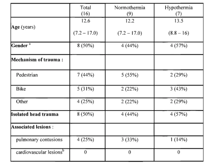
both groups (table I, page 33) with a median patient age of 12.7 years (range: 7.2 - 17.0).
TBl was the only physical problem in 50% (8/16 patients). Myocardial contusion,
myocardial laceration or rupture, or hemopericardium were never observed. No patients
with symptomatic congenital heart defects already treated for cardiac arrhythmias before
22
Among the 16 patients, 14 (88%) were transferred from another hospital after
stabilization. The median time between trauma and arrivaI to the study site was 2.8 hours
(range: 1.3 - 4.5 hours). Table II (page 34) describes baseline data. Patients in both groups
. were similar in terms of severity of brain trauma (initial Glasgow coma score in the emergency room, intracranial pressure (lCP) and cerebral perfusion pressure (CPP)
measured at intracranial monitoring insertion in the operating room and upon arrivai to the intensive care unit). Hemodynamie status based on vital signs in the emergency room
(blood pressure and heart rate) was also similar in the 2 groups. Median pediatric trauma
score was 3 (range: 2 - 5) in the NORMO group and 5 (range: 1 - 6) in the HYPO group, but the difference was not statistically significant (P=O.51).
Proto col compliance
ln the HYPO group, the length oftime from accident to the cooling process was 7.3 hours (range: 6.6 - 7.8 hours). Median time to reach the targeted temperature after initiation of
cooling was 3 hours (range: 1.7 to 11.5 hours). When the Holter recording began, ail
patients in the HYPO group were already hypothermie, with a median temperature of 32.9 oC (range: 31.6 to 34.4 oC). The temperatures reached and maintained during the Holter in
NORMO and HYPO groups are described in table III (page 35). At the end of the Holter
recording, patients in the HYPO group were still hypothermic.
Co-interventions
No difference in terms of vasopressors used or fluid boluses given was found
between patients in the HYPO and NORMO group. Sorne patients had central intravenous
right atrium for one patient; this patient did not present any arrhythmia. Ali other catheters
were in the desired position, confirmed by chest X-ray. No significant electrolytes
abnormalities were found.
Holter
Ali Holter monitors were installed without technical difficulties. Median time of recording was 24 hours (range: 21 to 36 hours). No Holter recording was stopped temporarily. The
median time between the trauma and the beginning of the Holter recording was 9.8 ho urs (range: 6.8 to 19.8 hours). Length oftime between trauma and start of Holter recording was
similar in the HYPO group (9.9 hours, range: 8.4 to 19.8 ho urs) and the NORMO group
(8.8 hours, range: 6.8 to 14.3 hours).
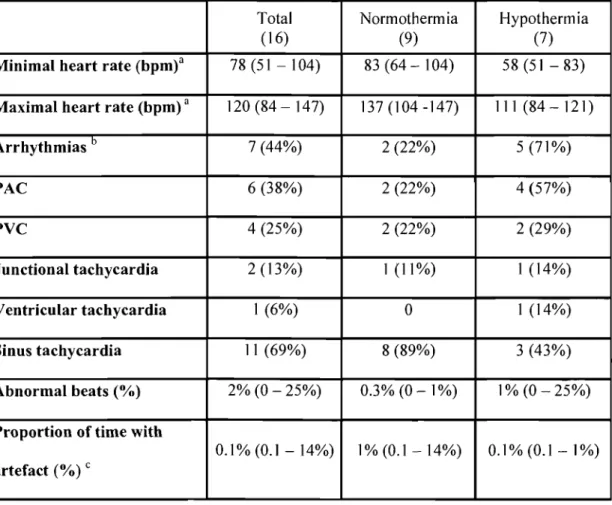
Overall, 44 % of patients (7/16) had at least one type of arrhythmia during the Holter recording; five patients had more than one type. In the NORMO group, 2/9 patients
had arrhythmias compared to 5/7 patients in the HYPO group (P=O.13; table IV, page 36).
The most frequent arrhythmias were isolated premature atrial contractions that did not
jeopardize hemodynamic status. Hypothermie patients had lower heart rates than normothermic patients (P=O.OI, table IV, page 36), but none had a severe bradycardia.
Two patients (one in each group) had junctional rhythms. One in the HYPO group
had occasional premature junctional depolarizations. The other one (NORMO group) had
an accelerated junctional rhythm alternating with sinus tachycardia. The junctional rhythm
was not recognized by the attending physician, and no specific treatment was given. While
this arrhythmia was present, the patient became hypotensive, which was attributed by the
attending physician to a barbiturate infusion. This patient also had isolated premature
24
Four patients had ventricular arrhythmias, three in the HYPO group and one in the NORMO group. Those ventricular arrhythmias were isolated or couplets of premature ventricular contractions. One patient in the HYPO group had a non-sustained monomorphic ventricular tachycardia without any hemodynamic instability.
No patient had asystole, AV block, atrial flutter or fibrillation, nor ventricular fibrillation. Since most arrhythmias were short-lived, we calculated the number of abnormal beats occurring overall in the recording, rather th an estimating the length of time with arrhythmias (table IV, page 36).
Overall, the median proportion of time considered as artefact in ail Holter recordings was 0.1 % (range: 0.1 to 14%). One patient had artefacts for 14% (3.5 hours) of his 24-hour Holter recording; this patient was in the HYPO group and also had isolated ventricular premature contractions.
ECG abnormalities
An ECG was do ne in 12 patients before temperature control was begun. Ali of the se patients were in a sinus rhythm. Six patients had an elevated or depressed ST segment (2 in the HYPO group and 4 in the NORMO group). The median QTc was 424 msec (range: 387 to 553 msec) in ail patients. The QTc was longer in the HYPO group before cooling (median 454 msec, range: 426 to 553 msec) compared to the NORMO group (median 404 msec, range: 387 to 434 msec, P< 0.01 between groups). Only two patients had a QTc greater th an 460 msec and both ofthese patients were in the HYPO group, before cooling.
After targeted temperature was obtained, an ECG was repeated in Il patients. Seven patients had elevated or depressed ST segment (5 in the HYPO group and 2 in the NORMO group). The QTc was longer in the HYPO group following cooling (median 497
msec, range: 447 to 581 msec) compared to the NORMO group (median 422 msec, range:
381 to 447 msec, P< 0.01 between groups). Five out of 6 hypothermic patients had an
abnormal QTc interval greater than 460 msec but no normothermic patient had an abnormal
QTc.
Other outcomes
The highest ICP during hypothermia was 36 mmHg (range: 22 to 91 mmHg) versus 34 mmHg (range: 22 to 81 mmHg) in NORMO group. The lowest CPP was 39 mmHg
(range: 0 to 52 mmHg) versus 42 mmHg (range: 9 to 61 mmHg) in the HYPO and NORMO group, respectively. Those differences were not statistically significant.
Seven of the sixteen patients (44%) developed pneumonia (NORMO group: 4
patients, HYPO group: 3 patients). Four patients (25%) died between 4 and 21 days after their admission into the PICU. In the HYPO group, two patients died of refractory septic
shock, 5 and 9 days after their arrivai and another one died of brain death associated with severe acute respiratory distress syndrome. In the NORMO group, the only death was
attributable to refractory intracranial hypertension. Length of stay in PICU and ventilator
free days at 28 days were similar in both groups (table V, page 37).
DISCUSSION
Traumatic brain in jury and arrhythmias
Electrocardiographic changes, arrhythmias and increased cardiac enzymes have been
reported in patients with TBI but no data are published on 24-hour Holter monitoring in
severe pediatric head trauma, nor in children exposed to moderate hypothermia therapy. In
26
arrhythmias in severe pediatric TBI patients. Most arrhythmias were of short duration and
did not alter the hemodynamic status. Overall, 44% of patients had at least one arrhythmia
during Holter monitoring: 2/9 normothermic patients and 5/7 hypothermic patients (P=O,13
between groups). The most frequent arrhythmias were isolated premature atrial contractions. Hypothermic patients had significantly lower heart rates compared to
normothermic patients. Arrhythmias can be explained by many causes other than head trauma and/or hypothermia therapy, such as pain, hypovolemia, and drugs; however, the
incidence of ail these possible risk factors of arrhythmias was similar in the HYPO and in the NORMO group.
We observed two patients who had episodes of more worrisome types of arrhythmias. [n the NORMO group, one patient had a persistent accelerated junctional
rhythm associated with prolonged hypotension. This arrhythmia was missed at the bedside, and it was only identified retrospectively in the Holter recording; the associated
hypotension was attributed by the attending physician to a barbiturate infusion, but we
cannot exclude the possibility that the instability was induced by the arrhythmia itself. In
the HYPO group, one patient had multiple isolated premature atrial contractions and a
non-sustained monomorphic ventricular tachycardia. The attending physician also missed this
arrhythmia, but it was not associated with hypotension.
This study revealed more arrhythmias in severe pediatric TBI patients th an what
was previously reported. Adelson et al. conducted a multicenter RCT looking at the safety
of moderate and short-term (32-33°C for 48 hours) hypothermia in severe pediatric TBl
[42]. They found arrhythmias in 8% of their normothermic and 22% of their hypothermic
First of ail, they did not use Holter recording; Holter is much more sensitive than standard bedside monitoring for detecting arrhythmias [65, 66]. As mentioned above, two of our patients had arrhythmias not detected at the bedside by health care providers using standard continuous cardiac monitoring.
Secondly, Adelson et al. did not define arrhythmias the same way as we did. They included sinus tachycardia as a significant dysrhythmia but it is unclear if they included abnormal supraventricular beats, supraventricular or junctional tachycardia, as we did. To be able to compare our results to theirs, we look back in our data to find how many patients had sinus tachycardia. We found that 8/9 normothermic patients had sinus tachycardia episodes compared to 3/7 in the hypothermic patients (P=0,10). Taking ail noted arrhythmias and adding episodes of sinus tachycardia, 13/16 patients (81 %) had at least one type of arrhythmias (89% of the normothermic and 71 % of the hypothermic patients). This is a lot more th an reported by Adelson et al. The reason why we chose to exclude sinus tachycardia in our primary outcome is that, out of a context of cardiac disease or surgery or primary sinus node disease in PIeU, we usually consider sinus tachycardia has a consequence of another variable rather than a primary arrhythmia to be treated. Knowing that hypothermia slows the heart rate, we assumed that there would be a higher heart rate within the normothermic group.
Adelson et al. found a trend towards an increase In cardiac arrhythmias in the
hypothermic group, and one patient was rewarmed before the end of the protocol because he had multiple premature ventricular contractions [42]. Despite this, the authors conc1uded that hypothermia was safe. We recommend further research on the effect of moderate hypothermia therapy on cardiac rhythm before to conclude.
28
It can be argued that Holter recording is too sensitive and can detect too many insignificant abnormal heart beats. A 24-hour Holter recording is indeed rarely free of
dysrhythmias in healthy children. Nagashima et al. reported the results of 360 ambulatory
electrocardiographic monitoring made in healthy children [67]. Supraventricular premature contractions was the most common type of arrhythmia detected, and was present in more
than half of ail children. Sorne children had premature ventricular contractions. No ventricular tachycardias, nor junctional accelerated rhythms were observed. Dickinson et
al. completed a similar study in healthy teenage boys [68]; premature ventricular contractions were found in 41 % ofthem, and short episodes ofventricular tachycardia were
recorded in 3%. ln our cohort, 38% of patients presented premature atrial contractions and 25% premature ventricular contractions. This is ev en less than what has been described in
healthy children. However, 2 patients (13%) presented a junctional rhythm and 1 patient ventricular tachycardia (6%). This is more than expected in healthy children.
To our knowledge, there is no study describing Holter monitoring in a PICU general
population. Thus, we cannot compare our observations to critically ill children without
head trauma. However, sorne case reports have been published on the occurrence of
malignant arrhythmias and ECG changes in children with severe TB! [14] [8, 69, 70]
Grosse-Worthmann et al. described an 8-year-old boy with severe head trauma who had
many supraventricular and ventricular extrasystoles, QTc prolongation, ventricular
fibrillation, accelerated junctional rhythm and supra-ventricular tachycardia without
echocardiographic evidence ofcongenital heart disease or major cardiac trauma [14].
Many mechanisms may explain the occurrence of dysrhythmias in severe head
trauma. The autonomic nervous system modulates cardiovascular function through many neurologically driven non-linear oscillatory systems involving the hypothalamus, brainstem
and spinal cord [71]. It is weil recognized that sorne dysregulation of these systems can occur after severe TBI. Such dysregulation could originate from the insult itself (the
trauma) or complications, su ch as increased ICP. There is evidence that the cardiac rhythm
is also controlled by the brain insular cortex; this control is lateralized, with a
predominance of sympathetic regulation on the right side of the brain, and a parasympathetic control originating from the left side [72-74]. Many studies done in
animaIs and patients with epilepsy, stroke or acute subarachnoid hemorrhage, have shown
that different arrhythmias are observed with insult on different sides of the brain. The severity of the autonomie dysfunction observed in patients with severe TBI is proportional
to the degree of neurologie dysfunction and it is correlated with worse outcomes [75, 76]. High plasma catecholamine levels may also be involved. An increased release of
norepinephrine has been documented in patients who have severe trauma, which was
correlated with the severity of the brain in jury [77, 78]. Such sympathetic activation can lead to tachycardia, increased myocardial oxygen demand, and myocardial ischemia.
Catecholamine-induced cardiac necrosis is a well-described entity. This phenomenon could
explain sorne ECG changes following brain in jury.
As reported in previous studies [58, 79], we found a prolongation of QTc interval,
which was even more marked in the HYPO group. QT interval extends from QRS complex
beginning to the end of the T wave [62]. It represents the time from onset of ventricular depolarization to completion of repolarization [63]. QT interval is considered prolonged
when the corrected QT interval is longer than 460 msec in children [63]. Prolongation of
QT interval has been identified as a risk factor for arrhythmias (torsades de pointe and
30
patients could not be attributable to any medications nor to any electrolyte abnormalities,
but it might be caused by the brain trauma itself and/or the hypothermia therapy.
Therapeutic hypothermia and arrhythmias
Without reaching a significant result, we found that more patients in the HYPO group had arrhythmias than in the NORMO group. The same phenomenon was observed
by Adelson et al [42]. Deep hypothermia (:'S 30°C) modulates ventricular repolarization;
prolongation ofQTc interval and inversion ofthe T-wave can result [25]. In animal studies, Osborn et al. also described an additional wave following the S-wave. This wave was
observed in ail animais but one who developed ventricular fibrillation later on. Thus deep hypothermia is arrhythmogenic, but it is unclear if this is also the case for moderate
short-term hypothermia therapy. The difference we observed might be attributable to chance al one, since the difference in the incidence rates of arrhythmia was not statistically
significant. It may also be a real effect of hypothermia, since no other explanations can be
found: severity of the trauma, highest lCP, electrolytes, and vasopressors used were indeed
simiJar in both groups. The cardiovascular safety of moderate hypothermia as a therapeutic
measure is not proven. In fact, the results of our study suggest there should be an increased
awareness to the presence and the severity of arrhythmias in patients exposed to moderate
hypothermia in future RCTs studying the efficacy of hypothermia as a treatment measure
of severe head trauma or post cardiac arrest.
Limitations of the study
This study has several limitations. First, the number of patients included in this