Direction des bibliothèques
AVIS
Ce document a été numérisé par la Division de la gestion des documents et des archives de l’Université de Montréal.
L’auteur a autorisé l’Université de Montréal à reproduire et diffuser, en totalité ou en partie, par quelque moyen que ce soit et sur quelque support que ce soit, et exclusivement à des fins non lucratives d’enseignement et de recherche, des copies de ce mémoire ou de cette thèse.
L’auteur et les coauteurs le cas échéant conservent la propriété du droit d’auteur et des droits moraux qui protègent ce document. Ni la thèse ou le mémoire, ni des extraits substantiels de ce document, ne doivent être imprimés ou autrement reproduits sans l’autorisation de l’auteur.
Afin de se conformer à la Loi canadienne sur la protection des renseignements personnels, quelques formulaires secondaires, coordonnées ou signatures intégrées au texte ont pu être enlevés de ce document. Bien que cela ait pu affecter la pagination, il n’y a aucun contenu manquant.
NOTICE
This document was digitized by the Records Management & Archives Division of Université de Montréal.
The author of this thesis or dissertation has granted a nonexclusive license allowing Université de Montréal to reproduce and publish the document, in part or in whole, and in any format, solely for noncommercial educational and research purposes.
The author and co-authors if applicable retain copyright ownership and moral rights in this document. Neither the whole thesis or dissertation, nor substantial extracts from it, may be printed or otherwise reproduced without the author’s permission.
In compliance with the Canadian Privacy Act some supporting forms, contact information or signatures may have been removed from the document. While this may affect the document page count, it does not represent any loss of content from the document.
Université de Montréal
Structure-function analysis of SOCS! mediated Growth arrest
Par Adrian MOQres
Département de Biochimie Faculté de Médecine
Mémoire présenté à la Faculté des études supérieures En vue de l'obtention du grade
Maîtrise (M.Sc.) en biochimie
Août 2007
Université de Montréal Faculté des études supérieures
Ce mémoire intitulé:
Structure function analysis of SOCS l-induced growth arrest
Présenté par : Adrian Moores
A été évalué par un jury compsé des personnes suivantes: Dr.Stephen Michnick Prési dent-rapporteur Dr. Gerardo Ferbeyre Directeur de recherche Dr. Léa Brakier-Gingras Membre du Jury 11
111
Résumé
Les suppresseurs de signalisation des cytokines (SOCS) sont une famille des protéines qui contrôlent la signalisation des cytokines par l'inhibition des JAK/STATs. Ils fonctionnent comme un adapteur pour la formation d'une ligase E3. De cette façon, des protéines SOCS aident à la dégradation des JAK et d'autres cibles, incluant d'autres SOCS. SOCS 1 est la protéine la plus étudiée de la famille. Elle a été identifiée comme étant nécessaire pour la fonctiori normale du développement des lymphocytes. Aussi SOCS 1 est hyperméthylé dans beaucoup des cancers et fonctionne comme un suppresseur de tumeurs. Les mécanismes précis de SOCS 1 comme suppresseur de tumeurs ne sont pas connus. La plupart des études se concentrent sur Je rôle de SOCS 1 dans la dégradation des kinases JAK et l'inhibition de la voie de signalisation de JAKISTAT.
Dans cette étude, nous avons démontré que la surexpression de SOCS 1 induit un arrêt permanent dans le cycle cellulaire par l'induction des mécanismes de sénescence. SOCS]
prévient la formation de colonies dans la lignée de cancéreuse des U20S et rend active la voie de signalisation de p53, peut-être par des interactions directes. Nous avons découvert un résidu nécessaire, le N 198, qui reste dans le « Cul box» de SOCS 1. Quand ce résidu a subi une mutation ou une perturbation, ceci inhibe la capacité de SOCS] à induire l'arrêt de croissance. Le résidu N 198 est impliqué dans la capacité de recrutement des cullins, ceci suggère que des séquences différentes dans le« Cul box» de SOCSI peuvent lui donner la capacité de se lier avec d'autres protéines ce qui lui donne la capacité d'induire un arrêt de croissance et de rendre active la voie de p53. D'autres membres de la famille des SOCS ne sont pas capables d'induire un arrêt de croissance, alors que SOCS5 semble fonctionner comme oncogène.
Mots Clés: SOCS: suppresseur de signalisation par les cytokines, JAK : kinase Janus, STA T : capteur de signal et activateur de transcription
IV
Abstract
The suppressors of cytokine signaling are a powerful family of proteins shown to negatively regulate cytokine signaling by inhibiting the JAKIST AT pathway. By acting as adaptor proteins and recruiting the modules of an active E3 ligase, SOCS proteins can degrade JAKs and a variety of other target substrates including other SOCS molecu les. SOCS 1 has been the most studied ofthe family and has been shown to be vital to normal lymphocyte development. As weil it has shown to be silenced via hypermethylation in a variety of cancers and been shown to contain the properties ofa tumor suppressor. The exact mechanisms ofSOCSI as a tumor suppressor are not known though most theories focus on the degradation of JAK's and inhibition of JAK/ST A T signaling.
Here we demonstrate SOCS 1 overexpression is able to induce permanent cell cycle arrest in primary cells by induction of the senescence machinery. It also prevents colony formation in the U20S sarcoma line and leads to activation of the p53 pathway, possibly through direct interactions. Also we have identified an essential residue, N 198, which when mutated or deleted leads to loss of ability to promote growth arrest. This residue is unique to SOCS 1 and lies within the Cul box domain of SOCS 1 and is involved in Cullin recruitment. This suggests that
differential sequences in the Cul box of SOCS 1 compared to other SOCS family proteins may allow it to bind to different cullins or other proteins that give it the ability to promote growth arrest in a variety of cells and activate the p53 pathway. They also hint towards the fact that SOCS 1 induced senescence and growth arrest is independent of its actions on the JAKIST A T pathway. Other members of the SOCS family investigated were not able to induce premature senescence, while sorne such as SOCS5 promoted cell proliferation.
Key words: SOCS: suppressor of cytokine signaling, JAK: Janus kinase, ST A T: Signal transducer and activator of transcription
v Table of Contents Résumé ... .iii Abstract. ... .iv Table of contents ... v List of figures ... ~ ... vi
Abbreviations list. ... vii
Chapter ] . Introduction 1.1 Cytokine signaling and Janus kinases ... 1
].2 STATS ... 3
].3 Discovery ofa SOCS family ofproteins ... .4
] .4 Role of SOCS 1 ... 6
1.5 SOCSI and the immune system ... 10
1.6 SOCS 1 acts through its SH2 domain ... .12
1.7 SOCSI as an E3Iigase ... .15
] .8 SOCS 1 in Cancer. ... 22
1.9 Regulation of SOCS 1 ... .26
Chapter 2. Materials and methods 2.] Cell culture ... 27
2.2 Retroviral gene transfer. ... 27
2.3 SA j3-Gal activity ... < • • • • • • • • • • • • • • • • • • • • • • • • • • • • • • • • • • • • • • • • • • • • • • • • • • • • • • • • 28 2.4 Immunofluorescence microscopy ... 28 2.5 SOCSI mutagenesis ... .29 2.6 Colony assays ... 30 2.7 Protein analysis ... 31 Chapter 3. Results ... 32 Chapter 4. Discussion ... 55 Chapter 5. Conclusion ... 62 Chapter 6. Acknowledgements ... 63 Chapter 7. References ... 64
VI
List of Figures
Figure 1 : SOCSI induced senescence in IMR90 fibroblast cells ... 38
Figure 2 : SOCSI prevents colony fonnation in the U20S sarcoma cellline ... .40
Figure 3 : DifferentiaI effects of SOCS family proteins on colony fonnation in U20S Cells ... 42
Figure 4 : CIS4/S0CS6 is incapable of inducing premature senescence in IMR90 cells ... 44
Figure 5 : Effects of domain mutations on SOCS 1 induced growth arrest.. ... .46
Figure 6: Activation of the p53 pathway by SOCSl ... .48
Figure 7 : Mechanism for instability in SOCS 1 domain mutants ... 50
Figure 8: Alignment of Cul boxes within the SOCS family ... 52
Figure 9: The N198 residue of SOCSI is necessary for SOCSI induced growth arrest. ... 54
VII
List of Symbols and abbreviations
% Percentage
Oc
Degrees Celsius fi Micro (l0-6) )lg Microgram ).lL Microlitre TM Trademark A AlanineAML Acute myeloid leukemia APC Antigen-presenting cell
ASKI Apoptosis signal-regulating kinase 1 ATP Adenosine 5'-triphosphate
bp Base pair
BSA Bovine serum albumin
C- Carboxy
cAMP Cyclic adenosine monophosphate CIS Cytokine inducible suppressor c-Kit Cytokine stem cell factor CD4 Cluster of differentiation 4 CD8 Cluster of differentiation 8 cDNA Complementary DNA
Cul CuIlin
D Aspartic Acid
DA Dalton
DMEM Dulbecco's Modified Eagle Media DMSO Dimethyl sulfoxide
DNA Deoxyribonuc1eic acid
dNTP Deoxyribonucleotide triphosphate
E Glutamic acid
Vlll
EST Expressed sequence tags EPO erythropoietin receptor
Erk2 Extracellular signal-regulated kinase 2
et al. And collaborators
F Phenylalinine
FGF Fibroblast growth factor FBS Fetal bovinse serum FBXOll F-box protein Il
G Glycine
g Gram
G-CSF Granulocyte colony-stimulating factor GAS Gamma activated sequences
GH Growth honnone
GST glutathione S-transferase HIF Hypoxia inducible factor HPV Human papillomavirus
1 Isoleucine
IRS Insulin Receptor substrates IFN Interferon
ISRE IFN response e1ements
IL Interleukin
JAB Janus kinase inhibitor/Janus kinase binding protein JAK Janus kinase
JHI Janus homology domain 1
K Lysine
KIR Kinase inhibitory region
KDa KiloDalton
Kbp Kilo base pair
L Litre
LPS M mM ml MAPK MHCr mRNA MTOC N N-n nm
NKT
ORF P PBS PCR P13K PMBL PML R Rb Rbx RNA S SA-~-Gal SH2 SH3 shRNA SDS SDS-PAGE Lipopolysaccharide Molaire (mol/litre) millimolaire millilitreMitogen-activated protein (MAP) kinases Major histocompatibility complex l Messenger RNA Microtubule-organizing center Asparagine Amino nana (10-9) nanometer
Natural killer T -cel! Open reading frame Proline
Phosphate buffered saline Polymerase chain reaction
Phosphatidylinositol-3-0H-kinase primary mediastinal B-ceIllymphoma Promyelocytie leukemia protein Arginine
Retinoblastoma protein Ring-finger catalytic protein Ribonucleie aeid
Serine
Senescence-assoeiated ~-galactiosidase Sre homology 2 domain
Sre homology 3 domain short hairpin RNA Sodium dodeeyl sulfate
Sodium dodeeyl sulfate polyacryladmide gel electrophoresis
STAT SOCS SSI-1 SUMO T TCR Tkip UBC
lNF
TYK2 VHL X-Gal ySignal transducer and activator of transcription Suppressor of cytokine signa1ing
ST AT -induced STAT inhibitor-l Small Ubiquitin-related modifier Threonine
T cell receptor
Tyrosine kinase inhibitor peptide Ubiquitin conjugating enzyme T umor necrosis factor
Tyrosine kinase 2
Von Hippel-Lindau tumor suppressor
5-Bromo-4-Chloro-3-indolyl-j3-D-gal ctopyranoside Tyrosine
Cytokine Signaling & Janus kinases
Cytokines are powerful signaling ligands used to communicate a signal from one cell to another and are capable of commencing multiple signaling cascades within a cell (1]. Cytokine signaling has been shown to play a large role in growth and differentiation though it is largely known for its role in immune responses such as wound healing and inflammation. They have also been demonstrated to play roles in development of the nervous system, and development during embryogenesis. Cytokines act in a similar manner to hormones as they are secreted and pass signais from one cell to the next, binding to a membrane bound receptor to commence their cascade within that cell. Most cytokines are sm aIl water-soluble proteins or glycoproteins and can be secreted by many different cell types. Four groups of cytokines have been classitied based on structure. The tirst group is classified as having a four a-helix bundle and can be further divided into subfamilies. The three subfamilies are; the interferons (IFN), activated in viral response, the interleukin-2 (IL-2) subfamily, which mediates signais to activate lymphocytes, and the interleukin-l 0 (IL-l 0) subfamily, which acts as an inhibitory molecule and prevents activation of other cytokine pathways such as the IFN and IL-2 pathways (2-7]. The a-helix bundle group makes up the largest and most weil known group of cytokines. The other three groups include the interleukin-l group (among the tirst cytokines discovered), interleukin-7 group and a group of cytokines simply classified as chemokines (8-11].
Each specifie cytokine has its own receptor that in tum is associated with a tyrosine kinase from the Janus kinase family (JAKs) [12-15]. The JAK family of kinases
consists of JAKl, JAK2, JAK3, and a fourth member, TYK2 [12, 13].lt is the JAKs that are responsible for downstream activation of aIl other kinases seen involved in cytokine signaling. AlI JAKs are similar in size, 120-130Kda. They are characterized by having a carboxy terminal kinase domain and pseudo kinase domains that precedes it. JAKs are almost ubiquitously expressed except for JAK3, which is found primarily in
haematopoietic ceIls.
JAK binding to cytokine receptors can take a variety offorms, which are generaIly c1assified into three patterns. JAK2 can bind constitutively, or in response to ligands, to single-chain receptors which then aggregate and cause a subsequent
aggregation of JAK2 [16, 17]. This leads to transphosphorylation of the kinase activation loops, activating the JAKs and increasing their catalytic activity. The now activated JAK2 is able to phosphorylate the receptor and target substrates recruited by the receptor, as well as autophosphorylate itself. A second model involves a pc_chain associated with a ligand binding a-chain, JAK2 associated with the pc_chain, which leads to its activation and signal transduction [17-19]. This is commonly seen in the lL-3 and 1L-6 families. The interferons and 1L-2 family require two chains to induce JAK signaling [20-24]. IFN-a & p have a p-chain, which associates with JAK2 and a a-chain that associates TYK2 [21-24]. IFN-y has an a-chain that associates with JAKI and a p-chain that associates with JAK2 [23]. This is similar to the 1L-2 receptor, where JAK2 binds to either a ligand specific a-chain or the proximal region of the p-chain and JAK3 binds to a shared Yc chain, the receptor aggregates bring the two JAKs together [20].
Though the mechanisms may vary slightly, the end goal of cytokine binding to a receptor is to cause aggregation of the JAKs which leads to their subsequent cross
activation and commences the signaling cascade [l, 12, 13, 18, 20]. JAKs can activate many signaling cascades by means of recruitment using SH2 domains oftarget
molecules. Such target molecules include RAS, phosphatidylinositol-3-0H-kinase (PI3K), as weIl as the signal transducers of activation and transcription (STA TS).
STATS
The ST A TS make up a well-known family of transcription factors heavily linked to cytokine/JAK signaling. ST A TS act upon target genes through different mechanisms as weB. The first mechanism was found in studies involving INF-a/~, these showed ST A Tl was phosphorylated in response to IFN and bound in a complex with p48 [25]. This caused ST A TIto enter the nucleus and cause transcription of genes containing IFN response elements (lSRE). Later it was shown IFN-y caused phosphorylation of ST A Tl which caused it to form a dimer which th an moves into the nucleus and binds to gamma activated sequences (GAS) in target genes and drives transcription [26]. As more ST A T molecules were cloned, this model became the widely accepted model for ST AT
activation, and it has since been shown that that almost aIl cytokines activate at least one STAT [13].
AlI ST AT members contain conserved SH2 domains as weIl as an SH3-like domain and a DNA binding domain [13, 27]. Phosphorylation of tyrosine residues in the carboxy terminus mediates both hetero and homo di mer formation via the SH2 domains [28]. Dimer formation leads to entry into the nucleus where transcription follows [26, 28]
ST A TS can also be activated by other serine/threonine kinases and have been linked to MAPK and the RAS signaling cascade [26,28-32].
Discovery of a SOCS Family of pro teins
Obviously, cytokines are involved in a variety of important signaling cascades and therefore must be carefully regulated. Constitutive activation of any growth or proliferation signal, as w~ll as a chronic inflammation response, can have dire
consequences for the cell, the worst being malignant transformation. As such, another family ofproteins known as the suppressors of cytokine signaling (SOCS) exist to negatively regulate the em~cts of cytokines through a variety ofmechanisms including direct inhibition oftheir receptors or the JAKs which activate them [33-37]. The first protein ofthis family was discovered in 1995 and named cytokine induced suppressor (CIS) [35].
Currently, there are eight known members of the SOCS family in humans (SOCSI-7 and CIS) [38]. By studying the primary amino acid sequences of the SOCS family members it has been revealed that there exists a high degree of similarity between pairs of proteins. For example, SOCS 1 is very similar to SOCS3 and thus they form a pair, SOCS2 and CIS make another, while SOCS6&7 and SOCS4&5 round out the pairings. SOCS family members a11 share a SH2 domain as well as conserved C-terminal SOCS box motif, but have variable N-terminals. The SOCS box domain has since been found in a variety of non-SOCS family proteins, over twenty so far [39]. The mechanisms
of inhibition by SOCS proteins varies from protein to protein, sorne ev en display multiple mechanisms.
SOCS 1 has become the most studied of the group. It was cloned and described by three different groups under different studies, which speaks of its versatility as a negative regulator of cytokine signaling. Naka et al. were trying to find other STA T family
members using an antibody for a sequence in the ST A T3 SH2 when they identified both CIS and a new gene, which they cloned and named Stat-induced ST A T inhibitor-1 or SSI-1 [37]. SSI-1 showed a 36% homology to the SH2 domain ofCIS, but none to ST A T3 or ST A T6, except for the phospho-tyrosine recognition site. A second group, looking for proteins that could bind to the JAKs directly through the JH1 domain, also managed to clone SOCS1, which they at the time had named JAB for Janus kinase binding protein·[34]. They too reported a protein with a SH2 domain sharing simi1arity to that of CIS (35%). A third group named the protein SOCS 1 after finding it by screening for genes encoding proteins that could inhibit IL-6 signa1ing [33]. They performed a screen of library of genes from the factor dependent cellline FDC-P 1. Upon infecting Ml cells with a retrovirus containing the cDNAs from the FDC-P 1 cells they found a 1.4 Kbp insert that rendered the Ml cells unresponsive to the IL-6 signal. They cloned the insert and also found it to be a relative of CIS and named it the suppressor of cytokine signaling 1 (SOCS1).
The early studies on SOCS 1 alllead to similar results that helped e1ucidate its role in cytokine signaling and shed sorne light on the mechanisms through which it works. The SOCS1 gene rests on a single exon and is located in mice on chromosome 16, close to the protamine gene cluster [33, 34]. It shares no homology to any of the protamine
genes, but rather corresponded to what was a then unknown ORF at the 3' ofthese genes [34]. It encodes a 212 amino acid prote in in mi ce and rats, 211 in humahs [33,34,37]. The human, mouse and rat copies of the protein are highly homologous sharing a 95-99% amino acid sequence [33]. SOCSI contains a SH2 domain at amino acids 79-167 and a SOCS Box domain at its C-terminus [33,34]. Using the sequence from SOCSI and expressed sequence tags (ESTs) Starr et al., were able to clone the first two other related SOCS proteins SOCS2 and SOCS3, which have similar SH2, and SOCS box domains as SOCS] [33]. SOCS1 expression is ubiquitous in most tissue types though it has been
repeatedly shown to have higher levels of expression in the thymus, spleen, testes and lung [33, 37].
Role of SOCS!
Investigations into the role of SOCS l in cytokine signaling clearly show its role as a negative regulator. When expressed in myeloid leukemia (M 1) cells that also expressed a Thrombopoietin receptor, SOCS 1 conferred a general resistance to cytokine signaling [33]. Furthermore these cells continued to grow and proliferate when
expressing SOCS 1, even when treated with a variety of cytokines including IL-6, IFN, Leukemia inhibitory factor (LIF) and Thrombopoietin (in Ml.mpl cells). Such treatment caused parental Ml cells to stop proliferation and form differentiated colonies [33, 37]. Treatment with dexamethasone did lead to differentiation in cells expressing SOCS 1, implying it acted only in the cytokine signaling pathways, and not the general
of the antiviral activities ofIFN [34]. Since SOCS 1 seemed to inhibit cytokine signaling, it was suspected that it acted as a negative feedback rnechanism so the effects of cytokine signaling on SOCS 1 induction were studied. In growth factor dependent celllines such as Ml and the hybridoma MH60 cellline, SOCSl was induced in response to treatment with
IL-6 and soluble IL-6 receptor [37]. This was repeated in the IL-4 dependent line CT4S after treatment with IL-4 as weIl as the G-CSF dependent NFS60 line with G-CSF treatment. Bone marrow ce Ils stirnulated with different cytokines are capable of not only inducing SOCS1, but also a variety of SOCS family members, depending on the cytokine
[33]. Also sorne cytokines are capable of inducing certain SOCS genes in specific cell lines but not others. IL-3 and activation of the erythropoietin receptor (EPO) are unable to induce SOCS1 in Tf-l and NFS60 cells. IL-6 induces CIS, SOCSl-3, in mi ce liver cells
but only C1S and SOCS1 in Ml ceIls, displaying a cell specific cytokine response in the
induction of SOCS family members.
Since SOCSI inhibits many cytokines, and most cytokine signaIing cascades signal through the JAKISTAT pathway, the interactions between SOCS 1 and the
JAKISTA T pathway were further studied. IL-6 and IL-4 are inhibited by SOCS l and are both capable of inducing SOCSl. Both act through the JAKIST A T pathway, IL-6 through ST A T3 and IL-4 through ST A T6. Studies of the SOCS l promoter revealed binding sequences for ST A T3 and ST A T6, implicating SOCS1 as a target gene for ST AT
signaling. It has since been shown that SOCS1 also has binding sites for ST A T5 as weIl
in its promoter. Ml cells transfected with SOCSl and STAT3 show induction ofSOCSI
rnRNA in response to IL-6. However when SOCS 1 is co-expressed with a ST A T3 mutant, in which a tyrosine reside phosphorylated by a JAK (Y705) is replaced with a
phenylalanine, no induction takes place, showing a requirement for activated ST AT signaling as a requirement for the induction of SOCS] [37].
Tyrosine phosphorylation ofSTAT3 and the Gp130 protein, which is part of the cytokine receptor and is phosphorylated by JAKs, were both reduced in Ml cells expressing SOCS]. IL-6 treatment is able to phosphorylate both STAT3 and Gp130 in normal Ml cells, though expression of SOCS] reduces this .. STAT3 and STAT5 tyrosine phosphorylation was also reduced in 293 cells expressing SOCS]. General ST AT
activation in response to many cytokines is greatly inhibited by SOCS 1; ST A T5 activation by EPO and ST A T3 activation by IL-6 is almost completely aboli shed in
SOCS] expressing Ml cells [34]. As well several STAT target genes su ch as the immunoglobin fragment Fcy recptor (FcyR) show a large reduction in expression in
SOCS] Ml cells.
SOCS 1 also pre vents ST AT molecules from forming dimers that enter the nucleus, preventing their activation ofDNA transcription [33]. In Ml cells treated with IL-6, the most common dimers observed are the SIF-A (STAT3 homodimer) and SIF-B (STATl/STAT3 heterodimer), the formation ofthese complexes is observed using electrophoretic mobility shift assays. In SOCS] expressing cells IL-6 treatment fails to induce formation ofthese dimers, as well it also blocks formation of the SIF-C STATI homodimer induced by IFN-y. Hence SOCSI is able to prevent tyrosine phosphorylation of ST A Ts, preventing them from forming an active heterodimer and blocking
transcription of ST AT target genes in the process.
SOCS 1 does not directly bind and inhibit the ST AT transcription factors. Instead it targets the JAKs that activate them and inhibits their signaling cascade. SOCS 1 directly
interacts with the JH 1 domain of JAK2 via its SH2 domain and has been shown to
interact with TYK2 as well [34,37]. However, JAK activity is needed for this interaction as SOCS 1 does not interact with a K882D kinase defective mutant copy of JAK2. When co-expressed in 293 cells JAK2 tyrosine phosphorylation levels are much lower than in control 293 cells not overexpressing SOCS]. However, phosphorylation at a key residue YI 007, is necessary to activate the JAK2 and needs to occur before SOCS 1 can bind. This implies that upon activation of the JAK2, SOCS 1 is able to binds to JAK2 and inhibit its autophosphorylation and subsequent phosphorylation of cytokine receptors, ST AT molecules and their target genes. Therefore, it is through inhibition of the activity of the JAKs that SOCS 1 confers a resistance to cytokines and negatively regulates their signaling cascades.
When JAK2 and ST A T3 are co-expressed in 293 cells, ST AT3 undergoes a rise in tyrosine phosphorylation. The same occurs when STAT5 is co-expressed with JAK2. If JAK2, SOCS 1 and STAT3/STAT5 are aIl expressed together, no phosphorylation of ST A T3 or ST AT5 occurs. Furthennore, SOCS 1 is capable of preventing phosphoryIation of JAK 1 and JAK3 in 293 cells as weil, indicating SOCS 1 is capable of regulating a broad spectrum of JAKISTAT signaling pathways other thanjust JAK2. SOCSl is able to inhibit IL-2 and IL-3 activation of the c-fos promoter as well [34]. Since STATS are not
required to activate c-fos but JAKS are, it demonstrates that SOCS 1 directly targets the JAKs not the ST A Ts themselves [34, 40].
The inhibitory mechanisms of SOCS 1 seem to be restricted specifically to the JAKISTAT pathway. Whereas SOCSl inhibits 1L-2 and lL-3 activation ofc-fos, it does nothing to prevent c-fos activation by cAMP (which follows a non cytokine activation
and does not involve the JAKs) [34]. When expressed in NII-l3T3 cells SOCSl does not alter tyrosine-phosphorylation of Erk2 and Shc in response to fibroblast growth factor (FGF). SOCS 1 binds the kinase domain of c-kit in yeast cells but fails to inhibit c-kit or the epidermal growth factor receptor. This demonstrates that SOCSl targets specifically the JAK tyrosine kinase activity and not tyrosine kinases in general.
SOCS] and the immune system
SOCS1 plays a central role in the deve10pment of the immune system. SOCS1 knockout mice show many phenotypes, among them T -cell associated lyrnphoma and large scale infiltration of macrophages, lymphocyte and eosinophils [41-43]. These mice die very young due to overactive IFNy signaling, the majority coming from T-cells [43]. They have been shown to suffer symptoms such as liver necrosis, myocarditis,
polymyositis and fatty degeneration. SOCS1 has been shown to induce resistance to IFNy when overexpressed [44]. In SOCS 1 knockout mice lacking T -cells or containing a knock out ofIFNy, the same rates of young lymphocyte-dependent lethality are not seen as they are in normal SOCS 1 -1-mice [43,45]. Macrophages isolated from SOCS 1 -1-rnice require little IFNy to bec orne activated, as compared to normal wild type mice [46]. As weB these rnice show higher levels ofnatural killer T-cells (NKT), which are implicated in the necrosis of the liver [42].
Regulation of T cell developrnent has been shown to be controlled directly by SOCS 1. SOCS 1 has been shown to be expressed in the thymus, especially during thyrnocyte development [33, 34, 37, 47, 48]. Expression ofSOCS1 is critical for the
proper development of CD4+ T cells [49]. Deficiencies of SOCS 1 lead to increases in CD8+ CD4- ceUs and CD4+CD8+ double positive cells, but leads to decreases in
CD4+CD8- cells [47,49]. This is caused through increased levels in IL-7 signaling, which protects double positive cells, and IL-5 which stimulates the proliferation of CD8+ CD4-celIs, both cytokines are usually negatively regulated by SOCSl [47,50,51]_ The number of total T lymphocytes in SOCSl deficient mice is also increased [52].
SOCS3 has also been shown to play a large role in regulation of macrophages and helper T~cells, placing the SOCS family ofproteins in the middle ofT-celI development and regulation of normal immune system development [53, 54].
Other immune cells such as dendritic cells are also under SOCS 1 control. Dendritic cells deficient in SOCS 1 are hypersensitive to interferon signaling and promote aberrant B-cell proliferation leading to the production of autoreactive T-cel1s [55, 56]. Silencing of SOCS 1 can enhance dendritic cell response and antigen presentation, this is beneficial for anti-tumor responses by enhancing cytotoxic t-cell responses [55-57]
SOCS 1 also helps in signaling within the innate immune system. Many foreign proteins such as v-E7 and bacterial LPS strongly induce SOCSl [58,59]. SOCSl
induction helps curtail the levels of inflammatory cytokines and chemokines secreted by activated antigen-presenting cells as weIl as negatively regulating LPS-induced
macrophage activation. SOCS 1 deficient mice have been shown to be unable to form a tolerance to LPS which can be lethal to them in certain cases [60]. Tumor necrosis factor alpha (TNF-a) is also regulated by SOCSl, giving SOCSl another branch of control over inflammation responses, such as TNF -a induced apoptosis, and regulation of cells within the immune system [61].
Regulation oftheimmune system is therefore dependant on SOCSI at many levels. SOCS 1 plays a central role in the development of T -cells as weil as their homeostasis. It also plays a role in reducing macrophage activation due to extemal stimuli and reduces dendritic cell activation and their ability to present antigens, thereby· inhibiting autoimmune responses. As weil SOCS 1 directly controls the levels of many inflammatory signaling pathways su ch as TNF-a giving it direct control over
inflammation and innate immune responses.
SOCS! acts through its SH2 domain
SOCS 1 acts through multiple mechanisms to inhibit the activity of JAKs. There are multiple lines of evidence supporting different mechanisms, which make use of the different structural domains of SOCS 1. Such diversity of mechanisms may seem
redundant but is perhaps necessary to ensure proper regulation of signaling cascades that control growth and proliferation as weil as many components of the immune system. It is also known that the different SOCS family member act through different mechanisms from each other. For example CIS inhibits cytokine signaling in a mechanism different from that of SOCS 1 and does not bind directly to the JAKs [36, 62].
One of the mechanisms through which SOCS 1 exerts control over JAKs is through direct binding to the autophosphorylation site through interactions with the JH 1 domain of JAK2 and the SH2 domain of SOCS 1 [34, 63]. Once cytokines bind to their receptors they induce phosphorylation of JAK2 at YI 007, a critical step in JAK2 activation [64]. Subsequent downstream JAK2 signaling in tum induces SOCSl, which
regulates the pathway in a negative feedback manner. SOCS I is able to bind to JAK2 phosphorylated at YI 007 with a high affinity but does not bind to unphosphorylated JAK2 or JAK2 mutants which contain mutations in the JHI domain (K882D) [34, 63]. Mutational analysis of SOCS I has revealed 3 regions necessary for complete binding and inhibition of JAK2 [63]. In order for SOCS I to bind to Y 1007 of JAK2 the SH2 domain, specifically a phospho-tyrosine-binding residue Arg 105, and a stretch of twelve amino acids (l68-G79) immediately N-terminal to the SH2 subdomain, dubbed the extended SH2 subdomain, are both required. A second set of twelve amino acids just N-terminal before the extended SH2 subdomain (N 56-67) are required for SOCS I to bind to JH I with high affinity and are necessary to inhibit JAK2 signaling. This region has been named the kinase inhibitory region (KIR).
The extended SH2 subdomain contains three aminoacid residues 168, L 75 and G79, which are highly conserved throughout the SOCS family and appear at the same position relative to the SH2 domain of STATs as weIl. Similarity between the SH2 domains of SOCS and ST A T family members may be important in SOCS I inhibition of JAK/STAT signaling as perhaps SOCS I could compete directly with STATs for binding sites with JAKs to reduce ST AT signaling. Mutating these conserved residues in (l68E and L75E) is enough to prevent the interaction between SOCSI and JHI and YI007 as weIl as reduce EPO-dependant ST A T5 signaling. Crystal structures of ST A T molecules show these conserved residues are involved in phospho-tyrosine binding in the SH2 domain [65].
The Kinase inhibitory region is not important to binding to YI 007 but is essential for inhibition of JAK2 signaling and mutations to the region greatly hamper JHI binding
[63]. Mutations to individual amino acids in the region prevented SOCSI from inhibiting EPO-dependent ST A T5 signaling. Furthermore eight of the twelve amino acids are also present in SOCS3, which can also bind JH1 domains and inhibits the JAKs, further implying their importance in SOCS/JAK interactions [63, 66]. Among these conserved amino acids were F56, F59, D64 and Y65. Mutations to these residues in particular greatly hampered SOCS 1 from binding to JH1. Mutations to F59 showed the greatest effect suggesting it is perhaps the more critical residue [63].
The current mechanism for SOCS 1 interaction with JAK2 involves the SH2 of SOCS1 recognizing the phosphorylated Y1007 of JAK2. The c1assical SH2 domain is involved in binding to this region. The extended SH2 subdomain aids in further binding to JAK2. Binding to Y 1007 allows the KIR to bind to the JH1 with high affinity and disrupt Jak2 signaling.
A 12 mer synthetic tyrosine kinase inhibitor peptide (Tkip) is able to mimic SOCS 1 by also inhibiting JAK2 signaling through binding of the autophosphorylation site [67]. Tkip is able to bind to JAK2 at YI 007 with a higher affinity than SOCS 1 and can bind to unphosphorylated JAK2 as weIl. Its binding to JAK2 is able to suppress IFN-y activitIFN-y, su ch as the upregulation ofMHC Class 1 molecules, induction of growth arrest, and EGFR autophosphorylation. Tkip actually acts much like the KIR region of SOCS 1 [68]. By acting in a mechanism very similar to that ofSOCS1 Tkip is also able to inhibit constitutive ST A T3 and IL-6 activated ST A T3 in prostate cancer cells (LN CaP and DU145) [69]. Injecting mice with Tkip also reduces allergic responses by inhibiting overactive cytokine signaling [70].
SOCS! as an E3 Ligase
Another mechanism commonly used to control cellular signaling is the
degradation of cellular receptors or other proteins involved in the cascade. Targeting of specifie proteins for degradation is a tightly controlled pro cess that requires complexes of enzymes working in unison. One of the better known pathways to protein degradation is the ubiquitin-proteasome pathway, which uses long chains ofubiquitin molecules to mark proteins for degradation [71]. Degradation is a two-step process. The first step requires the target protein to be covalently flagged with a long chain ofubiquitin, usually
accomplished by a complex ofproteins. The second step is the actual degradation of the protein by the 26 proteasome. The actual attachment of ubiquitin to a target substrate is a three-step process and involves many proteins. The first is an El activating enzyme, which activates the ubiquitin molecules. Once activated an E2 ubiquitin carrier or
ubiquitin conjugating enzyme (UBe), of which there are several, facilitates the transfer of the activated ubiquitin from the El to one of the E3 protein ligases. The substrate usually contains specifie sequences to ensure proper and specifie binding to the E3 ligase. The E3 ligase is responsible for completing the process and creating the covalent bond between the ubiquitin and the target protein. Usually, the first moiety is attached to a NH2 group
on a lysine residue, generating an isopeptide bond. Once the first moiety is attached the rest can attach to the proceeding moiety via its Lys48 residue. This allows the formation of a long poly-ubiquitin chain that serves as a flag, marking the prote in for later
as many more proteins and/or protein complexes that have E3 ligase activity are still being discovered.
Though the SH2 domain of SOCS 1 is responsible for binding to Jak2, it alone is not enough to inhibit JAK signaling [34]. A mutant copy ofSOCS1 lacking both C- and N- terminal domains also has no inhibitory affects on cytokine signaling demonstrated by its inability to inhibit c-fos activation by IL-2 or IL-3, which suggests that one or both of
these domains is necessary for complete SOCS 1 activity. The C-terminus of SOCS l, like aIl SOCS family members, contains a domain known as the SOCS box [33, 34, 37, 39, 72-76]. The SOCS box was once thought to be a unique domain to SOCS family members but has since been shown to be present in the C-terminal of a variety of different protein families inc1uding the Ras, WD-40 repeat, ankyrin repeat families and SPRY domain containing families [39, 77].
Within the SOCS box, there is a T/SLlMxxxC/SxxxVIL/I Elongin BC binding consensus sequence named the BC Box [77]. Elongin BC is a heterodimer protein
complex made up of the ubiquitin like Elongin B, and Elongin C, a protein that resembles the adaptor protein Skp in sequence [78, 79]. Elongin BC was first shown to be an
activator of the RNA Pol II elongation factor A (Elongin A) [80]. It has since been shown to also take part in the von Hippel-Lindau tumor suppressor complex (VHL) [81].
Mutations within, or deletions of the consensus sequence render Elongin A and VHL unable to bind Elongin BC [81-83]. Elongin BC is much more abundant in the cell than either Elongin A or VHL, which means it must have other activity or possible binding partners unrelated to the two [77]. It is now known that the Elongin BC complex can bind
not just SOCS l, but the BC box of ail proteins containing a SOCS box and that deletions or point mutations of the BC box inhibit the interaction [76, 77].
Endogenous levels ofElongin Band C can be immunoprecipitated from the 1ysates of cells stimulated with interferon and IL-6 against antibodies for the C- and N-tenninals of SOCS l, proving that this is a physiological interaction [77]. As well expressed Elongin Band C can be can be immunoprecipitated with antibodies against JAK2 and SOCS 1 in cells co-expressing JAK2 and SOCS 1. This Proves that Elongin BC is part of the SOCS 1 complex that binds to JAK2 and inhibits its signaling. Only a fonn of SOCS 1 with an intact BC box is capable of making the Elongin BC-JAK2 complex, although mutations in the BC Box do not disrupt SOCS 1-JAK2 binding.
The VHL tumor suppressor acts as an active E3 1igase [84]. In this complex Elongin BC acts as an adaptor to link VHL to a Cullin and a RIN G finger-containing protein (Rbx). The cullins are a family ofproteins that assemble with an Rbx molecule to fonn a module capable of catalyzing the transfer of ubiquitin from an E2 ubiquitin conjugating enzyme to the target substrate [85, 86]. There are currently five known cullin members in mammals (CuI1-5) [87]. The VHL protein acts as a subunit of a multi-protein complex able to recognize substrates for ubiquitination [84]. The VHL tumor suppressor shows similarity to the SKP-Cul1-F-box E3 ligases (SCF), and both show sorne similarity to an Elongin BC-CuI2-S0CS 1 complex. SOCS 1 also binds to Elongin BC, Elongin B is an ubiquitin like molecule, and Elongin C shares sequence similarity to the adaptor protein SKP1 [78, 79]. The F- box is a similar domain to the SOCS box. Studies into SOCS 1 showed that it too was able to bind to a Cullin/Rbx module [88]. This Elongin B/C-CuI2-S0CS 1 complex is capable of fonning GST -polyubiquitin chains by the E2
GST-ubiquitin conjugating enzyme Ubc5 when ATP, El activating enzyme Uba1 and GST-ubiquitinK48R are present, proving it acts as an active E3 ligase. The crystallized structures of a similar complex involving SOCS2 also defined an E3 ligase [75]. As weIl SOCS3 also forms such a complex, and upon activation with IL-6 sees its protein
expression peak within 60 minutes only to be near depleted by 120 minutes [76]. This drop off in SOCS3 levels is aboli shed by treatment with proteosomal inhibitors such as LLnL. This suggests that SOCS proteins form active E3 ligases that play a role in their subsequent degradation after induction.
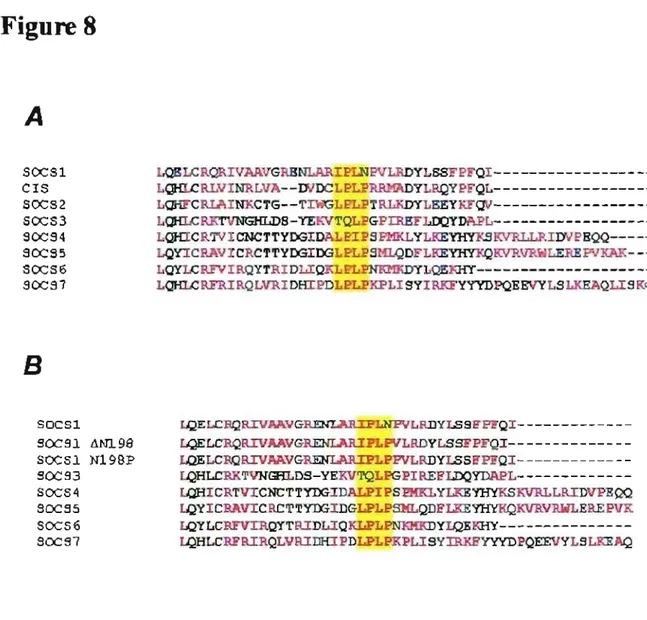
The specificity of the cullin binding is said to be due to a small stretch of amino acids located at the C-terminal of the SOCS box known as the Cul box [89]. Differences in the Cul box between VHL and SOCS family members lead VHL to specifically bind to endogenous Cul2-Rbx1 while SOCS-box proteins bind to endogenous CuI5-Rbx2. These differences allow VHL and SOCS family members to be grouped accordingly according to their Cul Box. VHL has a Cul2 box while the members of the SOCS family ofproteins contain a Cul5 box; the y bind to endogenous Cul5-Rbx2 and not CuI2-Rbx1.
A specific conserved amino acid sequence of LPxP within the Cul5 box appears necessary for Cul5 binding as mutating it abolishes it. While aIl SOCS members have a Cul5 box, SOCS1 oddly has an incompletely conserved Cul5 box. The Cul box of SOCS 1 contains the amino acid sequence IPLN instead of LPxP; this leads SOCS 1 to bind to CuI2-Rbx1, not CuI5-Rbx2, making it an exception within the SOCS family as the only member to do so. Changes in Cul5 expression cause no change in the
degradation ofVHL target substrate HIF-2a, however reducing Cul2 expression by usage of shRNA cause increases of HIF -2a. This suggests that each Cul member has a distinct
purpose or possibly has the ability to recognize different substrates. Ifthis is the case there could be sorne unique function conferred upon SOCS 1 as the only SOCS member that binds to Cu12.
There is plenty of evidence to suggest that the mechanism by which SOCS proteins inhibit cytokines lies in their ability to form ECS E3 ligases. SOCS 1 localizes to the microtubule organizing complex that is associated with the 20 S proteasome itself in a manner dependent on the SH2 domain [90]. SOCS 1 also binds directly to the microtubule organizing complex (MTOC). Such a direct link to the proteasome suggests SOCS 1 may help localize targets to the proteasome for degradation. SOCS 1 does in fact cause JAK 1 and VAV1 to localize to the MTOC and MTOC-associated 20S proteasome.
Many studies have suggested that SOCS 1 directly targets proteins for
degradation. SOCS 1 was shown to bind to the N- terminus of the guanine nucleotide exchange factor V AV, and reduce the formation of V AV -induced foci in NIH 3T3 cells [91]. SOCS1 also diminished the levels ofVAV within NIH 3T3 and COS celllines, and was ultimately found to induce ubiquitination ofVAV and onco-VAV. IFN-y induces expression of SOCS 1 and leads to a degradation of the viral oncogene E7 in HeLa and CaSki tumor lines [59]. E7 mRNA levels remain the same when SOCS 1 al one is expressed in these cells though E7 protein levels are diminished. SOCS 1 co-Iocalizes with E7 in the nucleus and can immunopreciptate with E7, suggesting a physical interaction. This interaction is dependent on the SOCS Box, but is uninterrupted by mutations of the SH2 domain. Like the interaction of SOCS 1 and VAY, tyrosine phosphorylation does not seem to play a role. SOCS 1 is also seen to promote
transfonnation by leading to E7 degradation. JAK kinases su ch as JAK2 are sometimes involved in oncogenic fusions, su ch as the fusion protein TEL-JAK2 [74]. In Ba/F3 cells transfonned with TEL-JAK2 expression of SOCS 1 or induction of SOCS 1 by treatment oflL-3leads to apoptosis. This response is not seen however in Ba/F3 cells transfonned with p21 0 Bcr-Abl, showing SOCS 1 activity to be specific to JAKs. SOCS 1 mutants lacking the SOCS box cannot suppress the growth ofTEL-JAK2 cells, despite the SOCS box not being necessary to bind to JAK2, suggesting that the SOCS box still plays a role in inhibition of JAKs [74, 92]. In 293 cells fulllength, WT SOCS 1 can reduce the levels of TEL-JAK2 and suppress TEL-JAK2 activation of ST A Ts. Treatment of proteasome inhibitors MG 132 and lactacystin protected TEL-JAK2 from SOCS 1 induced
degradation, suggesting it is proteasome dependent. Further studies showed that
phosphorylation of the JHl domain led to binding with SOCSI and led to de gradation of JAK2 preceded by SOCS 1 induced ubiquitination of TEL-JAK2. The SH2 domain of SOCS 1 is necessary for degradation ofTEL-JAK2. The SOCS box domain can be replaced with that of the protein CIS, but not the SOCS3 SOCS box. SOCS3 binds and inhibits TEL-JAK2, but does not induce its degradation, this is odd as both SOCS3 and CIS contain a different CulS box from SOCSI [66,89]. A dominant negative Cul-2 inhibits the Tel-JAK2 degradation, suggesting SOCS 1 induced degradation must work in an E3 ligase manner.
SOCS 1 also acts in the insulin signaling pathway and degrades insulin receptor substrates (lRS). In HEK293 cells, SOCS 1 associates with IRS 1 and IRS2 via
interactions that are significantly increased in response to insulin signaling. SOCS2 and SOCS3 were also found to interact with IRSI and IRS2. Interactions between SOCSI and
IRS leads to subsequent falls in the levels ofIRS 1 and IRS2 in HEK293, MCF7 breast cancer cells and ETE-LI adipocytes. Induction of SOCS 1 by treatment with
proinflammatory cytokines such as IL-6, TNFa, and IFN-y also led to reductions in levels of IRS 1 and IRS2. Expression of SOCS 1 in the liver of mice via adenovirus infection causes IRS 1 and IRS2 levels to fall until the infection is no longer detectable at which point IRS levels retum to normal. A SOCS 1 mutant missing the SOCS box is unable to regulate the same control over IRS 112 as can wild type SOCS 1. Mutations to key residues of the BC box cause a decrease in degradation of IRS but do not prevent SOCS 1 binding. This led Rui et al. to search for ubiquitination ofIRS 112. They found that SOCS 1 was indeed able to lead to ubiquitination ofIRS 112, although this could be prevented with treatment of MG 132 or lactcystin.
Colony formation induced by the granulocyte-colony stimulating factor, G-CSF, as weIl as its activation of ST A T3 and ST A T5, are also suppressed by SOCS 1 and SOCS3. However, both ~SOCS Box mutants of SOCS 1 and SOCS3 fail to cause any changes to colony formation or ST A T activation in response to G-CSF [94]. This also suggests a possible role of SOCS box mediated degradation of G-CSF or its downstream targets as weIl. Stabilization of phospho-ST AT5 by proteasomal inhibitors supports this model. The Apoptosis signal-regulating kinase 1 (ASK 1) is also a target of SOCS 1 induced degradation [95]. ASK 1 binds to SOCS 1 through its SH2 domain and is both ubiquitinated and degraded. It is known that TNF signaling stabilizes ASKI levels and prevents degradation while SOCS 1 acts as a negative regulator of TNF and promotes ASK 1 degradation.
These results have shown two powerful properties of SOCS 1. The first is to act on target substrates by fonning an E3 ligase with Elongin BC and a Cul-Rbx subunit in the SOCS box domain [76, 77, 88, 89]. It suggests the SH2 domain is used to recognize substrates which it than marks for proteosomal dependent degradation [59, 74, 91, 96]. Localization with the MTOC associated with the 20S proteasome only strengthens this argument [90]. It also shows another important property of SOCS l, the property of a tumor suppressor. It has already been established that SOCS 1 inhibits the JAK, a family of tyrosine kinases. Receptor tyrosine kinases are implicated in many cancers as
mutations which confer constitutive activity to them cause an over stimulation in many signaling cascades that promote growth and proliferation. However the results mentioned . above also show SOCS1 's ability to directly target other oncogenes su ch as EP7,
onco-V A V and the TEL-JAK2 fusion protein and directly lead to their destruction by targeting them for proteosomal degradation.
SOCS! in Cancer
Receptor tyrosine kinases have been identified as being proto-oncogenes. The JAKs as weIl can serve as oncogenes should their signaling go astray, or should they become involved in fusion proteins. SOCS 1 meanwhile demonstrates tumor suppressor properties [97]. Tumor suppressors are often inactivated in a variety of cancers either through gene silencing or mutations that encode a non-functioning protein. SOCS1 can actually prevent transformation of Ba/F3 ceIllines caused by Tel-JAK2 by inhibiting JAK2 and leading to its degradation [92]. Gene methylation of CpG islands and other
epigenetic modifications are seen in a variety of cancers, this often leads to silencing of tumor suppressor genes. SOCS1 methylation was first demonstrated in HCC cell lines (65%) [98]. Several groups have since repeated similar studiesshowing a high level of SOCS1 methylation in HCC, corresponding to a low level of expression and
accompanied with constitutive JAK2 activation [99-102]. Since th en it has been shown that SOCS1 is methylated in a variety of cancers. In 2003, it was shown that over halfthe patients sampled (53%) with newly diagnosed acute myeloid leukemia (AML) showed methylation of the promoter region of SOCS1 [103]. Multiple myeloma ceIllines such as the IL-6 dependant XG 1 and U266 lines show a low level of SOCS1 expression even in the presence ofIL-6 compared to controllines [104]. These ceIllines showed high levels of ST A T3 phosphorylation and a higher sensitivity to a chemical JAK inhibitor, AG490, which induced apoptosis. Samples taken from primary tumors of multiple myeloma also confirmed the results shown in cell culture as one study showed 62.9% of patients sampled to have SOCS1 methylation. This is also confirmed by the fact that SOCS 1 deficient mice die within 3-4 days due to a lymphoma [41,42]. Many human pancreatic cancers have also shown reductions in expression of SOCS1 [105]. This usually
correlated with a methylation of the 5' promoter region for SOCS1. While one group showed methylation of the SOCS1 promoter region in tumor samples, they found no mutations to markers within SOCS1. Breast and ovarian cancer displayed SOCS1
methylation as weIl but showed a pattern of di fferenti al hypermethylation between other members of the SOCS family [106]. SOCS1 was found to be hypermethylated in 4 out of 6 studied ovarian cancer cell lines and 8 out of Il breast cancer lines. Wh en actual tumors were studied, 23% of ovarian cancers had hypermethylated SOCS1 CpG islands,
while only 9% of the breast cancers showed the same result. Cells from the colorectal cancer line Hep3B, which have methylated DNA, also do not express SOCS] [107]. Also, SOCS 1 deficient mice showed a higher rate of developing tumors within their colon due to over inflammation in response to uninhibited IFN-y/Stat1 signaling [108]. Other groups have shown that SOCS 1 hypermethylation and subsequent loss of SOCS 1 expression confers growth-promoting effects in pancreatic cancers su ch as pancreatic ductal adenocarcinomas and intraductal papillary mucinous neoplasms [109]. Silencing of SOCS] can occur due to loss ofheterozygosity (LOH) and can lead to an increase of carcinogenesis in mice livers [110]. Hypermethylation of SOCS] in ail these cancers leads to a large reduction in expression, this effectively allows for constitutive JAK/ST AT signaling. Constitutive signaling of both the Janus kinases and ST A T
molecules has been largely linked to the formation of cancers in the past [111]. Silencing of SOCS] in these tumors can often be reversed by introducing agents that reverse DNA methylation, su ch as 5-azadeoxycytidine (5-aza-dC), mimic SOCS1 activity (Tkip), or directly inhibit the JAKs [69, 104, 106, 112]. Restoration of SOCS] expression or overexpression of SOCS] in these cancers often leads to growth inhibition or induction apoptosis. SOCS] mutations have been found in a significant number of tumors from Hodgkin, Reed-Stemberg, Hodgkin and classical Hodgkin tumors as weil as in primary mediastinal B-celllymphomas [113]. Mutations can range from out of frame mutations which lead to premature stop codons cutting off the SOCS box domain, to deletions in amino acids of the SH2 domain. These mutations lead to 10ss offunction of SOCS] and allow JAK2 signaling to persist, eventually resulting in an accumulation of active phospho-ST A T5 in the nucleus. In primary mediastinal B-celllymphoma (PMBL) cell
lines, MedB-1 and KaJ-pas11 06P, biallelic mutations to the coding region of SOCS 1 allow JAK2/STAT5 signaling to go unchecked [114,115]. In this cancer, JAK2 remains constitutively active although it shows only a normal physiologicallevel of expression. Instead JAK2 degradation is greatly impaired, hence the JAK2/ST A T5 signaling cascade is allowed to persist. Overexpressing SOCS 1 in MedB-1 celllines with this mutation leads to growth inhibition [114].
Our 1ab focuses on the study of premature senescence and growth arrest. Premature senescence is a permanent cell cycle withdrawl that can act as a tumor suppressor mechanism [116-118]. Senescence can be induced in response to oncogenic stress as a barrier to transformation. Previously, our 1ab demonstrated that a constitutively activated form ofSTAT5 (STAT5A1 *6) can induce premature senescence [119]. SOCS1 is highly upregulated in STAT5 senescent cells, implicating its involvement in
senescence. Other SOCS1 activators such as ~-IFN can also induce senescence [120]. Members of our lab have demonstrated that overexpression of SOCS1 in the IMR90 . fibroblast line Induces premature senescence. It is established that SOCS 1 is able to
induce a growth arrest in cells. However, the mechanisms by which it acts are completely unknown. SOCS 1 can indu ce p53 and SOCS 1 induced senescence in the IMR90 cellline was shown to be p53 dependent. SOCS 1 has the ability to bind to and inhibit JAKs and can form E3 ligases. Whether either ofthese functions is necessary for the induction of senescence or growth arrest is unknown. AIso, the role of the different domains of SOCS 1 is unknown at this time.
Regulation of SOCSl
The mechanisms by which SOCS 1 is regulated are not weil known. While many different stimuli for SOCS 1 induction are weil known and documented, the negative regulation of SOCS 1 remains for the most part a mystery. Tt is known that SOCS 1 can be targeted by other SOCS family proteins for degradation [121]. SOCS2 has been shown to have the ability to bind and inhibit SOCS 1 by targeting it for proteosomal degradation. Both SOCS6 and SOCS7 were later shown to have the ability to interact with aIl other SOCS molecules. Other reports suggest that SOCSI leads to its own auto degradation and that binding to the machinery of its E3 ligase such as the Elongin BIC complex lead to reductions in SOCS1 protein stability [76, 122]. However other reports have shown it is necessary to have binding of SOCS 1 to the Elongin BIC to promote stability and that interruptions in binding lead to proteasomal degradation [77, 123, 124]. The
interconnecting web of a family of SOCS proteins, each capable offorming active E3 ligases acting on one another and each with the ability to potentially self-regulate makes the exact mechanisms of regulation by means of how SOCS 1 is regulated by proteasomal degradation hard to elucidate, especially considering how little is known about the
majority of the other SOCS family members. Other groups have pointed to protein kinases that may regulate SOCS 1. The Pim family of prote in kinases was shown to phosphorylate SOCSl which 1eads to stabilization ofprotein levels [122]. It is possible that SOCS 1 undergoes other modifications which are used to regulate it at the protein level, including phosphorylation by other kinases, however as ofyet the se mechanisms remain mostly unknown.
Materials and methods:
Cel! culture
AlI IMR90 primary ceIllines and U20S sarcoma ceIllines were cultured in Dulbecco's Modified Eagle Media (DMEM) (GIBCO) supplemented with 10 % FBS (GIBCO) and
1 % Penicillin G-streptomycin sulfate (GIBCO).
Retroviral vectors and gene transfer
The foIlowing retroviral vectors were used, pLPC and its derivatives expressing SOCS1,
SOCS3, SOCS5, SOCS6, SOCS1 &Jax, SOCS1 &JC, SOCS1 ,dCul, SOCS1 LW 198 and SOCS1 N198P as weIl as the retroviral vector in pBABE and its derivatives hRAS and STA T5A 1 *6. Phoenix ceIls were plated to a density of2.5x104 ceIls / ml in a 10 mm culture plate (Coming) (l0 ml total volume) and transfected with retroviral vectors (20
/lg) and treated with 200 /lI of 5 mg/ml sodium butyrate the next day. Medium was changed 12 hours after sodium butyrate addition. 12 hours after the medium was changed and retro viral soups were coIlected and supplemented with 10% FBS and 4 mg/ml
polybrene. The supplemented soups were immediately added to primary ceIls which had been previously plated to a density of 8.0 x 10⁵ cells / ml in 10 cm culture plates
(Coming). Cells were treated with new retroviral soups every 6 hours for a total of 3 infections. Infected cells were selected in puromycin 2.5 /lg/ml (Bioshop) for 2 days.
SA fJ-Gal activity
IMR90 cells were infected as described above. Cells were given 3 days recovery period after selection and plated at a density of 2.5 xl 05 cells / ml 6 days post selection. Cells were than incubated in 5-bromo-4-chloro-3-indolyl-~-D-galctopyranoside (X-Gal) at pH 6.2 at 37°C until the negative control showed approximately 10% positive staining. The percentage of cells expressing SA-~-Gal was quantified by inspecting 100 cells per 10 mm plate three times.
Immunojluorescence microscopy
IMR90 cells were plated on cover slips in a 6-well plate at a density of2.5x104 cells / ml and fixed using 4% paraforrnaldehyde (Fisher Scientific) in PBS for 15 minutes at room temperature. Cells were washed several times in PBS and perrneabilized using 0.2% Triton X-100 (Fisher Scientific) in PBS with 3% bovine serum albumin (BSA) on ice for 5 minutes. Cells were than washed again several times with PBS/BSA and than treated with the primary antibody, anti-PML (Rabbit) prepared by Marie-France Gaumont-Leclerc for 1 hour at room temperature in a humidified chamber. After three more PBS/BSA washings cells were stained with Alexa-Flora Red, anti-rabbit secondary antibody (l :2000) (Molecular Probes) for 1 hour in a humidified chamber. Cells were than washed several times with PBS/BSA and counterstained with 4,6-diamidino-2-phenylindole (DAPI) at a concentration of 0.1 ~g/ml in PBSIBSA. Fluorescence microscopy was perforrned using an inverse fluorescence microscope (Nikon TE2000) and the Metamorph software (Molecular Devices). Images were prepared using
SOCS1 mutagenesis
The SOCS1 L1Box construct had already been prepared by Vivianne Calabresse. The rest of the SOCS1 mutants were made using PCR techniques. Ali reactions were performed in a Biometra T-gradient PCR Machine. Reactions were performed in 100 /lI total volume using 50 ng ofpLPC SOCS] as a template, 200 /lm dNTP, 50 !-lM ofboth primers, and 5% DMSO and Deep Vent Polymerase O'Jew England Biolabs) and it's Thermopol Buffer O'Jew England Biolabs). SOCS1,(jCul was created using a PCR using a sense primer (referred to as SOCS 1 sense primer) of
5 'GCGAA TTCTGA TGGTA GCACGCAACCA GGTG3 ' and an antisense primer 5 'GCGGGCTCGAGTCAGTTCTCGCGACCCACGGC3' that introduced a premature stop codon before the Cul Box.
SOCS L1BC was created using a two step process. First two small fragments were made. Fragment A was created using the SOCS 1 sense primer as used for SOCS1,(jCul and an anti-sense fragment that started at the beginning of the BC Box and contained a 15 bp overlap for fragment B, 5 'ACCCACGGCGGC CACCCGCACGCGGCGCTG3 '. Fragment B was created using a sense primer that had a ] 5 bp overlap to the tail of Fragment A and started after the BC Box,
5 'CAGCGCCGCGTGCGGGTGGCCGCCGTGGGT3 ' and a normal antisense primer for
SOCS1, 5 'CGCTCGA GTTCA GA TCTGGAAGGGGAAGGA3 '. The two fragments were than used as a template for a reaction using the normal SOCS 1 sense and antisense pnmers.
SOCS1 LfN198 and SOCS1 N198P were made in a similar fashion. First a fragment was made using the aforementioned SOCS 1 sense primer and an antisense primer
introducing a deletion of the 198P residue, 5 'GTCACGGAGTACCGGAAGAGG
GATGCGCGC3' this fragment was not fulliength SOCSl. A second fragment was made
using a forward primer that contained a 15 bp overlap to the first fragment and introduced a sense strand deletion mutation to the 198 P residue,
5 'GCGCGCATCCCTCTTCCGGTACTCCGTGAC3' and an antisense primer that started
after the SOCS 1 insert in the pLPC vector back bone, to produce a larger in sert th an 100 bp to facilitate cloning, 5 'CAGCTG TTCCATCTGTTCTTGGGC3 '. The two fragments
were than used as template for a PCR reaction using the normal SOCS 1 sense primer and a second SOCSI antÏsense primer (as the first did not work well with this reaction) 5 'GGGCCTCGA GTCA GA TCTGGA AGGGGAAGGA3' to give a fulIlength SOCSI
product with the ~N198 mutation. The N198P mutant was made in similar fashion using the SOCS 1 sense primer and aN 198P antisense primer
5 'GTCACGGAGTACCGGGGGAAGAGGGATGCGCGC3 J. Fragment B was generated
using a sense primer of 5 'GCGCGCA TCCCTCTTCCCCCGGTA CTCCGTGAC3 ' and the
pLPC antisense primer mentioned above. AlI mutants were sequenced at l'Institut de Recherche en Immunologie et en Cancérologie (IRIC) to make sure mutants were in the right places and that no other errors were introduced by the PCR process.
Colonyassays
U20S cells were transfected with 15 )lg ofDNA using the calcium phosphate method. Transfected cells were selected in puromycin l)lg 1 ml for 6 days. After which cells were given a 2 day recovery period for colonies to grow. Cells were then stained in 0.5 % Crystal violet to show colony formation. Cells were de-stained using 10% Acetic acid.
100 !lI of each sample was loaded to a 96 weIl plate and measured for absorbance at the 960 nm wavelength in a plate reader. Values were plotted on a bar graph and normalized to the control to show relative differences in growth. Standard error represents 1 standard deviation from the mean value.
Prote in analysis
Immunoblots were preformed using whole-celllysates obtained by first making cell pellets and than boiling them in Laemmli sample buffer. Samples of20 !lg ofprotein were resolved in SDS-polyacrylamide gel electrophoresis and transferred using the wet-transfer method to Immobilion-P membranes (Millipore). Antibodies used in the
immunoblots inc1ude anti-SOCS 1 (4H 1; 1: 1 000 Upstate), anti-p53 (catalog number 9282 1:1000 Cell Signaling Technology), anti-PS-15p53 (catalog number 9284 1:1000 Cell Signaling Technology), anti-p21 (catalog number 2949 1:1000 Cell Signaling
Technology), anti-RB (C-19, 1:250 Santa Cruz Biotechnology), anti-Mdm2 (2AI0; 1 :250; donated by A. Levine), anti-Tubulin (B-5-1-2; 1 :2,000; Sigma). Western blot . assays were performed using ECL detection (Amersham) or Lumilight detection system (Roche Applied Science).
Results:
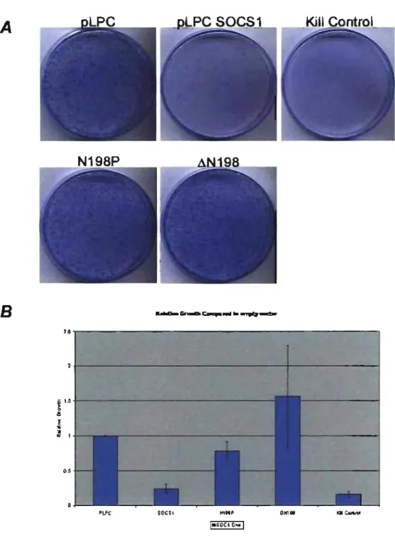
We were interested in exploring the full extent and mechanisms of SOCS 1-mediated growth arrest. First, we explored SOCS 1 growth arrest in primary cells. When infected into IMR90 fibroblasts SOCS 1 induces a permanent cell cycle aire st known as premature senescence. Senescent SOCS1 cells show a larger, flatter morphology and stain positive for the senescence associated ~-galactosidase akin to oncogenes such as RAS and STAT5A1 *6 (Figure lA) [125]. As well these cells show accumulation ofPML bodies within the nucleus (Figure lB). Accumulation ofPML bodies in the nucleus is often used as a marker of senescence [125].
Since premature senescence is a tumor suppressor pro gram it was interesting to see the effects of SOCS 1 on the growth of cancer cells directly. The sarcoma ceIlline U20S was transfected with SOCS1 and an empty vector. As shown in Figure 2 SOCS1
expression abolishes the ability ofU20S cells to form colonies and greatly hinders their growth. Hence SOCS 1 is also able to lead growth arrest within tumor ceIllines as weIl as inducing permanent cell cycle arrest in primary cells.
Members of the SOCS family ofproteins exist in similar pairs, SOCS1 and SOCS3 for example are more similar to each other than to other family members [38]. Sorne other SOCS members such as SOCS3 have also been demonstrated to play roles within a variety of cancers as weIl [106]. We wanted to see the effects of other SOCS family members on growth arrest so we repeated the colony assay in U20S using SOCS3, SOCS5, CIS4/S0CS6 as weIl. As demonstrated above SOCS1 completely
abolishes the ability ofU20S cells to fonn colonies as the SOCS1 transfected cells didn't fonn colonies as did the control plate (Figure 3B).
Although very similar in sequence to SOCS1, SOCS3 does not inhibit colony
fonnation in the U20S cell line. Though the number of relative colonies is slightly lower than that of the control plate, it is significantly higher than that of the SOCS1 plate
(Figure 3B). Surprisingly, SOCS5 over-expression actually causes a near two fold
increase in the number of colonies fonned (Figure 3B). This result was repeatable and suggests SOCS5 may actually play the role of an oncogene. The full role of SOCS5 is
largely unknown though it may function by actually interacting with the activity of other SOCS moJecules, as sorne crosstalk and interference between SOCS family members has been previously shown. [121]. SOCS6 was the only other member tested that showed the
ability to prevent colony fonnation that was significantly similar to SOCS1 (Figure 3A &
3B). When tested in primary cells however, SOCS6 is unable to induce premature
senescence (Figure 4). This suggests that SOCS6 maybe capable ofinducing growth arrest, but unable to induce a pennanent cell cycle arrest such as SOCS 1 through
induction of the senescence program.SOCS2 and SOCS7 as weil as CIS constructs were unavailable at this time.
Many roles of SOCS 1 are dependent on its SOCS box domain and its ability to fonn an active E3 ligase [73, 74, 91, 93]. E3 ligase activity ofSOCS1 requires an intact SOCS box to assemble the E3 ligase machinery. We wanted to investigate whether SOCS1 induced growth arrest was dependent on the SOCS Box as weIl. We made a series of SOCS box mutant lacking both the BC and Cul box subdomains, labelled as
SOCS box, SOCS1 Lillox, was donated to our lab and sub-cloned into a pLPC vector. These mutants were then transfected into U20S cells (Figure 5).
Deletions of the entire SOCS box have only minor effects on SOCS1 induced growth arrest in U20S (Figure 5). This points to a possible role of SOCS 1 in growth arrest that independent of its SOCS box and its E3 ligase activity. SOCS1 constructs containing deletions of the BC and Cul box also show little effect on growth arrest
(Figure 5). However these mutants provide no real infonnation to this regard as the levels of SOCS 1 in the cells are poorly expressed and cannot be detected through western blots (Figure 6A).
Sorne studies have shown SOCS 1 can target itself for auto degradation or that binding to the Elongin BC complex is necessary for stability ofSOCSl [77,123,124]. To see ifthis is the case with our SOCS mutants we treated transfected U20S cells with MG 132, a proteasome inhibitor, before collecting extracts. In these cells SOCS 1 levels are greatly enhanced compared to that ofuntreated cells (Figure 6B). This indicates that SOCS 1 lacking functional BC box and Cul box domains are targeted for proteasomal degradation whereas it appears SOCS1 constructs lacking the entire SOCS box domain are not (Figure 6A & B). SOCS1 LillC and SOCS1 LJCUL mutants still retain parts of the SOCS box and possibly still able to present epitopes or potential binding sites for other E3 ligases (such as other SOCS molecules) to recognize them and mark them for degradation (Figure 7 A). The SOCS1 LJbox mutant lacks the entire SOCS box and may present no epitope and could be therefore protected from degradation (Figure 7B).
It had previously been demonstrated in our lab that SOCS 1 induces the p53 pathway in fibroblasts and that SOCS1 senescence was p53 dependent (Malette et. al