marinus L.):
I. Neuroanatomy and physiology.
Journal: Journal of Comparative Neurology Manuscript ID: DOI: 10.1002/cne.24787
Wiley - Manuscript type: Accepted:
Research Article 20-Sep-2019
Keywords: Solitary Chemosensory Cells, Lamprey, Papillae, Microvilli, Merkel Cells, Taste, Central projections
Sensory cutaneous papillae in the sea lamprey (Petromyzon marinus L.):
1
I. Neuroanatomy and physiology.
2 3
4 Short Title: Cutaneous papillae in Lampreys
5
6 Gheylen Daghfous1,2, François Auclair1, Felix Blumenthal1, Tina Suntres3, Jessica Lamarre-
7 Bourret1, Masoud Mansouri1, Barbara Zielinski3,4 and Réjean Dubuc1,2,*
8
1 Groupe de Recherche sur le Système Nerveux Central, Département de neurosciences,
9
10 Université de Montréal, Montréal, QC, Canada
11 2 Groupe de Recherche en Activité Physique Adaptée, Département des sciences de l'activité
12 physique, Université du Québec à Montréal, Montréal, QC, Canada 13 3 Department of Biological Sciences, University of Windsor, Windsor, ON, Canada
14 4 Great Lakes Institute for Environmental Research, University of Windsor, Windsor, ON, Canada
15 16 17 18 19
20 *Correspondence: Réjean Dubuc, Ph.D., Professor, Département des sciences de l’activité 21 physique, Université du Québec à Montréal, PO Box 8888, section Centre-ville, Montréal, 22 Québec, H3C 3P8, Canada.
23 rejean.dubuc@gmail.com
Acknowledgments
24
25 The authors would like to thank Danielle Veilleux, Frédéric Bernard, and Christian Valiquette for 26 their technical assistance. We also thank Dr. Colin Favret from the Département des sciences 27 biologiques at the Université de Montréal, for letting us use his Nikon SMZ800 stereomicroscope 28 and Dr. Thomas Théry for help with this equipment. This study was supported by the Great Lakes 29 Fishery Commission as group grants to BZ and RD (54011, 54021, 54035, and 54067), by a grant 30 to RD from the Canadian Institutes of Health Research (15129), by individual grants to RD and BZ 31 from the Natural Sciences and Engineering Research Council of Canada (respectively 217435-01 32 and 03916-2014), and by a group grant from the Fonds de Recherche du Québec - Santé (FRQS, 33 5249). The funders had no role in the study design, data collection, data analysis, decision to 34 publish, or the preparation of the manuscript.
35 36
Conflict of Interest Statement
37
38 The authors wish to declare no conflict of interest. 39
40
Data Availability Statement
41
42 The data that support the findings of this study are available from the corresponding author upon 43 reasonable request. 44 45
Abstract
46 247 Molecules present in an animal's environment can indicate the presence of predators, food or 48 sexual partners and consequently, induce migratory, reproductive, foraging, or escape behaviors. 49 Three sensory systems, the olfactory, gustatory, and solitary chemosensory cell (SCC) systems 50 detect chemical stimuli in vertebrates. While a great deal of research has focused on the olfactory 51 and gustatory system over the years, it is only recently that significant attention has been devoted 52 to the SCC system. The SCCs are microvillous cells that were first discovered on the skin of fish, 53 and later in amphibians, reptiles, and mammals. Lampreys also possess SCCs that are particularly 54 numerous on cutaneous papillae. However, little is known regarding their precise distribution, 55 innervation, and function. Here, we show that sea lampreys (Petromyzon marinus L.) have 56 cutaneous papillae located around the oral disc, nostril, gill pores, and on the dorsal fins and that 57 SCCs are particularly numerous on these papillae. Tract-tracing experiments demonstrated that 58 the oral and nasal papillae are innervated by the trigeminal nerve, the gill pore papillae are 59 innervated by branchial nerves, and the dorsal fin papillae are innervated by spinal nerves. We 60 also characterized the response profile of gill pore papillae to some chemicals and showed that 61 trout-derived chemicals, amino acids, and a bile acid produced potent responses. Together with 62 a companion study, (Suntres, Daghfous, Dubuc, & Zielinski, this issue), our results provide new 63 insights on the function and evolution of the SCC system in vertebrates.
64 65
Keywords
66
67 Solitary Chemosensory Cells; Lamprey; Papillae, Microvilli; Merkel Cells; Taste; RRID:SCR_013672; 68 RRID:AB_2313574; RRID:AB_2336881; RRID:AB_2315147; RRID:SCR_011323; RRID:SCR_000903; 69 RRID:SCR_003210; RRID:SCR_014199; RRID:SCR_010279.
70
71
1. Introduction
72
73 Chemoreception is not restricted to olfaction and gustation (Daghfous, Green, Zielinski, & Dubuc, 74 2012; Finger, 1997; Hansen & Reutter, 2004; Kotrschal, 1996; Parker, 1912). Chemicals that are 75 present in the environment are also detected by specialized epithelial cells named “solitary 76 chemosensory cells” (SCCs). Bipolar epidermal cells thought to be sensory were initially described 77 in lampreys (Fahrenholz, 1936a, 1936b; Foettinger, 1876; Langerhans, 1873) and in ranid tadpoles 78 (Kölliker, 1885; 1886). However, it is only with the work of Whitear on the skin of teleost fish that 79 the innervation, and thus sensory nature, of these cells was demonstrated (Lane & Whitear, 1982; 80 Whitear, 1952, 1965, 1971). Subsequently, studies have reported putative SCCs on the barbels 81 and nasopharynx of hagfish (Braun, 1996; 1998; Braun & Northcutt, 1998), on the skin surface of 82 brook lampreys (Lampetra planeri Bloch) and river lampreys (Lampetra fluviatilis L.), including the 83 oral, gill pore, dorsal fin and genital regions (Baatrup & Døving, 1985; Fox, Lane, & Whitear, 1980; 84 Whitear & Lane, 1983a), on the skin, gills, and oropharynx of chondrichthyes and bony fish (Codina 85 et al., 2012; Hansen, Ghosal, Caprio, Claus, & Sorensen, 2014; Kotrschal, 1992; Kotrschal, 86 Krautgartner, & Hansen, 1997; Kotrschal, Peters, & Atema, 1989; Kotrschal, Whitear, & Adam, 87 1984, Kuciel et al., 2014; Peach, 2005; Peters, Kotrschal, & Krautgartner, 1991; Peters, Van 88 Steenderen, & Kotrschal, 1987; Silver & Finger, 1984; Whitear & Moate, 1994). Other studies have 89 described solitary chemosensory cells on the skin and in the oral cavity of amphibians (Koyama, 90 Nagai, Takeuchi, & Hillyard, 2001; Nagai, Koyama, Von Seckendorff Hoff, & Hillyard, 1999; Osculati 91 & Sbarbati, 1995; Whitear, 1976), and in the airways of reptiles and mammals (Finger et al., 2003; 92 Hansen, 2007; Saunders, Christensen, Finger, & Tizzano, 2014; Sbarbati, Crescimanno, Benati, & 93 Osculati, 1998; Sbarbati, Crescimanno, Bernardi, & Osculati, 1999; Sbarbati & Osculati, 2003; 94 Tizzano, Merigo, & Sbarbati, 2006; Tizzano et al., 2010). The study of SCC innervation and 4
95 physiology has been challenging because of the scarcity and widespread distribution of these 96 cells. Until recently, most knowledge regarding SCC innervation came from two teleost taxa: sea 97 robins (Prionotus carolinus) and rocklings (genera Ciliata and Gaidropsarus). In sea robins, SCCs 98 are concentrated on the free rays of the pectoral fins, and are innervated by spinal nerves 99 (Bardach & Case, 1965; Finger, 1982, 2000; Kotrschal, 1995; Morril, 1895). In rocklings, SCCs are 100 concentrated on the anterior dorsal fin and are innervated by a recurrent branch of the facial 101 nerve (Kotrschal, 1991, Kotrschal et al., 1984; Kotrschal, Royer, & Kinnamon, 1998; Kotrschal & 102 Whitear, 1988; Kotrschal, Whitear, & Finger, 1993b; Whitear & Kotrschal, 1988). More recent 103 studies have shown that the trigeminal nerve provides innervation to some of the SCCs located in 104 the distal nasal cavity of mammals and reptiles (Finger et al., 2003; Hansen, 2007; Tizzano et al., 105 2010). Information regarding the physiology and function of the SCCs is also limited. In sea robins, 106 SCCs detect amino acids, which act as feeding cues and promote foraging in this species (Bardach 107 & Case, 1965; Silver & Finger, 1984), whereas SCCs respond to fish mucus in rocklings (Peters et 108 al., 1991, 1987) and brook lampreys (Baatrup & Døving, 1985). In rocklings, SCCs are narrowly- 109 tuned to mucus from heterospecific fish and likely act as a predator-detection system (Kotrschal, 110 Peters, & Atema, 1993; Peters et al., 1991, 1987). Their function in brook lampreys is not clear. In 111 mammals, airway SCCs respond to bacterial signals (Tizzano et al., 2010). Their activation evokes 112 respiratory protective reflexes (i.e. coughing and sneezing) through the activation of trigeminal 113 fibers (Finger et al., 2003) and stimulates the secretion of antimicrobial peptides (Lee et al., 2014, 114 2017). SCC locations, innervation patterns, and functions thus differ greatly in the few species that 115 have been studied and the solitary chemosensory cell system may be highly specialized in these 116 species (Finger, 1997). While these studies have been informative with regards to SCCs in given 117 vertebrate taxa, the general role of this chemosensory system is still unknown.
118 The present study examines SCC localization, chemical sensitivity, and innervation in the 119 sea lamprey (Petromyzon marinus L.). A companion study, (Suntres et al., this issue), investigates 120 the biochemical properties and development of SCCs during the life cycle of the sea lamprey. 121 Together, these two studies advance our understanding of the solitary chemosensory system in 122 the most basal extant vertebrate group. Knowledge on the SCC system in lampreys is crucial to 123 bridge the gap between studies conducted in fish and mammals. Due to the basal phylogenetic 124 position of lampreys within vertebrates, this research sheds light on the general organization and 125 role of the SCC system in vertebrates.
126 127
2. Materials and Methods
128
129 2.1 Animals
130 Experiments were performed on 54 spawning phase adults and 6 newly-transformed sea 131 lampreys (Petromyzon marinus) of both sexes. Animals were collected from the Pike River 132 (Québec, Canada). Agents of the U.S. Fish and Wildlife Service (Vermont and Michigan, USA) and 133 the Department of Fisheries and Oceans (Sault Ste. Marie, Canada) provided us with the spawning 134 adults. The animals were kept in aerated fresh water maintained at 4-5°C. All surgical and 135 experimental procedures conformed to the guidelines of the Canadian Council on Animal Care 136 (CCAC) and of the animal care and use committee of the Université de Montréal, the Université 137 du Québec à Montréal, and the University of Windsor.
138
139 2.2 Anatomy and scanning electron microscopy (SEM)
140 Since cutaneous papillae contain a high density of SCCs in brook and silver lampreys (Baatrup & 141 Døving, 1985; Fox et al., 1980; Whitear & Lane, 1983a), the distribution of cutaneous papillae was 6
142 established in the sea lampreys by carefully examining the body surface of larval, newly 143 transformed, parasitic and spawning animals under a stereomicroscope. This examination was 144 either performed directly on live, anesthetized animals or on fixed tissue samples. Live specimens 145 were anesthetized using tricaine methanesulphonate (MS-222, 100 mg/l, E10521, Sigma-Aldrich 146 Canada, Oakville, ON, Canada) and placed in a water-filled dish under a Nikon SMZ800 147 stereomicroscope (Nikon Canada, Mississauga, ON, Canada). Tissue samples were collected from 148 deeply-anesthetized animals (MS-222, 200 mg/l) and quickly transferred into 4% 149 paraformaldehyde (PFA, O4042-500, Fisher Scientific Canada, Ottawa, ON, Canada) dissolved in
phosphate buffered saline (PBS, 0.1 M, NaCl 0.9%, pH 7.4) and stored at 4C. Fixed tissue samples 150
151 were examined and photographed with a Carl Zeiss Discovery V20 stereoscope fitted with an 152 AxioCam HRc camera running on Zen Digital Imaging for Light Microscopy Software V1.1.1.0 (Carl 153 Zeiss Canada, Toronto, ON, Canada, RRID:SCR_013672). Cutaneous papillae were counted on 154 fixed tissue samples under a stereomicroscope using an insect pin mounted on the tip of a Pasteur 155 pipette. For scanning electron microscopy, skin tissue samples were collected from the oral, nasal, 156 gill pore, and dorsal fin regions of deeply-anesthetized lampreys then quickly transferred into a 157 5% glutaraldehyde in 0.1 M sodium cacodylate fixative solution for at least 18 hours, rinsed in 158 0.1M sodium cacodylate buffer, and post fixed for 2 h in 2% osmium tetroxide in 0.1M sodium 159 cacodylate buffer. The skin samples were then dehydrated in an ethanol series of increasing 160 concentration, critical point-dried, and gold sputter-coated (Integrated Microscopy Biotron 161 Facility, Western University) before observation on a FEI Quanta 200 FEG SEM microscope (Great 162 Lakes Institute for Environmental Research, University of Windsor). Biometrics (length and width) 163 of cutaneous papillae were derived from low magnification electronmicrographs (SEMs).
164
165 2.3 Injection of neuroanatomical tracers into cutaneous papillae
166 For in vivo injections of papillae, anesthetized animals were laid down on their side in a dish and 167 kept moist by perfusing the dish with fish tank water. The papillae to be injected (only one type 168 per animal) were cut with fine scissors (McPherson-Vannas Scissors, 8cm, curved, 5mm blades, 169 #501234, World Precision Instruments, Sarasota, FL, USA) under a stereomicroscope and the 170 stumps were immediately covered with large crystals of biocytin (B-4261, Sigma-Aldrich Canada) 171 for 10-15 min. The injected papillae were then thoroughly rinsed with water. The animal was 172 transferred into an isolated fish tank until it recovered completely from anesthesia (around 60 173 min); it was then returned into a regular fish tank for 1 to 2 weeks to allow for axonal transport 174 of the tracer. At the end of this period, the animal was deeply anesthetized and decapitated 175 caudal to the 7th gill pore. The head was transferred into cold (8–10°C), oxygenated Ringer's (in
176 mM: 130 NaCl, 2.1 KCl, 2.6 CaCl2, 1.8 MgCl2, 4.0 HEPES, 4.0 dextrose, and 1.0 NaHCO3, at pH 7.4). 177 All tissue around the cranium was removed and the brain was exposed dorsally. For dorsal fin 178 papillae injections, a 5 cm-portion of the whole body (3 cm rostral and 2 cm caudal to the injection 179 site) was additionally collected and transferred into cold oxygenated Ringer's, where the tissue 180 ventral to the notochord was removed. After axonal transport of the tracer, the collected tissue
was fixed in PFA for 24 h at 4C and processed for biocytin histochemistry as described below. 181
182
183 2.4 Taste bud or cranial nerve injections
184 Injections of pharyngeal taste buds and whole nerve (nV or nIX/X) were carried out on semi-intact 185 preparations of newly-transformed lampreys. The animal was deeply anesthetized and 186 decapitated caudal to the heart before being transferred into Ringer’s. With the heart pumping, 187 the blood in the preparation was gradually replaced by cold, oxygenated Ringer’s solution. 188 Meanwhile, the brain was rapidly exposed dorsally, and decerebration achieved with a complete
189 transverse section just rostral to the mesencephalon. As the preparation recovered from 190 anesthesia, the gills began contracting again and followed a normal breathing rhythm.
191 The following protocol was used for nerve injections, to label their peripheral portion towards the 192 papillae (nV, n = 1; nIX/X, n = 1) or their central projections (nIX/X, n = 2). The nerves were cut 193 unilaterally at their exit from the brainstem and crystals of biocytin were applied between the 194 proximal and distal stumps of the cut nerves for 10-15 min. After thorough rinsing, the 195 preparation was transferred into a cooled chamber filled with 500 ml of re-circulated, oxygenated 196 Ringer’s for tracer transport overnight. The next day, the preparation was fixed with cooled PFA,
post-fixed for 24 h at 4C, and processed for the visualization of biocytin (see below). 197
198 For taste bud injections (n = 2), the preparation was further dissected by making a midline incision 199 of the ventral surface of the animal to gain access to the caudal part of the pharynx (sometimes 200 referred to as respiratory tube or as water tube), where the internal gill pores of the gill baskets 201 open and where the taste buds lie. Two or three taste buds caudal to the first and second internal 202 gill pores on one side were delicately disrupted with the pointed end of a pulled glass micropipette 203 to cut some of their innervating axons. Crystals of biocytin were immediately deposited on the 204 taste buds and left there to dissolve for 10-15 min. The taste buds were then rinsed thoroughly 205 and the preparation was transferred into a cooled chamber filled with Ringer’s, for tracer 206 transport overnight as described above. The next day, the central nervous system was dissected out and fixed by immersion in PFA for 24 h at 4C and processed for the visualization of biocytin 207
208 (see below). 209
210 2.5 Histology and histochemistry
211 Samples (skin tissue, whole heads, or isolated central nervous system preparations) were 212 transferred from PFA to a solution of 20% sucrose in phosphate buffer. Samples were
subsequently frozen by immersion in 2-methylbutane (O-3551, Fisher Scientific Canada) at -45C. 213
214 The preparations were embedded in Tissue-Tek OCT (#4583, Sakura Fineteck, Torrance, CA, USA; 215 diluted 1:10 with dH2O), and 25 µm-thick transverse sections were cut on a cryostat (Cryo-Cut
216 microtome, Model 845, AO Instrument Co., Buffalo, NY, USA). The sections were collected on 217 ColorFrost Plus microscope slides (Fisher Scientific, Canada) and air-dried overnight on a warming 218 plate set at 37°C. The following day, the tissue sections were rinsed 3 times 10 min with PBS and 219 then incubated for 120 min in PBS containing 0.3% Triton and one or two of the following: 220 streptavidin conjugated to Alexa Fluor 594 or 488 (Thermo Fisher Scientific Cat# S-11227, 221 RRID:AB_2313574, lot 1704463 and Cat# S11223, RRID:AB_2336881, lot 1694695; dilution 1:400, 222 Molecular Probes, Eugene, OR, USA) and/or phalloidin conjugated to Alexa Fluor 488 (Molecular 223 Probes Cat# A-12379, RRID:AB_2315147, lot: 1859640, dilution 1:100, Molecular Probes). The 224 sections were then rinsed 3 times 10 min with PBS and mounted with Vectashield with or without 225 DAPI (H-1000 or H-1200, Vector laboratories, Burlington, ON, Canada) or processed for 226 fluorescent Nissl staining with Neurotrace Green (120-minute incubation, diluted 1:200 in PBS at 227 room temperature) before mounting. The sections were then observed under epifluorescence 228 microscopy and photographed with an E600 microscope equipped with a DXM1200 digital camera 229 (Nikon Canada). The diameter of axons was measured with an intraocular microscale directly 230 under the microscope.
231
232 2.6 Electrophysiological recordings and chemical applications
233 Electrophysiological experiments were performed on in vitro isolated skin preparations pinned 234 down to the bottom of a recording chamber lined with Sylgard (Dow Corning, Midland, MI) and 235 continuously perfused with cold oxygenated Ringer's (~4 ml/min, total volume of chamber = 50 236 ml). Glass electrodes (ر50 µm) filled with Ringer’s solution were placed over the tip of one
237 papilla under visual guidance through a Wild M3C stereomicroscope (Wild, Heerbrugg, 238 Switzerland) to record multiunit activity. The signals were amplified (x10k) and filtered (100Hz- 239 1kHz band-pass) using an AM systems 1800 dual channel amplifier (AM systems Inc, Everett, WA) 240 and digitized using a Digidata 1322A interface running on Clampex V9.2 software (Axon 241 Instruments, Molecular Devices, Union City, CA, USA, RRID:SCR_011323). Chemical stimulation 242 was delivered through a small plastic tube (ر500 µm) positioned over the recorded papillae 243 connected to a 6-port injection valve (V-450, Upchurch Scientific, Oak Harbor, WA, USA). Test 244 solutions were loaded into the 100 µL sample loop of the injection valve and inserted into a 245 continuous flow (~4ml/min) of Ringer’s solution to avoid pressure variations. With this method, 246 delivery of the chemicals occurred over a period of approximatively 30 s. The test solutions were 247 delivered in a random order with a 5 min inter-stimulus interval, with three consecutive 248 applications for each stimulus. In other experiments, chemical stimulation was delivered through 249 a glass micropipette connected to a pressure ejection system (4s train, 4 Hz, 20 ms pulse duration, 250 ~4 psi, Picospritzer, General Valve, Fairfield, NJ). The inert dye Fast Green FCF (F99-10, Fisher 251 Scientific, Canada) was added to the solutions to monitor the delivery of the test solution to the 252 recorded papilla. Amino acids used in this study were all L-forms. All chemical substances tested 253 were diluted in Ringer’s and applied at 10-3 M except for the thawed trout water (unknown
254 concentration, referred to as “trout water” hereafter) and lamprey sex pheromones (3-keto- petromyzonol sulfate or 3kPZS, 10-5 M; 3-keto-allocholic acid or 3kACA; 10-5 M). The pH of these
255
256 solutions ranged from 6.9 to 7.4. Ejections of Ringer’s solution were used as a blank control in 257 each experiment. All chemicals except the pheromones and trout water were purchased from 258 Sigma-Aldrich Canada. Pheromones were a courtesy of Dr. Weiming Li (Michigan State University, 259 MI). Trout water was prepared by thawing a frozen rainbow trout in Ringer’s and filtering the 260 resulting solution (adapted from Baatrup & Døving 1985). Chemicals were kept as frozen
261 concentrated stock solutions and dissolved to their final concentration in Ringer's solution prior 262 to their use.
263
264 2.7 Data analysis and statistics
265 Electrophysiological signals were analyzed using Spike2 V5.19 (Cambridge Electronic Design, 266 Cambridge, UK, RRID:SCR_000903) and Clampfit V10.5 (pClamp, Molecular Devices, Union City, 267 CA, USA, RRID:SCR_011323,) software. Signal offsets were set to zero prior to any analysis. 268 Discharges were detected using an amplitude threshold set as five times the standard deviation 269 of the signal in control condition (Pouzat, Mazor, & Laurent, 2002). Raster plots and associated 270 peristimulus time histograms (PSTHs) were generated for each series of three consecutive 271 applications of a given chemical stimulus. PSTHs were computed over a period of 180 s with a bin 272 width of 1 s. The first 30 s served as the control period followed by 30 s of stimulation, and 120 s 273 post stimulus. The last 30 s of the post stimulus period were considered as the washout period. 274 The mean discharge frequency of each series of three applications over the control, stimulation, 275 and washout periods were compared using one-way repeated measures analysis of variance 276 followed by Holm-Sidak’s multiple-comparison post-hoc test or Friedman repeated measures 277 analysis of variance on ranks followed by Tukey’s multiple-comparison post-hoc test. Significant 278 changes in mean discharge frequency during control and chemical application were considered 279 as responses. An increase in mean discharge frequencies was classified as an excitatory response, 280 whereas a decrease was considered as an inhibitory response. Results are presented as mean ± 281 SD. Statistical analyses were performed using Sigma Plot V11.0 (Systat Software, San Jose, CA, 282 USA, RRID:SCR_003210). Statistical significance was set at p < 0.05. The majority of 283 photomicrographs and SEMs were adjusted for brightness and contrast, and the ones illustrating 284 phalloidin labeling of microvilli were also sharpened slightly, all using Photoshop software CS5
285 (Adobe Systems, San Jose, CA, USA, RRID:SCR_014199). Drawings and figure assembly was carried 286 out using Illustrator software CS5 (Adobe Systems, San Jose, CA, USA, RRID:SCR_010279). 287
288
3. Results
289
290 3.1 Location of cutaneous papillae
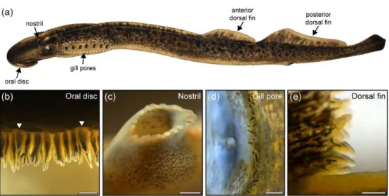
291 Newly-transformed lampreys had small papillae, which became more prominent during parasitic 292 stage, and reached full development in spawning stage. Therefore, the present investigation 293 focused on the spawning adults (for a detailed account of papillae development see Suntres et al. 294 in this issue). In spawning adult P. marinus, cutaneous papillae were found around the oral disc 295 (N = 25.6 ± 4.8), on the skin bordering the nostril (N = 16.6 ± 2.5), on the posterior margin of each 296 gill pore (N = 38.5 ± 6.7), and on the trailing edges of the dorsal fins (anterior: N = 329.3 ± 62.7; 297 posterior: N = 669.7 ± 166.0) (Fig. 1 and Table 1). Despite a generally similar form, the papillae 298 displayed some variations in size and shape at the different locations (Table 2).
299
300 3.2 Microvilli-bearing cells on oral, nasal, gill pore, and dorsal fin papillae
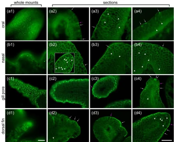
301 Tufts of microvilli emerged at the junction between the epidermal cells covering the 302 surface of oral, nasal, gill pore, and dorsal fin papillae viewed by SEM (Fig. 2). Tissue sections 303 labeled for F-actin with fluorescent phalloidin, showed that the microvilli extended from narrow 304 epidermal cells present in oral (n = 5), nasal (n = 7), gill pore (n = 5), and dorsal fin (n = 8) papillae 305 (Fig. 3a1,b1,c1,d1). Oral and gill pore papillae had the highest concentration of microvillar tufts in 306 both SEM (Fig. 2a1 and 2c1) and phalloidin labeled preparations (Fig. 3a1 and 3c1). Tufts of 307 microvilli were sparser on dorsal fin papillae (Fig. 3d1) and only a few were seen on the surface of 308 nasal papillae (Fig. 3b1). Phalloidin labeling was used to examine the morphology of the
309 microvillar cells. On sections, phalloidin labeled the actin core of microvilli intensely, slightly more 310 in the protruded portion of the microvilli than in the non-protruded portion, and it also labeled 311 the cell membrane of all cells in the papillae. Most of the microvilli-bearing cells had an elongated 312 piriform shape typical of SCCs (Fig. 3, magenta arrows) and they each bore many microvilli at their 313 apex, regardless of the type of papilla. The detailed microvillar organization was not examined 314 here, but SCCs on gill pore papillae appeared to bear the longest microvilli (Fig. 3c, all 12 315 photomicrographs in columns 2 to 4, including the inset in (b2), are at the same magnification). 316 In some animals, microvilli seemed absent from the surface of papillae, regardless of their location 317 on the body. In those cases, phalloidin only labeled cell apices without obvious protrusions. The 318 reasons for these differences are not clear.
319 In addition to SCCs, phalloidin also labeled Merkel cells (Fig. 3, arrowheads) as they bear 320 microvilli with a F-actin core at opposite poles of their cell body (Whitear & Lane, 1981). All 321 papillae investigated in our study contained Merkel cells. Dorsal fin and nasal papillae had a typical 322 aggregation of these cells at their tip (Figs. 3b2, inset, and 3d4 illustrate examples). Oral papillae 323 contained numerous Merkel cells spread out evenly in their epithelium, some located 324 superficially, and others deeper in the epithelium (Fig. 3a3-4), reminiscent of the epithelium 325 elsewhere on the body. A few Merkel cells were also seen on almost every gill pore papillae (Fig. 326 3c4 shows an example).
327 Our investigation of oral papillae also showed that fimbriae (n = 3, whole; n = 2 on 328 sections; see Fig. 1b), which are flat skin extensions with digit-like protrusions at their tip located 329 immediately behind the papillae, had numerous microvilli on their surface, in a manner 330 reminiscent of the oral papillae. On sections, the fimbriae also contained numerous Merkel cells, 331 like oral papillae.
332
333 3.3 Papillae respond to chemical stimulation
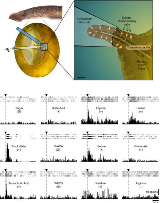
334 Responses to chemical stimulation were primarily investigated on gill pore papillae 335 because they showed, like oral papillae, the largest concentration of SCCs and the lowest number 336 of Merkel cells (see Fig. 3). The initial placement of the recording electrode over the papillae (Fig. 337 4, Top) generally produced a few discharges that subsided within a minute. A basal activity of the 338 papillae in the absence of chemical stimuli was present in most of the animals examined (n = 13). 339 Within one trial, 5-10 units were generally observed in the recording. The analysis was, however, 340 not performed on individual units (see materials and methods section for more details). Chemical 341 application over the papillae typically induced excitatory responses (i.e. a significant increase in 342 mean discharge frequency). However, in limited cases (4 trials in 2 animals), it produced a 343 significant decrease in mean discharge frequency (i.e. inhibitory responses). Figure 4 shows 344 examples of responses to different chemicals and Table 3 shows trial statistics. Trout water 345 repeatedly produced strong responses; it elicited excitatory responses in all but two animals 346 (11/13). The amino acids, glycine and proline, elicited excitatory responses in more than half of 347 the animals (7/12). Taurocholic acid, a bile acid, elicited excitatory responses in a little less than 348 half of the animals (5/13) and sialic acid, a mucus component, elicited responses in about one 349 third of the animals (3/10). Responses to the other amino acids tested (serine, glutamate, 350 arginine, histidine) were less frequent and sex pheromones (3kPZS, 3kACA) did not induce any 351 responses (Table 3). Serine, glutamate and arginine produced a few inhibitory responses. Our data 352 did not reveal any clear clustering of gill pore papillae based on their response profile to the 353 different chemical stimuli. The precise response profile of the other types of papillae (oral, nasal, 354 and fin) was not investigated here, but their chemosensory function was ascertained by puffing 355 trout water using a pressure ejection system over the papillae in a few animals (n = 4). In this 356 series of experiments, we tested whether the papillae responded to mechanical stimuli by puffing
357 jets of Ringer’s over the papillae. Gill pore papillae did not show responses, whereas mechanical 358 stimulation of oral, nasal or fin papillae sometimes produced discharges. When seen, short 359 duration bursts of only a few seconds (unlike responses to chemical stimulation) always 360 characterized the responses to mechanical stimulation of the papillae.
361
362 3.4 Innervation of papillae by ganglion cells and their central projections
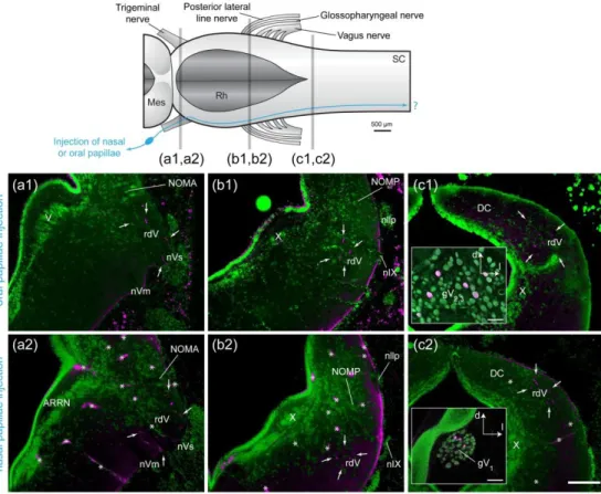
363 The four types of papillae investigated were all innervated by peripheral, bipolar ganglion 364 cells, never by dorsal cells within the central nervous system. Injections of tracer in oral papillae 365 of spawning adult lampreys (n = 2) labeled ganglion cells in the maxillomandibular part of the 366 trigeminal ganglion, while tracer injections in the nasal papillae of spawning adult lampreys (n = 367 2) labeled ganglion cells in the ophthalmicus profundus part of the trigeminal ganglion (Fig. 5). 368 For oral and nasal papillae, the central processes of the ganglion cells were coarse (3-4 µm). In 369 both cases, they entered the brainstem through the trigeminal sensory root (Fig. 5a1,a2) to join 370 the descending root of the trigeminal nerve (rdV) and reach the first spinal segment. Our 371 investigation did not include more caudal levels of the spinal cord. Injections (n = 2) of the cut 372 distal end of the trigeminal sensory nerve at its exit from the cranium labeled fibers that entered 373 the nasal and oral papillae (Fig. 6a,c). These injections also labeled fibers innervating the fimbriae 374 around the oral disc (Fig. 6b).
375 Axonal tracing experiments focused on papillae from gill pores 1 to 3 of spawning adult 376 lampreys (n = 6). Injections of tracer in papillae located at the first gill pore labeled cells in the 377 glossopharyngeal ganglion, while tracing from gill pore papillae located at the second and third 378 gill pores labeled cells in close relation to the main trunk of the vagus nerve. Occasional ganglion 379 cells were seen along the vagus nerve root innervating a gill, although not a great distance from 380 the main trunk of the vagus nerve. Their labeled central processes were coarse (around 4 µm) and
381 entered the brainstem through the glossopharyngeal nerve (first gill pore injections) or through 382 one of the roots of the vagus nerve (second and third gill pore injections). Upon entering the 383 brainstem, they immediately coursed towards a more dorso-medial position in the alar plate to 384 join a longitudinal tract (Fig. 7b1, white arrows). Most of them turned caudally in that tract (Fig. 385 7c1) to reach at least the first spinal segment. A small number of the entering axons joined the 386 same tract but coursed rostrally (Fig. 7a1). We could not determine in our material if these were 387 different axons or branches of some of the descending axons. Injections of the cut distal end of 388 the glossopharyngeal and vagus nerve at their exit from the cranium, labeled fibers that entered 389 the gill pore papillae (Fig. 6d, n = 2).
390 The central projections from gill pore papillae constitute only a portion of all the afferents 391 entering the brainstem by cranial nerves IX and X, as shown by the comparison to injections of 392 whole nerves IX and X (see Fig. 7a3-c3, injections of nIX and nX, n = 3). To compare their central 393 projections with those from taste buds, whose afferents are known to enter the brainstem 394 through cranial nerves VII, IX and X in many vertebrates (Finger, 1997), we injected taste buds in 395 the pharynx of newly-transformed animals (n = 2). These injections produced a labeling pattern 396 that differed from that obtained after gill pore papillae injections. First, the fibers innervating the 397 taste buds were of finer caliber (around 2 µm) and formed a longitudinal tract immediately after 398 entering the brainstem, at the lateral edge of the alar plate (Fig. 7b2). More fibers seemed to turn 399 caudally than rostrally in that tract, although the rostral projection was significant. From the point 400 of entrance of nIX to the level of the obex, fine varicose fibers left the longitudinal tract in a medio- 401 dorsal direction to terminate in a well-delineated nucleus, just adjacent to the tract, in the lateral 402 alar plate. This nucleus has been previously suggested to be the lamprey homologue of the 403 mammalian nucleus of the solitary tract (NTS), based on its location and neurochemical evidences 404 (Albersheim-Carter et al., 2016; Auclair, Lund, & Dubuc, 2004; Barreiro-Iglesias, Anadón, &
405 Rodicio, 2010; Pombal, López, de Arriba, González, & Megías, 2008; Pombal, López, de Arriba, 406 Megías, & González, 2006). The present data corroborate this homology hypothesis (see 407 Discussion). The ascending fibers coursed between the rdV and the vestibular area and 408 terminated at isthmic levels, just medial to the anterior octavomotor nucleus. The descending 409 fibers reached the level of the obex but did not seem to continue down to the spinal cord. 410 Injections of the cut distal end of the glossopharyngeal and vagus nerve at their exit from the 411 cranium, labeled fibers that innervated pharyngeal taste buds (Fig. 6e, n = 2). In one animal, the 412 skin dorsal to the 2nd and 3rd gill pores was carefully injected, making sure to spare neuromasts
413 in that area. The injection included the epidermis, dermis and the surface of the muscle layer. It 414 labeled ganglion cells close to the main trunk of the vagus nerve, but their central projections 415 entered the brainstem through the posterior lateral line nerve to terminate in the medial nucleus 416 of the octavolateral area. Other fibers travelling in the trunk lateral line nerve entered the 417 recurrent nerve. These fibers originated from ganglion cells located both in the intracapsular 418 ganglion and in the lateral portion of the anterior lateral line ganglion (see Koyama, Kishida, Goris, 419 & Kusunoki, 1990), and had central projections that terminated in the dorsal nucleus of the 420 octavolateral area. The skin injection also labeled spinal ganglion cells and dorsal cells within the 421 spinal cord, and ascending fibers were seen in the dorsal columns. These skin injections did not 422 label fibers resembling the ones that entered the brainstem through the vagus nerve after gill 423 pore papillae injections.
424 Tracer injections in 8 - 12 consecutive dorsal fin papillae of spawning adult lampreys (n = 425 3) labeled spinal ganglion cells on both sides of the animals. These cells were found either in the 426 dorsal root ganglions outside the spinal canal (Fig. 8a,b) or inside the spinal canal at different 427 levels along the dorsal roots (Fig. 8c,d). These injections never labeled dorsal cells (i.e. intraspinal 428 sensory ganglion cells) in our material. The ganglion cells innervating dorsal fin papillae were
429 bipolar with coarse axons (around 4 µm). The central processes from these cells entered the 430 dorsal spinal cord through the dorsal roots where they turned caudally and rostrally in the dorsal 431 funiculus. From the number of labeled ganglion cells and ascending and descending axons, it is 432 most probable that the entering axons gave rise to both an ascending and a descending branch, 433 although we could not see branching axons in our material. The individual descending branches 434 reached as far as 5 or 6 spinal segments more caudal than their own entrance point. Further 435 caudally, this translated into gradually fewer fibers in the dorsal funiculus until there were none 436 left. The ascending branches could be followed up to the last sections of our material (~ 3 cm 437 rostral to the injected dorsal fin papillae). Their numbers were similar to that of labeled ganglion 438 cells, which suggested that all ganglion cells projected at least this far rostrally. In 2 animals, 439 observations from sections of the brainstem failed to show labeled fibers from dorsal fin papillae 440 injections. The distance between the injected caudal dorsal fin and the brainstem was in the order 441 of 20 cm or so in these two animals, a distance that prevents efficient tract tracing in lampreys. 442 Contrary to the other types of papillae, we did not trace the fibers innervating dorsal fin papillae 443 from their exit from the spinal canal to the papillae.
444 445
4. Discussion
446
447 Despite recent advances (Finger et al., 2003; Hansen, 2007; Kirino, Parnes, Hansen, 448 Kiyohara, & Finger, 2013; Tizzano et al., 2006), information on the physiology and function of the 449 SCC system in vertebrates is limited. In this study, we established that SCCs are particularly 450 numerous on cutaneous papillae around the oral disc, nostril, gill pores, and on the dorsal fins in 451 the sea lamprey (Petromyzon marinus). We demonstrated that the oral and nasal papillae are 452 innervated by the trigeminal nerve, the gill pore papillae by the glossopharyngeal or vagus nerve, 19
453 and the dorsal fin papillae by spinal dorsal roots. We also characterized the chemical response 454 profile of gill pore papillae and showed that trout water and the amino acids, glycine and proline, 455 produced strong responses. A companion study, (Suntres et al., this issue), further investigates 456 the immunocytochemical properties and the development during ontogeny of the lamprey SCCs. 457 Together, these studies provide new insights on the function and evolution of the SCC system in 458 vertebrates.
459
460 4.1 Location of cutaneous papillae and SCC distribution
461 The first report of cutaneous papillae in lampreys long predates the discovery of SCCs. 462 Linnaeus (1758) described the mouth of P. marinus and L. planeri as “papilloſo”. He wrote about 463 L. planeri : “Behind the border of the mouth are numerous sharp papillae” (as cited in Turton, 464 1803). Since then, other authors have reported the presence of papillae (oral - Baatrup & Døving, 465 1985; Cook, Hilliard, & Potter, 1990; Fox et al., 1980; Langerhans, 1873; Lethbridge & Potter, 1981; 466 Potter, Lanzing, & Strahan, 1968; Whitear & Lane, 1983a; Woodland, 1913; gill pore- Dawson, 467 1905; Whitear & Lane, 1983a; fin – Langerhans, 1873; Whitear & Lane, 1983a) and have assessed 468 the papillae as a taxonomic character (Beamish, 2010; Khidir & Renaud, 2003). However, these 469 studies did not look systematically at the morphology, anatomy, and physiology of the papillae. 470 Cells that are now recognized as SCCs were first mentioned by Langerhans (1873) who 471 described hair-bearing bipolar cells on the body surface that were particularly numerous on oral 472 papillae (“Mundpapillen”) and dorsal fin papillae (“Flossenpapillen”) in L. planeri. The cells 473 described by Langerhans and their possible innervation were re-examined and discussed by 474 anatomists in the late 19th - early 20th century (Fahrenholz, 1936a, 1936b; Ficalbi, 1914;
475 Foettinger, 1876; Fusari, 1907; Marenghi, 1903; Retzius, 1892; Studnička, 1913; Tretjakoff, 1927).
476 Electron microscopy allowed researchers to demonstrate the precise association of these cells 477 with nerve fibers in lampreys (“oligovillous cells”, Fox et al., 1980; Whitear & Lane, 1983a). 478 Our investigation of SCCs in spawning phase P. marinus focused on papillae as they 479 contain a high density of SCCs in L. planeri and L. fluviatilis (Baatrup & Døving, 1985; Fox et al., 480 1980; Whitear & Lane, 1983a). We confirmed previous reports (Dawson, 1905; Whitear & Lane, 481 1983a) describing a fringe of papillae close to the gill pores and of papillae around the oral disc 482 (Baatrup & Døving, 1985; Khidir & Renaud, 2003). The number of papillae at these two locations 483 (Table 1) is consistent with observations in other lamprey species (Beamish, 2010; Cook et al., 484 1990; Khidir & Renaud, 2003). We also showed that P. marinus bears papillae on the tip of the 485 dorsal fins, as described in L. planeri (Fox et al., 1980; Whitear & Lane, 1983a). Moreover, we 486 provide the first description of papillae on the edge of the nostril.
487 The epidermis covering the papillae located at the different locations was thinner and 488 simpler than elsewhere on the body. It contained occasional granular cells but no skein cells, as 489 reported by Lane and Whitear (1980) and Cook et al. (1990) (see also Rodríguez-Alonso, Megías, 490 Pombal, & Molist, 2017). Numerous cells with the typical elongated piriform shape of vertebrate 491 SCCs (Tizzano & Finger, 2013) and harbouring a tuft of microvilli-like extensions were seen. Our 492 anatomical experiments showed that these microvilli-like extensions contain a F-actin core, 493 confirming their microvillous nature. These cells correspond to the “oligovillous cells” of Fox et al. 494 (1980) and Whitear and Lane (1983a). They were later recognized as lamprey SCCs (Whitear, 495 1992) and our results support this hypothesis. We found particularly numerous SCCs on oral and 496 gill pore papillae, comparatively less on dorsal fin papillae, and only a few were observed on nasal 497 papillae. In addition to SCCs, papillae also contained Merkell cells. These cells bore microvilli at 498 opposite poles of their cell body, a typical feature of lamprey Merkel cells (Takahashi-Iwanaga & 499 Abe, 2001; Whitear, 1989; Whitear & Lane, 1981). Previous studies reported Merkel cells in oral
500 papillae in L. planeri (Baatrup & Døving, 1985) and Geotria australis (Cook et al., 1990). Our 501 material provided evidence of numerous Merkel cells on oral, nasal, and fin papillae, as well as a 502 few on gill pore papillae (often 1-2 per papilla) in P. marinus.
503 Although, we did not look for the presence of SCCs on the rest of the body, we examined 504 all the tissue that was collected around the papillae. From these observations, we found a large 505 number of SCCs with microvilli, similar to those of the oral papillae on the fimbriae surrounding 506 the oral disk. The external wall of the nostril contained a few SCCs that became rare on the 507 epidermis away from the nostril. The internal wall of the nostril leading to the olfactory epithelium 508 also contained SCCs, but their microvilli were different from that of the papillae SCCs. These cells 509 had only a few (3 to 5) straight microvilli, with a short protruded portion and a long non-protruded 510 portion. The microvilli in these SCCs formed an elongated narrow V shape. Similarly, Fox et al. 511 (1980) originally described two types of SCCs on the basis of the organization of the microvilli in 512 L. planeri. However, in a subsequent paper, the authors argued that they were more likely distinct 513 examples from a continuous series (Whitear & Lane, 1983a). Gill pore structures (i.e. ectal valve 514 and central process) occasionally contained SCCs , but rarely on the thicker epidermis around the 515 gill pores. The epidermis on of the dorsal fins also contained a few SCCs. In general, we did not 516 find many SCCs on the epidermis of the head and body, as reported previously by others on L. 517 planeri (Whitear & Lane, 1983a).
518 The SCC distribution is variable from one vertebrate species to the other. In aquatic 519 vertebrates, they are widely distributed on the skin, gills, nasopharynx, or oropharynx (hagfish - 520 Braun, 1996, 1998; Braun & Northcutt, 1998; lampreys - Baatrup & Døving, 1985; Fox et al., 1980; 521 Whitear & Lane, 1983a; chondrichthyes - Peach, 2005; Whitear & Moate, 1994; bony fish - Codina 522 et al., 2012; Hansen et al., 2014; Kotrschal, 1992; Kotrschal et al., 1997, 1989, 1984; Kuciel et al., 523 2014; Peters et al., 1987, 1991; Silver & Finger, 1984; see Whitear, 1992 for a review). In terrestrial
524 vertebrates, on the other hand, SCCs seem restricted to specific anatomical locations (e.g. the 525 airways) (amphibians - Osculati & Sbarbati, 1995; Whitear, 1976; but see also for putative skin 526 SCCs Koyama et al., 2001; Nagai et al., 1999; reptiles - Hansen, 2007; mammals - Finger et al., 527 2003; Saunders et al., 2014; Sbarbati et al., 1998, 1999; Sbarbati & Osculati, 2003; Tizzano et al., 528 2010, 2006).
529 Chemical response profile and behavioral functionIn the present study, we have 530 examined the responses of papillae to chemicals that could potentially activate SCCs. We have 531 limited our study to chemicals that are known to activate chemosensory systems in lampreys. We 532 did not test all chemicals known to activate these systems in other species. Moreover, the 533 physiological experiments were limited to the responses of papillae in the gill region
534 We showed that gill pore papillae of sea lampreys respond strongly to trout water, to the 535 amino acids proline and glycine, and to the bile acid, taurocholic acid. Serine, sialic acid, and 536 glutamate also produced occasional excitatory responses. These results partially corroborate 537 those of Baatrup & Døving (1985) who electrophysiologically recorded from SCCs located around 538 the oral disc in L. planeri. They found responses to acetic acid, sialic acid, and trout water, but not 539 to proline and glycine (Baatrup & Døving, 1985). Several factors may contribute to these 540 differences. First, it is not entirely clear if Baatrup and Døving recorded from papillae or fimbriae 541 (see Cook et al., 1990 for details). If they recorded from fimbriae, their chemosensory response 542 profile may be different than the one from the papillae. We did not specifically investigate 543 fimbriae here, but we found that they contain SCCs with microvilli and that these cells are 544 innervated, contrary to earlier reports (Lethbridge & Potter, 1979). Second, oral and gill pore 545 papillae may have different chemosensory response profiles. Third, response profiles could be 546 species-specific as it is the case for the taste system that is tuned to the diet of the animal (Finger, 547 1997; Kasumyan & Døving, 2003). In both studies, trout water was a potent stimulus. The
548 stimulatory compounds of the trout water are not known yet. However, SCC responses to 549 individual chemicals from both our study and that of Baatrup & Døving (1985) suggest the 550 presence of amino acids, bile acids, and mucus-derived chemicals. These are likely to be found in 551 trout thawing water along with bacterial substances (see discussion below). This may indicate a 552 role in predator detection as teleosts can be predators of lampreys (Applegate, 1950; Potter, 553 1980; Vladykov, 1949) or it may indicate a role in feeding as sea lampreys feed on trout (Farmer, 554 1980; Lennon, 1954; Smith, 1971; Swink, 2003). Our findings support the latter hypothesis by 555 showing that amino acids (proline and glycine), which are generally considered as feeding- 556 associated cues in fish (Jones, 1989; Mackie & Mitchell, 1983; Mearns, 1986, 1989; Takeda, Takii, 557 & Matsui, 1984; Valentinčič & Caprio, 1994; for reviews see Finger, 1997; Kasumyan & Døving, 558 2003), induce excitatory responses in sea lamprey papillae. Moreover, these two amino acids are 559 concentrated in marine animal tissue (Beers, 1967; Carr, Netherton, Gleeson, & Derby, 1996) on 560 which sea lampreys feed. The responses to the bile acid taurocholic acid seen in the present study, 561 may corroborate a role of the SCC system in feeding as taurocholic acid is a potent stimulus of the 562 taste system in several fish species (Hara, 1994; Michel, 2005; Rolen & Caprio, 2008). However, 563 bile acids including taurocholic acid could act as social cues in fish (Li, Sorensen, & Gallahers, 1995; 564 Sorensen, Hara, & Stacey, 1991; Zhang & Hara, 2009). The failure of the lamprey pheromones 565 3kPZS or 3kACA to stimulate chemosensory responses in gill pore papillae could be due to the fact 566 that these sex pheromones are released at the gills (Siefkes, Scott, Zielinski, Yun, &, Li, 2003). This 567 would then result in a continuous stimulation of the receptors by the pheromones. The chemical 568 sensitivity of the lamprey SCC system is thus different from the olfactory system, which is highly 569 sensitive to basic amino acids (arginine and histidine) and pheromones (Green et al., 2017; Li, 570 1994) and at least partially different from that of the gustatory system that responds to
571 substances that are not stimulatory for the SCC system (sucrose, quinine) in L. planeri (Baatrup & 572 Døving, 1985).
573 Apart from lampreys, the chemical sensitivity of SCCs has been defined in only a few 574 species. In the sea robin (Prionotus carolinus), recordings of the spinal nerves innervating the free 575 pectoral fins rays that contain SCCs, showed responses to feeding-related cues such as prey 576 extract and amino acids (Silver & Finger, 1984). In rocklings (genus Ciliata and Gaidropsarus), the 577 SCCs of the anterior dorsal fin respond to body mucus of heterospecific fish, but not to classical 578 fish taste stimuli (amino-acids, etc.) or pheromones from congeners (Peters et al., 1987, 1991). In 579 the sea catfish (Plotosus japonicus), the maxillary barbels are extremely sensitive to pH variations 580 (Caprio, Shimohara, Marui, Harada, & Kiyohara, 2014) and to amino acids (Caprio et al., 2015). 581 However, the barbels contain both taste buds and SCCs which prevents the identification of the 582 sensory receptors involved (Caprio et al., 2014, 2015). In terrestrial vertebrates, SCCs have been 583 observed in amphibians, reptiles and mammals (Finger et al., 2003; Hansen, 2007; Sbarbati & 584 Osculati, 2003). However, their chemical sensitivity is only known in mammals where SCCs located 585 in the airways detect irritants and bacterial compounds via taste receptors (Finger et al., 2003; 586 Gulbransen, Clapp, Finger, & Kinnamon, 2008; Lin, Ogura, Margolskee, Finger, & Restrepo, 2008; 587 Tizzano et al., 2010). The detection of these substances by airways SCCs triggers different defense 588 mechanisms. The activation of SCC bitter and sweet taste receptors tunes the release of 589 antimicrobial peptide by adjacent epithelial cells (Lee et al., 2014, 2017). It also prevents harmful 590 airborne substances (irritants, toxins, etc.) to further enter the organism by evoking protective 591 respiratory reflexes (Finger et al., 2003) and by regulating fluid access to the vomeronasal organ 592 (Ogura, Krosnowski, Zhang, Bekkerman, & Lin, 2010). These findings suggest a third possible 593 interpretation of our results in lampreys. Indeed, the SCC-stimulating compound of the trout
594 water may be bacterial or irritants substances. Therefore the SCC system in lampreys could have 595 a similar role as in mammals.
596
597 4.2 Innervation of cutaneous papillae and central projections
598 Tracer injections in the different types of papillae demonstrated that the trigeminal nerve 599 innervates oral and nasal papillae, the glossopharyngeal and vagus nerves innervate gill pore 600 papillae, and spinal dorsal roots innervate dorsal fin papillae. Our tracing experiments did not 601 specifically label the sensory fibers innervating SCCs. Therefore, we cannot differentiate between 602 fibers innervating SCCs, Merkel cells, free nerve endings, or fibers innervating any other types of 603 receptors. However, all labeled fibers from one type of papillae entered the brainstem by the 604 same cranial nerve and had a similar projection pattern in the central nervous system. For 605 example, all labeled fibers from oral papillae which contain numerous SCCs and several Merkel 606 cells entered the brainstem through the trigeminal nerve and descended in the rdV to reach the 607 rostral spinal cord. However the size or diameter of the ganglion cells and axons differed. These 608 differences could relate to the different types of sensory fibers innervating the papillae, but more 609 information is needed to conclude on this matter. One exception to this pattern involves gill pore 610 papillae. On 2 animals out of 4, gill pore papillae injections labeled fibers that travelled in the 611 recurrent branch of the anterior lateral line nerve to enter the brainstem and terminate in the 612 dorsal nucleus of the octavolateral area, a nucleus mainly involved in electroreception (Bodznick 613 & Northcutt, 1981; Koyama, 2005; Ronan & Northcutt, 1987). Occasional putative skin 614 photoreceptors, referred to as “microvillous cells”, have been reported on gill pore papillae 615 (Whitear & Lane, 1983b). Others have described similar cells on the tail of larvae and showed their 616 innervation by lateral line fibers (Steven, 1951; Whitear & Lane, 1983b). There is thus a small 617 possibility that the labeled lateral line fibers in our material could be innervating such cells.
618 However, the fibers innervating photoreceptor cells from the tail area of larval lampreys enter the 619 brainstem through the posterior lateral line nerve and terminate in the medial nucleus of the 620 octavolateral area (Ronan & Bodznick, 1991). Another possibility is that the lateral line fibers were 621 accidentally labeled by lesioning a neuromast, some of which are located in the vicinity of gill pore 622 papillae, slightly dorsal between the gill pores.
623 The source of SCC innervation is often inferred from their location on the body. However, only a 624 few studies identified the nerves providing innervation to SCCs. In sea robins, spinal nerves 625 provide innervation to the SCCs of the pectoral fin rays (Finger, 1997; Silver & Finger, 1984). In 626 rocklings, the dorsal recurrent branch of the facial nerve provides innervation to the SCCs of the 627 anterior dorsal fin, whereas spinal nerves provide innervation to other sensory cells (Merkel) 628 located on the same structure (Kotrschal & Whitear, 1988; Whitear & Kotrschal, 1988). In rodents, 629 the trigeminal nerve provides innervation to SCCs in the distal part of the airways (Finger et al., 630 2003; Gulbransen, Silver, & Finger, 2008; Silver & Finger, 2009; Tizzano et al., 2010). Our results 631 in the lamprey support the view that cranial or spinal nerves providing cutaneous innervation to 632 the area where the SCCs are located also provide innervation to those SCCs (Fig. 9; Finger, 1997). 633 To our knowledge, the central projection pattern of the SCC system is only known from 634 the two teleosts species mentioned above, Prionotus and Ciliata. In Prionotus, the central 635 projections of the SCC system mirror those of an ascending somatosensory system, the dorsal 636 column system. The situation is different in rocklings where the SCCs are centrally connected like 637 an external taste system. Primary afferents terminate in the vagal lobe which, in turn, projects to 638 the secondary taste nucleus (Finger, 1997; Kotrschal & Finger, 1996). In lampreys, the central 639 projection pattern resembles that of the somatosensory system. The central process of the 640 trigeminal ganglion cells that innervate oral or nasal papillae courses in the descending root of 641 the trigeminal nerve (rdV). The central process of the glossopharyngeal or vagal ganglion cells that
642 innervate gill pore papillae courses caudally or rostrally in a longitudinal tract that is adjacent to 643 the rdV. Most of the fibers course caudally in that tract to reach the rostral spinal segments. A 644 few fibers course rostrally in the same tract to reach isthmic levels. We did not see any labeled 645 fibers in the rdV after gill pore papillae injections, suggesting that the fibers in the rdV in the whole 646 nIX/X labeling experiments do not innervate gill pore papillae. The central process of the dorsal 647 root ganglion cells that innervate dorsal fin papillae entered the dorsal columns through the 648 dorsal roots and then ran caudally and rostrally for a few segments. It is possible that the rostrally- 649 directed fibers reach brainstem levels directly, but it remains to be demonstrated.
650 We also specifically traced taste bud-innervating fibers from the periphery (i.e. taste buds) 651 to their central target. To our knowledge, this is the first time this has been done in lampreys since 652 previous studies were based on whole-nerve labeling (Fritzsch & Northcutt, 1993; Koyama, 2005). 653 The taste bud injections did not label fibers in the rdV as for whole nIX/X injections, suggesting 654 that the rdV fibers that course in nIX/X innervate structures other than taste buds and gill pore 655 papillae. We now show that the central projection of the lamprey taste system is different from 656 that of the SCC system. The fibers innervating the taste buds are of finer caliber and course in a 657 longitudinal tract located at the lateral edge of the alar plate. Along this tract, the fibers terminate 658 at all levels in a well-delineated nucleus, just medial to the tract, in the caudal hindbrain, but do 659 not reach spinal levels. Based on immunohistochemical data, previous studies have suggested 660 that this nucleus is the lamprey homologue of the mammalian nucleus of the solitary tract (NST) 661 (Auclair et al., 2004; Barreiro-Iglesias et al., 2010; Pombal et al., 2008, 2006; Robertson, Auclair, 662 Ménard, Grillner, & Dubuc, 2007). The present study strongly supports this hypothesis by showing 663 that this nucleus is the primary target of taste bud afferents in lampreys as in other vertebrates 664 (Beckstead, & Norgren, 1979; Contreras, Beckstead, & Norgren, 1982; Finger, 1987, 1997; 665 Hamilton & Norgren, 1984; Herrick, 1905; Norgren & Leonard, 1973). We also described that some
666 of the taste bud afferents ascend to terminate at isthmic levels. Other studies have reported such 667 ascending inputs from taste bud afferents in other species, but they only briefly discussed them 668 despite a potential role in central processing of taste inputs (rabbit: Hanamori & Smith, 1989; rat: 669 Hamilton & Norgren, 1984; frog: Hanamori & Ishiko, 1983; Matesz & Székely, 1978; chicken: 670 Ganchrow, Gentle, & Ganchrow, 1987).
671
672 4.3 Evolutionary considerations and conclusion
673 Taken together, our results show that lampreys, the most basal extant group of 674 vertebrates, possess a relatively well developed and distributed SCC system that is particularly 675 well represented on cutaneous papillae. Lamprey SCCs were found in all locations where they 676 have been reported in other species. Our results confirm that the cranial or spinal nerves 677 responsible for the cutaneous innervation of an area also provide innervation to SCCs in the same 678 area. The gill pore papillae respond mainly to trout-derived chemicals and amino acids, which 679 could indicate a role in feeding, in predator avoidance or in protective behaviors. The lamprey SCC 680 system appears less specialized than the highly specialized SCC systems observed in Prionotus and 681 Ciliata and may resemble the ancestral SCC system from which more specialized systems may 682 have arisen. The SCC system is also associated to a variety of behaviors in vertebrates: feeding, 683 predator avoidance, or detection of bacteria and irritants. Altogether, these differences between 684 taxa in the morphology, distribution, chemical sensitivity, and associated behavioral function raise 685 the question of the homology of SCCs among vertebrates. For instance, nasal SCCs in mammals 686 are often considered as components of an array of taste-like cells that are distributed throughout 687 the gut and airways (Saunders et al., 2014). These cells share molecular similarity and use a similar 688 transduction pathway (Saunders, Reynolds, & Finger, 2013). However, SCCs in fish do not seem 689 to use a G protein-coupled receptor transduction cascade as taste bud cells (Ohmoto, Okada,
690 Nakamura, Abe, & Matsumoto, 2011). Furthermore, important differences can exist among 691 species of the same vertebrate lineage. For example, in fish, SCCs located on the trunk are 692 innervated either by spinal or cranial nerves (Prionotus, Finger, 1997; Silver & Finger, 1984; 693 Gaidropsarus, Ciliata, Kotrschal & Whitear, 1988; Whitear & Kotrschal, 1988). So far, these 694 differences have been mainly attributed to phylogenetic differences. They may also be due to the 695 anatomical location of the SCCs. Indeed, in each species examined so far, the SCCs were located 696 on a different body location: anterior dorsal fin in Ciliata; fin rays in Prionotus, and airways in 697 mammals. The lamprey model offers the opportunity to test the hypothesis that SCC function may 698 be linked to their location. It will also provide a unique framework to shed light on SCC homology 699 in vertebrates and to compare the respective territories of the taste and SCC systems.
700 701
Author Contributions
702
703 GD, BZ, and RD designed the study; GD, FB, FA, TS, JL-B, and MM performed the experiments; GD, 704 FB, FA, analyzed the data; GD, FA, BZ and RD wrote the paper.
705 706
References
707
708 Albersheim-Carter, J., Blubaum, A., Ballagh, I. H., Missaghi, K., Siuda, E. R., McMurray, G., Bass, A. 709 H., Dubuc, R., Kelley, D. B., Schmidt, M. F., Wilson, R. J., & Gray, P.A. (2016). Testing the 710 evolutionary conservation of vocal motoneurons in vertebrates. Respiratory physiology & 711 neurobiology, 224, 2-10. doi:10.1016/j.resp.2015.06.010.
712 Applegate, V. C. (1950). Natural history of the sea lamprey (Petromyzon marinus) in Michigan. 713 Spec Sci Rep US Fish Wildl Serv, 55, 1-237. Retrieved from 714 http://name.umdl.umich.edu/2142335.0001.001
715 Auclair, F., Lund, J. P., & Dubuc, R. (2004). Immunohistochemical distribution of tachykinins in the 716 CNS of the lamprey Petromyzon marinus. Journal of Comparative Neurology, 479(3), 328-346. doi: 717 10.1002/cne.20324
718 Baatrup, E., & Døving, K. B. (1985). Physiological studies on solitary receptors of the oral disc 719 papillae in the adult brook lamprey, Lampetra planeri (Bloch). Chemical senses, 10(4), 559-566. 720 doi:10.1093/chemse/10.4.559
721 Bardach, J. E., & Case, J. (1965). Sensory capabilities of the modified fins of squirrel hake 722 (Urophycis chuss) and searobins (Prionotus carolinus and P. evolans). Copeia, 2, 194-206. 723 doi:10.2307/1440724
724 Barreiro-Iglesias, A., Anadón, R., & Rodicio, M. C. (2010). The gustatory system of lampreys. Brain, 725 behavior and evolution, 75(4), 241-250. doi:10.1159/000315151
726 Beamish, R. J. (2010). The use of gill pore papillae in the taxonomy of lampreys. Copeia, 2010(4), 727 618-628. doi:10.1643/CI-09-107
728 Beckstead, R. M., & Norgren, R. (1979). An autoradiographic examination of the central 729 distribution of the trigeminal, facial, glossopharyngeal, and vagal nerves in the monkey. Journal 730 of Comparative Neurology, 184(3), 455-472. doi: 10.1002/cne.901840303
731 Beers, J. R. (1967). The species distribution of some naturally-occurring quaternary ammonium 732 compounds. Comparative Biochemistry and Physiology, 21(1), 11-21. doi:10.1016/0010- 733 406X(67)90109-0
734 Bodznick, D., & Northcutt, R. G. (1981). Electroreception in lampreys: evidence that the earliest 735 vertebrates were electroreceptive. Science, 212(4493), 465-467. doi:10.1126/science.7209544
736 Braun, C. B. (1996). The sensory biology of the living jawless fishes: a phylogenetic assessment. 737 Brain, Behavior and Evolution, 48(5), 262-276. doi:10.1159/000113205
738 Braun, C. B. (1998). Schreiner organs: a new craniate chemosensory modality in hagfishes. Journal 739 of Comparative Neurology, 392(2), 135-163. doi:10.1002/(SICI)1096- 740 9861(19980309)392:2<135::AID-CNE1>3.0.CO;2-3
741 Braun, C. B., & Northcutt, R. G. (1998). Cutaneous exteroreceptors and their innervation in 742 hagfishes. In J. M. Jørgensen, J. P. Lomholt, R. E. Weber, & H. Malte (Eds.), The biology of hagfishes 743 (pp. 512-532). Dordrecht: Springer.
744 Caprio, J., Shimohara, M., Marui, T., Harada, S., & Kiyohara, S. (2014). Marine teleost locates live 745 prey through pH sensing. Science, 344(6188), 1154-1156. doi: 10.1126/science.1252697
746 Caprio, J., Shimohara, M., Marui, T., Kohbara, J., Harada, S., & Kiyohara, S. (2015). Amino acid 747 specificity of fibers of the facial/trigeminal complex innervating the maxillary barbel in the 748 Japanese sea catfish, Plotosus japonicus. Physiology & Behavior, 152, 288-294. doi: 749 10.1016/j.physbeh.2015.10.011
750 Carr, W. E., Netherton, III, J. C., Gleeson, R. A., & Derby, C. D. (1996). Stimulants of feeding 751 behavior in fish: analyses of tissues of diverse marine organisms. The Biological Bulletin, 190(2), 752 149-160. doi:10.2307/1542535
753 Codina, E., Loïc, K., Compere, P., Dragičević, B., Dulčić, J., & Parmentier, E. (2012). The barbel-like 754 specialization of the pelvic fins in Ophidion rochei (Ophidiidae). Journal of Morphology, 273(12), 755 1367-1376. doi:10.1002/jmor.20066
756 Contreras, R. J., Beckstead, R. M., & Norgren, R. (1982). The central projections of the trigeminal, 757 facial, glossopharyngeal and vagus nerves: an autoradiographic study in the rat. Journal of the 758 Autonomic Nervous System, 6(3), 303-322. doi:10.1016/0165-1838(82)90003-0
759 Cook, R. D., Hilliard, R. W., & Potter, I. C. (1990). Oral papillae of adults of the southern hemisphere 760 lamprey Geotria australis. Journal of Morphology, 203(1), 87-96. doi:10.1002/jmor.1052030109
761 Daghfous, G., Green, W. W., Zielinski, B. S., & Dubuc, R. (2012). Chemosensory-induced motor 762 behaviors in fish. Current Opinion in Neurobiology, 22(2), 223-230. 763 doi:10.1016/j.conb.2011.10.009
764 Dawson, J. (1905). The breathing and feeding mechanism of the lampreys. The Biological Bulletin, 765 9(1), 1-21. doi:10.2307/1535799
766 Fahrenholz, C. (1936a). Tastzellen und Tastorgane in der Neunaugenhaut. Zeitschrift fur 767 mikroskopisch-anatomische Forschung, 39, 116-134.
768 Fahrenholz, C. (1936b). Die sensiblen Einrichtungen der Neunaugenhaut. Zeitschrift fur 769 mikroskopisch-anatomische Forschung, 40, 323-380.
770 Farmer, G. J. (1980). Biology and physiology of feeding in adult lampreys. Canadian Journal of 771 Fisheries and Aquatic Sciences, 37(11), 1751-1761. doi:10.1139/f80-220
772 Ficalbi, E. (1914). Struttura del tegumento dei Petromizonti. Archivio italiano di Anatomia e di 773 Embriologia, 12, 596-827.
774 Finger, T. E. (1982). Somatotopy in the representation of the pectoral fin and free fin rays in the 775 spinal cord of the sea robin, Prionotus carolinus. The Biological Bulletin, 163(1), 154-161. 776 doi:10.2307/1541505


![[PDF] Cours Excel référence absolue et relatives | Cours Excel](data:image/gif;base64,R0lGODlhAQABAIAAAP///wAAACH5BAEAAAAALAAAAAABAAEAAAICRAEAOw==)