HAL Id: hal-00174488
https://hal.archives-ouvertes.fr/hal-00174488
Submitted on 1 Jun 2020
HAL is a multi-disciplinary open access
archive for the deposit and dissemination of
sci-entific research documents, whether they are
pub-lished or not. The documents may come from
teaching and research institutions in France or
abroad, or from public or private research centers.
L’archive ouverte pluridisciplinaire HAL, est
destinée au dépôt et à la diffusion de documents
scientifiques de niveau recherche, publiés ou non,
émanant des établissements d’enseignement et de
recherche français ou étrangers, des laboratoires
publics ou privés.
Role of transcription factor KLF11 and its
diabetes-associated gene variants in pancreatic beta cell
function
Bernadette Neve, Martin E Fernandez-Zapico, Vered Ashkenazi-Katalan,
Christian Dina, Yasmin H Hamid, Erik Joly, Emmanuel Vaillant, Yamina
Benmezroua, Emmanuelle Durand, Nicolas Bakaher, et al.
To cite this version:
Bernadette Neve, Martin E Fernandez-Zapico, Vered Ashkenazi-Katalan, Christian Dina, Yasmin
H Hamid, et al.. Role of transcription factor KLF11 and its diabetes-associated gene variants in
pancreatic beta cell function. Proceedings of the National Academy of Sciences of the United States
of America , National Academy of Sciences, 2005, 102 (13), pp.4807-12. �10.1073/pnas.0409177102�.
�hal-00174488�
Role of transcription factor KLF11 and
its diabetes-associated gene variants
in pancreatic beta cell function
Bernadette Nevea, Martin E. Fernandez-Zapicob, Vered Ashkenazi-Katalanc, Christian Dinaa, Yasmin H. Hamidd, Erik Jolye, Emmanuel Vaillanta, Yamina Benmezrouaa, Emmanuelle Duranda, Nicolas Bakahera, Valerie Delannoya, Martine Vaxillairea, Tiffany Cookc, Geesje M. Dallinga-Thief, Hans Janseng, Marie-Aline Charlesh, Karine Cle´menti, Pilar Galanj, Serge Hercbergj, Nicole Helbecquek, Guillaume Charpentierl, Marc Prentkie, Torben Hansend,
Oluf Pedersend,m, Raul Urrutiab, Danielle Melloulc, and Philippe Froguela,n,o
aCentre National de la Recherche Scientifique, Unite´ Mixte de Recherche 8090, Institute of Biology, Institute Pasteur de Lille, F-59019 Lille, France; bGastroenterology Research Unit, Saint Mary’s Hospital, Mayo Clinic, Rochester, MN 55905;cDepartment of Endocrinology and Metabolism, Hadassah
University Hospital, Jerusalem 91120, Israel;dSteno Diabetes Center and Hagedorn Research Institute, DK-2820 Copenhagen, Denmark;eMolecular Nutrition
Unit, Centre de Recherche du Centre Hospitalier l’Universite´ du Montreal, Notre-Dame Hospital, Montreal, QC, Canada H3J 1R4;fLaboratory of Vascular
Medicine and Metabolism andgDepartment of Clinical Chemistry, Erasmus Medical Center, 3000 CA, Rotterdam, The Netherlands;hInstitut National de la
Sante´ et de la Recherche Me´dicale U258, Paul Brousse Hospital, 94805 Villejuif, France;iDepartment of Nutrition-EA3502, Paris VI University, Institut
National de la Sante´ et de la Recherche Me´dicale, Avenir Hoˆtel-Dieu, 75004 Paris, France;jScientific and Technical Institute of Nutrition and Food (l’Institut
Scientifique et Technique de la Nutrition et Alimentation–Conservatoire National des Artes et Me´tiers), Institut National de la Sante´ et de la Recherche Me´dicale U557, Institut National de la Recherche Agronomique U1125, 75003 Paris, France;kDepartment of Epidemiology, Institut National de la Sante´ et
de la Recherche Me´dicale U508, Institute Pasteur de Lille, F-59019 Lille, France;lDepartment of Diabetes, Sud Francilien Hospital, 91100 Corbeil-Essonnes,
France;mFaculty of Health Science, University of Aarhus, DK-8000 Aarhus, Denmark; andnGenome Centre and Genomic Medicine, Imperial College,
London W12 0NN, United Kingdom
Edited by Donald F. Steiner, University of Chicago, Chicago, IL, and approved February 7, 2005 (received for review December 9, 2004)
KLF11 (TIEG2) is a pancreas-enriched transcription factor that has elicited significant attention because of its role as negative regu-lator of exocrine cell growth in vitro and in vivo. However, its functional role in the endocrine pancreas remains to be estab-lished. Here, we report, for the first time, to our knowledge, the characterization of KLF11 as a glucose-inducible regulator of the insulin gene. A combination of random oligonucleotide binding, EMSA, luciferase reporter, and chromatin immunoprecipitation assays shows that KLF11 binds to the insulin promoter and regu-lates its activity in beta cells. Genetic analysis of the KLF11 gene revealed two rare variants (Ala347Ser and Thr220Met) that segre-gate with diabetes in families with early-onset type 2 diabetes, and significantly impair its transcriptional activity. In addition, analysis of 1,696 type 2 diabetes mellitus and 1,776 normoglycemic subjects show a frequent polymorphic Gln62Arg variant that significantly associates with type 2 diabetes mellitus in North European popu-lations (ORⴝ 1.29, P ⴝ 0.00033). Moreover, this variant alters the corepressor mSin3A-binding activity of KLF11, impairs the activa-tion of the insulin promoter and shows lower levels of insulin expression in pancreatic beta cells. In addition, subjects carrying the Gln62Arg allele show decreased plasma insulin after an oral glucose challenge. Interestingly, all three nonsynonymous KLF11 variants show increased repression of the catalase 1 promoter, suggesting a role in free radical clearance that may render beta cells more sensitive to oxidative stress. Thus, both functional and genetic analyses reveal that KLF11 plays a role in the regulation of pancreatic beta cell physiology, and its variants may contribute to the development of diabetes.
insulin兩 polymorphisms 兩 TGF- 兩 type 2 diabetes
C
omponents of both the exocrine and endocrine pancreas are affected by diseases, e.g., pancreatic cancer and type 2 diabetes mellitus (T2DM), which severely compromise both the quality and span of human life. Both glandular compartments share the same cellular origin and early morphogenetic pathways, suggesting a close functional and pathophysiological relationship. For instance, the exocrine-specific transcription factor p48 and the endocrine-specific pancreatic duodenal homeobox gene 1 (PDX-1) are both expressed in the common cell precursor (1); and, under pathological condi-tions their compartmentalization may be lost, as exemplified by thedetection of PDX-1 in pancreatic cancer (2). In fact, T2DM is both a common feature and a risk factor for the subsequent development of pancreatic cancer (3, 4). The TGF--inducible transcription factor KLF11 regulates exocrine cell growth and behaves as a tumor suppressor in pancreatic cancer (ref. 5 and M.E.F.-Z. and R.U., unpublished observation). Because the TGF- signaling pathway is also a major regulator of endocrine cell fate (1, 6), the current study has been designed to define the role of the Sp1-like transcription factor KLF11 in the biology of the endocrine beta cell. Interestingly, several beta cell-specific genes display a potential binding site for Sp1 proteins in their promoter, suggesting that KLF11 is a potential endocrine regulator. Because the TGF- signaling pathway is crucial for pancreatic development and the impaired beta cell function observed in maturity-onset diabetes of the young (MODY) is caused by mutations in transcription factor genes that are also important in pancreas development [hepatocyte nuclear factors and PDX-1 (7), KLF11 may also be a candidate gene for predisposition to diabetes. Indeed, here we show that KLF11 is a glucose-induced regulator of the insulin gene and functional KLF11 gene variants are significantly associated with diabetes. These data, in combination with previous studies on KLF11, outline a molec-ular pathway important in both pancreatic cancer and diabetes.
Methods
Promoter Sequence Analysis. For EMSAs, purified GST-fusion proteins of KLF11 (8) were incubated in a buffer containing 4% glycerol, 1 mM MgCl2, 0.5 mM EDTA, 0.5 mM DTT, 50 mM NaCl, 10 mM Tris䡠HCl (pH 7.5), and 50g兾ml poly dI-dC (Promega) for 10 min at room temperature, and incubated for an additional 20 min with 0.035 pmol of end-labeled probes. The reactions were analyzed
This paper was submitted directly (Track II) to the PNAS office. Freely available online through the PNAS open access option.
Abbreviations: ChIP, chromatin immunoprecipitation; T2DM, type 2 diabetes mellitus; MODY, maturity-onset diabetes of the young; OGTT, oral glucose tolerance test; SOD, superoxide dismutase; LD, linkage disequilibrium; BMI, body mass index; PDX-1, pancreatic duodenal homeobox gene 1.
oTo whom correspondence should be addressed at: Genome Centre and Genomic
Medi-cine, Imperial College, Hammersmith Hospital, Du Cane Road, London W12 0NN, United Kingdom. E-mail: [email protected].
© 2005 by The National Academy of Sciences of the USA
by electrophoresis on 4% nondenaturing polyacrylamide gel fol-lowed by autoradiography. Site-directed mutagenesis of the GC box was generated by replacing each base with A, T, or C and densi-tometric units of each protein兾probe complex were determined by using NIH IMAGE 1.6.1 software. The relative binding specificity for an individual nucleotide at each position was calculated by using the following equation: (dNTP units)n兾(SdNTPs units)n, where n⫽ nucleotide position within the 9 bp GC box and is graphic-ally represented by the height of letters, using the program WEBLOGO(9).
Chromatin Immunoprecipitation (ChIP). At 24 h after transfection with KLF11 FLAG-tagged plasmids (8),TC3 cells were cross-linked with formaldehyde for 20 min at 25°C, harvested in SDS-lysis buffer (Upstate Biotechnology, Lake Placid, NY), and sheared to fragment DNA to⬇500 bp. Samples were then immunoprecipi-tated by using an agarose-conjugated anti-FLAG antibody (Sigma-Aldrich, St. Louis) or agarose beads alone at 4°C overnight, washed, and eluted by using the ChIP kit (Upstate Biotechnology). Crosslinks were removed at 65°C for 4 h, and immunoprecipitated DNA was purified by using phenol兾chloroform extraction and ethanol precipitation. Then, a 200-bp region of the insulin promoter was amplified by PCR, using the following primers 5
⬘-GGC-CATCTGCCTACCCACCC and 5
⬘-AGGCCCAAAGAG-GAGAGTACATAC and visualized by 2.5% agarose gel.
Reporter Assays.The KLF11 variants were generated following the recommendations of the Stratagene QuikChange kit. Then,TC3
cells were transfected by using TransFast transfection (Promega, Charbonnieres, France). The KLF11 constructs were cotransfected with the Gal4 reporter construct (10), a luciferase reporter vector containing six tandem repeats of KLF11-binding sites (10), the human insulin promoter, or catalase 1 promoter (5), and the luciferase activity was measured as described (5). Association of endogenous mSin3A with KLF11 was analyzed by immunoprecipi-tation assays (ref. 8 and Supporting Methods, which is published as supporting information on the PNAS web site).
Genetic Analysis and Study Population.The identification and geno-typing of genetic variants and calculation of logarithm of odds scores are described in Supporting Methods and Table 4, which is published as supporting information on the PNAS web site. The 96 normoglycemic control and 66 T2DM subjects of the pilot study were included in the familial study with 313 French cases from T2DM diabetes multiplex families that had at least one affected first-degree relative, and 313 control subjects (spouses) that were selected to match the cases for T2DM risk factors (Table 5, which is published as supporting information on the PNAS web site). The second study consisted of 1,383 consecutive T2DM patients and 1,463 control subjects older than 45 years of age from general populations (Table 5). Association was assessed by2analysis (11). Logistic regression and ANOVA were performed by using SPSS software (SPSS, Chicago). Tests for deviation from Hardy– Weinberg equilibrium (12) and for association were adapted from Sasieni (13).
Fig. 1. Role of KLF11 in pancreatic beta cells. (A) KLF11 mRNA expression in the beta cell lines Min6 andTC3 in two independent isolations of human pancreatic
islets, and in human beta cells isolated by zinc selection (14). (B) Quantative PCR measurement of KLF11 mRNA expression in both Min6 and INS832兾13 cells. (C) EMSA with the KLF11-GST fusion protein showing binding to the wild-type and mutated GC boxes. (Right) A representation of the relative percentage of DNA binding (Lower). (D) Position of the putative KLF11-binding sequence of the insulin promoter. (E) Binding of KLF11 to insulin promoter analyzed by ChIP assays inTC3 cells. (F) Luciferase assays of Flag-tagged KLF11 transiently cotransfected with the human insulin promoter reporter construct in TC3 cells cultured in high-glucose medium (n⫽ 6).*, significantly different (P⬍ 0.05).
Results
KLF11 Plays a Role in Glucose Signaling in Pancreatic Beta Cells.
KLF11 is expressed in human pancreatic islets and in the pancreatic beta cell lines HIT-T15, INS832兾13,TC3, and Min6 (Fig. 1A and data not shown). Similar to its inducible expression in exocrine cells, KLF11 mRNA is up-regulated by TGF- in beta cells (Fig. 1B). Under these conditions, mRNA insulin levels are significantly higher as compared with those at basal conditions. Moreover, high glucose levels induced KLF11 mRNA expression in beta cells (Fig. 1B). KLF11, as a member of the Sp1兾KLF family, has been predicted to bind to either GC-rich or CACC sequences. Data from random oligonucleotide binding and EMSA define the most prob-able GC-box sequence that binds KLF11 as T 兾G-GGGCGGG-G兾A (Fig. 1C). Interestingly, such a sequence is present in the promoter region of the insulin gene (Fig. 1D). Both ChIP and luciferase reporter assays showed that KLF11 binds and activates the human insulin promoter in beta cells under high-glucose levels (Fig. 1 E and F). Thus, these data indicate that in pancreatic beta cells KLF11 is inducible by glucose and up-regulates levels of insulin expression. Therefore, KLF11 may be involved in a positive regu-lation loop that is important in glucose homeostasis, raising the question whether aberrant KLF11 function may predispose to diabetes. To address this issue, we analyzed the association of KLF11 gene variants with diabetes.
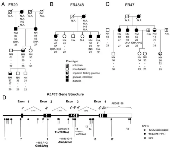
KLF11 Gene Variants Are Associated with Diabetes.Sequencing of the KLF11 gene in 190 probands of early-onset T2DM families iden-tified two rare KLF11 variants present in three distinct families, but absent in 313 late-onset T2DM patients and 313 normoglycemic subjects: A MODY-like family with four diabetic generations (FR29) and a bilinear transmission of T2DM in the second gen-eration presented the [⫹1,039 G⬎T (Ala347Ser)] variant that was
transmitted with diabetes兾glucose intolerance in all three genera-tions examined (Fig. 2A). Linkage analysis under a dominant inheritance MODY-like (12) and late-onset T2DM model (15) revealed the maximal, theoretically possible logarithm of odds scores (0.6 and 0.68, respectively). The evidence for linkage is not likely to be an at random observation, because 10,000 simulations under the no-linkage hypothesis resulted in an empirical P value of 0.030 and 0.035, respectively. The second rare variant [⫹659 C⬎T (Thr220Met)] was identified in two diabetic sibling of family FR4848, and in the three sisters of family FR47 with either glucose intolerance or frank diabetes (Fig. 2 B and C, respectively). How-ever, in the latter pedigree, this variant was absent in one diabetic family member with an onset of diabetes at a later age. Interestingly, the Ala347Ser variant is located within the third repressor domain of KLF11, and Thr220Met between two repressor domains. Bioin-formatics analysis [using PIX, a peptide identification system, which can be accessed at www.hgmp.mrc.ac.uk兾Registered兾Webapp兾 pix兾, and SOPMA (16)] predicted both variants alter the secondary protein structure of these domains, which may have functional implications on KLF11 transcriptional activity.
Analysis of the KLF11 gene-related sequences revealed 19 fre-quent KLF11 SNPs (minor allele frequency of ⬎5%, Fig. 2D). Differences between allele frequencies in normoglycemic and T2DM subjects were first assessed in a pilot study with a cutoff P value of 0.2 to prevent exclusion of potentially T2DM associated SNPs elusive in this pilot (Table 6, which is published as supporting information on the PNAS web site). SNP 1, 9, 16, and 17 showed a highly significant association with T2DM in the subsequently analyzed cohort (P⬍ 0.0002, Table 1 ). The T2DM-associated SNPs are in strong linkage disequilibrium (LD) with each other (D⬘ ⫽ 0.96–0.98), resulting in an association of combined haplotypes similar to that of individual SNPs (data not shown). To exclude that
Fig. 2. Identification of KLF11
vari-ants. (A) Pedigree of the MODY-like family with SNP [⫹1,039 G⬎T Ala347Ser]. (B and C) Pedigree of early-onset diabetes families with SNP [⫹659 C⬎T Thr220Met]. Below the symbols are genotype, age at medical examination, age of onset, diabetic treatment , and BMI are in-dicated. OHA, oral hypoglycemic agent; INS, insulin. Asterisks indicate presence of diabetic complications (e.g., neuropathy). (D) KLF11 gene structure with the position of SNPs. R, repressor domain; Zn, zinc finger domain.
the causal, T2DM-associated variants reside in a neighboring gene, we genotyped 47 additional SNPs (Fig 4, which is published as supporting information on the PNAS web site). Both the variants [KLF11 AS⫹ 2541 A⬎G] and [AK091299 UTR ⫹ 2422 A⬎T] in cilia-associated protein-1 (CYS1) that are in strong LD with the KLF11 variants, show a significant association with T2DM (Table 1). The T2DM-associated LD block of SNPs does not include other SNPs of CYS1, suggesting that the functional variant(s) is within KLF11 sequences. Thus, the underlying cause of association may result from combined effects of several KLF11 variants, or could depend on functionality of one SNP. A likely functional SNP is the nonsynonymous coding variant SNP9, which is responsible for a Gln to Arg conversion at amino acid 62, and which was predicted (PIX program) to affect the␣-helix structure in proximity of the first repressor domain that is responsible for the interaction with the corepressor mSin3A. Furthermore, this SNP displays a highly significant T2DM association that is independent of age, sex, and body mass index (BMI) differences between subjects (binominal
logistic regression, P⫽ 0.0003). Further analysis of 1,463 normo-glycemic and 1,383 T2DM subjects from North European popula-tions shows that the allele frequency of [KLF11⫹185 A⬎G (Gln62Arg)] in T2DM subjects was significantly higher than in controls (Table 2, P ⫽ 0.033). Overall, the initial and second case-control study results in a combined OR of 1.29 (Table 3, 95% CI of OR⫽ 1.12–1.49, P ⫽ 0.00033). The fraction of attributed risk for T2DM susceptibility of Gln62Arg in the followup study is 2% and 7% under an additive and a dominant model, respectively; and is similar to values recently reported for the CAP10 at-risk haplo-type (17). Thus, genetic analyses using family-based and case-control studies reveal that KLF11 variants have an association with early-onset diabetes and T2DM.
Biochemical and Functional Characterization ofKLF11 Variants.To assess the putative physiopathological link between T2DM and KLF11 variants, we analyzed the transcriptional regulatory activity of both the rare Thr220Met and Ala347Ser variants and the
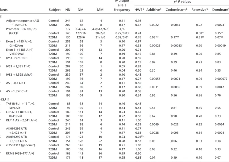
Table 1. SNP frequencies in French Caucasian case-control study
Variants Subject NN NM MM
Multiple allele frequency
2P values
HWE* Additive† Codominant‡ Recessive§ Dominant¶
KLF11
1 Adjacent sequence (AS) ⫺1,659 G⬎C Control 244 62 4 0.11 0.98 T2DM 202 88 8 0.17 0.67 0.0022 0.0084 0.22 0.0023 7 Promoter⫺86 del兾ins 5-5 5-4兾5-6 4-4兾4-6兾6-6 4兾6 (GCC)㛳 Control 145 127兾16 20兾2兾0 0.27兾0.03 0.24 0.08‡‡ 0.15‡‡ T2DM 130 129兾6 31兾1兾0 0.32兾0.01 0.76 0.03** 0.17†† 0.31§§ 0.07§§ 9 Exon 2⫹185 A⬎G, Gln62Arg Control 252 58 3 0.10 0.87 T2DM 211 95 7 0.17 0.33 0.00023 0.00083 0.20 0.00019 11 Exon 3⫹1185 A⬎T, Val395Val Control 202 96 13 0.20 0.71 T2DM 192 100 7 0.19 0.15 0.81 0.39 0.20 0.85 12 IVS3⫹976 T⬎C Control 198 96 14 0.20 0.59 T2DM 191 102 8 0.20 0.19 0.82 0.39 0.21 0.83 14 IVS3⫹1,331 T⬎C Control 282 30 1 0.05 0.83 T2DM 262 22 0 0.04 0.50 0.30 0.46 0.34 0.35
16 IVS3⫹1,398 del(A) Control 239 57 2 0.10 0.48
T2DM 192 93 7 0.17 0.27 0.00055 0.0021 0.09 0.000077 17 AS⫹343 G⬎T Control 240 64 2 0.11 0.56 T2DM 207 89 7 0.17 0.68 0.0031 0.0096 0.09 0.0047 18 AS⫹1,357 C⬎T Control 194 91 13 0.20 0.58 T2DM 195 101 9 0.20 0.34 0.96 0.56 0.36 0.76 Non-KLF11 2 TAF1B-SL1⫹16 T⬎G, Ser6Ala Control 88 138 64 0.46 0.48 T2DM 97 139 61 0.44 0.41 0.51 0.81 0.65 0.55 6 LBP32⫹1189 C⬎T, Control 180 111 14 0.23 0.63 lle419Val T2DM 183 108 12 0.22 0.50 0.67 0.90 0.70 0.73 17 KLF11 AS⫹2,541 A⬎G Control 240 61 3 0.11 1.00 T2DM 214 88 6 0.16 0.53 0.0069 0.022 0.32 0.0064
21 AK091299 UTR Control 245 59 4 0.11 0.77
⫹2,422 A⬎T T2DM 207 87 7 0.17 0.68 0.0028 0.095 0.34 0.0024
25 AK091299 UTR Control 174 123 10 0.23 0.04§§
⫹3,107 G⬎A T2DM 154 128 22 0.28 0.57 0.05 0.06 0.03 0.14
44 rs7587317 (genomic) Control 263 145 19 0.21 1.00
T2DM 180 108 16 0.17 1.00 0.08 0.22 0.10 0.33
47 RRM2 IVS8–177 A兾G Control 163 142 26 0.29 0.60
T2DM 171 118 17 0.25 0.65 0.07 0.19 0.10 0.07
Frequency analysis of variants showing a suggestive T2DM association in a pilot study (P⬍ 0.2). *2test for deviations from the Hardy–Weinberg equilibrium.
†2test for differences of allele frequencies between control and T2DM subjects.
‡2test for differences of genotype frequencies between control and T2DM subjects under a codominant model. §2test for differences of genotype frequencies between control and T2DM subjects under a recessive model. ¶2test for differences of genotype frequencies between control and T2DM subjects under a dominant model.
㛳Triallelic variant with four, five, and six GCC repeats that was analyzed in a 2⫻ 3 contingency table with genotype frequencies.
**Triallelic variant with four, five, and six GCC repeats that was analyzed with allele frequencies and a 2⫻ 6 contingency table with genotype frequencies.
††Analyses in the recessive model of 4-4 alleles vs. all others, and in the dominant model of 4-4, 4-5, and 4-6 alleles vs. 5-5 and 5-6 alleles. ‡‡Analyses in the recessive model of 6-6 alleles vs. all others, and in the dominant model of 5-6, 4-6, and 6-6 alleles vs. 5-5, 5-4, and 4-4 alleles.
§§Deviated from HWE, 3.2% of the genotypes were reanalyzed without observing errors. By using an Armitage’s trend test, which is robust to Hardy–Weinberg
(13), this SNP is associated with T2DM, P⫽ 0.04.
nonsynonymous SNP9 (Gln62Arg). Studies using the Gal4 reporter assay have previously shown that the N-terminal domain of KLF11 harbors three repression domains (8). Interestingly, 62Arg-, 220Met- and 347Ser-KLF11 variants demonstrate a significantly increased repression activity compared with the wild-type N-terminal KLF11-GAL4 chimeric construct (Fig. 3A). Similarly, the 62Arg- and 220Met-KLF11 variants show increased repression activity on the reporter plasmid containing repetitive KLF11-binding sites compared with wild-type control (data not shown). Interestingly, 62Arg-, 220Met-, and 347Ser-KLF11 are gain-of-function variants, suggesting they may affect corepressor affinity. Indeed, an increased binding of the corepressor mSin3A to 62-Arg KLF11 variant compared with wild type was observed by coimmu-noprecipitation (Fig. 3A). The Thr220Met and Ala347Ser muta-tions show no alteration in binding affinity, suggesting that other not-yet-identified corepressors may mediate the transcriptional effects of these proteins. Taken together, these results suggest that the association of impaired KLF11 function with diabetes may, under specific conditions, result from altered KLF11 transcriptional activity.
We also anticipated a direct link between the altered KLF11 function and the transcriptional regulation of the insulin gene. In the pancreatic beta cells, wild-type KLF11 induced the insulin promoter activity by 2-fold (Fig. 1F), but its activation was signif-icantly impaired by 62Arg-KLF11 variant under high-glucose levels (Fig. 3B). The 220Met- and 347Ser-KLF11 variants show no effect on the activation of the insulin promoter. Interestingly, under similar conditions, 62Arg-KLF11 variant showed a decreased in-duction of endogenous insulin mRNA expression compared with the wild-type protein (Fig. 3C). Thus, these results suggest that an altered 62Arg-KLF11 function may impair insulin levels by increas-ing the repression activity on this promoter. In fact, analysis of the available oral glucose tolerance tests (OGTTs) of 70 normoglyce-mic subjects from the familial case-control cohort show a
signifi-cantly decreased plasma insulin level at 60 and 120 min after an oral glucose load for AG-allele carriers compared with AA-allele carriers (Table 3 G and A allele encoding 62Arg- and wild-type KLF11, respectively). In addition, the normoglycemic AG allele carriers are lean and have increased insulin sensitivity that may protect them against impaired glucose tolerance. Analysis in pre-diabetic subjects also suggests that the AG allele is associated with decreased plasma insulin levels (Data not shown; P⬍ 0.05 at 30 and 90 min for 33 French AA allele vs. 12 AG allele carriers).
In addition, KLF11 may affect pancreatic beta cells function by modulating the expression of free radical scavengers such as superoxide dismutase (SOD)2 and catalase 1 that were recently identified as KLF11 target genes (5). These antioxidant enzymes have a very low expression level in pancreatic islets compared with other metabolic tissues, e.g., liver and skeletal muscle, and overex-pression of these enzymes protects beta cells against glucolipotox-icity (18). In pancreatic beta cells, KLF11 represses the promoter activity of the SOD2 (data not shown) and catalase 1 (Fig. 5, which is published as supporting information on the PNAS web site). The KLF11 variants show no changes in transcriptional activity of the SOD2 promoter (data not shown), but all three significantly increase repression of catalase 1 promoter under high-glucose levels (Fig. 5). Taken together, these results suggest that the association of altered functional KLF11 variants with diabetes may result from an altered KLF11 transcriptional activity.
Discussion
In this study, we addressed the questions as to whether KLF11 is involved in the regulation of pancreatic beta cell function, and whether it plays a role as gene modifier in predisposition to diseases of beta cell origin such as diabetes. Our study has yielded evidence that this is indeed the case. Regarding the biology of KLF11, our data support a model where in pancreatic beta cells the product of the KLF11 gene is induced by glucose to regulate in turn insulin
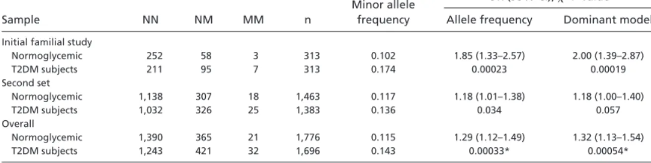
Table 2. Frequency of Gln62Arg KLF11 in additional population samples
Sample NN NM MM n
Minor allele frequency
OR (95% CI),2P value
Allele frequency Dominant model
Initial familial study
Normoglycemic 252 58 3 313 0.102 1.85 (1.33–2.57) 2.00 (1.39–2.87) T2DM subjects 211 95 7 313 0.174 0.00023 0.00019 Second set Normoglycemic 1,138 307 18 1,463 0.117 1.18 (1.01–1.38) 1.18 (1.00–1.40) T2DM subjects 1,032 326 25 1,383 0.136 0.034 0.057 Overall Normoglycemic 1,390 365 21 1,776 0.115 1.29 (1.12–1.49) 1.32 (1.13–1.54) T2DM subjects 1,243 421 32 1,696 0.143 0.00033* 0.00054*
NN, homozygous wild-type allele carriers; NM, heterozygous allele carriers; MM, homozygous mutant allele carriers; OR, odds ratio. *P value and combined OR for allelic effects calculated by a Mantel–Haenszel test (25).
Fig. 3. Functional characterization of
non-synonymous KLF11 variants. (A) GAL4 reporter assay with N-terminal KLF11-GAL4DBD con-structs cotransfected in CHO cells (n⫽ 4).
(Up-per) A representative Western blot of
coimmu-noprecipitation experiments (n⫽ 2) showing mSin3a binding of Flag-tagged KLF11 (WT and variants) or empty vector (control). (B) Lucif-erase assays of Flag-tagged KLF11 variants co-transfected with human insulin promoter re-porter constructs inTC3 cells (n ⫽ 3). (C) Real-time PCR quantification of insulin mRNA expression inTC3 cells cultured in high-glucose medium (25 mM) and transfected with control, WT KLF11, or 62R-KLF11. Values are corrected for GAPDH levels (n⫽ 4).*, signifi-cantly different (P⬍ 0.05).
transcription. Beside this important observation, further additional data point to an involvement of KLF11 in the biology of pancreatic beta cells. In these cells, KLF11 is also regulated by TGF-, a pathway playing a critical role in the development and homeostasis of both the exocrine and endocrine pancreas. In addition, KLF11 regulates key genes encoding scavengers of oxidative stress, includ-ing SOD2 and catalase 1. This observation is extremely important in the context of this study, because a tight control of oxidative stress is critical for maintaining the homeostasis of pancreatic beta cells. In fact, oxidative stress is believed to be involved in the progression of pancreatic beta cell dysfunction found in T2DM.
In light of these data, KLF11 is a truly valid candidate gene for genetic predisposition to T2DM. Indeed, our comprehensive ge-netic analysis by using family-based and case-control studies pro-vide epro-vidence for a causal association between three nonsynony-mous KLF11 variants, early-onset and polygenic T2DM. The overall P value for association of the KLF11 variant Gln62Arg with
T2DM in our study, including 1,696 T2DM and 1,776 normogly-cemic subjects, is significant (0.00033), and even the modest asso-ciation observed in the second study alone (0.03) is of similar order, as recently shown in replication studies of validated candidate genes for T2DM such as PPAR␥ (19) and KIR 6.2 (20). It remains to be established whether the 62Arg mutant is the only functional KLF11 variant among the group of late-onset T2DM-associated variants. For the three nonsynonymous variants, we showed that they affect the function of KLF11, and consequently, they may influence primary beta cell functions, and the balance between proliferation and differentiation of beta cells. Moreover, KLF11 has recently been shown to be up-regulated after refeeding in mouse skeletal muscles (21), suggesting a role for KLF11 in postprandial glucose metabolism of this tissue. In addition, the caveolin-1 gene that is highly expressed in adipose tissue is repressed by KLF11 in a cholesterol-dependent manner (22). The fact that caveolin-1 bind-ing to the insulin receptor stimulates both the kinase activity and recruitment of the insulin receptor to lipid rafts at the plasma membrane, and the fact that insulin receptor mutations impairing the calveolin binding result in T2DM (23), suggest that calveolin 1 is essential for insulin signaling. Therefore, we do not exclude that an altered KLF11 function in skeletal muscle, adipose tissue, or liver may also play a role in susceptibility to diabetes.
In light of the evidence, it is tempting to speculate on the possibilities of targeting KLF11 for the therapy of diabetes. The initial structural and molecular modeling data on mSin3A interac-tion with KLF11 (24), which shows that this interacinterac-tion differs from other transcription factors that bind to mSin3A, may present a fundamental step toward a rational drug design effort. Thus, further studies to unravel the mechanisms underlying the association of altered KLF11 function with the pathogenesis and severity of diabetes may also contribute with novel targets to the development of antidiabetic agents.
We thank all families and patients who participated to this study. We thank K. Borch-Johnsen and T. Drivsholm (Steno Diabetes Center and Research Center for Prevention and Health) for recruiting and charac-terization of part of the Danish control subjects. We thank F. Pattou and J. Kerr-Conte (Universite´ de Lille) for their kind gift of human pan-creatic islet and beta cell RNA. We thank T. Gurevitch, (Hadassah University Hospital), J. Hiddinga (Mayo Clinic), and C. Wachter and P. Boutin (Unite´ Mixte de Recherche 8090) for assistance with transfection experiments, pancreatic beta cell culture, and advice on the manuscript, respectively. We thank T. J. Woodage for providing TaqMan SNP genotyping assays. This work was supported by Mayo Clinic Cancer Center Specialized Program of Research Excellence Pancreatic Cancer Grant P50 CA10270 and National Institute of Health Grants DK52913 and DK56620 (to R.U.), Juvenile Diabetes Foundation Grant 1-2001-325 (to D.M.), the European Regional Development Fund, Region Nord-Pas de Calais (ARCir) (to B.N. and P.F.), the European Economic Com-munity (Genomic Integrated Force for Type II Diabetes 1998–2002), and Eli Lilly through the Lilly Consortium for Diabetes and Obesity.
1. Kim, S. K. & Hebrok, M. (2001) Genes Dev. 15, 111–127.
2. Song, S. Y., Gannon, M., Washington, M. K., Scoggins, C. R., Meszoely, I. M., Goldenring, J. R., Marino, C. R., Sandgren, E. P., Coffey, R. J. J., Wright, C. V. & Leach, S. D. (1999)
Gastroenterology 117, 1416–1426.
3. Fisher, W. E. (2001) World J. Surg. 25, 503–508.
4. Coughlin, S. S., Calle, E. E., Teras, L. R., Petrelli, J. & Thun, M. J. (2004) Am. J. Epidemiol.
159,1160–1167.
5. Fernandez-Zapico, M. E., Mladek, A., Ellenrieder, V., Folch-Puy, E., Miller, L. & Urrutia, R. (2003) EMBO J. 22, 4748–4758.
6. Sanvito, F., Herrera, P. L., Huarte, J., Nichols, A., Montesano, R., Orci, L. & Vassalli, J. D. (1994) Development (Cambridge, U.K.) 120, 3451–3462.
7. McCarthy, M. I. & Froguel, P. (2002) Am. J. Physiol. 283, E217–E225.
8. Zhang, J. S., Moncrieffe, M. C., Kaczynski, J., Ellenrieder, V., Prendergast, F. G. & Urrutia, R. (2001) Mol. Cell. Biol. 21, 5041–5049.
9. Crooks, G. E., Hon, G., Chandonia, J. M. & Brenner, S. E. (2004) Genome Res. 14, 1188–1190. 10. Kaczynski, J., Zhang, J. S., Ellenrieder, V., Conley, A., Duenes, T., Kester, H., van Der Burg,
B. & Urrutia, R. (2001) J. Biol. Chem. 276, 36749–36756. 11. Sham, P. C. & Curtis, D. (1995) Ann. Hum. Genet. 59, 97–105. 12. Mendell, N. R. & Simon, G. A. (1984) Ann. Hum. Genet. 48, 283–286. 13. Sasieni, P. D. (1997) Biometrics 53, 1253–1261.
14. Lukowiak, B., Vandewalle, B., Riachy, R., Kerr-Conte, J., Gmyr, V., Belaich, S., Lefebvre, J. & Pattou, F. (2001) J. Histochem. Cytochem. 49, 519–528.
15. Waeber, G., Delplanque, J., Bonny, C., Mooser, V., Steinmann, M., Widmann, C., Maillard, A., Miklossy, J., Dina, C., Hani, E., et al. (2000) Nat. Genet. 24, 291–295.
16. Geourjon, C. & Deleage, G. (1995) Comput. Appl. Biosci. 11, 681–684.
17. Cox, N. J., Hayes, M. G., Roe, C. A., Tsuchiya, T. & Bell, G. I. (2004) Diabetes 53, S19 –S25.
18. Robertson, R. P., Harmon, J., Tran, P. O. & Poitout, V. (2004) Diabetes 53, S119–S124. 19. Altshuler, D., Hirschhorn, J. N., Klannemark, M., Lindgren, C. M., Vohl, M. C., Nemesh,
J., Lane, C. R., Schaffner, S. F., Bolk, S., Brewer, C., et al. (2000) Nat. Genet. 26, 76–80. 20. Love-Gregory, L., Wasson, J., Lin, J., Skolnick, G., Suarez, B. & Permutt, M. A. (2003)
Diabetologia 46, 136–137.
21. Yamamoto, J., Ikeda, Y., Iguchi, H., Fujino, T., Tanaka, T., Asaba, H., Iwasaki, S., Ioka, R. X., Kaneko, I. W., Magoori, K., et al. (2004) J. Biol. Chem. 279, 16954–16962. 22. Cao, S., Fernandez-Zapico, M. E., Jin, D., Puri, V., Cook, T. A., Lerman, L. O., Zhu, X. Y.,
Urrutia, R. & Shah, V. (November 5, 2004) J. Biol. Chem., 10.1074兾jbc.M407941200. 23. Cohen, A. W., Combs, T. P., Scherer, P. E. & Lisanti, M. P. (2003) Am. J. Physiol. 285,
E1151–E1160.
24. Pang, Y. P., Kumar, G. A., Zhang, J. S. & Urrutia, R. (2003) FEBS Lett. 548, 108–112. 25. Mantel, N. & Haenszel, W. (1959) J. Natl. Cancer Inst. 22, 719–748.
26. Albareda, M., Rodriguez-Espinosa, J., Murugo, M., de Leiva, A. & Corcoy, R. (2000)
Diabetologia 43, 1507–1511.
27. Stumvoll, M., Mitrakou, A., Pimenta, W., Jenssen, T., Yki-Jarvinen, H., Van Haeften, T., Renn, W. & Gerich, J. (2000) Diabetes Care 23, 295–301.
Table 3. Analysis of normoglycemic Gln62Arg carriers during OGTT
AA allele AG allele P value†
No. of subjects 57 13
Age 56⫾ 1.4 51⫾ 3.7 0.82
Sex (M兾W) 25兾32 3兾10
BMI 24.9⫾ 0.7 21.4⫾ 2.0 0.057
OGTT time, min Glucose, mM兾l 0 5.1⫾ 0.1 5.0⫾ 0.1 0.53 30 8.1⫾ 0.2 7.3⫾ 0.5 0.14 60 7.1⫾ 0.3 6.3⫾ 0.7 0.25 90 5.8⫾ 0.2 5.4⫾ 0.4 0.51 120 5.1⫾ 0.2 4.7⫾ 0.3 0.36 ⌬AUCglu 170⫾ 19 116⫾ 43 0.22 Insulin, mU兾l 0 8.6⫾ 1.6 5.3⫾ 1.1 0.31 30 43.9⫾ 4.1 28.6⫾ 5.5 0.084 60 50.7⫾ 4.1 27.0⫾ 4.8 0.0081 90 38.1⫾ 4.1 26.5⫾ 5.1 0.21 120 36.6⫾ 4.4 18.3⫾ 3.6 0.049 ⌬AUCins 3,811⫾ 331 2,065⫾ 338 0.013
Assesement of beta cell function and insulin sensitivity‡
IG of 30 min 4.4⫾ 0.4 3.7⫾ 0.8 0.38
IG of 120 min 95.8⫾ 72.0 24.3⫾ 22.5 0.62
First-phase Stumvoll index 285.0⫾ 29.5 337.6⫾ 76.4 0.46 Second-phase Stumvoll index 99.4⫾ 5.8 107.9⫾ 14.8 0.54
ISI-Belfiore 0.13⫾ 0.03 0.08⫾ 0.04 0.50
ISI-Matsuda 229.6⫾ 41.3 220.7⫾ 56.1 0.93
ISI-Stumvoll 0.12⫾ 0.003 0.14⫾ 0.007 0.045
IG, insulinogenic index; ISI, insulin sensitivity index.
†Comparison of the mean by ANOVA. ‡Formulas are from refs. 26 and 27.