..
..
..
..
..
..
..
.
2D/3D echocardiographic determinants of
left ventricular reverse remodelling after
MitraClip implantation
Sara Cimino
1*, Viviana Maestrini
1, Donatella Cantisani
1, Valentina Petronilli
1,
Domenico Filomena
1, Massimo Mancone
1, Gennaro Sardella
1, Francesco Fedele
1,
Patrizio Lancellotti
2, and Luciano Agati
11
Department of Cardiovascular, Respiratory, Nephrological, Aenesthesiological and Geriatric Sciences “Sapienza” University of Rome, Policlinico Umberto I, Viale del Policlinico
155, 00161 Rome, Italy; and2
Department of Cardiology, GIGA Cardiovascular Sciences, Heart Valve Clinic, University of Lie`ge Hospital, CHU Sart-Tilman, Avenue de L’Ho`pital 1, 4000 Lie`ge, Belgium
Received 23 June 2018; editorial decision 29 September 2018; accepted 4 October 2018; online publish-ahead-of-print 8 November 2018
Aims The aim of this study was to describe incidence and determinants of left ventricular reverse remodelling (r-LVR) at 6 months follow-up after MitraClip implantation in patients with secondary severe mitral regurgitation (MR) and reduced left ventricular ejection fraction (LVEF).
... Methods
and results
Forty-five patients, undergoing MitralClip implantation with low ejection fraction and high surgical risk were enrolled in this study. Three of them died before the scheduled 6 months follow-up period and one patient had cardiac sur-gery due to MitraClip detachment. All patients underwent transthoracic 2D and 3D echocardiography before and 6 months after the procedure. A significant MR severity reduction and an improvement in New York Heart Association (NYHA) class were detected in all patients. The study population was divided in two groups according to the presence of r-LVR (51%, n = 23 patients) or not (non-rLVR group, 18 patients). Non-significant differences in MR aetiology and number of clips implanted were found. Left ventricular reverse remodelling patients showed sig-nificant lower values of logistic EuroSCORE and STS score, left ventricular end-diastolic volume index (LVEDV/i), right ventricular end systolic area, and pulmonary artery systolic pressure (PASp) at baseline evaluation. At multivari-able analysis, baseline PASp value resulted to be the only independent predictor of r-LVR [odds ratio 95% confi-dence interval 0.94 (0.89–0.99), P = 0.021]. In r-LVR patients, a significant improvement in LVEF and global longitu-dinal strain and a reduction in left atrial volume index were detected after 6 months, whereas in non-rLVR subgroup a significant increase in both LVEDV/i and left ventricular end-systolic volume index was observed at follow-up. ... Conclusion Even if a reduction of MR was detected in all patients after MitralClip implant, our findings suggest that end-stage
patients presenting with higher left ventricular volumes, logistic scores, and PASp may not benefit from the proced-ure at longer follow-up in terms of left ventricular function.
䊏 䊏 䊏 䊏 䊏 䊏 䊏 䊏 䊏 䊏 䊏 䊏 䊏 䊏 䊏 䊏 䊏 䊏 䊏 䊏 䊏 䊏 䊏 䊏 䊏 䊏 䊏 䊏 䊏 䊏 䊏 䊏 䊏 䊏 䊏 䊏 䊏 䊏 䊏 䊏 䊏 䊏 䊏 䊏 䊏 䊏 䊏 䊏 䊏 䊏 䊏 䊏 䊏 䊏 䊏 䊏 䊏 䊏 䊏 䊏 䊏 䊏 䊏 䊏 䊏 䊏 䊏 䊏 䊏 䊏 䊏 䊏 䊏 䊏 䊏 䊏 䊏 䊏 䊏 䊏 䊏 䊏 䊏 䊏 䊏 䊏 䊏 䊏 䊏 䊏 䊏 䊏 䊏 䊏 䊏 䊏 䊏 䊏 䊏 䊏 䊏 䊏 䊏 䊏 䊏 䊏 䊏 䊏 䊏 䊏 䊏 䊏 䊏 䊏 䊏 䊏 䊏 䊏 䊏 䊏 䊏 䊏 䊏 䊏 䊏 䊏 䊏 䊏 䊏 䊏 䊏 䊏 䊏 䊏 䊏 䊏 䊏 䊏 䊏 䊏 䊏 䊏 䊏 䊏 䊏 䊏 䊏 䊏 䊏 䊏 䊏 䊏 䊏 䊏 䊏 䊏 䊏 䊏 䊏 䊏 䊏 䊏 䊏 䊏 䊏 䊏 䊏 䊏 䊏 䊏 䊏 䊏 䊏 䊏 䊏 䊏 䊏 䊏 䊏 䊏 䊏 䊏 䊏 䊏 䊏 䊏 䊏 䊏 䊏 䊏 䊏 䊏 䊏 䊏 䊏 䊏 䊏 䊏 䊏 䊏 䊏 䊏 䊏 䊏 䊏 䊏 䊏 䊏 䊏 䊏 䊏 䊏
Keywords heart failure
•
mitral valve regurgitation•
percutaneous edge-to-edge valve repair•
MitraClipIntroduction
Mitral regurgitation (MR) is the second most prevalent valvular dis-ease in Europe. The disorder commonly evolves insidiously over many years, causing progressive left atrial (LA) and left ventricular
(LV) dilatation and consequent deterioration of LV contractile func-tion due to chronic volume overload. Despite optimal medical ther-apy, severe secondary MR confers a worse prognosis.1,2
Mitral valve (MV) surgery (repair or replacement)3is the current standard of care for patients with severe symptomatic primary MR; it
* Corresponding author. Tel:þ39 06 49979048; Fax: þ39 06 49979060. E-mail: sara.cimino@uniroma1.it
Published on behalf of the European Society of Cardiology. All rights reserved.VCThe Author(s) 2018. For permissions, please email: journals.permissions@oup.com.
..
..
..
..
..
..
..
..
..
..
..
..
..
..
..
..
..
..
..
..
..
..
..
..
..
..
..
..
..
..
..
..
..
..
..
..
..
..
..
.
has been shown to result in potential reverse LA and LV remodelling (r-LVR).4However, due to the high operative risk, limited data are available about therapeutic benefits of MV surgery in patients with heart failure and secondary MR. Current guidelines only advocate a Class II indication for MV surgery in symptomatic patients with chronic severe secondary MR and severely reduced LV ejection fraction (LVEF) (<30%).3–5
The MitraClip device has evolved as a promising interventional tool for MV repair in severe MR.6,7The first randomized controlled study (EVEREST II) demonstrated a superior safety of endovascular valve edge-to-edge repair when compared with MV surgery, with a similar improvement in clinical outcome.8,9MitraClip might be a valid therapeutic option for selected high-risk surgical patients with se-verely reduced LV function. Several studies demonstrated encourag-ing data about clinical and safety results after MitraClip implantation in this high-risk subset of patients, however, it is yet to know whether the reduction in MR leads to r-LVR, and ultimately improved progno-sis, in the long-term. We sought to evaluate the determinants of r-LVR at 6 months follow-up after MitraClip implantation.
Methods
Study population
From June 2014 to July 2017, 45 consecutive patients undergoing percutaneous MV repair with the MitraClip system were enrolled
prospectively in this study. Inclusion criteria were: (i) a diagnosis of severe secondary MR; (ii) reduced LV function (<45%); (iii) high surgi-cal risk; and (iv) New York Heart Association (NYHA) Class III or IV. Patients with MV morphological properties that would make MitraClip implantation unlikely or unsuitable were excluded.9,10The MitraClip procedure was explained to the patients, as well as alterna-tive options (medical treatment or high-risk MV surgery). The ‘Heart Team’ evaluated patients and conventional surgery was excluded in case of excessive morbidity and mortality (high logistic EuroSCORE or STS score, or excessive comorbidities).9The local ethics commit-tee approved this study, and all patients provided written informed consent.
Patients were on optimized medical therapy and were treated with percutaneous angioplasty and stent implantation, implantable cardioverter defibrillator, and cardiac resynchronization therapy devices prior to MitraClip therapy, if clinically indicated. The baseline and follow-up functional status was assessed according to the NYHA criteria.
Echocardiography
All enrolled patients underwent transthoracic two- and three-dimensional echocardiography (3DE) (Philips X5-1 Transducer, EPIQ7C) before and at 6 months after the procedure of percutan-eous MV repair. The presence of MR at baseline was qualified by col-our Doppler and quantified by the vena contracta width and the Proximal Isovelocity Surface Area method in accordance with the
...
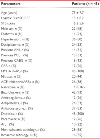
Table 1 Baseline characteristics
Parameters Patients (n 5 45) Age (years) 73 ± 7.7 Logistic EuroSCORE 13 ± 8.2 STS score 6 ± 5.6 Male sex, n (%) 22 (48) Diabetes, n (%) 11 (24) Hypertension, n (%) 36 (80) Dyslipidaemia, n (%) 24 (53) Previous AMI, n (%) 14 (31) Previous PCI, n (%) 15 (33) Previous CABG, n (%) 6 (13) CRF, n (%) 12 (26) NYHA III–IV, n (%) 45 (100) Nitrates, n (%) 20 (44) ACE-inhibitors/ARBs, n (%) 26 (58) Ivabradine, n (%) 1 (0.02) Beta-blockers, n (%) 42 (93) Anticoagulants, n (%) 12 (26) Antiplatelets, n (%) 24 (53) Antialdosterone, n (%) 37 (83) Diuretics, n (%) 45 (100) Pacemaker, n (%) 12 (26) AF, n (%) 10 (22) Non-ischaemic aetiology, n (%) 29 (65) Ischaemic aetiology, n (%) 16 (35)
ACE, angiotensin converting enzyme; AF, atrial fibrillation; AMI, acute myocardial infarction; ARBs, angiotensin II receptor blockers; CABG, coronary artery bypass grafting; CRF, chronic renal failure; NYHA, New York Heart Association; PCI, percutaneous coronary intervention; STS, Society of Thoracic Surgery Score.
...
Table 2 Echocardiographic parameters at baseline and at 6-month follow-up in the overall cohort of patients Parameters Baseline, mean 6 SD 6 months, mean 6 SD P-value Regurgitant volume (mL) 51 ± 14.3 27 ± 8 <0.001 EROA (cm2) 0.37 ± 0.15 0.19 ± 0.06 0.023 Annulus diameter (mm) 37 ± 4.4 35 ± 3.7 0.026 E wave (cm/s) 117 ± 49 148 ± 49 NS Septal e0(cm/s) 5.1 ± 1.5 4.34 ± 3.1 NS LVEF % 29 ± 11 29 ± 12 NS LVEDV/i (mL/m2) 96 ± 24 93 ± 32 NS LVESV/i (mL/m2) 68 ± 25 65 ± 30 NS LA Vol/i (mL/m2) 54 ± 19 49 ± 19.3 NS RA Vol/i (mL/m2) 41 ± 22.9 41 ± 31 NS RV ED area (cm2) 17± 4.7 18± 6.8 NS RV ES area (cm2) 11 ± 3.8 12 ± 5.8 NS RV FAC % 36 ± 8.6 36 ± 6 NS TAPSE (mm) 20 ± 3.6 21 ± 2.8 NS PASp (mmHg) 44± 15.7 44 ± 14 NS GLS % -6.4 ± 3.5 -7.3 ± 3.15 NS
ED, end-diastolic; ES, end-systolic; EROA, effective regurgitant orifice area; FAC, fractional area change; GLS, global longitudinal strain; LA Vol/i, left atrium vol-ume/index; LVEDV/i, left ventricular end-diastolic volume index; LVEF, left ven-tricular ejection fraction; LVESV/i, left venven-tricular end-systolic volume index; NS, not significant; PASp, pulmonary artery systolic pressure; RA Vol/i, right atrium volume/index; RV, right ventricle; SD, standard deviation; TAPSE, tricuspid annu-lar plane excursion.
..
..
..
..
..
..
..
..
..
..
..
..
..
..
..
..
..
..
..
..
..
..
..
..
..
..
..
..
..
..
..
..
..
..
..
..
..
..
..
..
..
..
..
..
..
..
2D/3D echocardiographic determinants of LV reverse remodelling
559
Figure 1Percentage of NYHA Class III–IV class at baseline and at 6 months follow-up.
...
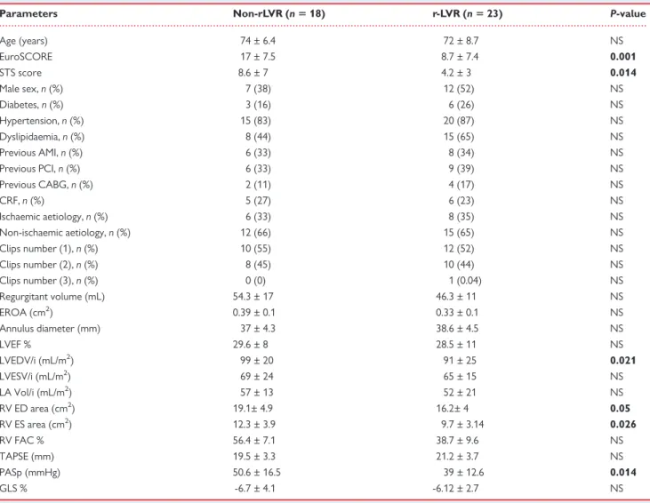
Table 3 Clinical and echocardiographic variables in r-LVR vs. non-rLVR patients at baseline
Parameters Non-rLVR (n 5 18) r-LVR (n 5 23) P-value
Age (years) 74 ± 6.4 72 ± 8.7 NS EuroSCORE 17 ± 7.5 8.7 ± 7.4 0.001 STS score 8.6 ± 7 4.2 ± 3 0.014 Male sex, n (%) 7 (38) 12 (52) NS Diabetes, n (%) 3 (16) 6 (26) NS Hypertension, n (%) 15 (83) 20 (87) NS Dyslipidaemia, n (%) 8 (44) 15 (65) NS Previous AMI, n (%) 6 (33) 8 (34) NS Previous PCI, n (%) 6 (33) 9 (39) NS Previous CABG, n (%) 2 (11) 4 (17) NS CRF, n (%) 5 (27) 6 (23) NS Ischaemic aetiology, n (%) 6 (33) 8 (35) NS Non-ischaemic aetiology, n (%) 12 (66) 15 (65) NS Clips number (1), n (%) 10 (55) 12 (52) NS Clips number (2), n (%) 8 (45) 10 (44) NS Clips number (3), n (%) 0 (0) 1 (0.04) NS Regurgitant volume (mL) 54.3 ± 17 46.3 ± 11 NS EROA (cm2) 0.39 ± 0.1 0.33 ± 0.1 NS Annulus diameter (mm) 37 ± 4.3 38.6 ± 4.5 NS LVEF % 29.6 ± 8 28.5 ± 11 NS LVEDV/i (mL/m2) 99 ± 20 91 ± 25 0.021 LVESV/i (mL/m2) 69 ± 24 65 ± 15 NS LA Vol/i (mL/m2) 57 ± 13 52 ± 21 NS RV ED area (cm2) 19.1± 4.9 16.2± 4 0.05 RV ES area (cm2) 12.3 ± 3.9 9.7 ± 3.14 0.026 RV FAC % 56.4 ± 7.1 38.7 ± 9.6 NS TAPSE (mm) 19.5 ± 3.3 21.2 ± 3.7 NS PASp (mmHg) 50.6 ± 16.5 39 ± 12.6 0.014 GLS % -6.7 ± 4.1 -6.12 ± 2.7 NS
AMI, acute myocardial infarction; CABG, coronary artery bypass grafting; CRF, chronic renal failure; ED, end-diastolic; ES, end-systolic; EROA, effective regurgitant orifice area; FAC, fractional area change; GLS, global longitudinal strain; LA Vol/i, left atrium volume/index; LVEDV/i, left ventricular end-diastolic volume index; LVEF, left ventricular ejec-tion fracejec-tion; LVESV/i, left ventricular end-systolic volume index; NS, not significant; PASp, pulmonary artery systolic pressure; PCI, percutaneous coronary intervenejec-tion; RV, right ventricle; STS, Society of Thoracic Surgery Score; TAPSE, tricuspid annular plane excursion.
Bold values represent statistical significance of P values.
..
..
..
..
..
..
..
..
..
..
..
..
..
..
..
..
..
..
..
..
..
..
..
..
..
..
..
..
..
..
..
..
..
..
..
..
..
..
..
..
..
..
..
..
..
..
.
current guidelines.3All patients were assigned a MR severity score of 1 (mild), 2 (mild to moderate), 3 (moderate to severe), or 4 (severe), according to the quantitative measure of the effective regurgitant ori-fice area (EROA) and regurgitant volume. The regurgitant volume was estimated as the EROA multiplied by the velocity time integral of the regurgitant jet. Procedural success was defined as the reduction of the MR severity score to 2 or less after clip implantation. The fol-lowing parameters were considered to evaluate the LV changes in size and function: LV end-diastolic and end-systolic volume indexed to body surface area (LVEDV/i and LVESV/i, respectively), and the LVEF, obtained using 3DE (full volume function). Right ventricular (RV) dimension and function and pulmonary artery systolic pressure (PASp) were also assessed according to guidelines.11 Two-dimensional speckle tracking analysis with global longitudinal strain (GLS) was also obtained in all patients. LV reverse remodelling was defined as a decrease >_10% in the LVESV/i at follow-up.12Reproducibility
Intra-observer and inter-observer variability for the 3D manual meas-urements of LVEF and LV volumes was assessed in a sample of 10 patients. Two investigators measured blinded the same 3DEcho loops, and one investigator repeated the analysis 1 week later, blinded to the previous measurements.
MitraClip procedure
All patients were undergone to endovascular edge-to-edge MV re-pair as previously described.9,10 All procedures were performed using the 24-Fr MitraClip device (Abbott Vascular, Santa Clara, CA, USA). All clips were implanted under general anaesthesia and the procedures were transoesophageal echocardiography guided. Haemostasis was achieved by compression of the vein for 12 h.
Patients were treated with double antiplatelet therapy after the inter-vention and oral anticoagulants where indicated.
Statistical analysis
Continuous variables are presented as mean ± standard deviation and were compared using Student’s t-test or the Mann–Whitney rank sum test for unpaired comparisons, as appropriate. The mean differences between the baseline and 6-month echocardiographic parameters are reported. The categorical variables are expressed as counts and percentages and were compared using the v2test or Fisher exact test, as appropriate. Differences were considered statis-tically significant when P < 0.05. Univariable and multivariable analyses were performed with backward method to assess determinants of r-LVR. Variables were included in the model when P < 0.10. Statistical analyses were performed using the Statistical Package for Social Sciences, version 23.0 (SPSS, Chicago, IL, USA). Interclass correlation coefficients (ICCs) were calculated to assess inter-observer and intra-observer agreement of 3DE measurements.
Results
Baseline characteristics
Baseline characteristics of the study population are depicted in Table 1. Mean age was 73 ± 7.7 years. All patients were in NYHA Class III–IV and about two-third of them had non-ischaemic cardio-myopathy. Cardiac death occurred in 3 (6%) patients before the scheduled follow-up; 1 patient underwent cardiac surgery after 3 months because of MitraClip detachment and 41 patients had a complete 6 months follow-up echocardiogram.
...
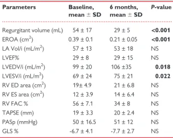
Table 4 Echocardiographic parameters at baseline and 6 months follow-up in non-rLVR group
Parameters Baseline, mean 6 SD 6 months, mean 6 SD P-value Regurgitant volume (mL) 54 ± 17 29 ± 5 <0.001 EROA (cm2) 0.39 ± 0.1 0.21 ± 0.05 <0.001 LA Vol/i (mL/m2) 57 ± 13 53 ± 18 NS LVEF% 29 ± 8 29 ± 15 NS LVEDV/i (mL/m2) 99 ± 20 106 ±35 0.018 LVESV/i (mL/m2) 69 ± 24 75 ± 21 0.022 RV ED area (cm2) 19± 4.9 21 ± 6.8 NS RV ES area (cm2) 12 ± 3.9 14 ± 6.4 NS RV FAC % 56 ± 7.1 34 ± 8 NS TAPSE (mm) 19 ± 3.3 20 ± 2.4 NS PASp (mmHg) 50 ± 16.5 51 ± 12 NS GLS % -6.7 ± 4.1 -7.7 ± 2.7 NS
ED, end-diastolic; ES, end-systolic; EROA, effective regurgitant orifice area; FAC, fractional area change; GLS, global longitudinal strain; LA Vol/i, left atrium vol-ume/index; LVEDV/i, left ventricular end-diastolic volume index; LVEF, left ven-tricular ejection fraction; LVESV/i, left venven-tricular end-systolic volume index; NS, not significant; PASp, pulmonary artery systolic pressure; RV, right ventricle; SD, standard deviation; TAPSE, tricuspid annular plane excursion.
Bold values represent statistical significance of P values.
...
Table 5 Echocardiographic parameters at baseline and at 6 months follow-up in r-LVR group
Parameters Baseline, mean 6 SD 6 months, mean 6 SD P-value Regurgitant volume (mL) 46 ± 11 25 ± 9.3 <0.001 EROA (cm2) 0.33 ± 0.1 0.19 ± 0.07 <0.001 LA Vol/i (mL/m2) 52 ± 21 44.9 ± 18 0.017 LVEF % 28.5 ± 11 32 ± 8.4 0.048 LVEDV/i (mL/m2) 91 ± 25 85 ± 23 <0.001 LVESV/i (mL/m2) 65 ± 15 57 ± 20 <0.001 RV ED area (cm2) 16.2 ± 4 17.1 ± 6 NS RV ES area (cm2) 9.7 ± 3.14 11.3 ± 5 NS RV FAC % 38.7 ± 9.6 36.6 ± 5.2 NS TAPSE (mm) 21.2 ± 3.7 22.4 ± 2.7 NS PASp (mmHg) 39 ± 12.6 39.9 ± 13 NS GLS % -6.12 ± 2.7 -8.5 ± 3.3 <0.001
ED, end-diastolic; ES, end-systolic; EROA, effective regurgitant orifice area; FAC, fractional area change; GLS, global longitudinal strain; LA Vol/i, left atrium vol-ume/index; LVEDV/i, left ventricular end-diastolic volume index; LVEF, left ven-tricular ejection fraction; LVESV/i, left venven-tricular end-systolic volume index; NS, not significant; PASp, pulmonary artery systolic pressure; RV, right ventricle; SD, standard deviation; TAPSE, tricuspid annular plane excursion.
Bold values represent statistical significance of P values.
..
..
..
..
..
..
..
..
..
..
..
..
..
..
..
..
..
..
..
..
..
..
..
..
..
..
..
..
..
..
..
..
..
..
..
..
..
..
..
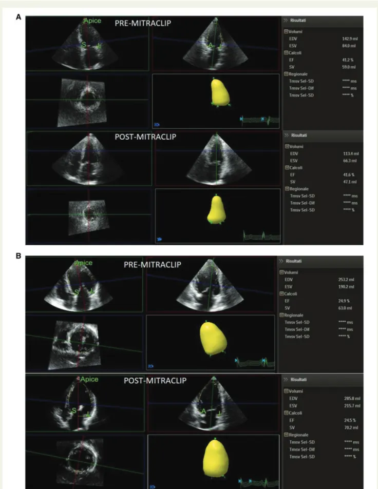
2D/3D echocardiographic determinants of LV reverse remodelling
561
Figure 2(A) LV volumes changes in a patient with r-LVR. (B) LV volumes changes in a patient without r-LVR.
..
..
..
..
..
..
..
..
..
..
..
..
..
..
..
..
..
..
..
..
..
..
..
..
..
..
..
..
..
..
..
..
..
..
..
..
..
..
..
..
..
..
..
..
..
..
..
..
..
..
..
..
..
..
..
..
..
..
..
Changes in NYHA class, MR severity and
LV volumes after MitraClip implantation
Echocardiographic parameters at baseline and at 6 months follow-up in the overall cohort of patients are depicted in Table2.By definition, all patients had severe MR at baseline. No significant changes were observed in LV volumes and LVEF after implant. Noteworthy, a sig-nificant MR reduction was observed after implantation in all patients. Similarly, a significant improvement in NYHA functional class was detected after MitraClip implantation, as showed in Figure1. LV re-verse remodelling occurred in 23 patients (56%), whereas 18 patients did not present reverse remodelling (non-rLVR 44%), as showed in Table 3. At baseline, non-rLVR patients showed higher values of logistic EuroSCORE, STS score, LVEDV/i, RV end systolic area, and PASp when compared with r-LVR subgroup. In these patients, a sig-nificant increase in both LVEDV/i and LVESV/i was observed at 6 months follow-up (Table4). On the contrary, r-LVR patients showed a significant improvement in LVEF and in GLS and a reduction in LA volume index after 6 months (Table5). Figure2shows an example of volumes changes in a patient with r-LVR (A) and a patient without r-LVR (B).
Determinants of LV remodelling
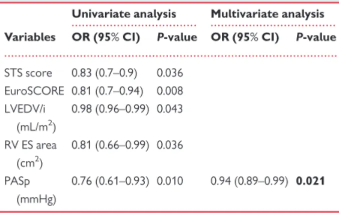
Table3compared patients with and without LVR. No significant dif-ferences in the MR aetiology and the number of clip implanted were found between the two groups. Non-significant MV stenosis and interatrial shunt was detected after the intervention in both groups. Univariable determinants of r-LVR were STS score (P = 0.036), EuroSCORE (P = 0.036), LVEDV/i (P = 0.043), RV end-systolic area (P = 0.043), and PASp (P = 0.010). On multivariable analysis, baseline PASp [P = 0.021; odds ratio 0.94, 95% confidence interval (CI) 0.89– 0.99] resulted to be the only independent predictor of r-LVR, as showed in Table6.
Reproducibility
Intra-observer agreement analysis showed an ICC of 0.981 (P < 0.001, 95% CI 0.92–0.996) for LVEF measurements, of 0.996
(P < 0.001, 95% CI 0.985–0.999) for LVEDV/i measurements, and of 0.998 (P < 0.001, 95% CI 0.992–0.999) for LVESV/i measurements.
Inter-observer agreement analysis showed an ICC of 0.938 (P < 0.001, 95% CI 0.75–0.996) for LVEF measurements, of 0.994 (P < 0.001, 95% CI 0.736–0.999) for LVEDV/i measurements, and of 0.997 (P < 0.001, 95% CI 0.986–0.999) for LVESV/i measurements (Table7).
Discussion
Secondary MR is a common finding in patients with heart failure with reduced ejection fraction and dilated LV, leading to progressive chamber dilatation, functional deterioration and increased mortality risk.13MitraClip is an effective procedure to reduce the cardiac over-load from severe MR. However, current criteria for subjects’ selec-tion are based on MV characteristic only, irrespective of LV and RV geometry and function.
This study showed that successful MitraClip procedure significantly reduced MR severity and improved functional NYHA class, in line with previously published data,14–17consistently with MR reduction. However, reverse remodelling occurred, at follow-up, only in 56% of patients with severe MR and low LVEF, with a parallel improvement in GLS. Lower pulmonary pressures, smaller LV volume and lower logistic risk scores were the main determinants of reverse LV remod-elling after MV repair. Previous studies showed significant benefits of MitraClip procedure in patients with preserved LVEF9,10and encour-aging data in terms of safety and feasibility in patients with reduced LV function.18Scandura et al.19observed a significant improvement in LVEF and a significant r-LVR in a population composed of both pri-mary and secondary MR. Rammos et al.20demonstrated both r-LVR and atrial remodelling with consequent improvement in GLS after MitraClip implantation in a series of patients with an average value of LVEF of 40.5 ± 2.5%. Pleger et al.15–18 also observed a significant r-LVR in patients with severely reduced LVEF.21In line with previous studies, this study confirms the good results in terms of MR reduction in the whole group of patients with low LVEF. However, percutan-eous MV repair is accompanied by reverse remodelling only in specif-ic subgroups of heart failure patients. As in our study, a signifspecif-icant
... ... ...
Table 6 Univariable and multivariable determinants of r-LVR
Univariate analysis Multivariate analysis
Variables OR (95% CI) P-value OR (95% CI) P-value
STS score 0.83 (0.7–0.9) 0.036 EuroSCORE 0.81 (0.7–0.94) 0.008 LVEDV/i (mL/m2) 0.98 (0.96–0.99) 0.043 RV ES area (cm2) 0.81 (0.66–0.99) 0.036 PASp (mmHg) 0.76 (0.61–0.93) 0.010 0.94 (0.89–0.99) 0.021
CI, confidence interval; ES, end-systolic; RV, right ventricle; PASp, pulmonary ar-tery systolic pressure; STS, Society of Thoracic Surgery Score.
Bold values represent statistical significance of P values.
...
Table 7 Intra- and inter-observer variability of echo-cardiographic measurements Variables Intra-observer agreement Inter-observer agreement LVEF (%) 0.981 (0.92–0.996), P < 0.001 0.938 (0.75–0.986), P < 0.001 LVEDV/i (mL/m2) 0.996 (0.985–0.999), P < 0.001 0.994 (0.736–0.999), P < 0.001 LVESV/i (mL/m2) 0.998 (0.992–0.999), P < 0.001 0.997 (0.986–0.999), P < 0.001
LVEDV/i, left ventricular end-diastolic volume index; LVEF, left ventricular ejec-tion fracejec-tion; LVESV/i, left ventricular end-systolic volume index.
..
..
..
..
..
..
..
..
..
..
..
..
..
..
..
..
..
..
..
..
..
..
..
..
..
..
.
2D/3D echocardiographic determinants of LV reverse remodelling
563
..
..
..
..
..
..
..
..
..
..
..
..
..
..
..
..
..
..
..
..
..
..
..
..
..
..
..
..
..
..
..
..
..
..
..
..
..
..
..
..
..
..
..
..
..
..
..
..
..
..
..
..
..
..
..
..
..
..
..
..
..
..
..
..
..
..
..
..
..
..
..
..
improvement in both 2D and 3D GLS after MitraClip implantation was recently demonstrated22only in patients with less RV impair-ment at baseline. We found that 44% of our subjects did not experi-ence a reverse remodelling at follow-up. These patients had higher risk scores, greater LV volume, more important RV impairment and increased pulmonary pressure at baseline. Most of them had progres-sion of LV dilatation over time. So, our data would be useful to iden-tify a subset of patients in which MitraClip intervention will not provide the significant expected benefits and will not change the nat-ural history of the heart failure progression and ultimately the prognosis.Limitations
Major limitation of the study is the short follow-up observation. The absence of events during follow-up precludes showing any associ-ation between the lack of reverse remodelling and outcome. It would be interesting to extend the follow-up to look at long-term outcome and possibly further cardiac changes. Another important limitation is the small sample size of the study that could mask other differences between groups.
Conclusions
Percutaneous MV repair using MitraClip system allows reduction in MR and NYHA. However, reverse remodelling occurs few months after the intervention only in patients with less severe baseline LV dilatation, lower pulmonary pressures, and lower logistic risk score. Our findings suggest that end-stage heart failure patients, presenting before the intervention with higher LV volumes and pulmonary pres-sure may not benefit from the procedure at long-term follow-up. Further studies with greater number of patients and longer follow-up are needed to confirm these data.
Conflict of interest: none declared.
References
1. Rossi A, Dini FL, Faggiano P, Agricola E, Cicoira M, Frattini S. Independent prog-nostic value of functional mitral regurgitation in patients with heart failure. A quantitative analysis of 1256 patients with ischaemic and non-ischaemic dilated cardiomyopathy. Heart 2011;97:1675–80.
2. Trichon BH, Felker GM, Shaw LK, Cabell CH, O’Connor CM. Relation of fre-quency and severity of mitral regurgitation to survival among patients with left ventricular systolic dysfunction and heart failure. Am J Cardiol 2003;91:538–43. 3. Baumgartner H, Falk V, Bax JJ, De Bonis M, Hamm C, Holm PJ et al.; ESC
Scientific Document Group. 2017 ESC/EACTS Guidelines for the management of valvular heart disease. Eur Heart J 2017;38:2739–91.
4. Song BG, On YK, Jeon ES, Kim DK, Lee SC, Park SW. Atrioventricular reverse remodeling after valve repair for chronic severe mitral regurgitation: 1-year fol-low-up. Clin Cardiol 2010;33:630–7.
5. Crestanello JA. Surgical approach to mitral regurgitation in chronic heart failure: when is it an option? Curr Heart Fail Rep 2012;9:40–50.
6. Feldman T, Wasserman HS, Herrmann HC, Gray W, Block PC, Whitlow P et al. Percutaneous mitral valve repair using the edge-to-edge technique: six month results of the EVEREST phase I clinical trial. J Am Coll Cardiol 2005;46:2134–40. 7. Tamburino C, Ussia GP, Maisano F, Capodanno D, La Canna G, Scandura S et al.
Percutaneous mitral valve repair with the MitraClip system: acute results from a real world setting. Eur Heart J 2010;31:1382–9.
8. Feldman T, Foster E, Glower DG, Kar S, Rinaldi MJ, Fail PS et al. EVEREST II Investigators. Percutaneous repair or surgery for mitral regurgitation. N Engl J Med 2011;364:1395–406.
9. Whitlow PL, Feldman T, Pedersen WR, Lim DS, Kipperman R, Smalling R et al. EVEREST II Investigators. Acute and 12-month results with catheter-based mitral valve leaflet repair: the EVEREST II (Endovascular Valve Edge-to-Edge Repair) High Risk Study. J Am Coll Cardiol 2012;59:130–9.
10. Feldman T, Kar S, Rinaldi M, Fail P, Hermiller J, Smalling R et al. EVEREST Investigators. Percutaneous mitral repair with the MitraClip system: safety and midterm durability in the initial EVEREST (Endovascular Valve Edge-to-Edge Repair Study) cohort. J Am Coll Cardiol 2009;54:686–94.
11. Lang RM, Badano LP, Mor-Avi V, Afilalo J, Armstrong A, Ernande L et al. Recommendations for cardiac chamber quantification by echocardiography in adults: an update from the American Society of Echocardiography and the European Association of Cardiovascular Imaging. Eur Heart J Cardiovasc Imag 2015;16:233–70.
12. Funaro S, La Torre G, Madonna M, Galiuto L, Scara` A, Labbadia A et al. Incidence, determinants, and prognostic value of reverse left ventricular remod-elling after primary percutaneous coronary intervention: results of the Acute Myocardial Infarction Contrast Imaging (AMICI) multicenter study. Eur Heart J 2009;30:566–75.
13. Patel JB, Borgeson DD, Barnes ME, Rihal CS, Daly RC, Redfield MM. Mitral regur-gitation in patients with advanced systolic heart failure. J Card Fail 2004;10: 285–91.
14. Franzen O, van der Heyden J, Baldus S, Schlu¨ter M, Schillinger W, Butter C et al. MitraClip therapy in patients with end-stage systolic heart failure. Eur J Heart Fail 2011;13:569–76.
15. Barth S, Hautmann MB, Kerber S, Gietzen F, Schade A, Deneke T et al. Hemodynamic improvement at three months follow-up after Mitraclip treatment in end-stage heart failure patients with functional mitral regurgitation. J Heart Valve Dis 2016;25:475–82.
16. Pleger ST, Chorianopoulos E, Krumsdorf U, Katus HA, Bekeredjian R. Percutaneous edge-to-edge repair of mitral valve regurgitation as a bail-out strat-egy in critically ill patients. J Invasive Cardiol 2013;25:69–72.
17. Pleger ST, Mereles D, Schulz-Scho¨nhagen M, Krumsdorf U, Chorianopoulos E, Rottbauer W et al. Acute safety and 30-day outcome after percutaneous edge-to-edge repair of mitral regurgitation in very high-risk patients. Am J Cardiol 2011; 108:1478–82.
18. Franzen O, Baldus S, Rudolph V, Meyer S, Knap M, Koschyk D et al. Acute out-comes of MitraClip therapy for mitral regurgitation in high-surgical-risk patients: emphasis on adverse valve morphology and severe left ventricular dysfunction. Eur Heart J 2010;31:1373–81.
19. Scandura S, Ussia GP, Capranzano P, Caggegi A, Sarkar K, Cammalleri V et al. Left cardiac chambers reverse remodeling after percutaneous mitral valve repair with the MitraClip system. J Am Soc Echocardiogr 2012;25: 1099–105.
20. Rammos C, Zeus T, Blazer J, Veulemans V, Hellhammer K, Niebel S et al. Left atrial and left ventricular function and remodeling following percutaneous mitral valve repair. J Heart Valve Dis 2016;25:309–19.
21. Pleger ST, Schulz-Scho¨nhagen M, Geis N, Mereles D, Chorianopoulos E, Antaredja M et al. One year clinical efficacy and reverse cardiac remodelling in patients with severe mitral regurgitation and reduced ejection fraction after MitraClip implantation. Eur J Heart Fail 2013;15:919–27.
22. Vitarelli A, Mangieri E, Capotosto L, Tanzilli G, D’Angeli I, Viceconte N et al. Assessment of biventricular function by three-dimensional speckle-tracking echo-cardiography in secondary mitral regurgitation after repair with the MitraClip system. J Am Soc Echocardiogr 2015;28:1070–82.