HAL Id: hal-03097549
https://hal.archives-ouvertes.fr/hal-03097549
Submitted on 5 Jan 2021HAL is a multi-disciplinary open access archive for the deposit and dissemination of sci-entific research documents, whether they are pub-lished or not. The documents may come from teaching and research institutions in France or abroad, or from public or private research centers.
L’archive ouverte pluridisciplinaire HAL, est destinée au dépôt et à la diffusion de documents scientifiques de niveau recherche, publiés ou non, émanant des établissements d’enseignement et de recherche français ou étrangers, des laboratoires publics ou privés.
Double hydrophilic block copolymers self-assemblies in
biomedical applications
Ayman El Jundi, Sytze Buwalda, Y. Bakkour, Xavier Garric, Benjamin
Nottelet
To cite this version:
Ayman El Jundi, Sytze Buwalda, Y. Bakkour, Xavier Garric, Benjamin Nottelet. Double hydrophilic block copolymers self-assemblies in biomedical applications. Advances in Colloid and Interface Science, Elsevier, 2020, 283, pp.102213. �10.1016/j.cis.2020.102213�. �hal-03097549�
Double hydrophilic block copolymers self-assemblies in
biomedical applications
Ayman El Jundi,1,2 Sytze J. Buwalda,1 Y. Bakkour,2 Xavier Garric,1 Benjamin Nottelet1*
1 IBMM, Univ Montpellier, CNRS, ENSCM, Montpellier, France
2 Laboratory of Applied Chemistry (LAC), Faculty of Science III, Lebanese University, P.O.
Box 826, Tripoli, Lebanon
∗ Corresponding author: benjamin.nottelet@umontpellier.fr
Abstract
Double-hydrophilic block copolymers (DHBCs), consisting of at least two different water-soluble blocks, are an alternative to the classical amphiphilic block copolymers and have gained increasing attention in the field of biomedical applications. Although the chemical nature of the two blocks can be diverse, most classical DHBCs consist of a bioeliminable non-ionic block to promote solubilization in water, like poly(ethylene glycol), and a second block that is more generally a pH-responsive block capable of interacting with another ionic polymer or substrate. This second block is generally non-degradable and the presence of side chain functional groups raises the question of its fate and toxicity, which is a limitation in the frame of biomedical applications. In this review, following a first part dedicated to recent examples of non-degradable DHBCs, we focus on the DHBCs that combine a biocompatible and bioeliminable non-ionic block with a degradable functional block including polysaccharides, polypeptides, polyesters and other miscellaneous polymers. Their use to design efficient drug delivery systems for various biomedical applications through stimuli-dependent self-assembly is discussed.
Keywords: Double-hydrophilic block copolymers; pH-responsive polymers; drug delivery systems; polyion complex micelles; degradable copolymers.
Abbreviations: AA, alginic acid; AEM; 2-aminoethyl methacrylate; Alb, albumin; APEG, acryloyl end-capped PEG; API, active pharmaceutical ingredient; ATRA, all-trans retinoic acid; ATRP, atom transfer radical polymerization; BIC, block ionomer complex; γCABεCL, -(carbamic acid benzylester)-ε-caprolactone; CAC, critical association concentration; CL, caprolactone; ClCL, chloro ε-caprolactone; CLSM, confocal laser scanning microscope; CMC, critical micelle concentration; CMD, carboxymethyldextran; COSamf, chitooligosaccharides with 5-anhydro-D-mannofuranose at their reducing end; CytC, cytochrome C; Dex, dextran; DG, diammonium glycyrrhizinate; DHBCs, double-hydrophilic block copolymers; DHCs, double hydrophilic copolymers; DIM, diminazene diaceturate; DL, drug loading; DLS, dynamic light scattering; DOTA, 1,4,7,10-Tetraazacyclododecane-1,4,7,10-tetraacetic acid; DOX, doxorubicin; DPcZn, dendrimer phthalocyanine zinc; DPP, diphenyl phosphate; DS, degree of substitution; DTT, dithiothreitol; EE, encapsulation efficiency; FA, folate ; FACS, flow cytometry analysis; Gd and Gd3+, gadolinium and gadolinium ion; GlcN, (1→4)-linked
units of 2-amino-2-deoxy-β-D-glucopyranose; HA, hyaluronic acid; LA, D,L-lactic acid; Lac-PEG , lactose-conjugated Lac-PEG; MA, methacrylic acid; MAL, L-malic acid; MH, minocycline
hydrochloride; mPEG, methoxy-poly(ethylene glycol); MRI, magnetic resonance imaging; MSA, mercaptosuccinic acid; MSC, mesenchymal stem cell; MTT, methotrexate; MTX, mitoxantrone; NAEP, 2-(N-acryloyloxy) ethylpyrrolidone; NMP, N-(2-methacryloylxyethyl) pyrrolidone; NPs, nanoparticles; NVPI, N-vinylphthalimide; PAA, poly(acrylic acid); PAE-API, poly(β-aminoester)-1-(3-aminopropyl)imidazole; PAEP, poly((2-(2-aminoethoxy)ethoxy) phosphazene); PAMPS, poly(2-acrylamido-2-methyl-1-propanesulfonic acid); PAMPSNa, poly(sodium 2-acrylamido-2-methylpropanesulfonate); PAPTAC, poly(3- acrylamidopropyltrimethylammonium chloride; PAsp, poly(L-aspartic acid); PAsp(DET), poly(L-aspartic acid) bearing a N-(2-aminoethyl)-2-aminoethyl group; PATMC, poly(5-allyloxytrimethylethylenecarbonate); PBYPCOOH, poly(phosphotriester)s bearing pendent carboxylic acids; PCCL, poly(6-acetoxyl-ε-caprolactone); PCEtOx, poly(2-carboxyethyl-2-oxazoline); PCL, poly(ε -caprolactone); P(CL-co-DCL), poly(ε-caprolactone-co-γ-dimethyl maleamidic acid-ε-caprolactone); PDEAEMA (poly(2-(diethylamino)ethyl methacrylate); PDMAEMA, poly(2-(dimethylamino)ethyl methacrylate); PDT, photodynamic therapy; PEC, polyelectrolyte complexes; PEG, poly(ethylene glycol); PEGMA, polyethylene glycol methacrylate; PEI, poly(ethyleneimine); PEO, poly(ethylene oxide); PEtOx, poly(2-ethyl-2-oxazoline); PGlu, poly(L-glutamic acid); PIC, polyion complex; PiPrOx,
poly(2-isopropyl-2-oxazoline); PLL, polylysine; PMA, poly(methacrylic acid); PMMA, poly(methyl methacrylate); P2MVP, methyl-2-vinyl pyridinium iodide); PNiPAM, poly(N-isopropylacrylamide); PPDO-, poly(phosphodiester)s with a negatively charged oxygen atom; PPIL, poly(4-N-piperilactone); PPLG, poly(γ-propargyl-L-glutamate); PSar, poly(sarcosine); PSMA, poly(styrene-alt-maleic anhydride); Pul, pullulan; PVA, poly(vinyl alcohol); PVAc, poly(vinyl acetate); PVAm, Poly(vinyl amine); PVCL, Poly(N-vinylcaprolactam); PVP, Poly(vinyl pyrrolidone); RAFT, reversible addition-fragmentation chain transfer; Rh, hydrodynamic radius; ROP, ring opening polymerization; ROS, reactive oxygen species; sCT, salmon calcitonin; siRNA, small interfering ribonucleic acid; SLS, static light scattering; TEM, transmission electron microscopy; TMC, trimethyl chitosan.
Contents
1. Introduction ... 3
2. Non-degradable DHBCs ... 6
3. Polysaccharide-based degradable DHBCs ... 16
4. Polypeptide-based degradable DHBCs ... 23
5. Polyester-based degradable DHBCs and others miscellaneous synthetic blocks ... 32
6. DHBCs for biomedical applications: unique advantages, current challenges and future perspectives ... 44
7. Concluding remarks ... 48
Declaration of Competing Interest ... 48
Acknowledgements ... 49
References: ... 58
1. Introduction
Block copolymers are a category of polymers in which the macromolecule consists of two or more distinct blocks of homopolymer. Block copolymers with two, three, and more blocks are called diblock, triblock, and multiblock copolymers respectively. Block copolymers can be further classified according to their architectural arrangement. For example, linear or star shaped block copolymers can be synthesized. The number of polymer types in block
copolymers may be equal to or lower than the number of blocks. For example, triblock copolymers may be constituted of three polymers (ABC triblock copolymers) or two polymers (ABA triblock copolymers).
The nature of the constituting blocks (hydrophilic or hydrophobic) gives rise to a further categorization within block copolymers. Amphiphilic block copolymers, which consist of at least one hydrophilic and one hydrophobic block, may self-assemble in water to form micelles or aggregates with a hydrophobic core surrounded by a hydrophilic shell. Micelles prepared from amphiphilic block copolymers have been studied primarily as controlled drug delivery systems as their hydrophobic core may serve as a depot for the solubilization of clinically relevant doses of hydrophobic drugs, as reviewed in several excellent publications [1–3]. In the last two decades, double-hydrophilic block copolymers (DHBCs), consisting of two or more water-soluble blocks of different chemical nature, have gained increasing attention. In many DHBCs one block (often based on poly(ethylene glycol) (PEG) or poly(ethylene oxide) (PEO)) only promotes solubilization in water, whereas the other block is responsive to an external stimulus or capable of interacting with another polymer or substrate. In aqueous solution under normal conditions, the two hydrophilic blocks are well solvated and DHBCs behave like water-soluble polymers with no amphiphilic characteristics. However, a variation of temperature, ionic strength or pH, as well as a complexation reaction may change the hydrophilicity of one of the homopolymer blocks into hydrophobicity, thereby introducing amphiphilicity, which in turn may lead to the formation of functional structures such as micelles.
Although the first synthesis of a DHBC dates back to 1972 [4], only during the last two decades the potential of DHBCs has been fully recognized for various applications. These include for example crystal growth modification, metal oxide particle stabilization, nanoparticle fabrication, controlled drug delivery and gene transfection (Scheme 1).
Scheme 1. Different type of double hydrophilic block copolymers and their applications.
Despite the increasing interest for this unique class of polymers, recent reviews dedicated to DHBCs are scarce. A comprehensive review concerning DHBCs dates back as far as 2001 [5]. Since then, a limited number of reviews have been published relating to the self-assembly processes of DHBCs, including recent examples of polyion complex (PIC) [6] and block ionomer complex (BIC) micelles [7] (Scheme 2), as well as a recent review dedicated to the self-assembly of completely water-soluble DHBCs in aqueous medium without application of external stimuli but at high concentration [8].
Scheme 2. Formation of Polyionic Complexes (PIC) (also known as Polyelectrolyte Complexes
The present review differs from these previous contributions as we aim at reporting and summarizing recent studies dedicated to the synthesis and the study of the behaviour of DHBCs in solution for biomedical applications. After discussing some recent examples of non-degradable DHBCs, and because biodegradability is in our view of first importance for biomedical polymers, we will subsequently categorize the various degradable DHBCs according to the chemical nature of the responsive or interacting polymer block of the DHBC. We will successively discuss DHBCs which contain polysaccharides, polypeptides, polyesters and other miscellaneous degradable blocks as functional blocks (Table 1). All these polymers are considered biodegradable and bioresorbable as they degrade to non-toxic products in vivo which can be excreted by the kidneys. Despite the existence of other alternatives (poly(2-oxazoline), poly(vinyl pyrrolidone) etc…), they are mostly combined with PEG as a solubilizing block due to its long track record in pharmaceutical formulation, its excretability via the renal pathway up to a molecular weight (MW) of approximately 30 kg/mol [9], and its medium to long-term biodegradability [10,11]. Finally, in a last part we will discuss the main biomedical applications taking advantage of these DHBCs and we will emphasize the remaining challenges and future perspectives associated with this class of copolymers.
2. Non-degradable DHBCs
Many of the non-degradable DHBCs reported in literature are based on a PEG block associated with a poly(acrylic acid) (PAA) or a poly(methacrylic acid) (PMA) block for the anionic ones, or poly(dimethylaminoethyl methacrylate) (PDMAEMA), and poly(ethylenimine) (PEI) for the cationic ones (Table 1). This type of copolymer offers the advantages of a straightforward synthesis as well as pH-responsiveness. Apart from PEG, also poly(vinyl pyrrolidone) (PVP), poly(vinyl caprolactam) (PVCL), poly(vinyl alcohol) (PVOH), poly(vinyl amine) (PVAm) and poly(oxazoline) (POx) have been used as water-soluble block in combination with different
responsive blocks. In the following paragraph examples of such non-degradable DHBCS proposed in various recent biomedical applications are discussed. A summary of the type of self-assemblies obtained with these DHBCs and their characteristics is provided in Table 2. Shin et al. reported on giant polymer vesicles (diameter 5-8 µm) which formed via self-assembly of double-hydrophilic PEG-PAA diblock copolymers at pH 2. This was attributed to protonation of the carboxylic groups on the PAA block (pKa of 4.5), thereby providing hydrogen bonding with itself and the PEG block resulting in aggregation of the polymer chains, thus providing a mechanism for phase segregation of the complex of otherwise soluble polymers. In contrast, no vesicle formation was observed at pH 9. To demonstrate that the polymer vesicles enable the triggered delivery of cargo molecules, Alexa Fluor 488 was incorporated by rehydrating the polymer film with a solution of the hydrophilic fluorophore at pH 2. Upon increase of the pH above 4.5, the encapsulated fluorophore was rapidly and completely released within 30 min due to dissociation of the vesicles [12].
Frangville et al. reported on nanoparticles of PEG-PAA crosslinked by gadolinium (Gd3+) ions and their use as magnetic resonance imaging (MRI) contrast agents. The formation of the hybrid inorganic/polymeric nanoparticles (20 nm in diameter) was ascribed to association between Gd3+ ions and negatively charged acrylic acid monomer units in the core, which is surrounded by a PEG corona. The integrity of the NPs was maintained even after significant dilution and over a large range of pH and ionic strength values. This was explained by the entanglement of PAA blocks, resulting in additional crosslinks that stabilize the nanoparticle. In vivo experiments showed that the Gd3+ / PEG-PAA nanoparticles provide both a higher intensity and a more persistent enhancement of vascular MRI signals, at one-third of the total Gd concentration of the commercially available contrast agent GdDOTA [13].
Raisin et al. reported the design of tripartite PIC micelles as original non-viral polymeric vectors suited for mesenchymal stem cell transfection with small interfering ribonucleic acid
(siRNA) using the non-degradable DHBC PEO-b-PMA. The formation of PIC micelles is based on the association of the DHBC with a cationic homopolymer (polylysine (PLL) or poly(ethyleneimine) (PEI)), and the siRNA can be associated with the core of micelles by complexation with the cationic polymer. The micelle formulations were designed to exhibit pH-triggered disassembly in an acidic pH range found in endosomes. The tripartite micelles were non-cytotoxic and exhibited an ability to deliver a siRNA targeting Runx2 and to induce efficient gene silencing in murine MSC [14].
In another example, Ramasamy et al. investigated the interaction of the cationic drugs doxorubicin (DOX) and mitoxantrone (MTX) with the anionic DHBC PEO-b-PAA. The primary amino group of DOX and two secondary amino groups of MTX were responsible for electrostatic interactions with the ionized carboxyl group (pKa ∼ 5) of PEO-b-PAA. The drug loading (DL) was 45 wt% for MTX and 70 wt% for DOX. A faster release of the two drugs was observed at pH 5 (typical of the environment of cancer cells) compared to the release at physiological pH (7.4). DOX PIC micelles showed a higher cellular uptake than MTX PIC micelles. Both complexes induced apoptosis of cancer cells and suppressed tumour growth to the same level as the respective free drugs, but they exhibited a prolonged blood circulation [15].
Poly(vinyl pyrrolidone) (PVP) is another example of non-ionic water-soluble block that has been used to prepare non-degradable DHBCs. PVP is largely used in pharmaceutical formulations due to its cytocompatibility and strong binding capacity via dipole interactions with drugs [16]. The next paragraph gathers the few recent examples of PVP analogues used to prepare DHBCs with a ionisable block. Gao et al. reported on a series of PVP-based DHBCs prepared by RAFT polymerization for drug delivery applications through the formation of PIC micelles with different drugs including coenzyme A and folic acid. This series of DHBCs embedded PVP-b-poly(styrene-alt-maleic anhydride) (PVP-b-PSMA),
PVP-b-poly(2-(dimethylamino)ethyl methacrylate) (PVP-b-PDMAEMA) and b-poly(2-acrylamido-2-methyl-1-propanesulfonic acid) (b-PAMPS) [17–19]. Another example consists of PVP-b-PMA also obtained by RAFT polymerization and used to control calcium carbonate (CaCO3)
morphologies with target applications in biomimetic mineralization [20]. More recently, Destarac et al. reported on another series of PVP-based DHBCs obtained by redox initiated aqueous RAFT/MADIX polymerization. This approach resulted in the synthesis of PVP-b-PAA, PVP-b-poly(sodium 2-acrylamido-2-methylpropanesulfonate) (PVP-b-PAMPSNa), and PVP-b-poly(3-acrylamidopropyltrimethylammonium chloride) (PVP-b-PAPTAC) foreseen for use in drug delivery and biological inorganic phase templating [21]. Another recent example of potential interest for the design of drug vectors includes a vinyl pyrrolidone analogue, namely 2-(N-acryloyloxy)ethylpyrrolidone (NAEP). NAEP was copolymerized by reversible addition-fragmentation chain transfer (RAFT) polymerization with 2-(diethylamino)ethyl methacrylate (DEAEMA) to yield PNAEP-b-PDEAEMA DHBCs that can lead to the formation of pH-responsive micelles [22]. In the same family of analogue, N-(2-methacryloylxyethyl) pyrrolidone (NMP) was copolymerized by RAFT polymerization with MA to prepare a family of well-controlled DHBCs able to stabilize and control the size of gold nanoparticles, that are largely used in biomedical applications [23], in a pH-dependent manner [24].
Poly(N-vinylcaprolactam) (PVCL) is also used for biomedical applications as a non-ionic water-soluble polymer. It is for example present in the BASF excipient Soluplus® that is proposed to formulate insoluble drugs. As such recent examples of PVCL-based DHBCs have been reported. Liang et al. prepared well-defined DHBCs of PVCL and PVP via RAFT polymerization [25]. PVCL is a thermoresponsive polymer with superior biocompatibility in comparison with PNiPAM [26]. The LCST could be lowered by increasing the PVCL segment length and raised by increasing the hydrophilic PVP segment length. Below the LCST the copolymers dissolved completely in aqueous solution, whereas they formed spherical micellar
or vesicular morphologies above the LCST. The size of the self-assembled structures could be controlled via the molar ratio of the PVCL and PVP segments. The PVCL-b-PVP copolymers showed non-toxic towards cancer cells. The group of Jerome reported on the cobalt-mediated radical polymerization of VCL initiated from a poly(vinyl acetate) (PVAc) based macroinitiator to yield PVAc-b-PVCL block copolymers [27]. These amphiphilic copolymers were subsequently hydrolysed to give poly(vinyl alcohol)-b-PVCL (PVA-b-PVCL) DHBCs. These polymers became amphiphilic when heated above their LCST in aqueous solution (36-42 °C, depending on their composition) due to dehydration of the PVCL sequence, leading to self-assembled spherical aggregates with a collapsed PVCL core and a soluble PVOH shell. In a follow-up paper the authors used the PVA-b-PVCL DHBCs to prepare nanogels by crosslinking the PVA corona above the LCST with a redox-responsive crosslinking agent [28]. The nanogels loaded with the hydrophobic model drug Nile red(NR) had sizes ranging from 50 to 100 nm and were able to release NR in the presence of the reducing agent dithiothreitol (DTT) thanks to cleavage of the disulphide bonds in the crosslinker (Figure 1a and 1b). In addition, the nanogels proved to be cytocompatible towards L929 fibroblasts and to be efficiently uptaken by MEL-5 cancer cells (Figure 1c), which show the potential of the PVA-b-PVCL nanogel system for intracellular drug release.
Figure 1. (a) Schematic illustration of the preparation, dissociation of PVA-b-PVCL
crosslinked nanogels, loading of Nile red (NR) and redox-triggered release behaviors (LCST: lower critical solution temperature, VPTT: volume phase transition temperature) ; (b) TEM images of the crosslinked nanogels (scale bar 200 nm) ; (c) Fluorescence microscopy images of the treated MEL-5 cells after 24 h incubation with the NR-loaded nanogel : (1) nuclei stained with DAPI (blue), (2) fluorescence pattern of NR (red), (3) contrast field pattern, (4) merged images of (1), (2) and (3) (scale bar: 100 mm) (adapted with permission from [28]).
This last example illustrates the use of PVCL in DHBCs, but also of PVA that is another example of non-ionic water-soluble polymer. Cobalt-mediated radical polymerization was also used for the preparation of PVA-b-PVP DHBCs [29] as well as pH-responsive PVA-b-PAA DHBCs [30]. The latter self-assembled into aggregates below pH 3 due to the protonation of the PAA carboxylic acid groups, which decreases PAA solubility and enables hydrogen bonding between PAA and PVA blocks. Moreover, PVA-b-PAA formed PIC micelles with poly(N-methyl-2-vinyl pyridinium iodide)-b-poly(ethylene oxide) (P2MVP-b-PEO) DHBCs at pH 8 through electrostatic interactions between the core-forming P2MVP and PAA blocks [31]. In the same family of vinyl polymers, Maki et al. prepared DHBCs having poly(vinyl amine)
(PVAm) blocks via sequential RAFT polymerization of N-vinylphthalimide (NVPI) and NiPAM, followed by deprotection of the PNVPI block [32]. Aqueous solutions of amphiphilic PNVPI-b-PNiPAM block copolymers and PVAm-b-PNiPAM DHBCs exhibited a LCST type phase transition between 25-32 °C and 53-75 °C, respectively, in accordance with the higher hydrophilicity of the PVAm-b-PNiPAM DHBCs. Although micelle formation was hypothesized for the block copolymers above the LCST, no characterization of these micelles was provided.
Poly(2-oxazoline)s (POx) are an important class of polymers that focuses a strong interest as they have been proposed as alternative to PEG in biomedical applications thanks to their tunable physicochemical properties, good water solubility and excellent biocompatibility [33]. As debates regarding their degradability are still ongoing [34], we chose in this review to present most POx-based DHBCs in this part dedicated to non-degradable DHBCs. Examples of POx DHBCs containing a second block of well-recognized degradability can be found in the corresponding subparts. Zschoche et al. prepared a series of di- and triblock copolymers with temperature-sensitive isopropyl-2-oxazoline) (PiPrOx) blocks and pH-sensitive poly(2-carboxyethyl-2-oxazoline) (PCEtOx) blocks [35]. The temperature- and pH-induced reversible phase transition converted these DHBCs into amphiphilic polymers allowing for self-assembly in aqueous solution. The temperature and pH values of the transitions as well as the nature of the formed aggregates (micelles or vesicles) could be controlled via the size and arrangement of the individual blocks. Other examples of POx-containing DHBCs include PEG-b-PEtOx star block copolymers [36], PEG-b-poly(2-methyl-2-oxazoline) diblock copolymers [37] and PEtOx-b-PVP diblock copolymers [38]. In these cases, no application was foreseen and studies aimed at studying the self-aggregation of these DHBCs that was observed without the
application of an external stimulus, suggesting that even in DHBCs subtle differences in hydrophilicity among the constituting blocks are sufficient to drive polymer aggregation.
As shown in this part, polyethers (PEG, PEO), vinyl polymers (PVP, PVCL, PVA…) and poly(2-oxazoline)s (PEtOx) are the non-ionic blocks classically used in non-degradable DHBCs. More exotic blocks like polydehydroalanine to yield PEG- or poly(acrylic acid)-b-polydehydroalanine DHBCs [39,40], synthetic glycopolymers in PEG-b-poly(mannose) [41], or poly((4-diethylamino)-(E)-stilbene)-alt-maleic acid)-b-poly(acryloyl morpholine) have been reported [42]. However, they remain largely unexploited in comparison to these classical families. In addition, and although the DHBCs discussed in this part represent promising systems that can potentially address several challenges in the biomedical domain, their non-degradability remains an issue as the polymers may accumulate in the body or, if elimination is possible, in the environment [43]. Therefore, in the remainder of this review we focus on degradable DHBCs containing polysaccharides, polypeptides or polyesters as functional blocks associated with biocompatible non-ionic and bioeliminable blocks, mainly PEG and PEtOx, whereas other non-ionic blocks raising concerns of potential toxicity, like PNiPAM, are not considered.
Table 2: Characteristics of the self-assemblies obtained with non-degradable double hydrophilic block copolymers.
DHBC Self-assemblies Stability
a Ref.
Type Preparation Characteristicsa
PEO-b-PAA Giant vesicles
gel-assisted rehydration
method at pH 2.3 Dh ̴ 5-8.0 µm / ξ potential ̴ -11
mV (at pH 2.4) ass. < pH 4.5 < disass. [12]
PEO-b-PAA Gd 3+-loaded PIC micelles addition of Gd3+ solution to PEO-b-PAA 0.1 wt% solution (R = 1) Dh ̴ 33 nm / ξ potential ̴ 0 mV disass. < pH 4. stable for DHBC concentrations of 0.1-10-4 wt% stable at 1M NaCl. [13]
PEO-b-PMA SiRNA-loaded PIC
micelles
(PLL/SiRNA) solution added to PEO-b-PMA solution (R = 1)
Dh ̴ 37-53 nm / ξ potential ̴ 3.5
to 7.9 mV (in water) and Dh ̴
101 nm (in PBS) / EE ̴ 90% disass. < pH 6. [14] PEO-b-PAA DOX- or MTX-loaded PIC micelles
mixing of drug and polymer aqueous solutions for 24h (D/P =0.25 or 0.5) Dh ̴ 100 nm / ξ potential ̴ 5 to -35 mV (at pH 7) / EE ̴ 90% / DL ̴ 45% (MTX) ̴ 70% drug release at pH 5. [15] PVP-b-PSMA & PVP-b-PDMAEMA Co A-loaded PIC micelles mixing of PVP-b-PSMA and PVP-b-PDMAEMA / Co A solutions, pH neutralization, dialysis Dh ̴ 100-130 nm / EE ̴ 60% / DL ̴ 10-29% drug release at pH 2 < pH 9 < pH 7.4. [17] PVP-b-PAMPS & PVP-b-PDMAEMA FA-loaded PIC micelles mixing of PAMPS/FA and PVP-b-PDMAEMA solutions, pH neutralization, dialysis Dh ̴ 170 nm / EE ̴ 85% / DL ̴ 21% drug release at pH 9 ≥ pH 7.4. [19] PVP-b-PMA templating of CaCO3 injection of Na2CO3 in PVP-b-PMA solution, pH adjustment at 10, addition of CaCl2 rhombohedral or mutilayered
PNAEP-b-PDMAEMA pH-responsive micelles direct dissolution at acidic pH followed by self-assembly upon pH increase Dh ̴ 50-100 nm disass. ≤ pH 3 ; ass. ≥ pH 10. [22]
PNMP-b-PMA micelles for Au NP templating direct dissolution in water Dh ̴ 100 nm (pH 4) Dh ̴ 200 nm (pH 2) ass. < pH 5.3. micellization temperatures ̴ 50-70°C. [24] PVCL-b-PVP thermo-responsive micelles direct dissolution in water Dh ̴ 100-260 nm ass. T > 42-46°C. [25] PVA-b-PVCL thermo-responsive and reducible nanogels direct dissolution in water and crosslinking with DPA
Dh ̴ 280-460 nm (DLS)
Dh ̴ 45-110 nm (TEM)
ass. T > 36-42°C.
nanogels stable upon dilution disass. with 10 mM DTT after 24h.
[28]
PVA-b-PAA
pH-responsive micelles and PIC micelles
direct dissolution in water (micelles), mixing with P2MVP-b-PEO aqueous solutions (PIC micelles)
Dh ̴ 200 nm (micelles)
Dh ̴ 30-40 nm (PIC micelles) ass. pH < 3 (micelles).
[30,3 1] PCEtOx-b-PiPrOx pH- & thermo-responsive micelles & vesicles direct dissolution in water (0.5 g/L) Dh ̴ 50-230 nm ass. 3.5 ≤ pH ≤ 5.2 at 60°C. ass. T ≥ 60°C. [35] PAA-b-PDHA loose nanoaggregates direct dissolution in water Dh ̴ 220 nm / ξ potential ̴ -5 mV (pH < 4) ass. pH < 4. [39]
Abbreviations : ass. assembly ; Co A coenzyme A ; disass. disassembly ; DOX doxorubicin ; DPA 3,3’-Dithiodipropionic acid ; DTT dithiothreitol ; FA folic acid ;MTX mitoxantrone ; PAA poly(acrylic acid) ; PAMPS poly(2-acrylamido-2-methyl-1-propanesulfonic acid) ; PAPTAC poly(3- acrylamidopropyltrimethylammonium chloride) ; PDHA poly(dehydroalanine) ; PDEAEMA (poly(2-(diethylamino)ethyl methacrylate) ; PDMAEMA poly(2-(dimethylamino)ethyl methacrylate) ; PEO poly(ethylene oxide); (PEO)s8 8 arms star-shaped poly(ethylene oxide) ; PMA poly(methacrylic acid) ; P2MVP poly(N-methyl-2-vinyl pyridinium iodide) ; PNAEP
poly(2-(N-acryloyloxy)ethylpyrrolidone); PNMP poly(N-(2-methacryloylxyethyl) pyrrolidone) ; PSMA poly(styrene-alt-maleic anhydride) ; PVP poly(vinyl pyrrolidone) ; R charge ratio between positive and negative charges.
a ranges of provided values correspond to characteristics obtained as a function of the blocks’ length of the used DHBCs and/or ratios of compounds. If not specified Dh
3. Polysaccharide-based degradable DHBCs
Polysaccharides are a broad class of naturally derived polymers (i.e. obtained from plants, animals or algae) that consist of monosaccharide units bound together by glycosidic linkages [44]. Polysaccharides display a linear or branched architecture and contain various functional groups such as carboxylic acid, amino and hydroxyl groups. These moieties are responsible for the hydrophilicity of many polysaccharides and offer numerous opportunities for chemical derivatization [45]. The molecular weight of polysaccharides may vary significantly (between hundreds and millions of Daltons), which further adds to the diversity of this polymer class [46]. In addition, owing to their native presence within the body, most polysaccharides have a very low toxicity and demonstrate good biocompatibility [47–50], while being enzymatically degradable down to their monomer or oligomer building blocks [51]. This unique combination of features explains their use as micellar systems for drug delivery which has been extensively reviewed [52,53]. Noteworthy, due to the chemical structure of polysaccharides most of their double hydrophilic copolymers (DHCs) have graft topologies rather than block topologies. Recent examples of such DHCs include chitosan-g-PEG [54–56], dextran grafted with poly((polyethylene glycol) methacrylate-co-aminoethyl methacrylate) [57] and alginic acid grafted with mPEG [58] that were all used to formulate drug loaded micelles. However, such graft copolymers, despite being double hydrophilic structures that offer interesting self-assembly behaviors are beyond the scope of this review that focuses on DHBCs and will not be further discussed. Examples of polysaccharide-based DHBCs where polysaccharides are used as functional and stimuli-responsive blocks (Table 1). A summary of the type of self-assemblies obtained with these copolymers and their characteristics is provided in Table 3.
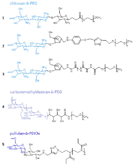
Chitosan is a polysaccharide that has been widely used in DHCs but more scarcely in DHBCs. Although chitosan is insoluble in water at physiological pH and other common solvents because
of its strong intra-molecular hydrogen bonding, its copolymerization with PEG or other hydrophilic polymers is known to disrupt the intra-molecular hydrogen bonding of chitosan, thus allowing its water solubilization. For this reason, and despite the fact that chitosan cannot be considered as a well solvated block on its own, the following structures can be considered as double hydrophilic structures. Ganji et al. reported on the synthesis of a chitosan-PEG DHBC (Figure 2 1) to be used as a thermosensitive gel. In a first step, the glycosidic bonds of the chitosan main chain were degraded with potassium persulfate to produce oligo-chitosan with a terminal carbonyl group at one scission end and a free radical at the other scission end. Then, the chain end with the free radical was reacted with acryloyl end-capped PEG (APEG, Mw= 2.103 g.mol-1) giving rise to a diblock-like copolymer structure. The molar ratio of PEG/chitosan
within the copolymer could be varied between 0.06 and 0.1 depending on the potassium persulfate concentration used to degrade chitosan. However, the MW of the chitosan segment was not really controlled, and it was also not clearly stated whether one or several acryloyl PEG chains can grow from the end radical which may lead to b-PEG or chitosan-b-P(APEG). A gelation time of ca. 10 min was observed at body temperature for concentrations as low as 2 w/v %., which confirmed that these chitosan-PEG DHBC could be used for biomedical application [59].
To yield similar DHBCs, Moussa et al. used fully N-deacetylated chitooligosaccharides with a 5-anhydro-D-mannofuranose at their reducing end (COSamf) to prepare various COS-based building blocks. COSamf with an average number of 22 repeating units of (1→4)-linked units of 2-amino-2-deoxy-β-D-glucopyranose (GlcN) were functionalized via reductive amination. In particular 4-(propargyloxy)aniline and adipic dihydrazide were selected to prepare alkyne or hydrazide functional COSamf and prepare 2 different COS-b-PEG diblock copolymers based on chain ends reactions. The first method used copper(I)-catalyzed azide alkyne cycloaddition (CuAAC) reaction between the alkyne-terminated COS block and a commercial mPEG-azide
(Mn=2000g/mol) to yield COS-b-PEG with Mn=6520 g/mol and Đ=1.26 (Figure 2 2). To get rid of the copper catalyst that may contaminate this first DHBC, the second strategy was based on the hydrazide condensation reaction between the hydrazide terminated COS block and a commercial mPEG-NHS ester (Mn=2000g/mol) to yield COS-b-PEG with Mn=6330 g/mol and Đ=1.15 (Figure 2 3). These COS-b-PEG DHBCs are foreseen to serve as cationic nanocarriers for the delivery of drugs [60].
Figure 2. Examples of degradable hydrophilic block copolymers based on polysaccharides.
Dextran is another example of a polysaccharide found in DHBCs. Winnik et al. synthesized various block copolymers of dextran (Mw = 8.3k or 14.7k) and PEG-NH2 (Mw = 3k or 7k) via
the specific oxidation of the dextran terminal aldehyde group and the covalent linkage of PEG-NH2 via a lactone aminolysis reaction. Conversion of the neutral diblock copolymers into
polyanions was achieved by carboxymethylation of the dextran block via side chain modification of dextran with chloroacetic acid, leading to carboxymethyldextran-PEG (CMD-b-PEG) block copolymers (Figure 2 4). The properties of CMD-b-PEG in aqueous solutions were analyzed by static and dynamic light scattering (DLS) showing a pH sensitive assembly [61]. In follow up studies, the authors evaluated the ability of (CMD)-b-PEG to encapsulate different drugs. The micellization was evaluated as a function of i) the ionic charge density or degree of substitution (DS) of the dextran block with carboxymethyl moieties and ii) the molar ratio of positive charges provided by the drug to negative charges provided by CMD-b-PEG. PIC micelles were formed with the cationic and water soluble diminazene diaceturate (DIM), an API used as antiparasitic agent, and CMD-b-PEG with various DS. Micelles with a charge ratio positive/negative = 2 had a DL ranging from 40 to 65 wt% with a hydrodynamic radius (Rh) ranging from 36 to 50 nm, depending on the MW and the DS of the CMD-b-PEG. The critical association concentration (CAC) was in the order of 15–50 mg/L for DIM/CMD-b-PEG with DS > 60 %, and 100 mg/L for DIM/CMD-b-PEG with DS∼30 %. Finally, PIC micelles with high DS and charge ratio = 2 allowed for a prolonged release of DIM in vitro compared with a solution of free drug [62]. In another application by the same group, the CMD-b-PEG was used to form PIC micelles with minocycline hydrochloride (MH), a semisynthetic tetracycline antibiotic with promising neuroprotective properties for the treatment of neuroinflammatory diseases. PIC micelles with Rh of 100 nm and 50 wt% loading of MH were obtained. The MH loaded PIC micelles showed a sustained release of drug from the micelle at physiological pH, thereby allowing to decrease inflammation in the murine microglia (N9) [63]. Lastly, CMD-b-PEG copolymers hydrophobized by n-dodecyl groups were used to encapsulate two aminoglycosides: paromomycin and neomycin at DL up to 50 wt%. PIC micelles were stable under physiological conditions (pH 7.4, 150 mM NaCl) in contrast with micelles formed by the unmodified CMD-b-PEG and exhibited reduced sizes (around 50 nm) compared to the
non-hydrophobized CMD-b-PEG (sizes in the range 75 to 100 nm). The minimal inhibitory concentration of the aminoglycosides encapsulated in PIC micelles was not altered as indicated by their ability to kill E.Coli in culture [64].
Brosnan et al. synthesized dextran and pullulan based DHBCs, namely dextran-b-poly (ethylene oxide) (Dex-b-PEO), pullulan-b-PEO (Pul-b-PEO), and dextran-b-poly(sarcosine) (Dex-b-PSar). The synthesis route involved reaction of one polysaccharide terminus, existing as an aldehyde group in the equilibrium state, with a hydroxy amine end group of mono-functionalized PEO or PSar, yielding a hydrolytically stable oxime bond between both polymer blocks. Each block had a MW of ca. 20 k yielding DHBCs with MW of ca. 40 k. A direct dissolution at low concentrations (0.1, 0.5, and 1.0 wt%) led after 7 days to the formation of self-assembled polymer vesicles with Rh in the range of 250 nm to 700 nm for the lowest and the highest concentrations, respectively. At higher concentrations, above 10 wt%, self-assembly yielded giant polymer vesicles, referred as “aquanelles” with sizes between 2 and 20 μm (Figure 3). These aquanelles’ solutions were stable over 7 days and due to their water permeability, the authors envisioned that they could be well suited for use as artificial cells [65].
Figure 3. Optical microscopy images of the giant double hydrophilic polymer vesicles referred
to as “aquanelles” prepared from Dex-b-PEO, Pul-b-PEO and Dex-b-PSar at 25 wt% (adapted with permission from [65]).
The self-assembly of pullulan-b-poly(2-ethyl-2-oxazoline) (Pull-b-PEtOx) was studied by
Willersin et al. The DHBC (Figure 2 5) was synthesized from a pullulan-alkyne (8–38 kg mol
-1) and a biocompatible azido-PEtOx (22 kg mol-1) by a CuAAC conjugation. The MW of the
Pull block was varied to study its impact on the self-assembly behavior. Sizes between 300 and 500 nm were measured by DLS and static light scattering (SLS) in dilute aqueous solution (0.1– 1.0 wt%) with an optimum ratio of 0.4/0.6 (Pull/PEtOx) for the assembly of copolymers in water. Larger particulate structures with sizes around 1 to 2 µm were observed by optical
Table 3: Characteristics of the self-assemblies obtained with polysaccharide-based degradable double hydrophilic block copolymers.
DHBC Self-assemblies Stability / Degradationa Ref.
Type Preparation Characteristicsa
PEG-b-chitosan Thermosensitive gel
dissolution of DHBC in PBS at 2 or 3 wt%
gelation time 6-11 min
sol < 35-40°C < gel. [59] PEG-b- carboxymethyldextran DIM-loaded PIC micelles MH-loaded PIC micelles DIM : mixing of DHBC and DIM Tris buffer solutions (pH 5.3, R = 2),
MH : mixing of DHBC and MH/CaCl2
Tris-buffer solutions (pH 7.4, R = 1) DIM : CAC 0.014-0.095 g/L / Dh ̴ 70100 nm / ξ potential ̴ -3.4 mV / DL ̴ 40-65% MH : Dh ̴ 200 nm (MH) / DL ̴ 50% dissa. ≥ 0.2 M NaCl. stable from pH 4 to 1.1
25% size increase over 2- month storage in Tris (pH 5.3).
stability over 1-month storage in Tris (pH 7.4). [61– 63] PEG-b-(n-dodecyl)-carboxymethyldextran PAR-loaded PIC micelles NEO-loaded PIC micelles mixing of DHBC and PAR or NEO PBS solutions (pH 7.4, R = 2.5) PAR : CAC 0.125 g/L / Dh ̴ 100 nm / DL ̴ 50% NEO : CAC 0.06 g/L /Dh ̴ 80-120 nm / DL ̴ 50%
PAR : stable at 100 mM NaCl. NEO : stable at 200 mM NaCl and stability > 3 months at 150mM NaCl and pH 7.4.
[64]
PEO-b-dextran PEO-b-pullulan
PSar-b-dextran giant vesicles
direct dissolution in water
Rh ̴ 250 to 700 nm for 0.1 to 1.0 wt% / Rh ̴ 2 to 20 µm for
10 to 25 wt% stability > 7 days. [65]
PEtOx-b-pullulan Nanoparticles direct dissolution in water
Dh ̴ 320-500 nm (minor
population with Dh ̴ 10-15 nm)
stability for pH 5 to 9 and 2M
NaCl. [66]
Abbreviations : ass. assembly ; disass. disassembly ; DIM diminazene diaceturate ; MH minocycline hydrochloride ; NEO neomycin ; PAR paromomycin ; PEG poly(ethylene glycol) ; mPEG monomethoxy-poly(ethylene glycol) ; PSar poly(sarcosine) ; R charge ratio between positive and negative charges.
a ranges of provided values correspond to characteristics obtained as a function of the blocks’ length of the used DHBCs and/or ratios of compounds. If not specified sizes
4. Polypeptide-based degradable DHBCs
Polypeptides have an inherent biocompatibility (with the exception of high concentrations and polycationic polypeptides with a high MW) [67] and possess a simple polymeric structure. They can form secondary structure motifs that can mimic protein behavior and introduce additional intermolecular forces such as hydrogen bonding [68,69].The incorporation of polypeptide sequences, such as pH-responsive poly(L-lysine) (PLL) and poly(glutamic acid) (PGlu), into DHBCs can endow them with additional structural versatility, tunable spatial arrangement of chain segments within self-assembled nanostructures, enhanced biocompatibility and broader applications in the field of biomedicines.
In this part we will discuss recent examples of DHBCs containing polypeptides as the functional block (Table 1). A summary of the type of self-assemblies obtained with these DHBCs and their characteristics is provided in Table 4.
Wu et al. reported on the synthesis of double hydrophilic PEtOx-b-PSar copolymers via a one-pot two-step approach (Figure 4). PEtOx–ammonium phosphate was first obtained by polymerization of 2-ethyl-2-oxazoline in the presence of the mild brönsted acid diphenyl phosphate (DPP), and was further used as macroinitiator for the ROP of sarcosine N-carboxyanhydride (Sar-NCA) [70].
Figure 1. One-pot synthesis of PEtOx-b-PSar diblock copolymers [70].
Salmanpour et al. synthesized poly(2-ethyl-2-oxazoline)-b-poly(benzyl-L-glutamate) (PEtOX-b-PbGlu) via cationic ROP of 2-ethyl-2-oxazoline, subsequent amine functionalization of PEtOX using 1-Boc-piperazine and finally N-carboxyanhydride polymerization of benzyl-L-glutamate [71]. PEtOX-b-poly(L-glutamic acid) (PEtOX-b-PGlu) DHBCs were obtained after removal of the protecting benzyl groups via hydrolysis. In contrast with PEtOX-b-PbGlu copolymers, which formed micelles in aqueous solution, PEtOX-b-PGlu was freely soluble in water as demonstrated with DLS. Chemical conjugation of the chemotherapeutic agent SN38 to the carboxylic acid groups of the PGlu block via carbodiimide mediated esterification resulted in PEtOX-b-PGlu-SN38 conjugates [72]. Thanks to the hydrophobicity of SN38 these polymer-drug conjugates self-assembled in aqueous solution into spherical particles of 90 nm. In vitro experiments with colorectal carcinoma cells demonstrated a higher cellular uptake and a higher cytotoxicity for polymer-conjugated SN38 than for free drug. However, the non-specificity of the hydrolysis reaction may result in premature drug release and side effects.
Also, two sets of double hydrophilic block copolymers with PEG and either poly(L-aspartic acid) (PAsp) or poly(L-glutamic acid) (PGlu) were successfully synthesized by Kasparova et al. via ring opening polymerization of their respective protected N-carboxyanhydride monomers using α-methoxy-ω-amino[poly(ethylene glycol)] (PEG-NH2) as macroinitiator
[73]. The resulting DHBCs were applied in the crystallization of CaCO3 and BaSO4.All DHBCs
with a minimum of 10 amino acids were shown to be effective in modifying crystal growth and promoting the formation of different crystal superstructures up to concentrations of 0.05 g/l, such as well-defined ball-shaped, extension and dumbbell particles between 2 and 10 μm in size. CaCO3 particles with prolonged stability of at least one year were obtained via an
aggregation of metastable vaterite nanoparticles.
Kataoka’s group developed a method based on charge-conversional PIC micelles, for the efficient delivery of protein into cytoplasm by a cationic DHBC composed of PEG and a cationic segment based on PAsp bearing a N-(2-aminoethyl)-2-aminoethyl group (PAsp(DET)) (Figure 5, middle row), that acts as a buffering moiety inducing endosomal escape with minimal cytotoxicity. This DHBC was associated with protein derivatives. They selected equine heart cytochrome c (CytC; Mw=12384 Da), an essential protein in the electron transfer of the mitochondria, as a model protein. CytC was modified with citraconic anhydride or cis-aconitic anhydride to increase the charge density and form anionic CytC derivatives, namely CytC–Cit and CytC–Aco (Figure 5, top row). DLS measurements showed the PIC micelles to have a unimodal size distribution with diameters of about 50 nm and PDI values of about 0.05, also at physiological salt concentration (150 mm NaCl). Spherical PIC micelles were formed at a N/C (amine/carboxylate) ratio of 2. Over 50 % of CytC–Cit was released from the PIC micelles within 4 hours at pH 5.5, whereas only 10 % was released after 8 hours at pH 7.4. Experiments with CytC–Aco showed similar release profiles but with a slower release. The intracellular distribution of the CytC derivatives after incubation for 24 h with HuH-7 hepatocyte-derived carcinoma cells was investigated (Figure 5, bottom row). The charge-conversional PIC micelles containing CytC–Aco or CytC–Cit showed an efficient release of CytC. It was assumed that the
polymer released from the PIC micelles could come into direct contact with the endosomal membrane to induce the efficient escape of the CytC into the cytoplasm [74].
Figure 2. Top row: schematic representation showing the preparation of charge-conversional
PIC micelles containing CytC derivatives and PEG–pAsp(DET). Middle row: chemical structures of PEG–pAsp(DET) and of PEG–pAsp(EDA-Suc). Bottom row: CLSM images of HuH-7 delivered by a) free native CytC control, b) succinyl CytC PIC non-charge conversional anionic derivative controls, c) Cyt–Aco PIC micelles, and d) Cyt–Cit PIC micelles after 24 h transfection. Each CytC derivative was labeled with Alexa Fluor 488 (green). The late endosome and lysosome were stained with Lyso-Tracker Red (red). CytC in the endosome was detected as yellow prior release and as green after release (adapted with permission from [74]).
The same group synthesized a similar DHBC PEG-SS-P(Asp(DET)) containing a biocleavable disulfide. The cationic DHBC was complexed with plasmid DNA (pDNA) yielding polyplex micelles with a size around 80 nm, which are stabilized by the hydrophilic PEG blocks. In contrast, aggregation was rapidly observed upon addition of 10 mM dithiothreitol (DTT) as a consequence of the disulfide reduction and PEG cleavage from the micelles. The gene transfection efficiency of the SS-P(Asp(DET)) micelles was higher than the one of PEG-P(Asp(DET)) micelles as a result of a much more effective endosomal escape thanks to the detachment of the PEG in the endosome [75].
Li et al. synthesized a mPEG-b-PGlu derivative bearing mercaptosuccinic acid (MSA) methoxypoly(ethylene glycol)-b-poly(-propargyl-L-glutamate-g-mercaptosuccinic acid) (mPEG-b-(PPLG-g-MSA)) (Figure 6 1) by combining the ROP of a clickable propargyl-Glu-NCA and the subsequent thiol-yne photoaddition of MSA. The self-assembly of the anionic polymer and cationic DOX.HCl in aqueous medium led to the formation of pH-responsive polymersomes with a size of 20 nm and 99 wt% EE. CLSM (Confocal Laser scanning microscope) and FACS (Flow cytometry analysis) studies confirmed that the FITC-labeled drug delivery polymersomes were taken up by A549 cells via endocytosis. Biodistribution studies in nude mice bearing A549 tumors showed less uptake of the polymersomes by the liver and kidneys compared to the free DOX.HCl, as well as a stronger fluorescence in the tumor (Figure 6 2). These results indicate that polymersomes loaded with DOX.HCl are able to modify the biodistribution of the drug and thereby reduce its systemic toxicity [76].
Figure 3. 1) Structure of mPEG-b-(PPLG-g-MSA) 2) Ex vivo DOX.HCl fluorescence images
showing the drug bio-distribution of A) free DOX.HCl and B) mPEG-b-(PPLG-g-MSA)-DOX HCl polymersomes in nude mice bearing A549 tumors at 2 and 24 h post-injection (adapted with permission from [76]).
Several studies report on the preparation of polypeptide-based PIC micelles for cancer treatment via photodynamic therapy, using either PEG-b-PAsp or PEG-b-PLL as DHBCs, depending on the nature of the photosensitizers [77,78]. Photodynamic therapy (PDT) involves systemic administration of porphyrin or phthalocyanine-based photosensitizers (PSs), followed by local photoirradiation of solid tumors with a specific wavelength light. As an example, PIC micelles formed via the electrostatic interactions between anionic dendrimer phthalocyanine zinc (DPcZn) and PEG-b-PLL (DPcZn/m) were prepared for use as an effective photosensitizer for photodynamic therapy at 650 nm. DPcZn/m PIC micelles had a size around 50 nm, which is suitable for intravenous administration. DPcZn and DPcZn/m exhibited an effective uptake of dissolved oxygen to generate reactive oxygen species ROS under light irradiation with an increase in photocytotoxicity as a function of irradiation time. However, after 60 minutes, DPcZn/m exhibited almost 100 times higher photocytotoxicity than free DPcZn, along with a
4 times higher cellular uptake for DPcZn/m due to the charge neutralization of the DPcZn by the micelles [79].
PEG-b-PLL DHBCs have also been proposed to yield MRI macromolecular contrast agents. Yokoyama et al. substituted ca. 50% of the lysine moieties of PEG118-b-PLL34 with DOTA
mono(N-hydroxysuccinimide ester) before complexation with Gd3+ ions. The resulting DHBC with 7 DOTA/Gd moieties formed micelles with a size of 43 nm and larger aggregates of 225 nm with a zeta potential of – 9.55 mV. The Gd-loaded micelles were able to circulate for 48h in colon 26-bearing CDF1 female mice blood stream with a significant accumulation in tumor tissues. This result demonstrates the applicability of this DHBC as a diagnostic tool, which was confirmed by a subsequent study using them for magnetic resonance lymphography [80,81].
In another application, PIC micelles were formed between the cationic antimicrobial peptide MSI-78 and the anionic PEG-b-PGlu DHBC to develop antimicrobial agents. The mean diameters of the spherical PIC micelles decreased with an increase in the length of the negatively charged PGlu block with sizes ranging from 80 to 120 nm, as observed by TEM. A sustained release of FITC-labeled MSI-78 was obtained from the PIC micelles for more than 40 hours. Importantly, these PIC micelles greatly decreased the hemolytic toxicity of MSI-78 to human red blood cells, without influencing its antimicrobial activity as shown with maintained MIC (minimum inhibitory concentration) values against Gram-negative E. coli and Gram-positive B. subtilis and S. aureus [82].
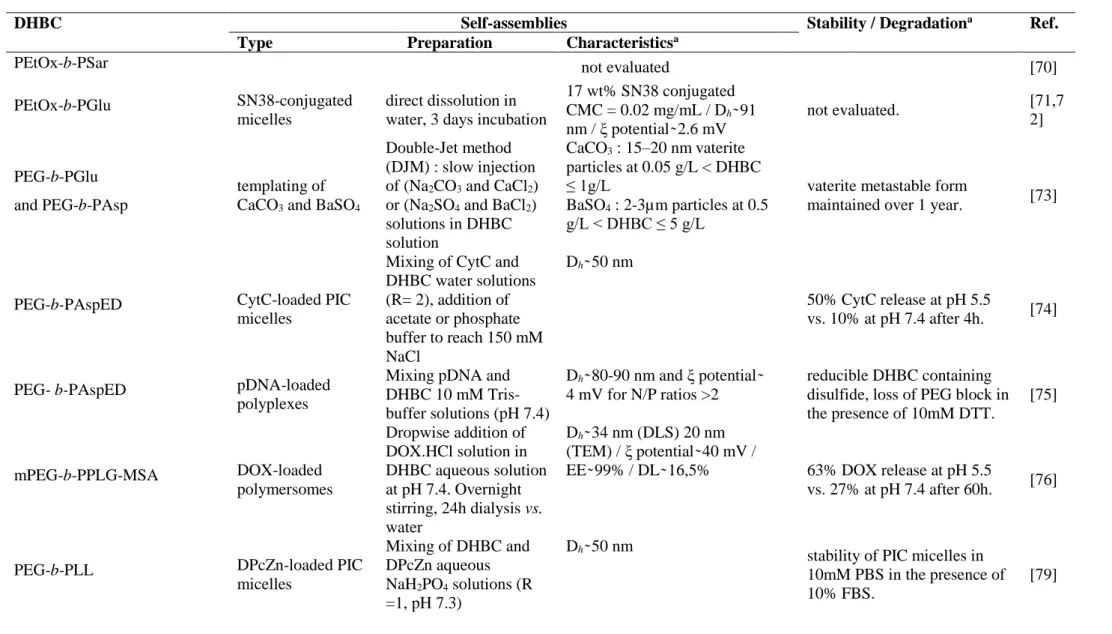
Table 4: Characteristics of the self-assemblies obtained with polypeptide-based degradable double hydrophilic block copolymers.
DHBC Self-assemblies Stability / Degradationa Ref.
Type Preparation Characteristicsa
PEtOx-b-PSar not evaluated [70]
PEtOx-b-PGlu SN38-conjugated
micelles
direct dissolution in water, 3 days incubation
17 wt% SN38 conjugated CMC = 0.02 mg/mL / Dh ̴ 91 nm / ξ potential ̴ 2.6 mV not evaluated. [71,7 2] PEG-b-PGlu and PEG-b-PAsp templating of CaCO3 and BaSO4
Double-Jet method (DJM) : slow injection of (Na2CO3 and CaCl2)
or (Na2SO4 and BaCl2)
solutions in DHBC solution CaCO3 : 15–20 nmvaterite particles at 0.05 g/L < DHBC ≤ 1g/L BaSO4 : 2-3µm particles at 0.5 g/L < DHBC ≤ 5 g/L
vaterite metastable form
maintained over 1 year. [73]
PEG-b-PAspED CytC-loaded PIC
micelles
Mixing of CytC and DHBC water solutions (R= 2), addition of acetate or phosphate buffer to reach 150 mM NaCl Dh ̴ 50 nm 50% CytC release at pH 5.5 vs. 10% at pH 7.4 after 4h. [74]
PEG- b-PAspED pDNA-loaded
polyplexes
Mixing pDNA and DHBC 10 mM Tris-buffer solutions (pH 7.4)
Dh ̴ 80-90 nm and ξ potential ̴
4 mV for N/P ratios >2
reducible DHBC containing disulfide, loss of PEG block in the presence of 10mM DTT. [75] mPEG-b-PPLG-MSA DOX-loaded polymersomes Dropwise addition of DOX.HCl solution in DHBC aqueous solution at pH 7.4. Overnight stirring, 24h dialysis vs. water Dh ̴ 34 nm (DLS) 20 nm (TEM) / ξ potential ̴ 40 mV / EE ̴ 99% / DL ̴ 16,5% 63% DOX release at pH 5.5 vs. 27% at pH 7.4 after 60h. [76]
PEG-b-PLL DPcZn-loaded PIC
micelles Mixing of DHBC and DPcZn aqueous NaH2PO4 solutions (R =1, pH 7.3) Dh ̴ 50 nm
stability of PIC micelles in 10mM PBS in the presence of 10% FBS.
PEG-b-PLL Gd3+-loaded micelles Dissolution of DHBC in 150 mM NaCl solution. Dh ̴ 43 & 225 nm / ξ potential ̴ -9.55 mV
stable in the presence of vacant DOTA groups leading to DOTA-DOTA interactions
[81]
PEG-b-PGlu AMP-loaded PIC
micelles
Mixing of DHBC and AMP aqueous solutions (1:1 molar ratio), dialysis 6h vs. water Dh ̴ 196277 nm / ξ potential ̴ -16 to -39 mV / EE ̴ 75-88% / DL ̴ 19-23% 80% release at pH 7.4 after 72h. [82]
Abbreviations : AMP antimicrobial peptide ; ass. assembly ; CytC cytochrom C ; disass. disassembly ; DTT dithiothreitol ; DOX doxorubicin ; DPcZn dendrimer phthalocyanine zinc ; FBS fetal bovine serum ; Gd3+ gadolinium ion ; PAsp poly(aspartic acid) ; PAspED poly(aspartic acid(ethylene diamine)) ; pDNA plasmid
DNA ;PEtOx, poly(2-ethyl-2-oxazoline) ; PGlu poly(L-glutamic acid) ; PLL poly(lysine) ; PPLG-MSA poly(γ-propargyl-L-glutamate-g-mercaptosuccinic acid) ; PSar poly(sarcosine) ; R charge ratio between positive and negative charges ; SN38 7-Ethyl-10-hydroxy camptothecin.
a ranges of provided values correspond to characteristics obtained as a function of the blocks’ length of the used DHBCs and/or ratios of compounds. If not specified sizes
5. Polyester-based degradable DHBCs and others miscellaneous synthetic blocks
Polyesters are polymers that can be hydrolyzed in accordance with the thermodynamic reversibility of the esterification reaction. This feature, associated with a recognized biocompatibility for some of them, e.g. those derived from lactic acid (LA), glycolic acid (GA) or -caprolactone (CL), explain their wide success in many biomedical applications such as implantable devices, drug delivery systems or scaffolds for tissue engineering. However, polyesters are known for their intrinsic hydrophobicity [83]. The lack of structural diversity of polyesters appears as an important limitation in terms of functionality and physico-chemical properties. As a consequence, various methodologies to introduce functional groups onto polyester backbones have been reported [84–88], for example to poly(-caprolactone) (PCL) functionalized with hydrophilic groups such as hydroxyl [89], carboxyl [90,91], or amino groups [92]. DHBCs containing such functional polyesters as functional block will be discussed in this part (Table 1). A summary of the type of self-assemblies obtained with these DHBCs and their characteristics is provided in Table 5.
Liu et al. synthesized a family of aminated DHBCs in 5 steps via ROP of -(carbamic acid benzylester)-ε-caprolactone (γCABεCL) in bulk using mPEG as macro-initiator to yield, after deprotection, mPEG-b-PACL that can be candidates for pH-sensitive drug delivery, especially for anionic hydrophilic drugs or genes (Figure 7 1). The authors studied the solution properties of the various DHBCs as a function of pH and PACL block length and highlighted by DLS the presence of unimers or aggregates with sizes ranging from 50 to 250 nm depending on the parameters previously cited [93].
By copolymerization of caprolactone (CL) and CL bearing reactive groups like chloro ε-caprolactone (ClCL) that can be derivatized after polymerization into an azido-PCL,
Charoongchit et al. obtained a clickable triblock (P(-N3-CL-co-CL)2-PEG copolymer that was
reacted with propargyltrimethyl ammonium iodide to yield the cationic (P(-TMA-CL-co-CL)2-PEG bearing trimethyl ammonium (TMA) side groups (Figure 7 2). The surface charge
of (P(-TMA-CL-co-CL)2-PEG particles was positive due to the grafted cationic ligand present
on the surface of the particles. The authors compared the particle size for different contents of cationic ligand but with constant PCL chain length, and found that the particle size increased with increasing the mol% of cationic ligand.[94] These cationic copolymers showed a capability to entrap enoxaparin, a low molecular weight heparin used in the treatment of deep vein thrombosis and pulmonary embolism [95], with 87% EE and 8% DL [94].
Figure 4. Degradable hydrophilic block copolymers based on polyester, polyphosphoester and
Gao et al. reported on the synthesis of PEG-b-poly(β-aminoester)-1-(3-aminopropyl)imidazole (PEG-PAE-API) by a Michael-type step polymerization between monoacrylated PEG, 1,6-hexanediol diaacrylate, 4,4’-trimethylene dipiperidine and 1-(3-Aminopropyl) imidazole. This copolymer (Figure 8, top) was used to encapsulate the model protein albumin (Alb). The Alb-loaded micelles, with sizes in the range of 50 to 70 nm, showed a charge conversion from neutral to positive when pH values were changed from 7.8 to 6.2, which is compatible with pH changes observed in cancerous tissue or ischemic tissues. The ability of this PEG-PAE-API to deliver protein in vivo in acidic tissues was assessed in a rat model of cerebral ischemia. Following intravenous injection with Cy5.5-Alb-loaded micelles a gradual increase in fluorescence signals of the brain ischemic area was observed (Figure 8, bottom), indicating that protein/PEG-PAE-API could be effective for targeting acidic environments and diagnostic imaging [96].
Figure 5. In vivo diffusion-weighted MRI (DW-MRI), near-infrared fluorescence (NIRF)
images and signal quantification. In the coronal cross-sectional NIRF images of rat brains of group A (rats injected with PEG-PAE-API-albumin-Cy5.5) (A) and group B (rats injected with albumin-Cy5.5) (B), albumin-Cy5.5 accumulation is clearly visible in the ischemic area of the
right hemisphere, which can be identified as hyperintense lesion on the DW-MRI, in comparison to the left hemisphere.( adapted with permission from [96]).
Mahmud et al. synthesized in 5 steps a family of PEO-b-poly(α-carboxyl-ε-caprolactone) DHBCs via ring opening polymerization of α-benzyl carboxylate-ε-caprolactone and CL with methoxy-PEO as an initiator, followed by catalytic debenzylation of the protected copolymer (Figure 7 3) [90]. The copolymer with 40% of carboxylated CL units assembled to spherical micelles with a CMC of 1.2×10⁻² mM and average diameters of 25 nm. According to the authors, this system could be used as delivery systems for the chemical conjugation, optimized solubilization, and controlled delivery of therapeutic agents.
Deng et al. synthesized methoxy poly(ethyleneglycol)-b-poly(ε-caprolactone-co-γ-dimethyl maleamidic acid-ε-caprolactone) (mPEG-b-P(CL-co-DCL) in 3 steps, having a polyester moiety carrying different amounts of acid-labile β-carboxylic amides (Figure 7 4) [97]. The copolymer formed stable micelles in water with diameters of 100 to 150 nm and with critical micellar concentrations (CMCs) of 3.2−6.3 μg/mL. The DL and EE of this copolymer for DOX were 3-4 times higher than those of amphiphilic copolymer mPEG-b-PCL micelles. The mPEG-b-P(CL-co-DCL) polymer micelles are negatively charged and stable in neutral solution, but, because of the hydrolysis of the β-carboxylic amides in acidic conditions (pH=6.0) the polymer becomes positively charged (Figure 7 4’). This negative to positive charge reversal triggered by the variation of the pH led to a very fast drug release under acidic conditions, and also improved the cellular uptake by electrostatic absorptive endocytosis. Also, the hydrolysis of the acidic group in the polyester upon pH decrease from 7.4 to 5.4 led to a faster release in acidic environment (already mentioned). The mPEG-b-P(CL-co-DCL) micelles showed a very low cytotoxicity up to a concentration of 1 mg/mL.
Zhuo et al. used a thiol-ene “click” reaction between pendent carbon-carbon double bonds of mPEG-b-poly(5-allyloxytrimethylethylenecarbonate) (mPEG-b-PATMC) and various thiol-bearing molecules to prepare four different acid modified copolymers mPEG-b-PATMC-g-SRCOOH (R = CH2, CH2CH2, (CH2)10 and CH(COOH)CH2), denoted as P1, P2, P3 and P4,
Figure 9 1) [98]. The micelles mean diameters determined by DLS for all the copolymers were below 130 nm, and by TEM the authors showed that the copolymer micelles were dispersed in spherical shape with average diameters from 25 to 35 nm (Figure 9 2). The negatively charged copolymers were used for the encapsulation of the positively charged drug DOX via synergistic hydrophobic and electrostatic interactions. The DL of the acid-modified copolymer micelles were all higher than 10 % and the EE were higher than 60 %. The DOX-loaded copolymer micelles showed a pH-dependent release behavior. After 50 hours, the release was limited to 20% at pH 7.4 against ca. 70% at pH 5.0. Overall, the copolymer P3 with the 11-mercaptoundecanoic moieties was the best candidate for DOX formulation as it showed a lower CMC value, smaller particle size, good stability and blood compatibility, as well as higher drug loading capacity. Moreover, cellular investigations revealed an efficient cancer cellular uptake and potent cytotoxic activity of DOX-loaded micelles based on P3 copolymer, probably due to a suitable hydrophobicity and charge density [98].
Figure 6. 1) Structure of 4 different acid modified copolymers mPEG-b-PATMC-g-SRCOOH.
2) Transmission electron microscope images of drug-free and drug-loaded polymeric micelles based on (A) P1, (B) P2, (C) P3 and (D) P4.( adapted with permission from [98]).
Zhang et al. prepared PEG-b-poly(lactic acid-co-malic acid) (Figure 7 5) copolymers via polycondensation between D,L-lactic acid (LA), L-malic acid (MAL), and monomethyl polyethyleneglycol using stannous chloride (SnCl2) as the catalyst [99]. The copolymer was
used to encapsulate the DOX via electrostatic interactions between the carboxyl side group of MAL units and the amino groups of DOX. The DL was 18.2% with good stability in aqueous solution, and TEM images showed spherical nanoparticles in a size range of 110-140 nm. The cumulative DOX release increased under acidic conditions because of the protonation of the acidic group in the MAL. This effect was however limited as after 60 hours, 80% of the DOX was released at pH 7.4, against 94% at pH 5.8.
Zhang et al. prepared a pH-responsive amphoteric block copolymer poly(6-acetoxyl-ε-caprolactone)-b-poly(4-N-piperilactone) (PCCL-b-PPIL) (Figure 7 6) by bulk ring-opening polymerization of 4-N-benzyl formate-piperilactone in the presence of the hydroxyl-terminated
poly(6-(p-methylbenzyl acetate)-ε-caprolactone) as macroinitiator, followed by removal of the protecting groups. The PCCL-b-PPIL copolymer contains carboxyl groups and secondary amine groups in each segment, leading to protonation of the secondary amine groups of the PPIL segments at pH < 5.7 and the formation of PCCL-core micelles. At pH > 6.8 the carboxyl groups of the PCCL segments were deprotonated and PPIL-core aggregates were formed. Noteworthy, the morphology of the self-assemblies changed from spherical at pH 5 with a diameter of 65 nm to worm-like micelle upon pH increase to 8. Thanks to the carboxyl and the secondary amine groups, fluorescent molecules were attached to the copolymer to form stimuli-responsive fluorescent materials [100].
Our group recently reported on a straightforward, 3-step synthetic strategy for the preparation of DHBCs with PCL blocks containing carboxylic acid, amine or hydroxyl functional moieties [101]. PEG-b-PCL copolymers were prepared via ROP of CL employing mPEG as macroinitiator, followed by post-polymerization functionalization of the PCL blocks with pendant alkyne groups using an anionic modification technique [102,103]. Reaction of the alkyne groups with mercaptosuccinic acid, 4,5-diamino-6-hydroxy-2-mercaptopyrimidine or 1-thioglycerol via thiol-yne photoaddition resulted in PEG-b-PCL copolymers with carboxylic acid, amine or hydroxyl functionalized PCL blocks, respectively (Figure 10a). For PEG1.9k
-b-PCL1.1k(OH)52 (containing 52% of hydroxyl groups with respect to CL units) no pH dependency
was expected and therefore its aqueous solution behavior was only tested at pH 7.4. This copolymer self-assembled into micelles above the CMC of 1 mg/mL with a diameter of 225 nm. PEG1.9k-b-PCL1.3k(COOH)55 and PEG1.8k-b-PCL1.4k(NH2)58 (containing 55% of carboxylic
acid and 58% of amine groups with respect to CL units, respectively) self-assembled into pH-responsive micelles with sizes ranging from ~190 nm (in case of ionized DHBCs) to ~130 nm (in case of non-ionized DHBCs). PEG1.9k-b-PCL1.3k(COOH)55 formed stable PIC micelles with
loss of electrostatic interactions between DOX and the carboxylic groups on the DHBC, leading to the release of the drug. The DOX loaded PIC micelles were highly cytotoxic towards MCF-7 cancer cells (Figure 10b), demonstrating the potential of this type of degradable DHBC for the intracellular delivery of electrostatically charged, hydrophobic drugs.
Figure 7. (a) Synthesis of carboxylic acid, amine or hydroxyl functionalized DHBCs from
mPEG-b-PCL in 2 steps and (b) illustration of mPEG-b-PCL(COOH)/DOX PIC micelles formation and their internalization in MCF-7 cancer cells (adapted with permission from [101]).
Zeynep et al. synthesized DHBCs composed of a PEO block and a polyphosphoester block by a combination of organocatalyzed ring opening polymerization, thiol–yne click chemistry and protection/deprotection methods. They prepared poly(phosphotriester)s bearing pendent carboxylic acids (PEO-b-PBYPCOOH) with an affinity for calcium by ROP of butynyl phospholane, as well as poly(phosphodiester)s with a negatively charged oxygen atom on each repeating monomer unit (PEO-b-PPDO-) by ROP of allyl phospholane (Figure 7 7 and 7’). The
authors exploited this family of DHBCs to formulate CaCO3 particles, that can be used for
encapsulation, and showed an efficient decrease of particle sizes by a factor of 6 while preventing their aggregation compared to formulations with hyaluronic acid (HA) [104]. In a follow up work, the authors reported on lysozyme-loaded CaCO3 particles prepared via a
supercritical CO2 process, where CO2 serves as a source of carbonate ions, using either
PEO-b-PBYPCOOH or HA as templating agent. With PEO-PEO-b-PBYPCOOH a twice higher loading of active lysozyme was obtained in the particles compared to HA. Furthermore, a smaller size and a deeper encapsulation of lysozyme in the particle core was observed as well as a more efficient incorporation of the protein (Figure 11) [105].
Figure 8. Confocal images of CaCO3 particles with lysozyme-FITC in the presence of (A) 0,1
% HA; (B) 0.1 % PEO-b-PBYPCOOH and (C) 1 % PEO-b-PBYPCOOH (adapted with permission from [105]).
In another study, poly(2-(2-aminoethoxy)ethoxy)phosphazene (PAEP) was coupled to folate-PEG-COOH or mfolate-PEG-COOH using a DCC/NHS activation (Figure 7 8). DNA was condensed by the resulting cationic PEG-PAEP DHBCs at various N/P ratios to form PEG-PAEP/DNA polyplexes that were compared with PAEP/DNA polyplexes. It was shown that the pegylation of the PAEP decreased the cytotoxicity toward Hela cells and improved the transfection efficiency [106].


![Figure 3. Optical microscopy images of the giant double hydrophilic polymer vesicles referred to as “aquanelles” prepared from Dex-b-PEO, Pul-b-PEO and Dex-b-PSar at 25 wt% (adapted with permission from [65])](https://thumb-eu.123doks.com/thumbv2/123doknet/12160651.312692/21.892.110.779.770.916/optical-microscopy-hydrophilic-vesicles-referred-aquanelles-prepared-permission.webp)

![Figure 1. One-pot synthesis of PEtOx-b-PSar diblock copolymers [70].](https://thumb-eu.123doks.com/thumbv2/123doknet/12160651.312692/25.892.137.694.103.314/figure-pot-synthesis-petox-b-psar-diblock-copolymers.webp)