HAL Id: tel-01205237
https://pastel.archives-ouvertes.fr/tel-01205237
Submitted on 25 Sep 2015
HAL is a multi-disciplinary open access
archive for the deposit and dissemination of sci-entific research documents, whether they are pub-lished or not. The documents may come from teaching and research institutions in France or abroad, or from public or private research centers.
L’archive ouverte pluridisciplinaire HAL, est destinée au dépôt et à la diffusion de documents scientifiques de niveau recherche, publiés ou non, émanant des établissements d’enseignement et de recherche français ou étrangers, des laboratoires publics ou privés.
Distributed under a Creative Commons Attribution - NonCommercial| 4.0 International License
Alexander S. Serov
To cite this version:
Alexander S. Serov. Modeling Oxygen Transport in the Human Placenta. Biological Physics [physics.bio-ph]. Ecole Polytechnique, 2015. English. �tel-01205237�
Thesis submitted in the fulfillment of the requirements for the degree of
Doctor of Philosophy in Physics at École Polytechnique by
Alexander Serov
Modeling oxygen transport
in the human placenta
Thesis defended on the 10th of July 2015 in front of the scientific jury:
Dr. Rohan LEWIS FM, University of Southampton Reviewer Prof. Jean-Frédéric GERBEAU INRIA Paris-Rocquencourt Reviewer Prof. James GROTBERG BME, University of Michigan Examiner Prof. Abdul BARAKAT LadHyX, École Polytechnique Examiner Prof. Mickaël TANTER INSERM, Institut Langevin, ESPCI Examiner
Dr. Denis GREBENKOV PMC, École Polytechnique Thesis supervisor Prof. Marcel FILOCHE PMC, École Polytechnique Thesis supervisor
English abstract
The efficient functioning of the human placenta is crucial for the favorable outcome of the pregnancy. This thesis aims at developing a mathematical model of respiratory gas exchange in the human placenta, which would improve our understanding of the relation between the structure and the function of the organ. Taking advantage of the precise 2D placental structure provided by the placental histology, we construct a 3D model of oxygen transport in the placenta by extending 2D histological cross-sections along the third dimension. The model simultaneously accounts for both diffusion and convention of oxygen in the intervillous space and allows us to predict the oxygen uptake of a placentone.
In the first part of the thesis, the diffusion-convection equation governing oxygen exchange is numerically solved for different densities of circular fetal villi in a placentone. These calculations provide estimations of the oxygen uptake of a placentone with an arbitrary villi density and demonstrate the existence of an optimal villi density maximizing the uptake. This optimality is explained as a trade-off between the incoming oxygen flow and the absorbing villous surface.
As a next step, the assumption of circular villi is relaxed and an approximate analytical solution is proposed for the diffusion-convection equation. It is shown that only two geometrical characteristics — the villi density and the effective villi radius — are required to predict the fetal oxygen uptake. Two combinations of physiological parameters that determine oxygen uptake in a given placenta are also identified: (i) the maximal oxygen inflow of a placentone, and (ii) the Damköhler number defined as the ratio of the transit time of the maternal blood through the intervillous space to a characteristic oxygen extraction time in a cross-section. Analytical formulas for fast and simple calculation of oxygen uptake are derived, and two diagrams of oxygen transport efficiency in an arbitrary placental cross-section are provided. The theory also suggests a method of how the results of artificial placenta perfusion experiments performed with no-hemoglobin blood can be recalculated to account for oxygen-hemoglobin dissociation.
Finally, an automatic image analysis method is developed allowing one to analyze large histological human placenta cross-sections and to determine areas, perimeters and shapes of villous, intervillous space and fetal capillary compartments. These data can then be used as input data for the model. This method is applied to 25 cross-sections from 22 healthy and 3 pathological pregnancies. By combination of the obtained data with the described efficiency diagrams, it is demonstrated that the villi density of a healthy human placenta lies within 10 % of the optimal value. The overall geometry efficiency of a healthy placenta was found to be rather low (around 30–40 %).
In a perspective, the presented model can constitute the base of a reliable tool of assessment of oxygen exchange efficiency in the human placenta from histological measurements post partum, or, in a longer term, from non-invasive in utero measurements.
French abstract
L’efficacité de fonctionnement du placenta humain joue un rôle crucial dans la santé du nouveau né. L’objectif principal de cette thèse est de développer un modèle mathéma-tique de l’échange des gaz respiratoires au sein du placenta humain, afin d’améliorer la compréhension de la relation entre la structure et la fonction de cet organe. En exploitant la structure détaillée du placenta fournie par les méthodes d’histologie placentaire, nous construisons un modèle 3D du transport d’oxygène dans le placenta en prolongeant la géométrie des coupes histologiques 2D le long de la troisième dimension. Ce modèle est capable de prendre en compte simultanément la diffusion et la convection de l’oxygène dans l’espace intervillaire.
Dans la première partie de la thèse, l’équation de diffusion-convection qui détermine l’échange d’oxygène est résolue numériquement pour diverses valeurs de densités de villosités à l’intérieur du placentone. Ces calculs fournissent une estimation de l’absorption d’oxygène d’un placentone pour une densité arbitraire de villosités, et permettent de mettre en évidence l’existence d’une densité optimale de villosités maximisant l’absorption d’oxygène. Cette optimalité peut être vue comme le résultat d’un équilibre entre l’arrivée d’oxygène par le flux entrant du sang maternel et l’absorption à la surface des villosités. À l’étape suivante, l’on s’affranchit de l’hypothèse d’une forme circulaire des villosités, et l’on propose une solution analytique approchée de l’équation de la diffusion-convection. Il est ensuite démontré que deux caractéristiques géométriques – la densité de villosités et le rayon efficace de villosités – suffisent à prédire l’absorption fœtale d’oxygène dans une géométrie donnée. La théorie identifie également deux combinaisons de paramètres phy-siologiques qui déterminent l’absorption d’oxygène : (i) le flux entrant maximal d’oxygène dans un placentone, et (ii) le nombre de Damköhler défini comme le rapport entre le temps de passage du sang maternel dans l’espace intervillaire et un temps caractéristique d’extraction d’oxygène dans une coupe transversale du modèle. Des formules analytiques permettant de calculer l’absorption d’oxygène d’une façon simple et efficace en sont dé-duites, et deux diagrammes d’efficacité du transport d’oxygène dans une coupe arbitraire de placenta sont tracés. La théorie analytique propose également une méthode permet-tant d’exploiter les résultats d’expériences sur la perfusion artificielle du placenta par un sang ne contenant pas d’hémoglobine, afin de prédire l’efficacité de transfert en intégrant cette fois l’interaction entre l’oxygène et l’hémoglobine.
Au final, nous présentons une méthode d’analyse de grandes coupes histologiques de placenta humain, dans le but de mesurer de façon automatique les aires, les périmètres et la morphologie des régions de villosités, de l’espace intervillaire et des capillaires fœtaux. Ces données peuvent ensuite être introduites dans le modèle afin d’estimer l’efficacité du placenta. La méthode est appliquée à 25 coupes placentaires provenant de 22 grossesses saines et de 3 grossesses pathologiques. La combinaison des données obtenues avec les diagrammes d’efficacité montre que toutes les valeurs mesurées de la densité de villosités se retrouvent en écart relatif à moins de 10 % de la valeur optimale. En revanche, l’efficacité globale correspond à une valeur assez basse (autour de 30–40 %).
Dans l’avenir, le modèle présenté peut constituer la base d’un outil fiable de l’évaluation de l’efficacité d’échange d’oxygène au sein du placenta humain à partir de mesures histologiques post partum, ou, à plus long terme, à partir de mesures non-invasives in utero.
Acknowlegements
The three and a half years of my Ph.D. studies at the PMC lab of École Polytechnique were an interesting challenge. The way I arrived here was not simple, and I have considered for a significant time the life-changing choices I had to make. However, I have to admit that finally I have never regretted the decisions made. Here I would like to thank all those without whom this experience would not have been so instructive, pleasant and diverse.
First of all, I would like to acknowledge the scientific as well as personal efforts Denis and Marcel have put into this project and into my development as a scientist. The progress of this placenta project is in a great part due to these two quite different researchers. I hope that this project is only a start of our long and fruitful collaboration. I would also like to thank all the members of the jury for having accepted to participate in the evaluation of the manuscript and in my Ph.D. defense, as well as for their instructive remarks. In particular, the two reviewers, Dr. Rohan Lewis and Dr. Jean-Frédéric Gerbeau, have performed a great amount of work analyzing the thesis manuscript and have provided very helpful and important suggestions.
I would like to thank Carrie for her patience in answering numerous basic biological questions (not at all evident for a physicist) and for introducing me to the field of experimental placental studies and to the international placenta community. I have also much enjoyed the scientific discussions we have had with Paul Brownbill and Rohan Lewis. This project also owes to the efforts of the three interns, Zhenzhen You, Nguyễn Đăng Khoa and Junior Goubalan, who have made great contribution to bringing this initially theoretical and computational project closer to practice.
The successful defense of the thesis would also not have been possible without the advice and support of the members of the PMC lab. I would especially like to thank the directors of the lab, Mathis and François, for having created and maintaining a good working atmosphere in the lab, as well as for their help with administrative questions and valuable discussions. Solving the administrative problems has also been made much easier for me thanks to the efforts of Anne-Marie, Eve, Élodie, Florence, Mme Cécile Vigouroux and the staff of the Direction des Relations Internationales (formerly Direction des Relations Extérieures who has funded this Ph.D. thesis). I would also like to express my gratitude to Anne and Yves for having made my moving to the new office smooth and pleasant, as well as for several delicious madeleines. The bug-shaped cookies of Anne L. were also great. Many other people of the lab (interns, Ph.D. students, post-docs and permanent staff of the lab), as well as friends outside the lab (Matthieu, Barbara, Salim, Jonghoon, Mahsa, Kami and many others) have contributed to making PMC, École Polytechnique and Paris a very pleasant place to work and stay.
Certainly, it would be hard to imagine the realization of the project without the support of my family who have closely followed all my progress and participated in the discussion of all important decisions. The “pot de thèse” would not have been so well organized, if it had not been for the careful selection of the products by my mother and for the preparation of the dishes by Галя, Серёжа, Kami and all the volunteers of the PMC.
– Maud, pour avoir été une super collègue de bureau et pour m’avoir parlé le plus de toutes les personnes du labo pendant son stage ;
– Julien, pour son soutien avec la partie informatique, pour les petites optimisations du code qu’il proposait au fil du projet, pour m’avoir accompagné pendant l’achat de la nourriture pour le barbecue et le repas de Noël du labo, ainsi que pour son aide avec le remplacement du disque dur de mon portable, ce qui l’a mis en panne pour un mois ;
– Наташу, за обучение секретам музыки, таким как умение слушать диалог двух рук, корректно определять музыкальные голоса или правильно писать слова мизинец, стаккато и мордент;
– Juliette, pour des discussions infinies des règles et des exceptions du français, des leçons de biologie, pour un accueil chaleureux septentrional, une balade dans la berdouille, ainsi que pour JJG et de savoureux thés goûtés et offerts ;
– Mahsa, pour les nombreux déjeuners ensemble, sa gaité et sa personnalité débor-dantes, ainsi que pour des idées exceptionnelles de post-doc ;
– Matthieu, pour des discussions de la science et du français, pour des balades insolites, ainsi que pour un week-end tout viril dans la Creuse ;
– Finally, Pani Kami, za to, że litery moich słów wyhodowały ogonki, za miły czas wspólnego burczenia w takim ciekawym języku, za spacery bez smyczy i po miastach europejskich, oraz za to, że nie dała mi Pani umrzeć bez słodkiego ani od czosnku.
English abstract . . . 2 French abstract . . . 3 Acknowlegements . . . 4 Contents . . . 7 Preface . . . 11 1 Introduction 13 1.1 Introduction . . . 13
1.1.1 Importance of placental studies and goals of the thesis . . . 13
1.1.2 Structure of this chapter . . . 14
1.2 Placental structure . . . 15
1.2.1 Placental classification by shape. . . 15
1.2.2 Placental classification by histological structure . . . 17
1.3 Human placenta structure. . . 21
1.3.1 Healthy pregnancies . . . 21
1.3.2 Pathological conditions . . . 24
1.4 Placental functions . . . 26
1.4.1 Placental transport function . . . 26
Passive and active transport . . . 26
Transferred substances . . . 29
1.4.2 Endocrine function. . . 30
1.4.3 Protective function. . . 31
1.4.4 Concluding remarks . . . 31
1.5 Experimental methods of placental studies . . . 31
1.6 Review of theoretical models of placental transport . . . 41
1.6.1 Overview of placenta modeling. . . 41
1.6.2 Respiratory gas exchange models. . . 42
1.6.3 Capillary–scale models. Flow patterns . . . 43
1.6.4 Morphometric diffusing capacity models . . . 46
1.6.5 Distributed parameters models . . . 48
1.6.6 Models based on histological placental sections . . . 51
1.6.7 Experimental validation of mathematical models of respiratory gas exchange in the placenta . . . 53
1.6.8 Concluding remarks . . . 53
1.7 Objectives and structure of the thesis . . . 54 7
2 Stream-tube placenta model 57
2.1 Model outline . . . 58
2.1.1 Model assumptions. . . 59
2.1.2 Dimensionless hydrodynamic numbers . . . 61
The Reynolds number (Re) . . . 61
The Péclet number (Pe). . . 63
The Schmidt number (Sc) . . . 63
The Damköhler number (Da). . . 64
2.2 Diffusion-convection equation. . . 65
2.2.1 Characteristic time scales of transfer processes in the human placenta65 2.2.2 Equilibrium between the bound and the dissolved oxygen . . . 65
2.2.3 Diffusive-convective transport of oxygen . . . 66
2.2.4 Linearization of the Hill equation . . . 67
2.2.5 Boundary conditions . . . 68
2.2.6 Conversion to a 2D eigenvalue equation . . . 69
2.2.7 General expression for oxygen uptake . . . 69
2.3 Model parameters . . . 70
2.3.1 Calculation of the model parameters . . . 70
2.3.2 Concluding remarks . . . 77
2.4 Numerical simulation . . . 77
2.4.1 Circular villi. Solution convergence and validation . . . 77
2.4.2 Results . . . 80
2.4.3 Comparison to the experimental data . . . 82
2.4.4 Comparison to the porous medium model . . . 83
2.4.5 Other ways of comparison . . . 86
2.4.6 Additional remarks . . . 87
2.5 Conclusions. . . 87
3 Approximate analytical solution 89 3.1 Mathematical formulation . . . 90
3.1.1 Form of the approximation . . . 90
3.1.2 Uptake at the infinite length . . . 90
3.1.3 Average concentration decay rate . . . 91
3.1.4 Dimensionless geometrical parameters . . . 93
3.1.5 The optimal cross-sectional geometry . . . 93
3.1.6 Optimal villi density . . . 95
3.1.7 Villi density efficiency . . . 97
3.2 Analytical results. . . 97
3.2.1 Oxygen uptake for circular villi . . . 97
3.2.2 Typical values of the parameters Da and F0 . . . 99
3.2.3 The parameter Da . . . 100
3.2.4 Advantages of the analytical theory . . . 101
4 Analysis of Histological Placental Cross-Sections 105
4.1 Manual placental histomorphometry . . . 106
4.1.1 Application of the STPM to histological placental cross-section . . 106
Geometrical parameters . . . .106
Physiological parameters. . . .107
Summary . . . .108
4.1.2 Histological placental cross-sections. . . 108
4.1.3 Point–intersection counting techniques . . . 110
4.2 Automatic placental segmentation . . . 112
4.2.1 “Ideal” segmentation and main segmentation challenges . . . 112
4.2.2 Thresholding and the initial segmentation . . . 114
4.2.3 Refining the segmentation . . . 117
Filtering out noise . . . .117
Discriminating fetal capillaries from the IVS. . . .118
Smoothing out the contours . . . .118
4.2.4 Accuracy of the segmentation algorithm. . . 120
4.2.5 Histomorphometrical measurements. . . 122
4.2.6 Code execution time as a function of image size . . . 124
4.3 Algorithm application to placental cross-sections . . . 125
4.3.1 Samples description . . . 125
4.3.2 Comparison of segmentation results for healthy and pathological placentas with the results obtained with manual histomorphome-trical techniques . . . 126
4.3.3 Efficiency diagrams. . . 128
4.4 Concluding remarks . . . 130
5 Conclusions and Further Development 133 5.1 Results summary. . . 133
5.2 Further development . . . 136
5.2.1 Development of the model . . . 136
5.2.2 Development of the field . . . 137
Appendices 141 A Hormones secreted by the placenta 142 B Typical parameters of placenta models 144 C Error estimation 148 D Typical problems of histomorphometrical placental studies 150 D.1 Inhomogeneity of the fetal villous tree . . . 150
D.1.1 Intraplacentone structural variations. . . 150
Trophoblast layer thickness. . . .152
D.1.2 Intraplacental structural variations . . . 157
D.2 Immersion fixation vs. perfusion fixation of placental samples . . . 157
D.3 Influence of the fixative on histomorphometrical measurements . . . 159 E Comparison of oxygen exchange efficiency at low and high altitudes 161
Preface
There’s nothing wrong with putting animal afterbirth in your hair
Customer recommendation of a placental shampoo (Power and Schulkin, 2012)
While working on this project, I was surprised to discover the multiple ways in which the placenta can be used after delivery. Those include ingestion of the placenta in the dried, frozen or ground into powder forms. Recipes can be found on the Internet instructing on how to make the most delicious placenta lasagne, placenta stew, placenta smoothie, placenta pizza, or a plazentaburger, and do-it-yourself placenta encapsulation kits can be ordered on Amazon. The placenta can also be burned, buried with a tree planted above it, converted into a teddy bear, or applied to different parts of the body to help you cope with the “baby blues”, make your hair lustrous and thick, your skin dry and improve the overall health and beauty.
This thesis is an attempt to demonstrate that in addition to all these options, a delivered placenta can also serve scientific purposes and help diagnose newborn health risks.
All remarks and questions about the work presented in this thesis can be sent to [email protected].
Introduction
1.1 IntroductionT
placenta is the only link between the mother and the growing fetus. It sup-plies the fetus with water, nutrients and oxygen, eliminates carbon dioxide and other wastes, performs barrier, metabolic and endocrine functions, thus fulfilling functions of multiple organs. Understanding the placental function is thus important to understand the fetal development in utero.While studying the placenta may help the assessment of baby health risks, this valuable and unique source of information about the development of the baby for the first nine months is usually discarded after birth as medical waste. At the same time, it is the only organ that the body does not need after birth and which can thus be extensively examined.
1.1.1 Importance of placental studies and goals of the thesis
The information the placenta may provide has been shown to be crucial for understanding the reasons of fetal maldevelopment and the main causes of fetal death (reported at the level of 6.05 fetal deaths / 1000 pregnancies after the 20th week of gestation in the US
in 2006, see MacDorma et al., 2012). For instance, Baergen (2007) has demonstrated that studying the placenta may not only reveal pregnancy-related problems that are due to an inherent placental abnormality, but often also lets ascertain the etiology of the injury and the corresponding time frame. Korteweg et al. (2009) analyzed 750 cases of singleton intrauterine fetal death (after 20 weeks of gestation) and have concluded that 64.9 % of death cases can be attributed to placental pathologies. Hutcheon et al.
(2012) have shown that low placental weight at birth significantly increases the risk of stillbirth, while high placental weight was related to a high probability of adverse neonatal outcomes.
Interestingly, the placenta does not only provide information on the fetal development in utero, but also allows one to make prediction on the future development of the newborn. For instance, Misra et al. (2012) have correlated the children intelligence coefficient at the age of 7 years with placental thickness and chorionic plate diameter. Other studies have provided evidence of correlation between the risk of hypertension and
coronary heart disease in adults and the placental weight, diameter and cross-sectional area (Barker, 1994, 1995, Eriksson et al., 2010). So understanding the placental function may not only shed light on adverse pregnancy outcomes, but also help identify children with increased health risks.
Past studies have shown that the transport function of the placenta is in significant part determined by the structure of the organ (Guilbeau et al., 1972, Hill et al., 1972,
Erian et al., 1977, Faber, 1977, Mayhew et al., 1984, Chernyavsky et al., 2010, Gill et al., 2011). The main goal of this thesis is to further extend our understanding of the relation between the placental structure and the transport function by constructing a mathematical model based on the details of placental morphology that can be obtained in a histological examination. This 3D model considers a part of the maternal blood flow in the intervillous space — a stream tube — crossed by fetal villi. In this stream-tube model, we will not only quantitatively estimate oxygen uptake, but will also determine an optimal geometry of the stream-tube that provides the maximal oxygen uptake. Starting with simple villi shapes, we will then advance to villi of arbitrary shape. Finally, we will thoroughly analyze how this model can be converted into a tool of medical diagnostics of placental exchange efficiency that would take 2D histological placental cross-sections as input.
1.1.2 Structure of this chapter
In the present chapter we provide an introduction to the placental studies for a reader who may be new to the subject. For this purpose, we first discuss the great variability of the placental shape and structure among the placental mammals. Two main principles of placental classification are considered: classification by shape and classification by histological structure of the organ.
Having provided an overview of inter-species placental differences, we further focus on the human placenta, which is of primary medical interest and which arguably has the most complicated pattern of maternal circulation. We briefly describe the structural organization of the human placenta and its main functions (transport, endocrine and protective). Special attention is given to its transport function, which is the principal topic of the present thesis.
After that, we review experimental techniques developed for placenta investigation including macroscopic and microscopic histological examination, micro-CT, T1– and T2– weighted contrast MRI, confocal laser scanning microscopy and ultrafast ultrasound tech-niques. We discuss the data that can be obtained with each method and the corresponding limitations. Our analysis will demonstrate that due to ethical reasons and the fact that histological and physiological placental features commonly co-vary, the experimental tech-niques are not able to provide a complete picture of intraplacental transport processes. A complementary point of view on this question is offered by mathematical models. In the vast field of placenta models, we direct our attention to the models of respiratory gas exchange. On one hand, it is explained by the short time within which the adverse effects of compromised respiratory gas exchange are observed in the fetus. On the other hand, we are interested in the special focus of this kind of models on the geometry
of the placenta as an exchanger (rather than on transport-related biochemical reactions). Other directions of the placenta modeling (such as modeling of the placenta shape, villi branching or glucose and amino acid exchange) are also listed.
The first models of respiratory gas exchange in the human placenta date back to Barron
(1951) and Lamport (1954). To let the reader understand the positioning of the present study in the context of the previous models, we analyze the main directions of the placental respiratory gas exchange modeling, of which we distinguish four:
• capillary-scale models and flow patterns studies, • morphometric diffusing capacity models,
• distributed parameters models,
• models based on histological sections.
After a short review of the models, we discuss the possible ways of validation of the models and analyze their main limitations. This analysis of placental models was published in Filoche et al. (2015). In the last section, the structure of subsequent chapters is outlined.
1.2 Placental structure
The placenta is an organ developed only by mammals during pregnancy. It is formed when the fetal membranes (blue and black in Fig. 1.1) come into close contact with the endometrial lining of the uterus (red in Fig. 1.1). However, not all mammals can produce a placenta, which is one of the basic criteria for distinguishing three classes of mammals: eutherians (placental mammals), metatherians (or marsupials, nonplacental mammals having abdominal pouches) and monotremes (nonplacental egg-laying mammals like the platypus and the echidna). Although eutherian mammals share many similar features, there exist considerable interspecies differences (especially with regards to the placental structure and morphology), which yielded multiple placenta classification schemes. In this section we will review the two basic classifications (by the placental shape and by histological structure) following King (1992), Wooding and Burton (2008), Power and Schulkin (2012). For the details of other classifications, the reader is referred to Ramsey (1982).
1.2.1 Placental classification by shape
The growing fetus is surrounded by four fetal membranes: the chorion, the amnion, the allantois and the yolk sac (Fig. 1.1). The chorion is the external embryonic membrane in direct contact with the maternal tissue. It often forms chorionic folds or villi that significantly increase the exchange surface. The placenta classification by shape is based on the distribution of outgrowths of the chorion on the mature gestation sac. According to this criterion, four major placental categories are recognized (Fig. 1.2):
1. A diffuse placenta completely surrounds the fetus, and the chorion is distributed over most of the surface of the gestation sac. As a consequence, almost all the
Figure 1.1. Synoptic representation of the fetal membranes (black and blue) that may contribute to the
formation of the placenta. The maternal tissues are shown in red (the endometrial lining in contact with the fetal chorion) and brown (the rest of the uterus). Reproduced from Benirschke et al. (2006)
Pig Horse Cow, sheep Deer Dog, cat Human Monkey Rodent Diffuse placentas Cotyledonary placentas Zonary placentas Discoid placentas placental zone placenta locations placenta caruncles
Figure 1.2. Major categories of placentas according to their shape (second column). The corresponding
shape of the uterus and placental location in it (third column) in it are also shown. Modified from Wooding and Burton (2008)
placental surface attaches to the uterine wall. This placenta type is seen in pigs, horses, whales, dolphins, hippopotamuses and some primitive primates (lemurs and lorises).
2. A cotyledonary placenta attaches to the uterus in multiple discrete locations, which form circular or oval patches on the chorionic sac. The maternal attachment sites are called caruncles, and the placental sections which attach to the uterine wall are termed cotyledons. A maternal attachment site together with a corresponding placental cotyledon form what is called a placentome (Power and Schulkin, 2012). Note however that nowadays the terms cotyledons, placentones (-omes) or lobules are often used interchangeably (Chernyavsky, 2011), especially in application to the human placenta. In this thesis we will mainly use the term placentone. The cotyledonary placental organization can be found in ruminants (e.g., cows, sheep), deer, sloths, armadillos. Horses have microcotyledons in discrete sections of their diffuse placenta (Power and Schulkin, 2012). Placentones are also found in the discoid human placenta (Benirschke et al., 2006).
3. A zonary placenta forms a band or a girdle around the fetus usually located in the equatorial plane. In some species it completely encircles the fetus, while in others the band is incomplete or forms two half-bands. Zonary placenta is found in all carnivores (e.g., canines, felines, bears, seals), elephants, rock hyraxes and aardvarks. Among these species, dogs and cats have complete bands, while ferrets and raccoons have two half bands.
4. A discoid (or discoidal) placenta has a shape close to a disk, in which chorionic villi are distributed. This placenta type is found in rodents, bats, tenrecs, giant anteaters, red panda and anthropoid primates, including humans.
The placental classification by shape is straightforward and convenient, but it does not describe four functionally different categories, since numerous functional variations exist within the same shape categories (King, 1992, Wooding and Burton, 2008, Power and Schulkin, 2012).
1.2.2 Placental classification by histological structure
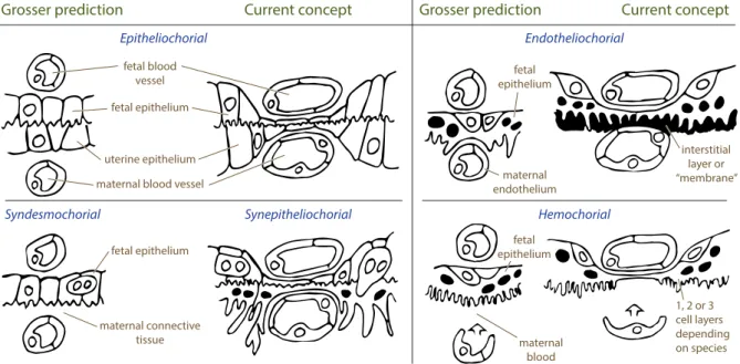
An alternative placental classification, by histological structure, is based on the number and origin of tissue layers separating the maternal and fetal circulations. These characteristics are closely related to the exchange properties of the placenta, which makes such cate-gorization more functional. This classification was originally proposed by Grosser (1909,
1927) and later modified by Mossman (1937), Owers (1960).
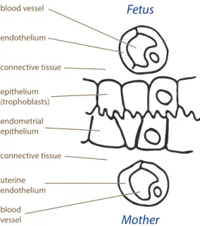
The maximal number of different tissue layers separating the maternal and fetal blood circulation is six: maternal endothelial, connective and epithelial tissue layers and fetal endothelial, connective and epithelial tissue layers (Fig. 1.3). However, with the evolutionary progress, some species have lost one, two or three (i.e. all) maternal tissue levels. The Grosser’s classification distinguishes four main placental types which arise as a consequence of this tissue layers reduction (Fig. 1.4). The names of the categories are
Fetus blood vessel endothelium connective tissue epithelium (trophoblasts) endometrial epithelium connective tissue uterine endothelium blood vessel Mother
Figure 1.3. Illustration of the six potential tissue layers separating the maternal and fetal circulations:
maternal endothelial, connective and epithelial tissue layers and fetal endothelial, connective and epithelial
tissue layers. Modified from Wooding and Burton (2008)
defined by the maternal tissue layer, which is in contact with the fetal chorion after the placental implantation and early differentiation of fetal organs are completed (see also Fig. 4.5 and Table 4.1 in Benirschke et al., 2006):
1. In epitheliochorial placentas no tissue layers have been lost, so that the uterine epithelium is in contact with the chorion. Such placental organization is observed in pigs, horses, deer, dolphins and whales.
2. In synepitheliochorial (syndesmochorial) placentas the uterine epithelium is removed, and the maternal connective tissue is in contact with the chorion. Such placentas are typical for ruminants.
3. In endotheliochorial placentas the uterine epithelium and the maternal connective tissue are removed, and the maternal capillary endothelium is apposed to the chorion. Such placentas can be seen in carnivores (e.g., cats and dogs).
4. In hemochorial placentas, all intervening maternal tissues are removed, and the maternal blood directly bathes fetal trophoblasts. Such placentas are found in rodents, rabbits, insectivores and anthropoids (including humans).
Having proposed this histological classification, Grosser suggested that the number of tissue layers separating the maternal and fetal circulations determines the thickness of the placental membrane and therefore the placental transport properties. However, with the discovery of active, facilitated and endocytic transport (discussed later), this hypothesis became oversimplified. For instance, immunoglobulin is transferred differently from the mother to the fetus in humans and guinea pigs (see King, 1992).
It was also reported that more tissue layers do not necessarily make the placental membrane thicker: for example, in the epitheliochorial placenta of the pig, maternal capillaries indent the uterine epithelium and fetal capillaries indent fetal trophoblasts, so
fetal blood vessel fetal epithelium fetal epithelium fetal epithelium fetal epithelium maternal endothelium maternal blood maternal connective tissue uterine epithelium maternal blood vessel
Grosser prediction
Epitheliochorial Endotheliochorial
Hemochorial Syndesmochorial Synepitheliochorial
Current concept Grosser prediction Current concept
interstitial layer or “membrane” 1, 2 or 3 cell layers depending on species
Figure 1.4. Major categories of placentas according to histological placental structure. The original suggestion
of Grosser (1909, 1927) is accompanied to the right by a revised version (Wooding and Burton, 2008). The revised version takes into account the fact that fetal and maternal capillaries indent into the chorionic and uterine epithelium respectively during growth, which significantly decreases the diffusion distance between the two circulations. Modified from Wooding and Burton (2008)
that the diffusional distance between the two circulations is significantly reduced (King,
1992). With these restrictions kept in mind, the Grosser’s classification still stays a convenient reminder of which placental tissue layers should be taken into account while working with one or another species.
The differences in the histological structure of the placenta across species find their reflection in the organization of the maternal and fetal circulations in the placenta. In many species, the maternal blood and the fetal blood pass through the placenta in blood vessels which come into close contact in the exchange region to allow for substance transfer. Such organization of the organ is natural for epitheliochorial (Fig. 1.5a), syn-desmochorial and endotheliochorial placentas, where at least one maternal tissue layer separates the maternal and fetal placental vascular systems (King, 1992). In hemochorial placentas, there are no maternal blood vessels in the exchange region. In labyrinthine hemochorial placentas, the maternal space is organized in a kind of labyrinth, the archi-tecture of it resembles that of blood vessels (Figs 1.5b, 1.6a, see Battaglia and Meschia,
1986, King, 1992). However, the structure of the non-labyrinthine anthropoid primate placenta (monkeys, apes and humans, see Figs 1.6b, 1.7a) is quite different with maternal blood forming a blood basin into which fetal chorionic villi are immersed. Such structural organization significantly complicates the description of transport properties of the human placenta due to the intricate maternal blood flow pattern and chorion arrangement. This thesis focuses on the human placenta because of of its primary medical interest, and also because of the greater complexity of its structure compared to other species.
chA epM aM vM vF aF F g g M (a) TT FA FV FC AS UV VS MA MV (b)
Figure 1.5. (a): Arrangement of the maternal and fetal blood vessels in microcotyledons of the
epithe-liochorial placenta of the horse. Small brown arrows mark the countercurrent organization of the maternal and fetal blood flows, currently supposed in the horse placenta. Notations: maternal (M), fetal (F), maternal artery (aM), maternal vein (vM), fetal artery (aF), fetal vein (vF), chorioallantois (chA), uterine epithe-lium (epM), endometrial glands (g). Modified from Silver et al. (1973), Battaglia and Meschia (1986), based on the data by Tsutsumi (1962). (b): Arrangement of the maternal and fetal blood vessels in the rabbit placenta. Maternal blood enters the exchange area via maternal arteries (MA) that feed large arterial sinuses (AS). Efferent arteries proceed from these sinuses toward the fetal surface of the placenta and open into trophoblastic tubules (TT) of the placental labyrinth. In these tubules the blood flows towards the decidua, where it collects in venous sinuses (VS) that are drained by large veins in the uterine wall (UV) and mesometrium (MV). Fetal arteries (FA) penetrate deep into the placenta and then break up into fetal capil-laries (FC) in which fetal blood runs back towards fetal umbilical veins (FV). Modified from Carter (1975), based on the original drawing by Mossman (1926)
a Mouse
b Human
Trophoblast giant cell Spongiotrophoblast
Maternal blood space Labyrinth:
syncytiotrophoblast, chorionic trophoblast, blood vessels, stroma
Extravillous cytotrophoblast Column cytotrophoblast Chorionic villi: syncytiotrophoblast, villous cytotrophoblast, blood vessels, stroma
Fetal blood vessel
Fetal endothelial cell Maternal blood sinus Syncytiotrophoblast Mononuclear trophoblast cell Maternal blood Mesenchyme Fetal endotherial cell
Fetal blood vessel Syncytiotrophoblast Villous
cytotrophoblast
Figure 1.6. Comparative anatomy of the mouse and the human placenta. (a) Structure of the mouse
placenta. The details of the fetal-maternal interface in the labyrinth are provided in the inset. (b) Structure of the human placenta. The inset image shows a cross-section of a fetal villus. One can see that in the mouse placenta the maternal blood and the fetal blood are confined to capillaries, while in the human placenta fetal villi are directly immersed in a basin of maternal blood. Reproduced from Rossant and Cross
Basal plate Umbilical cord Umbilical vein Umbilical arteries Trophoblast Amnion Chorion Villus Maternal vessels Stratum spongiosum Placental
septum Limiting or boundary layer
Marginal sinus
(a) (b)
Figure 1.7. (a): Structure of the human placenta (modified from Gray, 1918). The bottom side is the basal
plate which attaches to the uterus; the upper side is the chorionic plate, where the umbilical cord connects to the placenta. Typical location and orientation of a histological placental section is marked by a dashed blue line. (b): Location and structure of villous trees inside the human placenta. The chorionic plate is facing up. The direction of the maternal blood flow is marked by arrows. According to the modern views, the maternal blood enters the placentone near the center of a villous tree and leaves the intervillous space between the villous trees (Benirschke et al., 2006). The adjacent villous trees may overlap. Reproduced from Kaufmann (1985b), Benirschke et al. (2006)
1.3 Human placenta structure
1.3.1 Healthy pregnancies
A healthy human placenta is a disk with an average diameter of 22 cm (Benirschke et al.,
2006, p. 13). The two faces of the disk are called the chorionic plate (the one from which the umbilical cord stems, Fig. 1.8a) and the basal plate (the one which attaches to the uterus wall, Fig. 1.8b). The average thickness of the delivered organ is 2.5 cm and the average weight is 470 g (Benirschke et al., 2006, p. 13).
In what concerns the internal structure, the maternal portion of a term human placenta is a basin into which maternal blood is brought by numerous spiral arteries and which is drained by decidual veins (Fig. 1.7a; Benirschke et al., 2006). It is remarkable that after many years of human placenta studies, the exact number of spiral arteries and decidual veins perfusing the placenta is still unknown. The review by Panigel and Pascaud (1968) cites the following numbers: spiral arteries — from 50 to 120 and orifices of spiral arteries — from 100 to 500. There are also around 30 (Klein, 1890) decidual veins, which form around 80 venous openings (Franken, 1954). The low precision of these numbers is explained by the difficulty to find the orifices of the vessels and to distinguish between arteries and veins after the delivery (see Haller, 1968, Benirschke et al., 2006, p. 251–2).
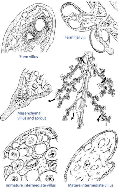
The fetal portion of the human placenta is a tree of villi immersed into the maternal blood. As a tree, it has many generations of branches (up to 30, mean value around 10, see Benirschke et al., 2006) which differ by their caliber, stromal structure, vessel structure and location within the villous tree (Fig. 1.9). According to these criteria, five villi types are distinguished: stem villi, immature intermediate villi, mature intermediate villi, terminal villi and mesenchymal villi (for the details of classification see Kaufmann et al., 1979,
(a) (b) (c)
Figure 1.8. (a), (b): Appearance of the human placenta: the chorionic plate (a) and the basal plate (b).
It can be seen that the basal surface is subdivided into placental lobules of various size by an interrupted net of dark grooves. Reproduced from Benirschke et al. (2006). (c): Subdivision of the human placenta into maternal and fetal lobules. The borderlines of the maternal lobules are marked by red lines (drawn from the grooves at the basal surface), 29 fetal trees are shaded in blue (obtained by a radiograph of the placenta after injection of a radiopaque medium into the fetal vessels). Although there is a good agreement in the locations of the fetal and the maternal lobules, one to three fetal lobules can project on the same maternal lobule. Reproduced from Kaufmann and Scheffen (1992), Benirschke et al. (2006), based on the photographs by Boyd and Hamilton (1970)
Sen et al., 1979). Scanning electron photographs of mature intermediate and terminal villi are shown in Fig. 1.10. The characteristic diameters (calibers) of the villi are: stem villi, 300–3000 μm; immature intermediate villi, 60–200 μm; mature intermediate villi, 40–80 μm; terminal villi, 30–80 μm.
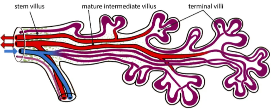
Inside each villus, fetal capillaries conduct fetal blood from the umbilical arteries to the umbilical vein, without admixture with the maternal blood stream (Fig. 1.11). All exchange processes between the mother and the fetus take place at the surface of the fetal villi. It mainly occurs in the smallest branches of the tree — terminal and mature intermediate villi, — in which capillaries are located close to the villous membrane and the membrane itself is thin (Benirschke et al., 2006, p. 121).
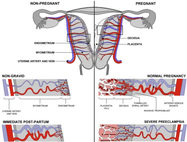
The human placenta is normally considered as subdivided into functional units (placen-tones, also called lobules or cotyledons, Fig. 1.8c). It is estimated that a healthy human placenta counts from 40 to 60 placentones (Benirschke et al., 2006, p. 161). Fetal trees tend to overlap, and placental septa do not completely separate one placentone from another, but rather mark their approximate boundaries (Figs 1.7b and 1.8c). Each placentone normally has a villi-free central part (a central cavity, with a characteristic di-ameter of 1 cm, as estimated from figure 4 of Aifantis, 1978), which, as some researches believe, is formed by the mechanical force of a “jet” of maternal blood spurting from the spiral arteries (Ramsey et al., 1963, Borell et al., 1958, Benirschke et al., 2006, p. 163–4). However, recent calculations predict that due to trophoblast invasion and remodeling of the spiral arteries, the velocity of the “jet” is significantly lower (∼10cm/s) than 1–2 m/s estimated earlier (Burton et al., 2009). So the mechanisms of formation of the central cavity are as yet unclear.
The intervillous space (IVS) is drained by decidual veins which tend to be located at the periphery of the placentone (at the basal plate or in the lower one-third of placental septa; see Benirschke et al., 2006, p. 161, p. 252 and Fig. 1.7a). The ideal arrangement with one spiral artery opening near the center of the villous tree is
Figure 1.9. Schematic representation of the cross-sections of the main types of fetal villi (stem villi, immature
intermediate villi, mature intermediate villi, terminal villi and mesenchymal villi) and their location in the villous tree. Note that villi cross-sections are shown at different magnifications. Reproduced from Kaufmann and Scheffen (1992)
Figure 1.10. Scanning electron photographs of different villous regions. (A): A single mature intermediate
villus. (B): A tip of a mature intermediate villus featuring rich branching into terminal villi. (C): A group of terminal villi, with the central one showing a typical constricted neck region and a dilated final portion. Reproduced from Kaufmann et al. (1979), Benirschke et al. (2006)
Figure 1.11. Location of fetal vessels inside a branch of a villous tree. Color code: blue — fetal arteries
and arterioles (with low oxygen content), lilac — fetal villous capillaries and sinusoids (terminal dilations), red — fetal venules and veins (with high oxygen content), green — stroma of the parent mature intermediate villus. Reproduced from Benirschke et al. (2006)
however rarely observed. It normally occurs only near the periphery of the placental disk, where placentones are small, and one maternal lobule approximately corresponds to one villous tree (Benirschke et al., 2006, p. 252). Near the center of the placental disk, maternal lobules visible from the basal plate normally contain one to four overlapping villous trees (Fig. 1.7b), and the precise location of maternal vessels in these lobules is unclear.
The maternal circulation starts in the human placenta only around 10th–12th week
of gestation, after the maternal spiral arteries have been invaded and remodeled by fetal trophoblasts (see Fig. 1.12 and Benirschke et al., 2006, Burton et al., 2009). The delayed onset of the maternal circulation is believed to protect the developing fetal tissues from the high velocity of the maternal blood flow and high partial pressures of oxygen. In the human placenta, the volumetric blood flow in the umbilical vein (i.e., the total fetal placental blood flow) is reported to be around 36 ml/min at 20 weeks of gestation, and around 265 ml/min at 40 weeks (Acharya et al., 2005). Per 1 kg of fetal weight these numbers give 88ml/(min · kg) at 20 weeks and 66ml/(min · kg) at 40 weeks. Analysis of umbilical blood samples provides the approximate arterial (0.11± 0.06ml O2/ml blood) and venous (0.21±0.07ml O2/ml blood) oxygen content of the
fetal blood at term (Acharya and Sitras, 2009). Combination of these data yield fetal oxygen uptake at term around 5.6±3.2ml O2/(min · kg) or, equivalently, (4.2±
2.4) ·10−6mol/(s · kg) in SI units (Acharya and Sitras, 2009). At the same time, the maternal uterine blood flow at term (to the uterine and placental tissues) is reported to be 500±200ml/min (Metcalfe et al., 1955). The uterine oxygen consumption was then estimated at the level of 25±13ml O2/min or 7.5±3.8ml O2/(min · kg)
per kilogram of fetal weight. In SI units it amounts to (19 ±10) ·10−6mol/s or (5.6±2.8) ·10−6mol/(s · kg) respectively. Therefore, fetal oxygen uptake corresponds to around 75 % of the uterine oxygen consumption at term.
1.3.2 Pathological conditions
Numerous studies have reported alterations in the structure of the human placenta in pathological cases. The most typical pathologies and pathological conditions are (Benirschke et al., 2006):
Figure 1.12. Remodeling of the maternal spiral arteries in pregnancy. Note the dilation and the funneled
outlets of the spiral arteries as well as the formation of arterio-venous shunts that persist in the immediate
post-partumperiod. Non-dilated spiral arteries of severe pre-eclampsia are also shown. Color code: arteries —
red, veins — blue. See also Fig. 1.20. Reproduced from Burton et al. (2009)
• Diabetes mellitus: a group of diseases characterized by abnormalities of glucose metabolism.
• Pre-eclampsia, also called pregnancy-induced hypertension (PIH): a pregnancy disor-der characterized by high maternal blood pressure and increased amount of protein in the urine.
• Placenta accreta, placenta increta, placenta percreta: cases of abnormally strong attachment of the placenta to the uterine myometrium (the uterine muscular level), which may cause heavy bleeding after the delivery. The three pathological conditions are distinguished by the depth of placenta invasion into the uterine wall.
• Placenta previa: an abnormality when the placenta overlies the internal cervical os of the uterus, effectively obstructing the birth canal, which may cause bleeding before the delivery.
• Placental abruption: a pregnancy complication, when the placental lining separates from the uterus before the labor.
• Umbilical cord abnormalities. • Placental infections.
• Pathologies of placental membranes usually associated with abnormal vascularization of the chorionic plate.
• Maternal anemia: low maternal RBCs count. • Maternal smoking, alcohol abuse or drug addiction.
To this list should be added the intra-uterine growth restriction (IUGR) and small for gestational age (SGA fetus) condition, which are symptoms which can result from numerous pathologies, rather than pathologies themselves. Typical placental structure variations reported for some of these pathologies are collected in Table 1.1. Note that it is often hard to distinguish whether these alterations are directly caused by a pathology or are rather a placental adaptation to it.
1.4 Placental functions
Placenta is a multifunctional organ combining transport, endocrine, protective and metabolic functions, each of which is briefly reviewed below. For more details, the reader is referred to the reviews by Gude et al. (2004), Desforges and Sibley (2010). Note also that there exist many interspecies differences in the placental function.
1.4.1 Placental transport function
The placental transport function much depends on the organization of the blood flow in the placenta. Since the circulation of the maternal blood in the placenta is established only at the end of the first trimester of pregnancy (Benirschke et al., 2006), the transport function in early pregnancy significantly differs from that in late pregnancy. During the first trimester, the nutrition of placental and embryonic tissues is histiotrophic with trophoblast phagocytosis of endometrial glandular secretions including glycogen and a variety of glycoproteins (Burton et al., 2002). In other words, the nutriments (proteins and carbohydrates) are absorbed by the surface of the developing embryo. After the formation of the placenta and the onset of the maternal circulation, the maternal blood comes into contact with terminal fetal villi, the nutrition becomes hemotrophic and all transport occurs across the placental membrane (which comprises the membranes and the cytoplasm of all tissue layers making it up, see Section 1.2.2).
Passive and active transport
According to energy requirements and the ability of the transported substance to diffuse through the membrane, all transmembrane transport processes are divided into passive and active transport processes (Sadava et al., 2007, pp. 105–125). Passive transport does not require input of chemical energy and comprises diffusion, facilitated diffusion, filtration and osmosis:
• Diffusion follows the concentration gradient and is possible only for those substances to which the membrane is permeable. Since the phospholipids that make up the bilayer of biological membranes have hydrophilic heads and hydrophobic fatty acid
Table 1.1. Reported alterations of the human placenta structure in some pathological cases. The up arrow
denotes the increase of the area, perimeter or branching of a structural component, while the down arrow marks their decrease. The up and down arrows put together mean that variation in both directions was observed or that the direction of change was not reported
Placenta size Villous tissue IVS Exchange surface Villous membrane thickness Villous tree branching Fetal capillary bed References
Diabetes ↑ ↑ ↑ ↑ ↑ ↑ Teasdale (1981, 1983,
1985a), Boyd et al.
(1986), Teasdale and Jean-Jacques (1986), Jirkovská
et al. (1994), Mayhew
(1996), Mayhew and Bur-ton (1997), Jirkovská et al.
(2002, 2012), Jauniaux and Burton (2006), Nelson et al. (2009), Higgins et al.
(2011), Dubova et al. (2011) Well-controlled diabetes ↑ ↑
/
↓ Mayhew et al. (1994),Mayhew and Sisley (1998),
Mayhew and Jairam (2000),
Maly et al. (2005)
Pre-eclampsia ↓ ↓ ↑ ↑ Boyd and ScottTeasdale (1987),(1985Resta), et al. (2006), Rainey and Mayhew (2010)
Maternal
anemia ↓ ↓ ↓ Mayhew and Burton (1997)
High altitude placentas
↓ ↑ Lee and Mayhew (1995),
Mayhew (1996), Mayhew
and Burton (1997)
IUGR ↓ ↓ ↓ ↓ ↓ ↓ Teasdale (1984), Teasdale
and Jean-Jacques (1988),
Mayhew et al. (2003,
2004b), Egbor et al.
(2006), Rainey and May-hew (2010), Almasry and Elfayomy (2012)
Smoking
mothers ↓ ↑
/
↓ ↓ van der Velde et al.Mayhew (1996), Mayhew(1983), and Burton (1997)Figure 1.13. Facilitated diffusion across the cell membrane. Both ion channels and carrier proteins are
shown. Image taken from the public domain
tails, only small non-polar and non-charged molecules (e.g., oxygen or carbon dioxide) can freely cross the membrane.
• Facilitated diffusion is a spontaneous process of transport of molecules and ions across a biological membrane via specific integral transmembrane proteins, which follows the concentration gradient. Small molecules pass through channels in trans-membrane proteins (ion channels), large molecules bind to transtrans-membrane proteins and cause conformation changes (carrier proteins, Fig. 1.13). Most of the integral transmembrane proteins are gated, i.e. they can open and close under the influence of special chemical signals. Facilitated diffusion has been observed for sodium and chloride ions, glucose, amino acids and lipids, as well as for other substances. • Filtration is the process of crossing the membrane by water and solutes under the
hydrostatic pressure produced by the cardiovascular system and can occur against the concentration gradient.
• Osmosis refers to the process of spontaneous crossing of a semi-permeable membrane by solvent molecules from the region of low solute concentration into the region of high solute concentration, which tends to equalize solute concentrations on both sides of the membrane. In particular, in osmosis solvent molecules can spontaneously cross the membrane against the solvent concentration gradient. The physical nature of osmosis lies in the repulsion of the solute by the membrane which then transfers its momentum to the solvent. This repels the solvent and diminishes the solvent flux across the membrane from the compartment where solute concentration is high (see
Joos and Freeman, 1951, Kramer and Myers, 2013). Water being the main solvent in the living organisms, the term osmosis is generally used in biological sciences to describe osmotic transport of water.
Active transport describes the movement of molecules across a biological membrane, which either requires energy in the form of adenosine triphosphate (ATP) molecules (primary active transport, Fig. 1.14a) or relies on electrochemical potential difference at the membrane created by ion pumps (secondary active transport, Fig. 1.14b). Active transport is typically associated with substances that cell needs in high concentrations, such as sodium and potassium ions, glucose and amino acids.
(a) (b)
Figure 1.14. Active transport across a biological membrane. (a): Primary active transport requires an
ATP molecule for moving molecules across the membrane (example of the sodium–potassium pump). (b): Secondary active transport relies on electrochemical potential difference across the membrane created by ion pumps (example of a sodium ion pump moving an amino acid against the concentration gradient). Images taken from the public domain
Transferred substances
A short review of the main substances crossing the human placenta barrier and the corresponding transport mechanisms (passive or active) is provided below:
• Respiratory gases. The placental membrane is highly permeable to respiratory gases (oxygen, carbon dioxide), which are transferred passively by diffusion. Due to the high membrane permeability, the transfer rate may be limited by the availability of the gases at both sides of the membrane (flow-limited) rather than by the permeability of the membrane itself (diffusion-limited, see discussion in Faber, 1995). One should also take into account that 99 % of oxygen in blood is transported bound to hemoglobin (Hb), and association-dissociation processes also contribute to the effective permeability of the placental membrane. Interestingly, in human the oxygen affinity of the fetal hemoglobin is higher than that of the maternal hemoglobin (the phenomenon also called a shift in oxygen dissociation curves), which some researchers believe to facilitate oxygen transport from the mother to the fetus and carbon dioxide transfer from the fetus to the mother (Gude et al., 2004). However, it has been argued that under the conditions of flow-limited transfer (which seem to hold for respiratory gases in the human placenta), the shift in the dissociation curves does not give a significant transport advantage (see discussion in
Faber, 1995).
• Glucose. Glucose is the main source of energy for the growing fetus. Since little glucose is synthesized by the fetus, it is mainly transferred from the maternal circu-lation. Glucose transport across the placenta occurs via protein-mediated facilitated diffusion involving a number of membrane-located glucose transporters (GLUTs). Glucose molecules are first transferred across syncytiotrophoblast membranes facing the maternal circulation, then across syncytiotrophoblast basement membranes, then to fetal endothelial cells and finally to the fetal blood. The passage of syncytiotro-phoblasts is believed to be the rate-limiting step for this process (Baumann et al.,
• Amino acids. The growing fetus requires over 20 different amino acids, which are used by the fetus as building blocks for proteins or are metabolized. It was demonstrated that the concentration of many amino acids is up to 4 times higher in the fetal circulation than in the maternal indicating active transport (Yudilevich and Sweiry, 1985). Multiple amino acid transporters can be classified into two large families of heterodimeric and monodimeric transporters (Gude et al., 2004).
• Lipids. Lipids (including free fatty acids, triacylglycerols, phospholipids, glycolipids, sphingolipids, cholesterol, cholesterol esters and fat-soluble vitamins) can be trans-ported across the placental membrane by both passive and active transport. In the maternal plasma, most of the lipids are transported bound to proteins, from which they should be released before crossing the membrane. For instance, fatty acids are bound to serum albumin in the maternal blood, but at the maternal surface of the placenta they are released from the complexes by lipoprotein lipase. Free fatty acids, being lipophilic, can then passively diffuse through the syncytiotrophoblast membranes. Alternatively, they can cross it by binding to membrane-located cytoso-lic fatty acid binding proteins, which allow for regulation of the net flux of fatty acids (Haggarty, 2002). Once in the trophoblast cytoplasm, the free fatty acids can bind to cytosolic binding proteins, be transported out of the trophoblast, or be oxidized or esterified (Gude et al., 2004).
• Water. Water is believed to passively cross the placenta by filtration and osmosis, with the transfer rate defined by the hydrostatic and osmotic pressures (Stulc, 1997). However, water transfer may be facilitated by water-channel-forming integral protein expressed in the trophoblasts (Stulc, 1997).
• Inorganic ions, minerals and vitamins. Most of these substances are actively trans-ported across the placental membrane (Shennan and Boyd, 1987, Sibley et al.,
2002, Gude et al., 2004), although passive transport has been reported, for in-stance, for sodium and chloride at least in some species (Stulc, 1997).
Note finally that the placenta should not be considered as a simple conduit for the exchanging substances: it also consumes oxygen, nutriments and excretes metabolic waste products.
1.4.2 Endocrine function
The placenta secretes endocrine, paracrine and autocrine factors which may be released both into the maternal and fetal circulations. The main of them are progesterone, estrogens, human chorionic gonadotrophin, human placental lactogen, placental growth hormone, chorionic thyrotropin, chorionic corticotropin, cytokines, chemokines, eicosanoids, autacoids and acetylcholine. More details on synthesis and function of these hormones can be found in Appendix A.
1.4.3 Protective function
Although some xenobiotics can be transferred by some of the placental transport mecha-nisms, the placenta disposes of several protective mechanisms which diminish fetal expo-sure to dangerous substances. On one hand, special export pumps can be found in the maternal-blood-facing membrane of the fetal trophoblasts, which include multidrug resis-tance protein 1, several members of the multidrug resisresis-tance-associated protein family, placenta-specific ATP-binding cassette proteins, breast cancer resistance protein and mi-toxantrone resistance-associated protein (Marin et al., 2003, Gude et al., 2004). On the other hand, the placenta contains a number of cytochrome P450 and other phase I and phase II enzymes that can metabolize xenobiotics (Pasanen, 1999). However, there are many substances (such as alcohol, thalidomide, many anticonvulsants, lithium, warfarin and isotretinoin) that can still cross the placenta and have adverse effects on the fetus (Gude et al., 2004). The placenta also lets maternal immunoglobulin G class antibodies cross the placental barrier, which provides the baby with passive immunity (Gude et al., 2004). Interestingly, no generally accepted answer has yet been found to the question of why the placenta and the baby are not rejected by the immune system of the mother ( Mof-fett and Loke, 2004). It is however clear that separation of the maternal and fetal circulations considerably simplifies this function since the maternal immune cells are mainly in contact with only fetal trophoblast cells, which allows these cells to develop special strategies to evade maternal immune attack (including expression of non-typical antigens and complement regulatory proteins, see Bulla et al., 2003).
1.4.4 Concluding remarks
One can see that the sophisticated internal structure of the human placenta is related to its complex function. For instance, the combination of the protective and transport functions requires separation of the fetal and maternal circulations and at the same time a capacity for a high exchange rate. This naturally leads to a large exchange surface, which is organized in a form of a tree (like in the lungs). Thus, our understanding of physiological processes taking place in the placenta may be incomplete as long as we do not account for the role of the geometrical structure of the organ.
1.5 Experimental methods of placental studies
Modern technologies provide numerous ways to study the placenta. However, few measurements are ethical in the human placenta, to the extent that even oxygen content in the umbilical arteries and vein (which is a general and imprecise characteristic of the placental oxygen transport function) is rarely available. Moreover, the internal placental structure has as yet been only obtained after birth, but even for a delivered human placenta no high-resolution 3D structure is available. In what follows, we summarize experimental techniques and experimental data they can provide on the placentas of different species, which were obtained in special setups or can be gathered on a regular basis:
1. Macroscopic measurements, such as placenta weight, placenta–to–fetus weight ratio, placental diameter and thickness can be obtained after birth (see for example Salafia et al., 2005, 2012, Misra et al., 2012). These measurements, however, do not provide complete information on the placental functions.
2. Histological placental sections can be obtained after birth and be microscopically analyzed to diagnose pathologies. Histomorphometrical measurements can also be manually performed on these sections (see Aherne and Dunnill, 1966, Laga et al.,
1972, 1973, Teasdale, 1978, 1980, 1981, 1982, 1983, 1984, 1985a,b, 1987,
Teasdale and Jean-Jacques, 1985, 1986, 1988, Sen et al., 1979, van der Velde et al., 1983, Boyd, 1984, Boyd and Scott, 1985, Boyd et al., 1986, Jack-son et al., 1987a,b, Barker et al., 1988, Karimu and Burton, 1993, Mayhew et al., 1993b, 1994, 2003, 2004a,b, Mayhew, 2002, 2006a,b, 2008, 2009,
Mayhew and Wadrop, 1994, Mayhew, 1996, Mayhew and Burton, 1997,
May-hew and Sisley, 1998, Mayhew and Jairam, 2000, Jirkovská et al., 1994, Lee and Mayhew, 1995, Maly et al., 2005, Egbor et al., 2006, Huppertz et al.,
2006, Jauniaux and Burton, 2006, Nelson et al., 2009, Rainey and Mayhew,
2010, Dubova et al., 2011, Higgins et al., 2011, Samson et al., 2011, Almasry and Elfayomy, 2012, and references therein). Typical values of the measured histological characteristics are collected in Table 1.2. These studies have con-vincingly demonstrated that the placental structure plays an important role in the organ’s function. Figure 1.15 illustrates the basic idea: a typical sample taken from a healthy placenta (Fig. 1.15b) contains intervillous space (IVS) and villous tree sections in “healthy” proportions (which allow efficient function), whereas pre-eclamptic (Fig. 1.15a, disproportionally large IVS, rare villi) and diabetic (Fig. 1.15c, denser and larger villi) cases exhibit very different geometries.
Theoretically, a stack of serial 2D placental cross-sections could be taken with a small step from the same region of a placenta, and these sections could then be reconstructed in 3D to provide the 3D arrangement of the villi. Although such reconstruction has been performed on a scale of a single or several villi with the confocal laser scanning microscopy (see Fig. 1.16 and Jirkovská et al., 2012), to the best of our knowledge, no studies have reported performing this operation on a large scale, using histological placental cross-sections.
Note however that the placental morphometry obtained after birth may not exactly represent the situation in vivo because of the detachment of the placenta from the uterine wall, cease of maternal blood circulation, mechanical stress of the vaginal delivery and fixation procedures (Bouw et al., 1976, Sisson, 1978, Kaufmann,
1985a, Illsley et al., 1985, Burton et al., 1987, Burton and Palmer, 1988, Mayhew and Burton, 1988, Mayhew et al., 1993b, Luckhardt et al., 1996, Mayhew, 2008). These problems are reviewed in more details in Appendix D on p. 150. For these reasons, the placental histology cannot provide complete information on the organ functions, although it can yield valuable indications.
200 μm (a) 200 μm (b) 200 μm (c)
Figure 1.15. Typical microphotographs of the human placenta: (a) pre-eclamptic placenta (rarefied villi,
reduced exchange surface);(b)healthy placenta;(c)diabetic placenta (dense villi, reduced surface accessibility). White space is the intervillous space (IVS), normally filled with the maternal blood, which has been washed away during the preparation of the slides (some residual red blood cells are still present). The dark shapes are cross-sections of fetal villi. The lighter regions inside them correspond to fetal capillaries, and the darker regions at their boundaries are the syncytiotrophoblast boundary layer. The sections are H&E stained and have been taken in the direction from the basal to the chorionic plate (see Fig. 1.7a). For more details on the components of placental cross-sections refer to Fig. 4.2 on p. 109
Figure 1.16. Three dimensional reconstructions (b, c) of the villous capillary bed in a healthy human
placenta based on a set of confocal laser scanning microscopy images like the one shown in (a). The images (b) and (c) are two different views of the same reconstructed 3D structure (a) taken at different angles. Reproduced from Jirkovská et al. (2012)