HAL Id: dumas-01314985
https://dumas.ccsd.cnrs.fr/dumas-01314985
Submitted on 12 May 2016
HAL is a multi-disciplinary open access archive for the deposit and dissemination of sci-entific research documents, whether they are pub-lished or not. The documents may come from teaching and research institutions in France or abroad, or from public or private research centers.
L’archive ouverte pluridisciplinaire HAL, est destinée au dépôt et à la diffusion de documents scientifiques de niveau recherche, publiés ou non, émanant des établissements d’enseignement et de recherche français ou étrangers, des laboratoires publics ou privés.
Visual field of the fellow eye of non-arteritic ischemic
anterior neuropathy
Nischal Aryal Charles
To cite this version:
Nischal Aryal Charles. Visual field of the fellow eye of non-arteritic ischemic anterior neuropathy. Human health and pathology. 2014. �dumas-01314985�
AVERTISSEMENT
Ce document est le fruit d'un long travail approuvé par le
jury de soutenance et mis à disposition de l'ensemble de la
communauté universitaire élargie.
Il n’a pas été réévalué depuis la date de soutenance.
Il est soumis à la propriété intellectuelle de l'auteur. Ceci
implique une obligation de citation et de référencement
lors de l’utilisation de ce document.
D’autre part, toute contrefaçon, plagiat, reproduction illicite
encourt une poursuite pénale.
Contact au SID de Grenoble :
thesebum@ujf-grenoble.fr
LIENS
LIENS
Code de la Propriété Intellectuelle. articles L 122. 4
Code de la Propriété Intellectuelle. articles L 335.2- L 335.10
http://www.cfcopies.com/juridique/droit-auteurUNIVERSITE JOSEPH FOURIER FACULTE DE MEDECINE DE GRENOBLE
Année: 2014
Visual Field of the fellow eye of non-arteritic ischemic anterior
neuropathy
THÈSE
PRESENTEE POUR L’OBTENTION DU DOCTORAT EN MEDECINE DIPLÔME D’ETAT
Nischal ARYAL CHARLES
THÈSE SOUTENUE PUBLIQUEMENT À LA FACULTE DE MEDECINE DE GRENOBLE * Le 16 Octobre 2014
DEVANT LE JURY COMPOSE DE
Président du jury: Monsieur le Professeur Jean Paul ROMANET Directeur de thèse: Monsieur le Professeur Florent APTEL Membre: Monsieur le Professeur Christophe CHIQUET
Madame le Docteur Diane BERNHEIM
*La Faculté de Médecine de Grenoble n’entend donner aucune approbation ni improbation aux opinions émises dans les thèses ; ces opinions sont considérées comme propres à leurs auteurs.
PUPH 2014-2015
N° poste HARPEGE
N° poste ministère
Occupation Actuelle Discipline universitaire
2304 0100 ALBALADEJO Pierre Depuis 01/09/2008 Anesthésiologie réanimation 2321 0054 APTEL Florent depuis le 01/09/2014 Ophtalmologie 2315 0174 ARVIEUX-BARTHELEMY Catherine Depuis de 01/09/2007 chirurgie générale 2431 0024 BALOSSO Jacques Depuis 01/09/2003 Radiothérapie 2301 0919 BARRET Luc Depuis 01/10/1992
Médecine légale et droit de la santé
2290 0003 BENHAMOU Pierre Yves Depuis 01/09/2003 Endocrinologie, diabète et maladies métaboliques 2495 0031 BERGER François Depuis 01/09/2001 Biologie cellulaire 2324 0042 BETTEGA Georges Depuis le 01/09/2013 Chirurgie maxillo-faciale, stomatologie 2456 0039 BONAZ Bruno Depuis 01/09/2001 Gastro-entérologie, hépatologie, addictologie 5179 1453 BOSSON Jean-Luc Depuis 01/01/2006
Biostatiques, informatique médicale et technologies de communication 2416 0011 BOUGEROL Thierry Depuis 01/09/1998 Psychiatrie d'adultes 2372 0013 BOUILLET Laurence Depuis 01/09/2012 Médecine interne SURNOMBRE 9/10/013 BRAMBILLA CHRISTIAN Depuis 01/10/1989 Pneumologie SURNOMBRE 28/12/012 BRAMBILLA Elisabeth Depuis 01/10/1993
Anatomie et cytologie pathologiques
2302 0048 BRICAULT Ivan Depui 01/09/2011
Radiologie et imagerie médicale
2365 0900 BRICHON Pierre-Yves Depuis 01/10/1993 Chirurgie thoracique et cardio-vasculaire 2294 0043 CAHN Jean-Yves Depuis 01/09/2004 Hématologie 2405 0017 CARPENTIER Françoise Depuis 01/09/1997 Thérapeutique, médecine d'urgence
2310 0020 CARPENTIER Patrick Depuis 01/10/1990 Chirurgie vasculaire, médecine vasculaire 2425 0030 CESBRON Jean-Yves Depuis 01/09/1999 Immunologie 2308 0110 CHABARDES Stephan Depuis 01/09/2010 Neurochirurgie 2535 0725 CHABRE Olivier Depuis 01/09/2002 Endocrinologie, diabète et maladies métaboliques 2316 0061 CHAFFANJON Philippe Depuis 01/09/2005 Anatomie 2340 0134 CHAVANON Olivier Depuis 01/09/2006 Chirurgie thoracique et cardio-vasculaire 2297 0190 CHIQUET Christophe Depuis 01/09/2007 Ophtalmologie 2343 0233 CINQUIN Philippe Depuis 01/10/1992
Biostatiques, informatique médicale et technologies de communication
3551 1414 COHEN Olivier Depuis 01/09/2003
Biostatiques, informatique médicale et technologies de communication 5237 0103 COUTURIER Pascal Depuis 01/09/2007 Gériatrie et biologie du vieillissement 2306 0077 CRACOWSKI Jean-Luc Depuis 01/09/2009 Pharmacologie fondamentale, pharmacologie clinique 2313 0879 DE GAUDEMARIS Régis Depuis 01/07/1992
Médecine et santé au travail
2322 0032 DEBILLON Thierry Depuis 01/09/2003 Pédiatrie 8855 0084 DEMATTEIS Maurice Depuis 01/09/2010 Addictologie SURNOMBRE 26/03/012 DEMONGEOT Jacques Depuis 01/10/1989
Biostatiques, informatique médicale et technologies de communication 2403 0022 DESCOTES Jean-Luc Depuis 01/09/1997 Urologie 2369 0041 EPAULARD Olivier Depuis le 01/09/2014 Maladies infectieuses, maladies tropicales 4068 1437 ESTEVE François Depuis 01/09/2004 Biophysique et médecine nucléaire 2361 0034 FAGRET Daniel Depuis 01/10/1992 Biophysique et médecine nucléaire 2493 0055 FAUCHERON Jean-Luc Depuis 01/09/2001 chirurgie générale 2447 0047 FERRETTI Gilbert Depuis 01/09/2000
Radiologie et imagerie médicale
2317 0005 FEUERSTEIN Claude Depuis 01/07/1992
5178 1452 FONTAINE Eric Depuis 01/01/2006
Nutrition
2411 0028 FRANCOIS Patrice Depuis 01/09/1998
Epidémiologie, économie de la santé et prévention 9594 0072 GARBAN Frédéric Depui 01/09/2011 Hématologie, transfusion 2491 0053 GAUDIN Philippe Depuis 01/09/2001 Rhumatologie 2296 0052 GAVAZZI Gaetan Depui 01/09/2011 Gériatrie et biologie du vieillissement 2300 0065 GAY Emmanuel Depuis 01/09/2004 Neurochirurgie 2368 0004 GODFRAIND Catherine Depuis le 01/09/2013
Anatomie et cytologie pathologiques (type clinique) 2318 9901 GRIFFET Jacques Depuis 01/03/2010 Chirurgie infantile SURNOMBRE 17/01/012 HALIMI Serge Depuis 01/10/1990 Nutrition 2339 0102 HENNEBICQ Sylviane Depuis 01/09/2012 Génétique et procréation 2305 0027 HOFFMANN Pascale Depuis 01/09/2012 Gynécologie obstétrique 2384 0012 HOMMEL Marc Depuis 01/09/1995 Neurologie 2404 1317 JOUK Pierre-Simon Depuis 01/09/1997 Génétique 2364 0014 JUVIN Robert Depuis 01/10/1993 Rhumatologie 5235 0069 KAHANE Philippe Depuis 01/09/2007 Physiologie 2508 0050 KRACK Paul Depuis 01/09/2003 Neurologie 2338 0912 KRAINIK Alexandre Depuis 01/09/2009
Radiologie et imagerie médicale
2344 1009 LABARERE José Depuis 01/09/2012
Département de veille sanitaire
5176 1480 LANTUEJOUL Sylvie Depuis 01/09/2008
Anatomie et cytologie pathologiques
2507 0015 LECCIA Marie-Thérèse Depuis 01/09/2002 Dermato-vénéréologie 2392 0006 LEROUX Dominique Depuis 01/09/1996 Génétique 2303 0064 LEROY Vincent Depuis 01/09/2007 Gastro-entérologie, hépatologie, addictologie
SURNOMBRE 10/05/013 LETOUBLON Christian Depuis 01/05/1992 chirurgie générale 2401 0033 LEVY Patrick Depuis 01/09/1997 Physiologie SURNOMBRE 05/11/012 MACHECOURT Jacques Depuis 01/10/1989 Cardiologie 2329 0184 MAGNE Jean-Luc Depuis 01/07/1990 Chirurgie vasculaire 5233 3755 MAITRE Anne Depuis 01/09/2007
Médecine et santé au travail
2500 0056 MAURIN Max Depuis 01/09/2002 Bactériologie - virologie 2330 0021 MERLOZ Philippe Depuis 01/10/1991 Chirurgie orthopédique et traumatologie 2332 0007 MORAND Patrice Depuis 01/09/2007 Bactériologie - virologie 2323 0018 MOREAU-GAUDRY Alexandre Depuis 01/09/2013
Biostatiques, informatique médicale et technologies de communication 2362 0071 MORO Elena Depuis 01/09/2012 Neurologie 2293 0046 MORO-SIBILOT Denis Depuis 01/09/2005 Pneumologie 2380 0010 MOUSSEAU Mireille Depuis 01/09/1994 Cancérologie 2331 0001 MOUTET François Depuis 01/10/1990
Chirurgie plastique, reconstructrice et esthétique, brûlogie 2311 0062 PALOMBI Olivier Depui 01/09/2011 Anatomie 2325 0026 PARK Sophie Depuis le 01/09/2013 Hémato - transfusion 2379 0019 PASSAGIA Jean-Guy Depuis 01/09/1994 Anatomie
2393 0009 PAYEN DE LA GARANDERIE Jean-François Depuis 01/09/1996 Anesthésiologie réanimation 2486 0889 PELLOUX Hervé Depuis 01/09/2001 Parasitologie et mycologie 2291 0057 PEPIN Jean-Louis Depuis 01/09/2004 Physiologie 2395 1318 PERENNOU Dominique Depuis 01/04/2008
Médecine physique et de réadaptation
2312 0051 PERNOD Gilles Depuis 01/09/2007
2314 0060 PIOLAT Christian Depuis 01/09/2009 Chirurgie infantile 2381 0671 PISON Christophe Depuis 01/09/1994 Pneumologie 2333 0044 PLANTAZ Dominique Depuis 01/09/2003 Pédiatrie 2412 0038 POLACK Benoît Depuis 01/09/1998 Hématologie 2327 0112 POLOSAN Mircea Depuis le 01/09/2013 Psychiatrie d'adultes 2414 0066 PONS Jean-Claude Depuis 01/09/1998 Gynécologie obstétrique 2334 0058 RAMBEAUD Jacques Depuis 01/07/1991 Urologie 2367 0911 REYT Emile Depuis 01/10/1992 Oto-rhino-laryngologie 2307 1117 RIGHINI Christian Depuis 01/09/2010 Oto-rhino-laryngologie 2335 0023 ROMANET J. Paul Depuis 01/10/1991 Ophtalmologie 2336 0025 SARAGAGLIA Dominique Depuis 01/07/1992 Chirurgie orthopédique et traumatologie 0063 SAUDOU Frédéric Depuis le 01/09/2014 Biologie cellulaire 2298 0913 SCHMERBER Sébastien Depuis 01/09/2005 Oto-rhino-laryngologie 2326 0037 SCHWEBEL Carole Depuis 01/09/2012 Réanimation médicale 11273 8888 SCOLAN Virginie Depuis le 01/09/2013
Médecine légale et droit de la santé
2320 0059 SESSA Carmine Depuis 01/09/2005 Chirurgie vasculaire en disponibilité pour un an 2341 1026 STAHL Jean-Paul Depuis 01/10/1992 Maladies infectieuses, maladies tropicales 2337 0002 STANKE Françoise Depuis 01/09/2011 Pharmacologie fondamentale 2328 0045 TAMISIER Renaud Depuis 01/09/2013 Physiologie 2319 0088 TONETTI Jérôme 01/09/2007 au 31/12/2010 Chirurgie orthopédique et traumatologie 2370 0074 TOUSSAINT Bertrand Depuis 01/09/2008
Biochimie et biologie moléculaire
2428 0016 VANZETTO Gérald Depuis 01/09/1999
2430 0035 VUILLEZ Jean-Philippe Depuis 01/09/1999
Biophysique et médecine nucléaire
9591 0117 WEIL Georges Depui 01/09/2011
Epidémiologie, économie de la santé et prévention 2292 0878 ZAOUI Philippe Depuis 01/09/2002 Néphrologie 2377 0036 ZARSKI Jean-Pierre Depuis 01/09/1994 Gastro-entérologie, hépatologie, addictologie
MCU-PH 2014-2015
N° poste HARPEGE N° posteministère Occupation Actuelle Discipline universitaire
2444 0081 Depuis le 01/09/2014 BIDART Marie Biologie cellulaire
2354 0118 BOISSET Sandrine
Depuis 01/09/2012 Agents infectieux
2350 0137 BONNETERRE Vincent Depuis 01/09/2011 Médecine et santé au travail
2371 1226 Depuis 01/10/1993 BOTTARI Serge Biologie cellulaire
2489 0090 Depuis 01/09/2012 BOUZAT Pierre Réanimation
2513 0131 BRENIER-PINCHART M.Pierre Depuis 01/11/2001 Parasitologie et mycologie
2492 0099 BRIOT Raphaël
Depuis 01/09/2009 Thérapeutique, médecine d'urgence
3549 1415 CALLANAN-WILSON Mary Depuis 01/09/2002 Hématologie, transfusion
2349 0087 Depuis le 01/09/2013 DECAENS Thomas Gastro-entérologie, Hépatologie
2524 0114 DERANSART Colin
Depuis 01/09/2004 Physiologie
2427 0136 Depuis 01/09/2009 DETANTE Olivier Neurologie
2356 0138 Depuis 01/09/2012 DIETERICH Klaus Génétique et procréation
2488 0956 DOUTRELEAU Stéphane
Depuis le 01/09/2014 Physiologie
2378 0132 DUMESTRE-PERARD Chantal Depuis 01/09/2004 Immunologie
2424 0109 Depuis 01/10/2009 EYSSERIC Hélène Médecine légale et droit de la santé
2497 0082 FAURE Julien
Depuis 01/09/2008 Biochimie et biologie moléculaire
2383 0089 Depuis 01/09/1995 GRAND Sylvie Radiologie et imagerie médicale
2410 0130 Depuis 01/09/2012 GUZUN Rita nutrition, éducation thérapeutique Endocrinologie, diabétologie, 2351 0113 Depuis 01/10/1991 LAPORTE François Biochimie et biologie moléculaire
2391 0073 LARDY Bernard
Depuis 01/09/2007 Biochimie et biologie moléculaire
2402 0097 LARRAT Sylvie
Depuis 01/09/2009 Bactériologie, virologie
2496 0078 LAUNOIS-ROLLINAT Sandrine Depuis 01/09/2001 Physiologie
2426 0068 LONG Jean-Alexandre
Depuis le 01/09/2013 Urologie
2422 0086 Depuis le 01/09/2013 MAIGNAN Maxime Thérapeutique, médecine d'urgence
2352 0133 MALLARET Marie-Reine
Depuis 01/08/1992
Epidémiologie, économie de la santé et prévention
2347 0124 Depuis le 01/09/2013 MARLU Raphaël Hématologie, transfusion
2538 0085 MAUBON Danièle
Depuis 01/09/2010 Parasitologie et mycologie
2348 0123 MC LEER (FLORIN) Anne Depuis 01/09/2011 Cytologie et histologie
2353 0119 Depuis 01/10/1992 MOUCHET Patrick Physiologie
5521 1454 PACLET Marie-Hélène Depuis 01/09/2007 Biochimie et biologie moléculaire
2445 0162 Depuis 01/02/2008 PAYSANT François Médecine légale et droit de la santé
5201 1481 PELLETIER Laurent Depuis 01/01/2006 Biologie cellulaire
2440 0120 Depuis 01/09/2003 RAY Pierre Génétique
2494 1399 Depuis 01/09/2001 RIALLE Vincent Biostatiques, informatique médicale et technologies de communication 4803 0920 ROUSTIT Matthieu
Depuis le 01/09/2013
Pharmacologie fondamentale, pharmaco.clinique, addictologie
2357 0111 ROUX-BUISSON Nathalie Depuis 01/09/2012 Biochimie, toxicologie et pharmacologie 2400 0106 Depuis 01/09/2005 SATRE Véronique Génétique
2359 1010 STASIA Marie-Josée
Depuis 01/08/1992 Biochimie et biologie moléculaire
2413 0079 SEIGNEURIN Arnaud
Depuis le 01/09/2013
Epidémiologie, économie de la santé et prévention
REMERCIEMENTS
À mes Maîtres et aux Membres de mon jury
Monsieur le Professeur Jean Paul ROMANET
Pour votre dévouement à la pédagogie, vos avis éclairés et la chance que vous m’avez offerte d’apprendre { vos côtés.
Monsieur le Professeur Christophe CHIQUET
Pour votre rigueur, votre passion pour la recherche, et aussi pour votre écoute dans les moments de doutes.
Monsieur le Professeur Florent APTEL
Pour m’avoir accompagné dans plusieurs travaux : mémoire de DES, cette thèse. Pour votre professionnalisme et vos topos éclairés dans le domaine où vous excellez : le glaucome.
Madame le Docteur Diane BERNHEIM
Pour avoir accepté d’être membre de mon jury. A ton amitié, tes conseils, ton soutien et ton enthousiasme débordant. Et bien sûr pour mes premiers pas de la chirurgie
ophtalmologique et pour ma première phaco avec toi.
Aux acteurs de cette thèse
Monsieur le Professeur Jean Louis PEPIN et son équipe pour m’avoir permis de réaliser cette étude en collaboration avec leur service.
Monsieur le Professeur Renaud TAMISIER, Madame le Docteur Rita GUZUN et Madame le Docteur Natalia SIYANKO pour avoir directement participé au recrutement des patients.
Madame Sandrine BOUZON ainsi que Madame Marie PEETERS et Madame Sandrine LAUNOIS pour leur investissement dans le recrutement des patients et la coordination entre les deux services.
Mademoiselle Julie ROBERT pour le recueil de données et les calculs. Madame Nathalie ARNOL pour la partie statistique.
À mes Maîtres en Ophtalmologie
Au Dr Karine PALOMBI : A ton écoute, ta patience et ta zénitude et aussi pour ma première cryo indent’ gaz à tes côtés !
Au Dr Ralitsa HUBANOVA : pour l’amour du travail bien fait.
Au Docteur Olivier SAVY : je ne pourrais jamais assez te remercier pour tout ce que tu as fait pour moi.
Au Docteur Addel CHIBANI : pour ta générosité.
Au Docteur Aurélie COMBEY -DE LAMBERT : hâte d’être collègue.
Au Docteur Tiffany LACHARME : mes premiers pas en Ophtalmologie avec toi reste un grand plaisir.
Au Docteur Pierre ALBINET : votre humilité, votre dévouement et au respect que vous accordez { votre interlocuteur restera pour moi une grande source d’inspiration. Docteur Jacques FAURE : aux discussions et pour les aides opératoires que j’ai pu effectuer à vos côtés.
Docteur Viviane VINH : Pour tes conseils précieux et ta gentillesse et pour les chocolats de Noël.
Docteur Rouxendra HERA : pour les matinées de rétine Méd. à tes côtés. Docteur Mathieu TONINI : pour ta sympathie et ton amitié.
Docteur Elisabeth REANARD : pour le plaisir du travail partagé.
Docteur Hafide KHAYI : pour tes avis précieux face à des cas difficiles.
Et également aux Docteurs: Denis CHEVALIER, Jean Yves MILLET, Evelyne LEBLOND, Brigitte GONZALVEZ, Pierre PEGOURIE, Dominique SATGER, Marc BRU, Christian NOEL Sylvie BERTHEMY PELLET. Vous m’avez tous transmis une part de vos connaissances immenses.
Anciens et actuels co internes
Youssef : c’est toujours plaisant de te retrouver… surtout { Paris ! Ahmed : content de t’avoir connu
Eva : à ta bonne humeur. Magali : pour ta jovialité.
Joséphine : { notre amitié, tous mes souhaits pour l’heureux événement à venir. Antoine : pour ton calme magistral.
Caroline : une fille parfaite !
Mathilde : à notre complicité et ton agréable compagnie.
Cécile : pour notre parcours commun et tant d’années d’amitié solide. Thierry : pour tes blagues et ton esprit rieur.
Benjamin : pour ta perspicacité et notre entente au travail et au-delà ! Marc Antoine : pour les bons moments de détente passés ensemble. Georges : pour ta nature aidante.
Frédéric : pour ton dynamisme au travail et en dehors. Perrine C : pour tes blagues politiquement incorrectes. Rachel : garde ton sourire agréable !
Julie K : grande épicurienne et éternelle élégante. Adrianne : esprit vif et plein d’entrain.
Olivier : notre dandy, reste comme tu es.
Perrine R : à ton attitude positive en toutes circonstances. Floriane : discrète mais efficace.
Julie B : sensible et empathique. Bruno : choisis ce qui te plaît.
Oualid : tu te débrouilles bien, bonne route.
À tout le personnel
du 8eM, 8e G, bloc d’Ophtalmologie de CHU, C1, B2, bloc ambulatoire de CH de Chambéry : un grand merci pour votre gentillesse et votre bonne humeur. A nos orthoptistes Laetitia BANEY, Sandrine MAFFRE, Maïté BARALDI et Armelle COSTA d’avoir intercalés les champs visuels des patients recrutés pour ma thèse avecgentillesse.
A Madame Bernadette RASSAT et Madame Catherine TARANTINI pour nous faciliter les tâches de la vie hospitalière et bien plus…
A tous les médecins qui ont fait preuve de bienveillance à mon égard et qui m’ont transmis les valeurs du travail :
Dr. Damien DUPREZ, Dr. Olivier OULIE, Dr. Jean François MESTRALLET, Dr. Pierre MORATI, Pr. Jean Jacques RAMBAUD, Pr Jean Louis DECOTES, Dr. Caroline THUILLIER et Dr. Jean Alexandre LONG
A mes Parents et ma Famille en France : c’est l’occasion de vous témoigner toute ma gratitude. Sans vous, ma vie ne serait pas ce qu’elle est aujourd’hui.
A mes Parents et ma Famille au Népal : vous me manquez A Nitya : Je t’aime.
A mes amis : la vie est plus agréable avec vous
Julien et Amandine, Thibault et Stéphanie, Greg et Aurélie, JTZ et Laure, Olivier et Amélie, Cucu et Virginie, Christophe et Valérie, Vincent et Aurélie, Neets, Loïc, Junior A tous mes amis « Sudistes »…malgré la distance vous êtes toujours dans mon cœur
Champ visuel controlatéral de l'œil atteint de neuropathie optique ischémique antérieure aigue
RESUME
OBJECTIF : Evaluer si le syndrome d’apnées du sommeil (SAS) est responsable
des anomalies du champ visuel des yeux adelphes des sujets atteints de neuropathie optique ischémique antérieure aigue (NOIAA).
METHODES : Etude transversale prospective comparant le champ visuel d’yeux
adelphes de sujets atteints de NOIAA et atteints d’un SAS au champ visuel de sujets contrôles atteints de SAS appariés pour la sévérité de SAS. Un examen
ophtalmologique complet incluant un champ visuel Humphrey 24.2 Sita-standard ainsi qu’un examen général avec une polysomnographie nocturne ont été réalisés chez tous les patients. Les atteintes du champ visuel ont été classées selon la classification de l’Ischemic Optic Neuropathy Decompression Trial (IONDT).
RESULTATS : A partir d’une cohorte de 78 patients avec NOIAA adressées à un
centre tertiaire et ayant bénéficié d’un examen polysomnographique, 34 yeux
adelphes de NOIAA ont été comparés à 34 yeux contrôles des sujets atteints de SAS appariés pour la sévérité du SAS (IAH moyen de 35,5 11,6 événements par heure contre 35,4 9,5 respectivement, p=0,63). Après ajustement pour l’âge et l’index de masse corporelle, tous les paramètres de champ visuel étaient significativement différents dans les deux groupes : déviation moyenne MD (-4,5 ± 3.7 dB contre -1,3 ± 1,8 dB respectivement, p<0,05), visual field index VFI (91,6 ± 10 % contre 97,4 ± 3,5 % respectivement, p=0.002), pattern standard deviation PSD (3,7 ± 2,3 dB contre 2,5 ± 2 dB respectivement, p=0.015), nombre de sujet avec au moins un déficit selon la classification IONDT (20 vs 10, respectivement, p<0,05).
CONCLUSIONS : Les anomalies périmétriques des yeux adelphes de NOIAA ne
sont pas seulement expliquées par la présence d’un syndrome d’apnées du sommeil. Ce résultat suggère que la plupart des épisodes de NOIAA symptomatiques sont précédés d’épisodes de NOIAA sub-cliniques se traduisant uniquement par des déficits du champ visuel, et que la NOIAA n’est pas un accident vasculaire aigu survenant sur un œil jusque-là sain.
MOTS CLES : NOIAA, Œil adelphe, Champ visuel, Polysmonographie, Syndrome
Visual field of the fellow eye of non-arteritic ischemic anterior neuropathy
Nischal Aryal-Charles, MSc1,2,Christophe Chiquet, MD, PhD1,2,3, Renaud Tamisier, MD, PhD1,3, Jean-Louis Pépin, MD, PhD1,3, Antoine Lesoin, MD1,2, Jean-Paul Romanet, MD1,2, Florent Aptel, MD, PhD1,2,3
1 Ŕ Université Grenoble Alpes, Grenoble, F-38041, France
2 Ŕ Department of Ophthalmology, University Hospital, CHU Grenoble, Grenoble, F-38043, France
3 Ŕ INSERM U1042, Lab Hypoxia and Physiopathology, Université Grenoble Alpes, Grenoble, France
Financial support: This research received grants from the foundation for medical research “Fondation de l’Avenir” (ETO-578).
Conflicts of interest: No conflicting relationship exists for any author Running head: Non-arteritic ischemic optic neuropathy and visual field Keywords: Non-arteritic ischemic optic neuropathy, Fellow eye, Visual Field, Polysomnography, Obstructive sleep apnea
Word count text: 3320
Corresponding author and address for reprints: Florent Aptel
Clinique Universitaire d’Ophtalmologie CHU de Grenoble
38043 Grenoble cedex 09, France Tel.: + 33 (0) 4 76 76 55 48
Fax: + 33 (0) 4 76 76 75 70 E-mail: faptel@chu-grenoble.fr
ABSTRACT
Purpose: To evaluate whether obstructive sleep apnea (OSA) is responsible for the visual field defects found in the fellow eye of subjects with non-arteritic ischemic optic neuropathy (NAION).
Methods: Prospective cross sectional study comparing the visual fields of the fellow eyes of NAION subjects with OSA to the visual fields of control OSA patients
matched for the severity of OSA. History, comprehensive ophthalmological examination including Humphrey 24.2 Sita-Standard visual field examination and general examination including overnight polysomnography were performed for all patients. Visual fields defects were classified as per the classification of the Ischemic Optic Neuropathy Decompression Trial (IONDT).
Results: From a cohort of 78 subjects with anterior ischemic optic neuropathy referred to a tertiary center and undergoing polysomnography to detect OSA, 34 unaffected fellow eyes were compared to 34 control eyes of subjects with OSA matched for severity of OSA (mean AHI 35.5 ± 11.6 versus 35.4 ± 9.48 events per hour, respectively, p= 0.63). After adjustment for age and body mass index, all the parameters of the visual field were significantly different in the NOIAA fellow eye and control group, including mean deviation (-4.5 ± 3.7 vs -1.3 ± 1.8 dB, respectively, p<0.05), visual field index (91.6 ± 10 vs 97.4 ± 3.5 %, respectively, p=0.002), pattern standard deviation (3.7 ± 2.3 vs 2.5 ± 2 dB, respectively, p=0.O15), and number of subjects with at least one defect of the IONDT classification (20 vs 10, respectively, p<0.05).
CONCLUSION
OSA does not explain alone the visual field defects frequently found in the fellow eyes of NAION patients. This could suggest that episodes of subclinical NAION frequently occur before the onset of a symptomatic episode.
INTRODUCTION
Non-arteritic ischemic optic neuropathy (NAION) is one of the most common acute optic neuropathies, which causes sudden and painless loss of vision in
individuals aged 50 years and above (1) (2). The incidence is about 10 persons per 100 000 (3) (4).
The pathophysiology of this condition is still unclear (5), although different risk factors like advanced age (6), systemic hypertension (HT) (7), diabetes mellitus (DM) (8) (9), arteriosclerosis (7), hypercholesterolaemia (10), nocturnal systemic
hypotension (11) and a small optic cup to disc ratio have been identified (12) (13) (14).
A large cohort study, the Ischemic Optic Neuropathy Decompression Trial (IONDT) (15), has shown that the unaffected fellow eye of the NAION affected eyes frequently presents with various visual field defects. They are often superior and/or inferior arcuate defects. The reasons of these defects are not yet explained and different hypothesis were put forward.
First hypothesis was the learning effect, but this seems to be unlikely, because most of these defects remain unchanged during the course of the study. Secondly, the presence of media opacity was raised. Neither this is relevant, because cataract may eventually give rise to diffuse depression but rarely arcuate defects, which in general signifies abnormality of retinal ganglion cells. The third hypothesis suggested a history of subclinical NAION on the fellow eye, with no sequels of optic neuropathy in the fundus.
Numerous studies have demonstrated that the patients presenting with NAION often have an obstructive sleep apnea (OSA). Mojon et al. in 2002 (16) and Palombi et al. in 2006 (17) found that 71% and 89% of patients respectively presenting NAION had OAS. In 2007, Li J. et al concluded that the patients presenting with the sleep disorders of OSA had an odds ratio of 2.62 for the NAION compared to the general population (18).
Other studies also highlighted that the OSA patients without glaucoma or any other eye disease have visual field defects and/or localized or diffuse thinning of the retinal nerve fiber layer (19) (20) (21).
As many recent studies have suggested a strong association between OSA and NAION, the main goal of the present study is to evaluate if OSA is responsible for the high prevalence of visual field defects found in fellow eyes of NAION. We compared the visual fields of fellow eyes of NAION subjects with OSA to the visual field of controls OSA patients matched for the severity of OSA.
PATIENTS AND METHODS
All patients provided both verbal and written informed consent to have their data recorded. Data collection was authorized by the Commission Nationale
Informatique et Liberté (CNIL), the French information technology and personal data protection authority. Ethics committee approval to analyze the data was obtained from an institutional review board (IRB 5891 no. 2013-25). Described research adhered to the tenets of the Declaration of Helsinki.
STUDY GROUP
78 patients referred to Grenoble University Hospital with NAION were prospectively included from 2002 to 2010. All patients underwent overnight
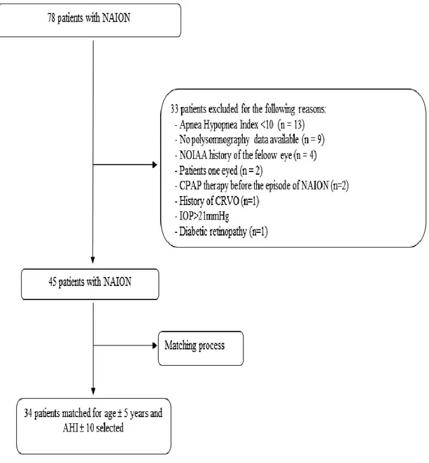
polysomnography (PSG). A total of 45 patients with unilateral NAION with no ocular abnormalities on the fellow eye and who had an AHI > 10 on PSG recording were included (see figure 1).
Diagnostic criteria of NAAION include: (1) history of sudden (less than 14 days) painless loss of vision; (2) documentation of optic disc edema at the time of visual loss (3); visual field defects consistent with optic-disc pathology; (4) no evidence of another neurologic or ocular disorder that may be responsible for optic disc edema and visual impairment; (5) exclusion of arteritic anterior ischemic optic neuropathy (22).
The diagnosis of arteritic anterior ischemic optic neuropathy was made on the basis of a combination of clinical and laboratory signs: the onset of clinical signs of giant cell arteritis, elevated erythrocyte sedimentation rate and C-reactive protein, white papilledema, occlusion associating the cilio-retinal arteries, choroidal ischemia on fluorescein angiography and/or a positive histological examination of the temporal artery biopsy. Patients presenting with diffuse or sectorial atrophy and/or whiteness of the contralateral optic nerve were not included in the study.
A comprehensive general clinical and ophthalmic examination with measurement of visual acuity, anterior segment and fundus examination, and a measurement of intraocular pressure using a Goldmann applanation tonometer were performed. Photographs (30°) of the papilla were taken to measure the diameter of the disc and its cup (vertical cup/disc ratio). Keratometry and measurement of the optical correction allowed the ocular magnification to be corrected with Littman’s algorithm.
After screening, patients underwent fluorescein angiography (Topcon angiograph, Topcon 50 IA camera, Tokyo, Japan) and a visual field examination (HumphreyTM automated field analyzer 24-2 SITA-standard, Zeiss, Dublin, California, USA). The visual field was considered reliable and healthy only if the false negatives and false positives were below 25%, and if the loss of fixation was less than 20%.
An arbitrary score of the mean deviation (MD) of -34 decibels was assigned to patients with excessive loss in visual acuity and in whom the entire visual field could not be attained. Visual field defects were classified under 13 categories as defined by Feldon et al in the IONDT study (20). Single eye may have more than one type of defect.
The assessment systematically included repeated measurements of daytime blood pressure (8 times/ day), laboratory tests for biological inflammatory syndrome (erythrocyte sedimentation rate and C-reactive protein), dyslipidemia (total
cholesterol, triglycerides, HDL cholesterol), and measurement of fasting blood glucose. An examination of the carotid arteries was performed with an ultrasound of the supra-aortic trunks (HP Sonos 2500® scanner, equipped with a probe of 7.5 MHz, Hewlett-Packard, Santa Clara, California, USA). Smoking status was
ascertained by questioning, with estimated number of cigarettes smoked per day and the number of years of smoking.
A nocturnal polysomnogram and the Epworth questionnaire were
systematically performed to detect sleep apnea syndrome. As polysomnography is the gold standard for documenting abnormal events occurring during sleep, a continuous recording was made using C3/A2-C4/A1-Cz/01 electrodes. Sleep was rated manually according to standard criteria. The airflow rate was measured using nasal pressure along with nasal and buccal thermistor measurements. The
assessment of respiratory effort was made using the thoracic and abdominal movements and the transit time of the pulse. The measurement of blood oxygen saturation was carried out using a pulse oximeter (Biox-Ohmeda 3700, Ohmeda, Liberty Corner, NJ, USA). Sleep apnea was defined as an apnea-hypopnea index (AHI) of 10 or more per hour.
CONTROL GROUP
The control group consisted of 40 consecutive patients who were referred for their first time polysomnography recording in the pneumology department of the same hospital and who had an OSA with AHI >10.
A comprehensive general clinical and ophthalmic examination with measurement of visual acuity, anterior segment and fundus examination, and a measurement of intraocular pressure using a Goldmann applanation tonometer was performed. Photographs (30°) of the papilla were taken to measure the diameter of the disc and its cup (vertical cup/disc ratio). Keratometry and a measurement of the optical correction allowed the ocular magnification to be corrected with Littman’s algorithm.
After screening, patients underwent a visual field examination (HumphreyTM automated field analyzer 24-2 SITA-standard, Zeiss, Dublin, California, USA). The visual field was considered reliable and healthy only if the false negatives and false positives were below 25%, and loss of fixation was less than 20%. Visual field
defects were classified under 13 categories as defined by Feldon et al (20). Smoking status was ascertained by questioning, with an estimate of the number of cigarettes smoked per day and the number of years of smoking.
A nocturnal polysomnogram and the Epworth questionnaire were
systematically performed to detect sleep apnea syndrome. As polysomnography is the gold standard for documenting abnormal events occurring during sleep, a
continuous recording was made using C3/A2-C4/A1-Cz/01 electrodes. Sleep was rated manually according to standard criteria. The air flow rate was measured using nasal pressure along with nasal and buccal thermistor measurements. The
assessment of respiratory effort was made using the thoracic and abdominal movements and the transit time of the pulse. The measurement of blood oxygen saturation was carried out using a pulse oximeter (Biox-Ohmeda 3700, Ohmeda, Liberty Corner, NJ, USA). Sleep apnea was defined as an apnea-hypopnea index (AHI) of 10 or more per hour.
Patients who had any ocular pathologies that may interfere with visual field test i.e.: glaucoma, myopia >-5 D, tilted optic disc, optic disc drusens and systemic diseases with possible effects on the visual field were excluded.
STATISTICS
The following parameters of the visual field were collected: presence or absence of defect defined in the IONDT classification, the type of defect if present, test time, foveal sensitivity (FS), number of fixation loss (FL), mean deviation (MD), visual field index (VFI), pattern standard deviation (PSD), false positive rate (FP), false negative rate (FN), total number of points out of 52 points of visual field with their probability symbols <5%, 2%, 1% and <0.5%.
One OSA patient was matched to maximum three NAION patients. The
controls were matched in a masked fashion for age (± 5 years) and AHI (± 10 events per hour). This matching allowed us to select 17 (34 eyes) out of 40 OSA without NAION and 34 out of 46 NAION (34 eyes) patients.
Description of the constructed models:
Common characteristics: age, body mass index (BMI), AHI and sex were compared between two groups of patients without adjustment but the matching was respected.
Continuous data:
As data were correlated within the strata (and because each patient without NAION had two repeated measures, for each eye), generalized estimating equations (GEE) were used to estimate the effects properly adjusted for non-independent contributions. To take into account the strata and two repeated measures for each patient without NAION, categorical data were studied with conditional logistic regression models.
Statistical analyses were conducted with the SAS 9.1 (SAS institute, Cary, NC, USA) software package. All tests were conducted in a bilateral manner. Differences were considered statistically significant when the probability (p) of a type I error was below 5%. Continuous variables were tested for normality using the Shapiro-Wilk test.
RESULTS
PATIENTS CHARACTERISTICS
Among a total of 78 patients with NAION seen in the University Hospital of Grenoble between 2002 and 2010, 46 patients with OSA were included. A flow chart showing the reasons for exclusion is illustrated in figure 1. Considering the matching criteria, a total of 34 eyes out of 45 fellow eyes with unilateral NAION were matched to 34 out of 80 eyes of 40 controlled OSA patients. The mean AHI in NAION patients was 35.5 ± 11.6 versus 35.4 ± 9.5 in control (p = 0.6305). The mean age of patients was slightly but significantly higher in NAION patients than in control: 69.8 ± 8.2 versus 63.9 ± 9.9 (p < 0.001).
Figure 1. Flow chart showing the reasons for exclusion of patients with NAION.
NAION: Non-arteritic ischemic optic neuropathy, CPAP: continuous positive airway pressure therapy, CRVO: central retinal vein occlusion, IOP: intraocular pressure, AHI: apnea-hypopnea index.
Figure 2. Examples of visual field of subjects with NAION and SAS controls.
(C) Example of inferior arcuate defect in fellow eye of NAION patient (left) and control patient (right)
Table 1. Demographic and ophthalmic characteristics.
NAION Controls p value
Number of patients 34 17 Age 69.8 ± 8.2 63.9 ± 9.9 < 0.001 Sex Male Female 20 12 0.413 14 5 BMI 26.9 ± 4.3 29.4 ± 5.5 0.0433 IAH 35.5 ± 11.6 35.4 ± 9.5 0.6305
Visual acuity (log MAR) 1.27 ± 0.9 0.05± 0.1 <0.05
Systemic Hypertension 55.8% 64.7% 0.687 Diabetes 23.5% 17.6% 0.630 Cerebral Stroke 2.9% 0 0.475 Cardiac ischemia 2.9% 23.5% <0.05 Dyslipidemia 35.3% 41.2% 0.733 Tobacco 11.8% 11.8% 0
NAION: non-arteritic ischemic optic neuropathy. BMI: body mass index. AHI: apnea-hypopnea index.
VISUAL FIELD
After adjustment for age and body mass index, all the parameters of the visual field were significantly different in the NAION fellow eye and control group, except loss of fixation: mean deviation (-4.5 ± 3.7 vs -1.3 ± 1.8, respectively, p<0.05), visual field index (91.6 ± 10 vs 97.4 ± 3.5, respectively, p=0.002), pattern standard deviation (3.74 ± 2.27 vs 2.45 ± 1.96, respectively, p=0.0019), number of subjects with at least one defects of the IONDT classification (20 vs 10, respectively, p<0.05) (see Tables 3 and 4)
Table 3. Visual field parameters (parameters with normal distribution).
Controls (n = 34)
NAION (n = 34)
Mean ± SD Mean ± SD Mean inter group difference Mean intra strata difference 95%CI intra strata difference P Value Time 5.16 ± 0.91 6.5 ± 1.23 1.34 1.17 0.73-1.61 <.0001 PSD 2.45 ± 1.96 3.74 ± 2.27 1.28 1.25 0.46-2.04 0,0019 N° points < 5% 9.50 ± 9.47 25.79 ± 16.14 16.29 14.73 9.25-20.21 <.0001 N° points < 2% 5.76 ± 7.41 17.88 ± 14.95 12.12 11.85 7.03-16.68 <.0001
NAION: non-arteritic ischemic optic neuropathy. SD: standard deviation. CI: confidence interval. PSD: pattern standard deviation
Table 4. Visual field parameters comparisons (parameters with non-normal distribution).
NAION vs Controls NAION (n = 34) Controls (n = 34) OR 95% CI P-Value FS > median (35) versus ≤ 33.5 (32; 36) 36 (33.8; 37.3) 0.37 0.11-1.30 0.1213 FL > median (0.13) versus ≤ 0.1 (0; 0.2) 0.07 (0; 0.3) 1.14 0.40-3.29 0.8091 VFI > median (97) versus ≤ 95 (90.8; 97.5) 98 (97; 99) 0.09 0.02-0.43 0.0024 MD > median (-2.045) versus ≤ -3.4 (-6.5; -1.9) -1.1 (-2.2; -0.02) 0.06 0.01-0.46 0.0075 FP > median (1) versus ≤ 2 (0; 5.3) 1 (0; 2.3) 3.22 1.00-10.40 0.0504 FN > median (2.5) versus ≤ 5 (0; 9.5) 0 (0; 4.5) 3.02 0.96-9.54 0.0597 N° points < 1% > median (5) versus ≤ 9 (4.8; 21.5) 0.5 (0; 5) 8.37 2.26-31.05 0.0015 N° points < 0.5% > median (2) versus ≤ 3 (1; 15) 0 (0; 2.3) 5.14 1.61-16.40 0.0057 IONDT classification defect: yes vs no 20 (58.8%) 10 (29.4%) 4.10 1.28-13.16 0.0179 Diffuse depression, yes vs no 10 (29.4%) 3 (8.8%) 2.96 0.73-12.03 0.1286 Superior arcuate, yes vs no 5 (14.7%) 4 (11.8%) 1.19 0.21-6.89 0.8452
Second and third columns: data are presented with median (25-75 percentiles) or number and percent for categorical data. NAION: non- arteritic ischemic optic neuropathy. OR: odds ratio. CI: confidence interval. FS: foveal sensitivity. FL: fixation loss. VFI: visual field index. MD: mean deviation. FP: false positive rate. FN: false negative rate. IONDT: Ischemic Optic Neuropathy Decompression Trial.
DISCUSSION
The aim our study was to find out if OSA can explain visual field defects noted on the fellow eye in patients presenting unilateral NAION. For this purpose, we compared two groups, one who already had a NAION and diagnosed with OAS by polysomnograhy to those who were recently diagnosed as OAS and never had an ophthalmic problem that may interfere with the outcome of visual field. Those who were already having CPAP therapy for their known OSA before the episode of NAION were also excluded so that the eventual benefit of this therapy may not interfere with the visual field.
After constituting two groups (46 NAION and OSA patients and 40 OSA without NAION), they were matched for the age ± 5 years and for the AHI ± 10. This allowed us to select 34 eyes of NAION patients and 34 eyes of 17 OSA patients. The two groups were strictly similar regarding the severity of OSA (35.5 ± 11.6 for NAION
vs 35.4 ± 9.5 for OSA, p=0.63). Male patients were present in comparable proportion
in each group. The two groups were also similar regarding most of the cardiovascular risk factors. Only the rate of ischemic cardiopathy was significantly higher in OSA patients.
The two groups were slightly different for age (69.8 ± 8.2 years for NAON patients versus 63.9 ± 9.9 for OSA controlled patients, p<0.0001) and body mass index (26.9 ± 4.1 for NAON patients versus 29.4 ± 5.5 for OSA controlled patients, p<0.05). To take this into account, visual field comparisons were adjusted for age and body mass index.
In our study, all the parameters concerning the visual fields were significantly different in two groups, with the fellow eye of NAION having significantly more visual field abnormalities and defects than the eyes of OSA patients matched for the
severity of OSA. Superior arcuate (5/20) and inferior arcuate (7/20) defects were the most common defects found in the fellow eyes of the patients with unilateral NAION.
Scherer et al (15), in one of the reports of the IONDT pointed out that almost three quarters of non-study eye i.e. the fellow eye of unilateral NAION at the baseline presented mild superior and/or inferior arcuate defects. They also remarked that the
defects improved in almost 25% of eyes in 6-month follow up visit but did not disappear completely, suggesting the existence of true defects. The authors in the study tried to explain these defects as a result of media opacity. However this reason may not explain the arcuate defects, which in general are the results of ganglion cell death. They also compared visual fields for eyes of randomized patients according to the presence of vascular conditions, but taking this criteria into account, there were no changes in the distribution of defect pattern type from baseline to either follow up visits. At that time, the authors had not evaluated whether the defects were caused by the existence of OSA.
In our study, visual field defect as defined in the IONDT classification was present in (25/45) eyes of initially selected NAION patients and (20/34) matched NAION patients. Since the patients were not followed up, the evolution of their visual field defects is not known.
Tsang et al (20) have also found that the moderate to severe OSA is
associated with a higher incidence of visual field defect compared to an age matched non-OSA patients. They demonstrated two visual field global indices i.e. MD and PSD significantly higher in patients with OSA to those matched for the age and without OSA. The patients in these two arms showed statistically significant
difference on the optic disc glaucomatous changes i.e. cup: disc ratio greater than 0.5, signs of pathological disc changes like vertical cupping, rim notching, disc haemorrhage, or disc swelling (26.39 against 6.78%). The authors hypothesized a ganglion cells loss in OSA patients without glaucoma.
Mojon et al (19) equally found an association between SAS and visual field defects. They found that the respiratory disturbance index correlated positively with the mean visual field defect (r = 0.81, P< 0.05) and with the visual field loss variance (r = 0.78, P<0.05). They also found relative arcuate visual field defects in OSA patients suggesting an optic neuropathy prevailing in those patients.
Three studies have shown that most of the NAION patients present with an OSA. Mojon et al (16) had reported in 2002 in a prospective study 71% (12/17) of NAION patients had an OSA compared to age and sex matched population. 15 out of
higher in male than female (22). Palombi et al (17) in their prospective study in 2006, found that 89% (24/27) of patients presenting a NAION had an OSA. The risk ratio for a NAION patient to have sleep apnea was 4.9 compared to the general population (p<0.001). And the prevalence rate of OSA was 1.5 to 2 fold more than the rate of other identified risk factors classically associated with NAION i.e. hypertension, diabetes etc. Recently, these results were consolidated by Jian Li et al (18). NAION patients were significantly more likely to report symptoms and characteristics
consistent with SAS than controls (OR 2.62; 95% CI 1.03 to 6.60) when adjusted for medical and health behavior characteristics on a telephone survey based on Sleep Apnea scale of the Sleep Disorders Questionnaire (SA-SDQ).
In our study, all the visual field parameters were significantly different between two groups (NAION with OSA patients and OSA patients matched for the severity of the OSA), thus suggesting that the defects found in the fellow eye of NAION are not explained alone by the presence of OSA and there are other unknown reasons behind these findings. To date, the pathophysiology and natural history of NOIAA is not fully known. The results of the present study could suggest that episodes of subclinical NAION frequently occur before the onset of a symptomatic episode, and that NAION is not a sudden vascular event occurring on one eye so far unharmed.
REFERENCES
1. Rucker JC, Biousse V, Newman NJ. Ischemic optic neuropathies. Current opinion in neurology. 2004;17(1):27-35. Epub 2004/04/20.
2. Repka MX, Savino PJ, Schatz NJ, Sergott RC. Clinical profile and long-term implications of anterior ischemic optic neuropathy. American journal of
ophthalmology. 1983;96(4):478-83. Epub 1983/10/01.
3. Johnson LN, Arnold AC. Incidence of nonarteritic and arteritic anterior
ischemic optic neuropathy. Population-based study in the state of Missouri and Los Angeles County, California. Journal of neuro-ophthalmology : the official journal of the North American Neuro-Ophthalmology Society. 1994;14(1):38-44. Epub 1994/03/01.
4. Hattenhauer MG, Leavitt JA, Hodge DO, Grill R, Gray DT. Incidence of nonarteritic anterior ischemic optic neuropathy. American journal of ophthalmology. 1997;123(1):103-7. Epub 1997/01/01.
5. Knox DL, Kerrison JB, Green WR. Histopathologic studies of ischemic optic neuropathy. Transactions of the American Ophthalmological Society. 2000;98:203-20; discussion 21-2. Epub 2001/02/24.
6. Hayreh SS, Joos KM, Podhajsky PA, Long CR. Systemic diseases associated with nonarteritic anterior ischemic optic neuropathy. American journal of
ophthalmology. 1994;118(6):766-80. Epub 1994/12/15.
7. Salomon O, Huna-Baron R, Kurtz S, Steinberg DM, Moisseiev J, Rosenberg N, et al. Analysis of prothrombotic and vascular risk factors in patients with
nonarteritic anterior ischemic optic neuropathy. Ophthalmology. 1999;106(4):739-42. Epub 1999/04/14.
8. Jacobson DM, Vierkant RA, Belongia EA. Nonarteritic anterior ischemic optic neuropathy. A case-control study of potential risk factors. Archives of ophthalmology. 1997;115(11):1403-7. Epub 1997/11/21.
9. Lee MS, Grossman D, Arnold AC, Sloan FA. Incidence of nonarteritic anterior ischemic optic neuropathy: increased risk among diabetic patients. Ophthalmology. 2011;118(5):959-63. Epub 2011/03/29.
10. Deramo VA, Sergott RC, Augsburger JJ, Foroozan R, Savino PJ, Leone A. Ischemic optic neuropathy as the first manifestation of elevated cholesterol levels in young patients. Ophthalmology. 2003;110(5):1041-6; discussion 6. Epub 2003/05/17.
11. Hayreh SS, Zimmerman MB, Podhajsky P, Alward WL. Nocturnal arterial hypotension and its role in optic nerve head and ocular ischemic disorders. American journal of ophthalmology. 1994;117(5):603-24. Epub 1994/05/15.
12. Beck RW, Savino PJ, Repka MX, Schatz NJ, Sergott RC. Optic disc structure in anterior ischemic optic neuropathy. Ophthalmology. 1984;91(11):1334-7. Epub 1984/11/01.
13. Burde RM. Optic disk risk factors for nonarteritic anterior ischemic optic neuropathy. American journal of ophthalmology. 1993;116(6):759-64. Epub 1993/12/15.
14. Jonas JB. Optic disc morphology and NAION. The British journal of ophthalmology. 2009;93(6):703. Epub 2009/05/28.
15. Scherer RW, Feldon SE, Levin L, Langenberg P, Katz J, Keyl PM, et al. Visual fields at follow-up in the Ischemic Optic Neuropathy Decompression Trial: evaluation of change in pattern defect and severity over time. Ophthalmology.
2008;115(10):1809-17. Epub 2008/05/20.
16. Mojon DS, Hedges TR, 3rd, Ehrenberg B, Karam EZ, Goldblum D, Abou-Chebl A, et al. Association between sleep apnea syndrome and nonarteritic anterior ischemic optic neuropathy. Archives of ophthalmology. 2002;120(5):601-5. Epub 2002/05/11.
17. Palombi K, Renard E, Levy P, Chiquet C, Deschaux C, Romanet JP, et al. Non-arteritic anterior ischaemic optic neuropathy is nearly systematically associated with obstructive sleep apnoea. The British journal of ophthalmology. 2006;90(7):879-82. Epub 2006/03/25.
18. Li J, McGwin G, Jr., Vaphiades MS, Owsley C. Non-arteritic anterior ischaemic optic neuropathy and presumed sleep apnoea syndrome screened by the Sleep Apnea scale of the Sleep Disorders Questionnaire (SA-SDQ). The British journal of ophthalmology. 2007;91(11):1524-7. Epub 2007/05/17.
19. Mojon DS, Mathis J, Zulauf M, Koerner F, Hess CW. Optic neuropathy associated with sleep apnea syndrome. Ophthalmology. 1998;105(5):874-7. Epub 1998/05/21.
20. Tsang CS, Chong SL, Ho CK, Li MF. Moderate to severe obstructive sleep apnoea patients is associated with a higher incidence of visual field defect. Eye (Lond). 2006;20(1):38-42. Epub 2005/01/15.
syndrome. Graefe's archive for clinical and experimental ophthalmology = Albrecht von Graefes Archiv fur klinische und experimentelle Ophthalmologie.
2011;249(4):585-93. Epub 2010/10/20.
22. Hayreh SS. Ischemic optic neuropathy. Progress in retinal and eye research. 2009;28(1):34-62. Epub 2008/12/10.