Review Article
Chronic Maxillary Rhinosinusitis of Dental Origin:
A Systematic Review of 674 Patient Cases
Jerome R. Lechien,
1,2Olivier Filleul,
1Pedro Costa de Araujo,
1Julien W. Hsieh,
3Gilbert Chantrain,
4and Sven Saussez
1,41Laboratory of Anatomy and Cell Biology, Faculty of Medicine, UMONS Research Institute for Health Sciences and Technology,
University of Mons (UMons), Avenue du Champ de Mars 6, B7000 Mons, Belgium
2Laboratory of Phonetics, Faculty of Psychology, Research Institute for Language Sciences and Technology, University of Mons (UMons),
B7000 Mons, Belgium
3Laboratory of Neurogenetics and Behavior, Rockefeller University, 1230 York Avenue, New York City, NY 10065, USA
4Department of Otorhinolaryngology, Head, and Neck Surgery, CHU Saint-Pierre, Faculty of Medicine,
Universit´e Libre de Bruxelles (ULB), B1000 Brussels, Belgium
Correspondence should be addressed to Sven Saussez; sven.saussez@hotmail.com
Received 19 December 2013; Revised 6 March 2014; Accepted 11 March 2014; Published 8 April 2014 Academic Editor: Charles Monroe Myer
Copyright © 2014 Jerome R. Lechien et al. This is an open access article distributed under the Creative Commons Attribution License, which permits unrestricted use, distribution, and reproduction in any medium, provided the original work is properly cited.
Objectives. The aim of this systematic review is to study the causes of odontogenic chronic maxillary rhinosinusitis (CMRS), the
average age of the patients, the distribution by sex, and the teeth involved. Materials and Methods. We performed an EMBASE-, Cochrane-EMBASE-, and PubMed-based review of all of the described cases of odontogenic CMRS from January 1980 to January 2013. Issues of clinical relevance, such as the primary aetiology and the teeth involved, were evaluated for each case. Results. From the 190 identified publications, 23 were selected for a total of 674 patients following inclusion criteria. According to these data, the main cause of odontogenic CMRS is iatrogenic, accounting for 65.7% of the cases. Apical periodontal pathologies (apical granulomas, odontogenic cysts, and apical periodontitis) follow them and account for 25.1% of the cases. The most commonly involved teeth are the first and second molars. Conclusion. Odontogenic CMRS is a common disease that must be suspected whenever a patient undergoing dental treatment presents unilateral maxillary chronic rhinosinusitis.
1. Introduction
Chronic rhinosinusitis (CRS) is the most frequent pathology
in USA, since it affects 33.7 million people each year [1],
representing nearly 14% of the American population [2].
According to various reports, a dental origin is found in 5 to
40% of cases of chronic maxillary rhinosinusitis (CMRS) [1,3,
4]. CMRS is defined by the presence of ongoing rhinosinusal
symptoms for at least 12 weeks [5, 6]. Its incidence is
consistently growing and it is more frequent among women
[7]. The majority of CMRS patients are between 30 and 50
years old. From an anatomic perspective, maxillary sinus are air-filled cavities situated laterally to the nasal fossae and communicate with them through an ostium which is approximately 4 millimetres in diameter and vulnerable
to occlusion during mucosal inflammation [8]. The
maxil-lary sinus anatomical relationships involve the dental roots inferiorly, explaining the easy extension of the infectious
processes from some teeth to the maxillary sinus [3,9]. The
paranasal sinuses and the whole nasal fossae are covered with a ciliated pseudostratified epithelium. The essential role of this epithelium is the secretion of respiratory mucus and its movement to the nasopharynx, ensuring elimination of sinus secretions towards the nasal fossa. Normal mucociliary clearance requires an adequate permeability of the sinus
ostium as well as good secretory and ciliary functions [10].
From a pathophysiological point of view, CMRS is due
to a temporary and reversible mucociliary dyskinesia [11],
which could be favoured by several factors: gastroesophageal
reflux disease [12], atmospheric pollution [13], smoking [14],
Volume 2014, Article ID 465173, 9 pages http://dx.doi.org/10.1155/2014/465173
nasosinusal polyposis [15], arterial hypertension [15], dental infections, anatomic malformations such as septal deviations,
concha bullosa, allergic reactions, and immune deficits [16–
20]. Odontogenic CMRS occurs when the Schneiderian
membrane is irritated or perforated, as a result of a dental infection, maxillary trauma, foreign body into the sinus, maxillary bone pathology, the placing of dental implants in the maxillary bone, supernumerary teeth, periapical granu-loma, inflammatory keratocyst, or dental surgery like dental
extractions or orthognathic osteotomies [3,21]. Among the
CMRS induced by foreign bodies, one might distinguish between exogenous or, less frequently, endogenous foreign bodies. The most frequent types of exogenous foreign bodies
are endodontic material used in dental obturation [9]; these
foreign bodies can trigger an inflammatory response and an
alteration of the ciliary function [22, 23]. A CMRS caused
by a dental infection can take two different routes to spread the infection. It can extend into the sinus through the pulp chamber of the tooth, causing an apical periodontitis. If the “tooth height” is altered due to a chronic infection and destruction of the tooth socket, we call it a marginal periodontitis.
Once the drainage is compromised by mucosal oedema, sinus infection may start involving various microorganisms. In bacteriological studies, it is well recognised that anaerobes can be isolated in up to two-thirds of patients who have CRS,
mostly in the setting of a polymicrobial infection [24].
𝛼-hemolytic Streptococcus spp., microaerophilic Streptococcus spp., and Staphylococcus aureus are predominant aerobes and the predominant anaerobes are Peptostreptococcus spp.
and Fusobacterium spp. [3]. There is a difference between
the bacteriology of odontogenic CMRS and that of other cases; however, in clinical practice, taking an uncontaminated bacteriological sample might turn out to be difficult. In addition, fungal superinfections are frequent and increased by immunodeficiency, diabetes mellitus, sinus radiotherapy
and, excessive antibiotic and corticosteroid use [10,17, 25].
Dental amalgams may sometimes contain minerals such as zinc oxide, sulphur, lead, titanium, barium, calcium salts,
and bismuth that may accelerate fungal growth [17].
Micro-biological findings often reveal Aspergillus fumigatus and, more rarely, Aspergillus flavus, which may be much more
aggressive [17,25,26]. Different theories are put forward to
explain those aspergillus superinfections. Following a French etiologic hypothesis, an Aspergillus infection would also be odontogenic, requiring an oroantral fistula to allow sinus contamination. Other hypotheses favour a mixed origin or strict aerogenic contamination via heavy spore inhalation
over an extended period of time [22,27]. CMRS is clinically
characterised by a variable association of symptoms includ-ing anterior or posterior, unilateral or sometimes bilateral discharge (purulent, watery, or mucoid), sinus or dental pain, nasal obstruction, hypo- or anosmia facial headaches that intensify in the evening while bending, halitosis, and
occasionally coughing [17]. Even if there is no significant
difference between classic and odontogenic CMR, anterior discharge, sinus pain, nagging pain of the upper teeth of the damaged side that increases during occlusion and tooth mobilisation, and halitosis seem to be more frequent in
the latter [21,25]. Percussion of the causal tooth may reveal
an abnormal sensitivity, unless endodontic filling has been performed. Most cases are unilateral, although bilateral cases
have been described as well [7]. The time interval between
symptoms onset and the causal dental procedure may be highly variable: according to Mehra and Murad, 41% of patients developed CMRS in the following month, 18% between one and three months after the procedure, 30% from three months to one year, and 11% of patients after
more than one year [8]. Computed tomography (CT) of the
sinus is essential. Some authors also recommend the Valsalva
test for diagnosing an oroantral communication [10]. Most
of the literature concerning odontogenic CMRS consists of either prospective or retrospective reports, and the guidelines on how to deal with the disease are often based on expert opinions.
2. Materials and Methods
2.1. Aim. The aim of this review is to define the aetiologies of odontogenic CMRS and the teeth involved.
2.2. Literature Search and Data Extraction. The literature was reviewed independently by three different authors (Jerome R. Lechien, Pedro Costa de Araujo, and Julien W. Hsieh) to minimise inclusion biases. The authors were not blinded to the study author(s), their institutions, the journal, or the results of the studies. The search for articles was done through
PubMED, Cochrane Library, and EMBASE (Figure 1). It
included all articles written in English, French, and other lan-guages and published between January 1980 and January 2013. We focused only on published papers. The keywords used were “odontogenic, chronic, maxillary sinusitis, dental, cyst, foreign body, iatrogenic, and periodontitis.” The initial 190 references (including case reports, retrospective and prospec-tive studies) were manually sorted to extract all descriptions of patients meeting the diagnostic criteria of chronic maxil-lary rhinosinusitis proposed by the European position paper
on rhinosinusitis and nasal polyps 2012 [6]. Methodologic
quality was assessed by the authors to determine the validity of each study. When important data were missing in some studies, the first author (Jerome R. Lechien) tried to contact the authors to obtain the additional information. In addition, references were obtained from citations within the retrieved articles. To avoid multiple inclusions of patients, we checked for the age, gender, author, and geographic area, whenever they were available. If a patient was described in more than one publication, we used only the data reported in the larger and more recent publication. Patient demographic data, age, gender, and the teeth involved in odontogenic cases were only recorded on the basis of individual data; if it was impossible to obtain these data from the authors, they were considered missing.
2.3. Inclusion and Exclusion Criteria. The diagnosis of CMRS was based on;
(1) the presence of ongoing rhinosinusal symptoms for at least 12 weeks secondary to a clearly identified dental
Combination of keywords Custom date range:
J.R.L. P.C. J.H.
Articles screened, identified by database searching, and assessed for
eligibility (n = 190)
Discussion among J.R.L., P.C., and J.H.
PubMED Cochrane library EMBASE
Studies included in the
Exclusion criteria Inclusion criteria systematic review (n = 23) Retrospective uncontrolled case studies (n = 10) (n = 6) (n = 6) (n = 1) Case reports uncontrolledProspective
studies
Case-control study January 1980–January 2013
Figure 1: Flow chart shows the process of article selection for this study.
cause (including traumatic, iatrogenic, tumour, and dental infectious);
(2) the diagnosis of CMRS should be confirmed by computed tomography or by panoramic radiography. Concerning periodontal infections, they were defined as clearly identified infections around the teeth that were con-comitant of CMRS. Immunocompromised patients, cases of acute and subacute rhinosinusitis, and unclear causes of dental origin and cases where the type of rhinosinusitis is not clear were excluded.
3. Results
Our database search yielded 190 articles. From these, we selected 23 articles, including 6 isolated case reports, 10 ret-rospective uncontrolled case studies describing 389 patients, 6 prospective uncontrolled studies describing 192 patients,
and one case-control study describing 91 patients [11,15,22,
23, 26–44]. The description of all articles and ventilation
of cases is displayed in Table 1. Among the 23 papers, 18
were published in English, two in both English and Spanish, and three in French. Fifty-four percent of all patients were women, and average patient age at diagnosis was 45.6 years (ranging between 12 and 81 years). The different aetiologies
found in the literature search are summarized in Figure 2.
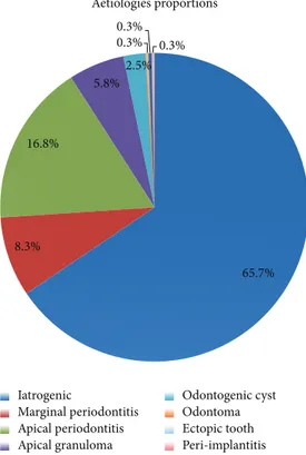
Based on the 674 patients for whom it was displayed, iatro-genic causes were the most frequent, accounting for 65.7% of cases of described odontogenic maxillary rhinosinusitis. They included impacted tooth after dental care, artificial implants, dental amalgams in the sinus,and oroantral fistula. They were followed by apical periodontal pathologies, accounting for 25.1% of the cases. Apical periodontal pathologies include apical periodontitis (16.8%), apical granulomas (5.8%), and odontogenic cysts (2.5%). Unfortunately, the paucity of clini-cal descriptions limited the data of the involved teeth to only
236 cases. Nevertheless, as shown inFigure 3, the first and
second molars were the most commonly affected teeth when reported, representing 35.6% and 22% of cases, respectively. They were followed by the third molar (17.4%) and the second premolar (14.4%).
4. Discussion
The aim of our study was to describe the aetiologies of odontogenic CMRS, the teeth involved, and age and sex distribution. To our knowledge, this paper is the first review studying the causes of CMRS. Further descriptions of CMRS causes were displayed in consecutive case series. In a case series of 70 patients with odontogenic CMRS, published by
T a ble 1: G eneral ch ar ac te ri st ics o f the st udies. G eneral ta b le des cr ib in g th e st udies cha rac ter ist ics (inc ludin g ca teg o ry o f evidence fo llo win g th e E u ro pea n po si ti o n pa pe r o n rh in o si n u si ti s an d n as al po ly ps 2007 re co mm en da tio n s [ 6 ]) n u m b er o f cas es, aet io log y, middle ag e, se x, an d the in vo lv ed te et h. CA: ca teg o ry o f evidence ,N A :n o t av ai la b le . A u th o rs Y ea r R ev ie w L an gua ge St u d y design CA 𝑛 to t A et io log y 𝑛 M iddle ag e (ra n ge d) Se x F Se x M To o th 𝑛 Li n d ahl et al . [ 39 ] 19 81 A ct a O tol ar yn gol E ng li sh Pro sp ec tive ca se se ri es III 29 M ar gi n al pe ri od o n ti ti s A p ical p er io d o n ti ti s Iat ro ge n ia 13 14 2 52 42 40 NA NA C anina 1st p re mo la r 2nd p re mo la r 1st m o la r 2nd m o la r 2 5 11 17 10 Me le n et al . [ 22 ] 19 86 A ct a O tol ar yn gol E ng li sh Pro sp ec tive ca se se ri es III 9 9 Iat ro ge n ia M ar gi n al pe ri od o n ti ti s Gr an u lom a api ca l 17 43 39 48 NA NA 2nd Incisi va C anina 1st p re mo la r 2nd p re mo la r 1st m o la r 2nd m o la r 3r d m o la r 1 4 11 23 56 34 9 Flign y et al . [ 27 ] 19 91 A n n O to -L ar yn g F re n ch Pro sp ec tive ca se se ri es III 14 Ia tr og enesis 14 4 2 (22–6 0 ) NA NA NA Li n et al . [ 40 ] 19 91 E ar N os e Th ro at J E n glish Retr os p ecti ve ca se se ri es III 16 Ia tr og enesis 16 11 –6 0 41 2 C anina 1st m o la r 2nd m o la r 3r d m o la r 1 5 3 2 The voz et al. [ 28 ] 2000 Sc h w ei z M ed Wo ch en sc h r Fre n ch Retr os p ecti ve ca se se ri es III 10 Ia tr og enesis 10 4 8 NA NA NA D o ud Galli et al . [ 29 ] 20 01 Am J R hino log y E n glish Retr os p ecti ve ca se se ri es III 14 Ia tr og enesis 14 (21 – 80) 10 4 NA Lo pa ti n et al . [ 30 ] 200 2 L ar yn gosc o pe En gli sh Retr os p ecti ve ca se se ri es III 70 Ia tr og enesis O d o n to ge nic cy st 60 10 (1 6–6 2) NA NA 3r d m o la r 26 C edin et al . [ 31 ] 2005 Br az J Ot o rhino la ryn go l En gl ish Retr os p ecti ve ca se se ri es III 4 Ia tr o ge nesis 4 N A NA NA NA N imig ea n et al . [ 32 ] 2006 B-ENT E n gli sh Retr os p ecti ve ca se se ri es III 12 5 A p ical p er io d o n ti ti s Ia tr og enesis 99 26 46 (1 2– 81 ) 69 56 NA Se lm an i an d A sha mmak hi [ 33 ] 2006 J cra nio fac surger y En gl ish Pro sp ec tive ca se se ri es III 13 Ia tr og enesis 13 45 (26–8 1) 85 NA U ginci us et al . [ 15 ] 20 0 6 St o m at o logi ja E n glish Retr os p ecti ve ca se se ri es III 13 6 Ia tr o ge nesis 13 6 N A NA NA NA Ma ca n et al . [ 41 ] 2006 D en to m axi llo-facial R adio log y En gl ish C as e R ep o rt III 1 Ia tr o ge nesis 1 61 10 NA
Ta b le 1: C o n ti n u ed . A u th o rs Y ea r R ev ie w L an gua ge St u d y design CA 𝑛 to t A et io log y 𝑛 M iddle ag e (ra n ge d) Se x F Se x M To o th 𝑛 Sr ini vas a P ras ad et al . [ 42 ] 20 07 In dia n J D en t R es En gl ish C as e R ep o rt III 1 E ct o p ic to o th 1 45 10 3r d m o la r 1 Me n si et al . [ 34 ] 20 0 7 OOOO E E n gl is h C as e Co n tr o l II B 91 Ia tr o ge n es is 91 N A NA NA NA Co st a et al . [ 23 ] 2007 Oral an d M ax ill o fa ci al Su rg er y En gl ish Pro sp ec tive ca se se ri es III 17 Ia tr og enesis O d o n to ge nic cy st P er i-imp la nt is 8 7 2 NA NA NA NA Cr esp o del H ier ro et al . [ 35 ] 2008 Ac ta Ot o rri n o la ri n go l Es p Sp an ish/Eng lish C as e R ep o rt III 1 O do n to m a 1 24 10 NA Ro dr igues et al . [ 11 ] 2009 Me d O ra lP at o l Oral Cir B ucal En gl ish C as e R ep o rt III 1 Ia tr o ge nesis 1 62 01 NA B o det Au gu st ´ıe ta l. [ 36 ] 2009 Ac ta Ot o rri n o la ri n go l Es p Sp an ish/Eng lish Retr os p ecti ve ca se se ri es III 10 Ia tr og enesis 10 N A NA NA NA Andr ic et al . [ 37 ] 20 10 OOOO E E n gl is h Retr os p ecti ve ca se se ri es III 14 Ia tr og enesis 14 4 0 59 1st p re mo la r 1st m o la r 2nd m o la r 3r d m o la r 1 6 5 2 H ajiioa n no u et al . [ 26 ] 20 10 J L ar yn go lO to l E n gli sh Pro sp ec tive ca se se ri es III 4 Ia tr o ge nesis 4 N A NA NA NA L ec hien et al . [ 38 ] 20 11 Re vue m edicale de B rux elles Fre n ch Retr os p ecti ve ca se se ri es III 2 Ia tr o ge nesis 2 36 20 NA Mo h an et al . [ 43 ] 20 11 N at io n al Jo ur nal of Ma xi ll of ac ia l Su rg er y E n gl is h C as e re p or t II I 1 E ct opi c to ot h 1 28 10 3r d m o la r 1 Kh o n sa ri et al . [ 44 ] 20 11 Re v St C hir M ax ill o fa c Eng lish C as e rep o rt III 1 O ste o ma 1 52 10 NA N o te :t he incidence o f o ro an tra l co mm unica tio n is estima ted at 0,58% o f all p remo la r an d m o la r extrac tio n (r ef ); o n e ca n assume th at th e incidence o f o ro an tra l fist u la is ev en sma ller ,k eep in g in m ind tha t so m e oro an tr al com m u n ic at ions w er e tre at ed im m ed ia te ly aft er th ei r cr ea ti on or h ea le d sp on ta n eou sl y. re f = P u n w ut ik or n J, W ai k aku lA .
65.7% 8.3% 16.8% 5.8% 2.5% 0.3% 0.3% 0.3% Aetiologies proportions Iatrogenic Marginal periodontitis Apical periodontitis Apical granuloma Odontogenic cyst Odontoma Ectopic tooth Peri-implantitis
Figure 2: Aetiology of odontogenic CMRS. The main cause of odontogenic CMRS is iatrogenic and accounts for 65.7% of cases. Apical periodontal pathologies (including apical periodontitis, api-cal granulomas, and odontogenic cysts) and marginal periodontitis follow them and account for 25.1% and 8.3%, respectively. Peri-implantitis, ectopic tooth, and odontoma remain rare causes of odontogenic CMRS.
Lopatin et al., an exogenous foreign body from the teeth was found in 10 cases (14%), of which 7 dental amalgam fillings and 3 dental packings, and an endogenous foreign
body (i.e. a tooth root) in 11 cases [30]. Thirty-nine patients
(56%) also presented an oroantral fistula. Although rare, the foreign body was sometimes inserted in the sinus through
trauma or accident [32]. In another case series of 125 patients
suffering from odontogenic CMRS, the main aetiology was periapical chronic periodontitis (79% of patients), followed by complications of endodontic treatment (21% of cases)
[32]. In addition, in two prospective studies of Melen et al.
and Lindahl et al., most cases of CMRS were secondary to a dental infectious process such as marginal periodontitis
and apical diseases [22, 39]. We compared the results of
their studies with ours, specifically looking at aetiologies. Our results are consistent with the study of Lopatin et al., showing a majority of iatrogenic causes in comparison with infectious aetiologies. Our results in favor of the iatrogenic cause can be explained in part by the high proportion of studies reporting only a large number of iatrogenic etiology
[15,27,29,34,37]. However, patient selection criteria were not
described in most of the studies. Therefore, we were unable to control for selection bias, and our study may be subject to under- and overreporting bias. Finally, in a case series written by Krause et al., focusing on the foreign bodies found in any of
the sinus, 60% of all foreign bodies were found to be
iatrogenic and 25% of industrial accidents [45]. The sinuses
affected were mainly the maxillary (75%) and frontal sinus (18%), foreign bodies in ethmoidal or sphenoid sinus being rare. Several studies found in the literature are limited by different biases. So the size of the clinical series is often relatively small, which may allow for undetected infrequent variants, and the retrospective design of the studies included did not let us make incidence estimations. Putting these limitations aside, the larger size of the sample studied allows for a better description of the pathology than what could be made based on a single case series, something crucial for a frequently overlooked condition. Concerning the gender distribution, our data show that women (57%) were slightly more affected by odontogenic CMRS than men (43%). Most clinical series are also characterized by a ratio in favour
of women [29, 32, 33]. Among the dental characteristics
found in the literature, information about the teeth involved is rare. Indeed, apart from the study of Lopatin et al. who reported involvement of the third molar, only three publications accurately investigated the teeth involved. The prospective study from Melen et al. shows that the most commonly involved teeth are the first (40.6%) and second
molars (24.6%) [22]. Even with a smaller sample, Andric
et al. observed similar proportions in their retrospective analysis where first and second molars account for 42%
and 35%, respectively [37]. Finally, Lindahl et al. reported a
higher proportion of the first molar (38%), followed by the
second premolar (24%) and second molar (22%) [39]. These
results can easily be explained by the preferential anatomical relationships between the floor of the maxillary sinus and the various teeth concerned (premolars, first and second molars). These proportions are similar in our study. However, our work is limited by the retrospective nature of most reports, which may include selection bias in the overall description.
The diagnosis of unilateral chronic maxillary RS required systematic dental examination and sinus computed
tomog-raphy (CT) [22]. CT, with reconstructions following the axial
and coronal planes, classically reveals sinus filling or a chronic mucous swelling associated with a reaction to foreign body
[46,47]. Interestingly, secondary aspergillosis, which is often
associated with dental foreign body and appeared as a luminal opacity, can be misinterpreted as calcified dental amalgam. Other types of sinus opacities include ectopic tooth frag-ments, calcified retention cysts, osteoma, condensing osteitis, calcified polyps, odontomas, osteosarcomas, cementomas,
bone fibrous dysplasia, and metastases of carcinoma [10].
Odontogenic CMRS is managed by both a medical and surgical approach. The first step consists of addressing the dental pathology and the second is a functional endoscopic sinus surgery. Starting with the dental intervention allows for the elimination of the origin of the infection as well as the removal of any newly introduced foreign sinus material in the same sinus endoscopy. Usually, the stomatologist or dental practitioner repeats the endodontic treatment or proceeds to an extraction. Addressing the sinusal component with func-tional endoscopic sinus surgery allows for the removal of the foreign bodies with a curved aspiration or a curved forceps and opening the sinus cavities for a better drainage. Minimal
2nd Canina 1st 1st molar 2nd molar 3rd molar Total 0.4 3 7.2 14.4 35.6 22 17.4 100 0 20 40 60 80 100 120 N u m b er o f t eet h ( n) Involved teeth Proportions premolar 2nd premolar incisive
Figure 3: Involved teeth. The first and second molars were the most commonly affected teeth representing 35.6% and 22% of cases, respectively. They are followed by the third molar (17.4%), the second premolar (14.4%), and the first premolar (7.2%). The canina (3%) and the second incisiva (0.4%) remain rare and occasional.
invasive endoscopic sinus surgery [23] is safer [48], quicker
[3], has less impact on the sinus mucus clearance, provokes
less bleeding, and allows for a shorter hospitalisation time
[49]. The endoscopic approach is also recommended to treat
Aspergillus infections with the exception of invasive mycotic complications. Medical treatment is based on decongestants and antibiotics selected with bacterial cultures.
5. Conclusion
Odontogenic CMRS is a frequent ENT pathology. Our review summarized the current clinical knowledge about aetiologies, teeth involved, gender, and age of this clinical entity. This condition affects women slightly more than it affects men. Patients are relatively young, given the average age of 45 years. Iatrogenic cause is the most common aetiology, and thus medical and dental practitioners should keep it in mind whenever a patient presents unilateral RS after dental treatment. The first and second molars are the most affected teeth, and the diagnosis is based on a combination of nasal endoscopy and CT, which usually displays sinus filling and intraluminal opacity. Managing odontogenic CMRS requires collaboration between the ENT specialist, the dental practi-tioner, the stomatologist, and the radiologist. The treatment always starts with the dental treatment, and then the removal of the foreign body is achieved by endoscopic route. Even if a complete cure is achieved in most cases, clinical follow-up remains critical for this pathology.
Conflict of Interests
The authors declare that there is no conflict of interests regarding the publication of this paper.
Acknowledgments
S. Chhem and F. E. H. Sleiman (English M.D. students) are acknowledged for the collaboration in proofreading of the paper.
References
[1] B. F. Marple, J. A. Stankiewicz, F. M. Baroody et al., “Diagnosis and management of chronic rhinosinusitis in adults,”
Postgrad-uate Medicine, vol. 121, no. 6, pp. 121–139, 2009.
[2] L. Chee, S. M. Graham, D. G. Carothers, and Z. K. Ballas, “Immune dysfunction in refractory sinusitis in a tertiary care setting,” Laryngoscope, vol. 111, no. 2, pp. 233–235, 2001. [3] P. A. Clement and F. Gordts, “Epidemiology and prevalence
of aspecific chronic sinusitis,” International Journal of Pediatric
Otorhinolaryngology, vol. 49, supplement 1, pp. S101–S103, 1999.
[4] I. Brook, “Sinusitis of odontogenic origin,” Otolaryngology—
Head and Neck Surgery, vol. 135, no. 3, pp. 349–355, 2006.
[5] P. Schleier, C. Br¨auer, K. K¨uttner, A. M¨uller, and D. Schumann, “Video-assisted endoscopic sinus revision for treatment of chronic, unilateral odontogenic maxillary sinusitis,” Mund-,
Kiefer- und Gesichtschirurgie, vol. 7, no. 4, pp. 220–226, 2003.
[6] W. J. Fokkens, V. Lund, and J. Mullol, “EP3OS 2007: European position paper on rhinosinusitis and nasal polyps 2007. A summary for otorhinolaryngologists,” Rhinology, vol. 50, no. 1, pp. 1–12, 2007.
[7] J. D. Osguthorpe and J. A. Hadley, “Rhinosinusitis: current concepts in evaluation and management,” Medical Clinics of
North America, vol. 83, no. 1, pp. 27–41, 1999.
[8] P. Mehra and H. Murad, “Maxillary sinus disease of odonto-genic origin,” Otolaryngologic Clinics of North America, vol. 37, no. 2, pp. 347–364, 2004.
[9] Y. Ariji, E. Ariji, K. Yoshiura, and S. Kanda, “Computed tomographic indices for maxillary sinus size in comparison
with the sinus volume,” Dentomaxillofacial Radiology, vol. 25, no. 1, pp. 19–24, 1996.
[10] O. Arias-Irimia, C. Barona-Dorado, J. A. Santos-Marino, N. Mart´ınez-Rodr´ıguez, and J. M. Mart´ınez-Gonz´alez, “Meta-analisis of the etiology of odontogenic maxillary sinusitis,”
Medicina Oral, Patologia Oral y Cirugia Bucal, vol. 15, no. 1, pp.
e70–e73, 2010.
[11] M. T. Rodrigues, E. D. Munhoz, C. L. Cardoso, C. A. de Freitas, and J. H. Damante, “Chronic maxillary sinusitis associated with dental impression material,” Medicina Oral, Patologia Oral y
Cirugia Bucal, vol. 14, no. 4, pp. E163–E166, 2009.
[12] M. M. Al-Rawi, D. R. Edelstein, and R. A. Erlandson, “Changes in nasal epithelium in patients with severe chronic sinusitis: a clinicopathologic and electron microscopic study,”
Laryngo-scope, vol. 108, no. 12, pp. 1816–1823, 1998.
[13] C. M. Tammemagi, R. M. Davis, M. S. Benninger, A. L. Holm, and R. Krajenta, “Secondhand smoke as a potential cause of chronic rhinosinusitis: a case-control study,” Archives of
Otolaryngology—Head and Neck Surgery, vol. 136, no. 4, pp.
327–334, 2010.
[14] L. Mfuna Endam, C. Cormier, Y. Boss´e, A. Filali-Mouhim, and M. Desrosiers, “Association of IL1A, IL1B, and TNF gene poly-morphisms with chronic rhinosinusitis with and without nasal polyposis: a replication study,” Archives of Otolaryngology—
Head and Neck Surgery, vol. 136, no. 2, pp. 187–192, 2010.
[15] P. Ugincius, R. Kubilius, A. Gervickas, and S. Vaitkus, “Chronic odontogenic maxillary sinusitis,” Stomatologija, vol. 8, no. 2, pp. 44–48, 2006.
[16] S. J. Zinreich, D. E. Mattox, D. W. Kennedy, H. L. Chisholm, D. M. Diffley, and A. E. Rosenbaum, “Concha bullosa: CT evaluation,” Journal of Computer Assisted Tomography, vol. 12, no. 5, pp. 778–784, 1988.
[17] R. Matjaz, P. Jernej, and K.-R. Mirela, “Sinus maxillaris myce-toma of odontogenic origin: case report,” Brazilian Dental
Journal, vol. 15, no. 3, pp. 248–250, 2004.
[18] L. J. Newman, T. A. E. Platts-Mills, C. D. Phillips, K. C. Hazen, and C. W. Gross, “Chronic sinusitis: Relationship of computed tomographic findings to allergy, asthma, and eosinophilia,”
Journal of the American Medical Association, vol. 271, no. 5, pp.
363–367, 1994.
[19] H. F. Krause, “Allergy and chronic rhinosinusitis,”
Otolaryngology—Head and Neck Surgery, vol. 128, no. 1,
pp. 14–16, 2003.
[20] D. P. Kretzschmar and C. J. L. Kretzschmar, “Rhinosinusitis: Review from a dental perspective,” Oral Surgery, Oral Medicine,
Oral Pathology, Oral Radiology, and Endodontics, vol. 96, no. 2,
pp. 128–135, 2003.
[21] C. Rudack, F. Sachse, and J. Alberty, “Chronic rhinosinusitis— need for further classification?” Inflammation Research, vol. 53, no. 3, pp. 111–117, 2004.
[22] I. Melen, L. Lindahl, L. Andreasson, and H. Rundcrantz, “Chronic maxillary sinusitis. Definition, diagnosis and rela-tion to dental infecrela-tions and nasal polyposis,” Acta
Oto-Laryngologica, vol. 101, no. 3-4, pp. 320–327, 1986.
[23] F. Costa, E. Emanuelli, M. Robiony, N. Zerman, F. Polini, and M. Politi, “Endoscopic surgical treatment of chronic maxillary sinusitis of dental origin,” Journal of Oral and Maxillofacial
Surgery, vol. 65, no. 2, pp. 223–228, 2007.
[24] I. Brook, “Microbiology of acute and chronic maxillary sinusitis associated with an odontogenic origin,” Laryngoscope, vol. 115, no. 5, pp. 823–825, 2005.
[25] A. C. Pasqualotto, “Differences in pathogenicity and clini-cal syndromes due to Aspergillus fumigatus and Aspergillus flavus,” Medical Mycology, vol. 47, supplement 1, pp. S261–S270, 2009.
[26] J. Hajiioannou, E. Koudounarakis, K. Alexopoulos, A. Kotsani, and D. E. Kyrmizakis, “Maxillary sinusitis of dental origin due to oroantral fistula, treated by endoscopic sinus surgery and primary fistula closure,” Journal of Laryngology and Otology, vol. 124, no. 9, pp. 986–989, 2010.
[27] I. Fligny, G. Lamas, F. Rouhani, and J. Soudant, “Chronic max-illary sinusitis of dental origin and nasosinusal aspergillosis. How to manage intrasinusal foreign bodies?” Annales
d’Oto-Laryngologie et de Chirurgie Cervico-Faciale, vol. 108, no. 8, pp.
465–468, 1991.
[28] F. Thevoz, A. Arza, and B. Jaques, “Dental foreign bodies sinusi-tis,” Schweizerische Medizinische Wochenschrift, supplement 125, pp. 30S–34S, 2000.
[29] S. K. Doud Galli, R. A. Lebowitz, R. J. Giacchi, R. Glickman, and J. B. Jacobs, “Chronic sinusitis complicating sinus lift surgery,”
The American Journal of Rhinology, vol. 15, no. 3, pp. 181–186,
2001.
[30] A. S. Lopatin, S. P. Sysolyatin, P. G. Sysolyatin, and M. N. Melnikov, “Chronic maxillary sinusitis of dental origin: is external surgical approach mandatory?” Laryngoscope, vol. 112, no. 6, pp. 1056–1059, 2002.
[31] A. C. Cedin, F. A. De Paula Jr., E. R. Landim, F. L. P. Da Silva, L. F. De Oliveira, and A. C. Sotter, “Endoscopic treatment of the odontogenic cyst with intrasinusal extension,” Revista Brasileira
de Otorrinolaringologia, vol. 71, no. 3, pp. 392–395, 2005.
[32] V. R. Nimigean, V. Nimigean, N. Mˇaru, D. Andressakis, D. G. Balatsouras, and V. Danielidis, “The maxillary sinus and its endodontic implications: clinical study and review,” B-ENT, vol. 2, no. 4, pp. 167–175, 2006.
[33] Z. Selmani and N. Ashammakhi, “Surgical treatment of amal-gam fillings causing iatrogenic sinusitis,” Journal of Craniofacial
Surgery, vol. 17, no. 2, pp. 363–365, 2006.
[34] M. Mensi, M. Piccioni, F. Marsili, P. Nicolai, P. L. Sapelli, and N. Latronico, “Risk of maxillary fungus ball in patients with endodontic treatment on maxillary teeth: a case-control study,”
Oral Surgery, Oral Medicine, Oral Pathology, Oral Radiology and Endodontology, vol. 103, no. 3, pp. 433–436, 2007.
[35] J. Crespo del Hierro, M. Ruiz Gonz´alez, M. Delgado Portela, E. Garc´ıa del Castillo, and J. Crespo Serrano, “Compound odontoma as a cause of chronic maxillary sinusitis,” Acta
Otorrinolaringologica Espanola, vol. 59, no. 7, pp. 359–361, 2008.
[36] E. Bodet Agust´ı, I. Viza Puiggr´os, C. Romeu Figuerola, and V. Martinez Vecina, “Foreign bodies in maxillary sinus,” Acta
Otorrinolaringologica Espanola, vol. 60, no. 3, pp. 190–193, 2009.
[37] M. Andric, V. Saranovic, R. Drazic, B. Brkovic, and L. Todor-ovic, “Functional endoscopic sinus surgery as an adjunctive treatment for closure of oroantral fistulae: a retrospective analysis,” Oral Surgery, Oral Medicine, Oral Pathology, Oral
Radiology and Endodontology, vol. 109, no. 4, pp. 510–516, 2010.
[38] J. Lechien, V. Mahillon, E. Boutremans et al., “Chronic maxillary rhinosinusitis of dental origin: report of 2 cases,” Revue Medicale
de Bruxelles, vol. 32, no. 2, pp. 98–101, 2011.
[39] L. Lindahl, I. Melen, C. Ekedahl, and S. E. Holm, “Chronic maxillary sinusitis. Differential diagnosis and genesis,” Acta
Oto-Laryngologica, vol. 93, no. 1-2, pp. 147–150, 1981.
[40] P. T. Lin, R. Bukachevsky, and M. Blake, “Management of odontogenic sinusitis with persistent oro-antral fistula,” Ear,
[41] D. Macan, T. ´Cabov, P. Kobler, and ˇZ. Bumber, “Inflammatory reaction to foreign body (amalgam) in the maxillary sinus mis-diagnosed as an ethmoid tumor,” Dentomaxillofacial Radiology, vol. 35, no. 4, pp. 303–306, 2006.
[42] T. Srinivasa Prasad, G. Sujatha, T. M. Niazi, and P. Rajesh, “Dentigerous cyst associated with an ectopic third molar in the maxillary sinus: a rare entity,” Indian Journal of Dental Research, vol. 18, no. 3, pp. 141–143, 2007.
[43] S. Mohan, H. Kankariya, B. Harjani et al., “Ectopic third molar in the maxillary sinus,” National Journal of Maxillofacial
Surgery, vol. 2, no. 2, pp. 222–224, 2011.
[44] R. H. Khonsari, P. Corre, P. Charpentier, and P. Huet, “Maxillary sinus osteoma associated with a mucocele,” Revue de
Stomatolo-gie et de ChirurStomatolo-gie Maxillo-Faciale, vol. 112, no. 2, pp. 107–109,
2011.
[45] H. R. Krause, J. Rustemeyer, and R. R. Grunert, “Foreign body in paranasal sinuses,” Mund-, Kiefer- und Gesichtschirurgie, vol. 6, no. 1, pp. 40–44, 2002.
[46] H. Lund, K. Gr¨ondahl, and H. G. Gr¨ondahl, “Cone beam computed tomography evaluations of marginal alveolar bone before and after orthodontic treatment combined with premo-lar extractions,” European Journal Oral Sciences, vol. 120, no. 3, pp. 201–211, 2012.
[47] J. J. Cymerman, D. H. Cymerman, and R. S. O’Dwyer, “Evalua-tion of odontogenic maxillary sinusitis using cone-beam com-puted tomography: Three case reports,” Journal of Endodontics, vol. 37, no. 10, pp. 1465–1469, 2011.
[48] X. Dufour, C. Kauffmann-Lacroix, F. Roblot et al., “Chronic invasive fungal rhinosinusitis: two new cases and review of the literature,” The American Journal of Rhinology, vol. 18, no. 4, pp. 221–226, 2004.
[49] M. H. Dahniya, R. Makkar, E. Grexa et al., “Appearances of paranasal fungal sinusitis on computed tomography,” British
Submit your manuscripts at
http://www.hindawi.com
Stem Cells
International
Hindawi Publishing Corporationhttp://www.hindawi.com Volume 2014
Hindawi Publishing Corporation
http://www.hindawi.com Volume 2014
Hindawi Publishing Corporation
http://www.hindawi.com Volume 2014
Behavioural
Neurology
Endocrinology
International Journal of Hindawi Publishing Corporationhttp://www.hindawi.com Volume 2014
Hindawi Publishing Corporation
http://www.hindawi.com Volume 2014
Disease Markers
Hindawi Publishing Corporation
http://www.hindawi.com Volume 2014
BioMed
Research International
Oncology
Journal ofHindawi Publishing Corporation
http://www.hindawi.com Volume 2014
Hindawi Publishing Corporation
http://www.hindawi.com Volume 2014
Oxidative Medicine and Cellular Longevity
Hindawi Publishing Corporation
http://www.hindawi.com Volume 2014
PPAR Research
The Scientific
World Journal
Hindawi Publishing Corporation
http://www.hindawi.com Volume 2014
Immunology Research
Hindawi Publishing Corporation
http://www.hindawi.com Volume 2014
Journal of
Obesity
Journal ofHindawi Publishing Corporation
http://www.hindawi.com Volume 2014
Hindawi Publishing Corporation
http://www.hindawi.com Volume 2014
Computational and Mathematical Methods in Medicine
Ophthalmology
Journal ofHindawi Publishing Corporation
http://www.hindawi.com Volume 2014
Diabetes Research
Journal ofHindawi Publishing Corporation
http://www.hindawi.com Volume 2014
Hindawi Publishing Corporation
http://www.hindawi.com Volume 2014 Research and Treatment
AIDS
Hindawi Publishing Corporation
http://www.hindawi.com Volume 2014
Gastroenterology Research and Practice
Hindawi Publishing Corporation
http://www.hindawi.com Volume 2014