Blockade of Ethanol-Induced Potentiation of Glycine Receptors
by a Peptide That Interferes with G
␥ Binding
Leonardo Guzman, Gustavo Moraga-Cid, Ariel Avila, Maximiliano Figueroa,
Gonzalo E. Yevenes, Jorge Fuentealba, and Luis G. Aguayo
Laboratory of Neurophysiology, Department of Physiology (L.G., G.M.-C., A.A., G.E.Y., J.F., L.G.A.) and Laboratory of Biophysics, Department of Biochemistry and Molecular Biology (M.F.), University of Concepcio´n, Concepcio´n, Chile Received August 14, 2009; accepted September 21, 2009
ABSTRACT
The large intracellular loop (IL) of the glycine receptor (GlyR) interacts with various signaling proteins and plays a funda-mental role in trafficking and regulation of several receptor properties, including a direct interaction with G␥. In the present study, we found that mutation of basic residues in the N-terminal region of the IL reduced the binding of G␥ to 21⫾ 10% of control. Two basic residues in the C-terminal region, on the other hand, contributed to a smaller extent to G␥ binding. Using docking analysis, we found that both basic regions of the IL bind in nearby regions to the G␥ dimer, within an area of high density of amino acids having
an electronegative character. Thereafter, we generated a 17-amino acid peptide with the N-terminal sequence of the wild-type IL (RQH) that was able to inhibit the in vitro binding of G␥ to GlyRs to 57 ⫾ 5% of control in glutathione S-transferase pull-down assays using purified proteins. More interestingly, when the peptide was intracellularly applied to human embryonic kidney 293 cells, it inhibited the G ␥-mediated modulations of G protein-coupled inwardly recti-fying potassium channel by baclofen (24⫾ 14% of control) and attenuated the GlyR potentiation by ethanol (51⫾ 10% versus 10⫾ 3%).
The glycine receptor (GlyR) is a member of the ligand-gated ion channel (LGIC) superfamily, and along with acetylcholine nicotinic receptors, serotonin receptors, and ␥-aminobutyric acid (GABA)Areceptors, GlyR conforms the Cys-loop family. Its activation by glycine causes a rapid in-crease in Cl⫺ conductance, resulting in postsynaptic mem-brane hyperpolarization and leading to an effective inhibi-tory response. GlyRs are the main inhibiinhibi-tory LGICs in spinal cord and brain stem (Legendre, 2001), thus explaining their role in pain transmission, motor control, and cardiovascular and respiratory regulations. GlyRs are composed of ␣1 through␣4 and  subunits, which are arranged as pentamers with all the ion-permeating function residing in the␣ sub-units (Moss and Smart, 2001). The receptor can assemble as a functional membrane protein only with the combination of ␣1 subunits, making the examination of molecular
determi-nants for receptor activation, blocking, and modulation straightforward (Legendre, 2001). Each of the receptor sub-units presents a large extracellular amino terminal domain, four transmembrane␣ helixes (TM1–4), and a large intra-cellular loop (IL) between TM3 and TM4. The IL is relevant for GlyR function because it was reported that it can be modulated by activation of intracellular cAMP-dependent protein kinase and protein kinase C (Aguayo et al., 1996; Tapia et al., 1997). GlyR modulation and the direct interac-tion with specific intracellular domains in the receptor have been recently documented (Yevenes et al., 2003, 2006). Fur-thermore, recent studies showed that, similar to nicotinic acetylcholine receptor (Fischer et al., 2005), the GlyR func-tion can be directly modulated by G␥. Together, these re-sults clearly link this inhibitory receptor to specific intracel-lular signal transduction pathways.
Although it is recognized that taurine, -alanine, Zn2⫹, neurosteroids, picrotoxin, and strychnine are important li-gands acting on GlyR, their mechanism and sites of action are not well understood (Young and Snyder, 1974; Prince and Simmonds, 1992; Laube et al., 1995; Wu et al., 1997). Fur-thermore, previous studies have described that the function This work was supported in part by the National Institutes of Health
National Institute on Alcohol Abuse and Alcoholism [Grant AA15150]; Fondo Nacional de Desarrollo Cientifico y Tecnologico [Grant 11080145]; and CONICYT Bicentenario [Grant 7].
Article, publication date, and citation information can be found at http://jpet.aspetjournals.org.
doi:10.1124/jpet.109.160440.
ABBREVIATIONS: GlyR, glycine receptor; LGIC, ligand-gated ion channels; GABA,␥-aminobutyric acid; TM, transmembrane; IL, intracellular loop; GIRK, G protein-coupled inwardly rectifying potassium channel; GST, glutathione S-transferase;ARK, -adrenergic receptor kinase; GRK, G protein-coupled receptor kinase; NTIL, N-terminal intracellular loop; WT, wild type; CTIL, C-terminal intracellular loop; HEK, human embryonic kidney; ct, C-terminal.
THEJOURNAL OFPHARMACOLOGY ANDEXPERIMENTALTHERAPEUTICS Vol. 331, No. 3
Copyright © 2009 by The American Society for Pharmacology and Experimental Therapeutics 160440/3537423
JPET 331:933–939, 2009 Printed in U.S.A.
ence of guanine nucleotides and regulators of G protein acti-vation (Aguayo and Pancetti, 1994; Aguayo et al., 1996; Tapia et al., 1997). Moreover, recent findings from our labo-ratory showed that G␥ is a main factor for potentiation of the GlyR by low concentrations of ethanol (Yevenes et al., 2008).
Although there is not a well defined consensus sequence within the several proteins that interact with G␥, the pres-ence of basic amino acids (Arg, Lys) appears to be essential (Krapivinsky et al., 1998; Cantí et al., 1999). In the case of GlyRs, mutations in basic amino acids in the IL decreased both the interaction and modulation of G␥ with GlyRs (Yevenes et al., 2006). Therefore, using biochemical, electro-physiological, and in silico techniques, we decided to examine the ability of several regions of the␣1GlyR IL to directly interfere with G␥ signaling. We identified a heptadecapep-tide with the sequence of the N-terminal region of the IL that was able to inhibit the effects of G␥ on two effectors, GABAB activation of G protein-coupled inwardly rectifying potas-sium channel (GIRK) and, more interestingly, the ethanol-induced potentiation of GlyRs.
Materials and Methods
Plasmids and Constructions. All the chemicals and reagents were purchased from Sigma-Aldrich (St. Louis, MO), and the molec-ular biology reagents were from New England Biolabs (Ipswich, MA) unless otherwise indicated. Expression vectors for GABAB1 and
GABAB2were provided by Dr. Andres Couve (University of Chile,
Santiago, Chile). The GIRK1 plasmid was used as the template for glutathione S-transferase (GST) fusion protein construction using polymerase chain reaction products designed for the insertion in the pGEX-5⫻3 vector (GE Healthcare, Little Chalfont, Buckingham-shire, UK). The plasmid encoding -adrenergic receptor kinase [ARK; G protein-coupled receptor kinase (GRK2)], GIRK1, and GIRK4 were provided by Dr. Stephen Ikeda (National Institutes of Health, Bethesda, MD). The heptadecapeptide RQH (RQHKELLR-FRRKRRHHK) and its scrambled analog RQHsc (REKHRLKHR-FKHRLRQR) were purchased from GenScript Corporation (Piscat-away, NJ).
Molecular Modeling and Docking. The secondary structure of IL fragments was obtained from the SCRATCH web page (http:// www.igb.uci.edu/tools/scratch) (Cheng et al., 2005), three-dimension-ally modeled with Pymol (http://pymol.sourceforge.net), and energet-ically minimized in GROMACS (http://www.gromacs.org/) (Van Der Spoel et al., 2005) with a molecular dynamic of 1 ns in the Gromos96 43bl power field. The following fragments of the IL were used: N-terminal [(NTIL)-wild type (WT), from Arg309 to Lys392], C-termi-nal (CTIL-WT, from Arg382 to Arg392), and their mutant versions (NTIL-5A, 316-320A and CTIL-2A, 385-386A). For G␥ (Protein Data Bank1tbg), molecular docking was performed using ZDOCK software (http://zlab.bu.edu/zdock/index.shtml) in an angular step of 6° (Chen et al., 2003). Two thousand docking results were clustered into 10 groups using CLUSPRO server (http://nrc.bu.edu/cluster/) (Comeau et al., 2004). The structure of the complex representing the main cluster was minimized energetically with a molecular dynamic of 1 ns under the same conditions used for the fragments alone. The free energy calculation was determined with FastContact server (http://structure.pitt.edu/servers/fastcontact/) (Camacho and Zhang, 2005). The surface electrostatic potential was calculated with APBS software (http://apbs.sourceforge.net/) (Baker et al., 2001). All the
ARK C terminus, and GIRK1 (amino acids 184–501) were sub-cloned in the vector pGEX-5⫻3 (GE Healthcare). GST fusion pro-teins were expressed in Escherichia coli BL21 bacteria using 50M isopropyl-D-1-thiogalactopyranoside. After 4 h, the cells were
col-lected and sonicated in lysis buffer (phosphate buffer, 1% Triton X-100, protease inhibitor mixture II; Calbiochem, San Diego, CA). Subsequently, the proteins were purified using a glutathione resin (Novagen, Madison, WI)). Normalized amounts of GST fusion protein were incubated with purified G␥ protein (10 ng; Calbiochem). In-cubations were done in 800l of binding buffer (200 mM NaCl, 10 mM EDTA, 10 mM Tris, pH 7.4, 0.1% Triton X-100, and protease inhibitor mixture II) at 4°C for 1 h. The beads were washed five times in binding buffer, and bound proteins were separated on 12% SDS-polyacrylamide gels. Bound G␥ was detected using an anti-G antibody (1:1000; Santa Cruz Biotechnology, Inc., Santa Cruz, CA) and a chemiluminescence kit (PerkinElmer Life and Analytical Sci-ences, Waltham, MA). The complete IL in fusion with GST and GST alone were used as positive and negative controls, respectively. Fi-nally, the relative amount of G␥ was quantified by densitometry. G detection on Western blots was considered direct evidence for G␥ binding.
Electrophysiology. For experiments with GIRK channels,
hu-man embryonic kidney (HEK) 293 cells were cultured using standard methodologies and cotransfected with plasmids encoding the GABAB receptor subunits GABAB1 (fused to green fluorescent protein), GABAB2, GIRK1, and GIRK4 using Lipofectamine 2000 (Invitrogen, Carlsbad, CA). Expression of green fluorescent protein was used as a marker for positively transfected cells, and recordings were made after 18 to 24 h. Whole-cell recordings were performed using a holding potential of⫺60 mV. Patch electrodes were filled with 120 mM KCl, 10 mM 1,2-bis(o-aminophenoxy)ethane-N,N,N ⬘,N⬘-tet-raacetic acid, 10 mM HEPES, pH 7.4, 4 mM MgCl2, 2 mM ATP, and 0.5 mM GTP with or without 200M RQH peptide. The external solution contained 125 mM NaCl, 30.0 mM KCl, 3.0 mM CaCl2, 1.0 mM MgCl2, 10 mM HEPES, pH 7.4, and 10 mM glucose. The ampli-tude of the potassium current was measured using a short pulse (4 –5 s) of 10M baclofen every 2 min during 16 min. A nonrelated peptide (EVHHQKL) was used as a control at the same concentration. We used this small peptide to bolster the capacity to access the intracel-lular milieu and to interact with G␥. For the recording of ethanol-mediated potentiation on GlyRs, a previously described methodology was used (Yevenes et al., 2003, 2006). Ethanol was coapplied with glycine (15M), and the results were expressed as percentage of potentiation at 15 min. Although ethanol effects on GlyRs are ap-parent with 10 mM (Aguayo et al., 1996; Yevenes et al., 2008), we decided to use 100 mM to increase the noise to signal ratio, facilitat-ing the statistical analysis and comparison with previous work from our and other laboratories. A scrambled peptide with the same amino acid composition (previously described) was used as control.
Data Analysis. Statistical analyses were performed using
anal-ysis of variance, and the results are expressed as the arithmetic mean⫾ S.E.M. Values of P ⬍ 0.05 were considered statistically significant. Origin 7.0 (OriginLab Corp., Northampton, MA) soft-ware was used for all the statistical analyses.
Results
Binding of G␥ to IL Fragments. It has been previously
established that the GlyR IL (between TM3 and TM4) plays an important role in channel modulation through its inter-actions with signal transduction proteins (Smart, 1997; Yevenes et al., 2006). For example, it was found that GTP␥S
enhances the glycinergic current and that this effect was blocked by the expression of G␥ scavengers, such as ct-ARK (Pitcher et al., 1992). This result supports single-channel recordings that showed an increased open single-channel probability in the presence of G␥ (Yevenes et al., 2003). A more recent study indicated that truncation of the central region of the IL (Glu326 –Gln382) did not affect the regula-tion by G␥ (Yevenes et al., 2006). However, mutations in the clusters of basic residues in the IL (Arg316 to Lys320, Lys385, and Lys 386) attenuated the potentiation caused by G␥. Therefore, to examine the contributions of these two motifs in the interaction with G␥, we constructed GST fu-sion proteins in wild and mutant forms and studied their ability to directly bind G␥ using GST pull-down assays. Figure 1 shows a scheme with the different regions of the IL used for construction of GST fusion proteins.
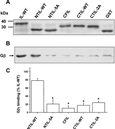
Figure 2A illustrates purified fusion proteins that were visualized in a brilliant blue-stained SDS-acrylamide gel. The binding of G␥ dimer to these proteins was subsequently analyzed with Western blot (Fig. 2B, see the experimental procedures for details). The data show that the NTIL is sufficient to display a significant G␥ dimer binding (78 ⫾ 5% of control) and that this binding was strongly diminished (21⫾ 12%) by mutating the positive residue cluster (from position 316 to 320) to alanine. Moreover, the data show that G␥ was unable to bind the central fragment of the IL (Fig. 2, B and C). These results support previous studies showing that direct binding of G␥ to the GlyR and its subsequent channel modulation were diminished by mutations in basic residues (Yevenes et al., 2006). On the other hand, the C-terminal region (CTIL-WT) in fusion with GST did not present a significant binding to G␥ in the pull-down assays
Fig. 1. Scheme of the GlyR topology and the sequences of recombinant fragments. A, schematic topology of the human␣1 GlyR subunit. B, alignment
of intracellular fragments used for GST pull-down assays and molecular docking. Underlined are the sequences used for molecular modeling and docking. IL-WT, full␣1 IL; NTIL-WT, N-terminal (17 amino acids) region of IL; NTIL-5A, mutant version with alanine substitutions; CFIL, central fragment (326 –381) of IL; CTIL-WT, C-terminal region of IL; CTIL-2A, mutant version. The sequence of NTIL-WT served as template for the RQH peptide.
Fig. 2. Binding of G␥ to different GlyR IL regions. A, brilliant blue
staining of purified GST fusion proteins. B, binding of G␥ to IL-WT and fragments detected as Western blots from GST pull-down experiments. C, quantification of G␥ binding to different intracellular fragments. The bars represent the mean⫾ S.E.M. obtained from at least three different experiments. The level of G␥ binding to NTIL-WT was significantly different (ⴱ, P ⬍ 0.05).
(Fig. 2C). In a previous study, using the entire loop, we reported that this region contributed to G␥ binding (Yevenes et al., 2006). With these in vitro results, it was evident that the N-terminal region was comparatively more important for G␥ binding than the central or the C-terminal regions of the IL.
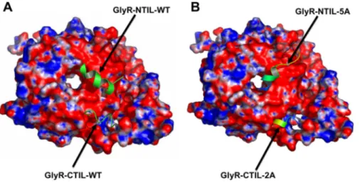
We then used computational modeling to obtain theoretical information about the contribution of these two GlyR IL regions (Fig. 1) for the binding of G␥ (Fig. 3). According to secondary structure prediction, both IL regions were modeled as ␣ helices. By using this technique, we found that both fragments fit in nearby regions of the G␥ dimer, within an area of G␥ that has a high density of amino acids with an electronegative character (as shown in red). The values for ⌬G calculated in the in silico analysis showed that the N-terminal region of the IL binds to G␥ with a higher strength than the C-terminal region, with values of ⫺20.63 and ⫺12.15 kcal/mol, respectively. In addition, the docking stud-ies with the alanine-substituted IL fragments to G␥ showed large reductions in⌬G values (⫺1.44 and ⫺12.00 kcal/mol for N- and C-terminal regions, respectively). These results sup-port the conclusion that the N-terminal region is very
rele-vant for G␥ binding, and it could be an area of choice for the design of a G␥-blocking peptide.
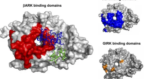
Peptide Derived from the GlyR IL N-Terminal Re-gion Sequence Interfered with G␥ Binding to Several Effectors. We synthesized a small peptide, referred to as RQH (see Fig. 1), to test its capacity to interfere with the binding of the IL to G␥ and with activation of some effectors. Micromolar concentrations (0.2 and 2 M) of this peptide were able to inhibit the binding of G␥ to the whole IL of the GlyR in GST pull-down assays to 74 ⫾ 4 and 57 ⫾ 5%, respectively, compared with control (Fig. 4A). No higher level of binding inhibition was found with 20M RQH. However, showing the specificity of the peptide inhibition, we found that the same concentrations of the scramble peptide were unable to alter the binding of G␥ to the IL (Fig. 4A). More-over, the RQH peptide was able to interfere with the binding of G␥ to two other effectors coupled to GST, such as GST-ct-ARK and GST-GIRK138. Comparison of the predicted binding position of RQH in G␥ crystal structure indicated that the regions for binding of RQH,ARK, and GIRK1 are partly overlapped (Fig. 5). These data are in agreement with previous crystallographic studies of G␥ in complex with formed using crystallographic three-dimensional data of G␥ (blue and red represent regions of positive and negative electrostatic potential, re-spectively) as described under Materials and
Methods. A, WT N- and C-terminal fragments
(Arg309 –Lys325 and Arg382–Arg392) interact with high⌬G binding in different regions on the highly electronegative surface of G␥. B, mutant N- and C-terminal fragments [316-320A (5A) and 385-386A (2A)] interact with G␥ with a reduced ⌬G binding (see text).
Fig. 4. RQH reduced G␥ binding to GlyR and other dimer effectors. A, binding of G␥ to the full IL (GST-IL-WT) was reduced by RQH. The graph shows the quantification of G␥ binding to GST-IL-WT in the presence of RQH (dashed bars) or a scrambled peptide (RQHsc, white bars). Data are from four dif-ferent GST pull-down experiments that were quantified and plotted as percentage of control condition, without any peptide (gray bar) (ⴱ,
P⬍ 0.05 compared with control). B, reduction of G␥ binding to ct-ARK and GIRK1138by RQH at the shown micromolar concentrations. Similar results were obtained in three different experiments.
ct-ARK and G␣i, as well as mutational analysis of GIRK (Ford et al., 1998; Li et al., 1998). A similar conclusion was reached comparing regions of binding for GIRK1 (Ford et al., 1998) and the RQH peptide on G␥ (Fig. 5).
RQH Blocks G␥-Mediated Activation of GIRK and Ethanol Potentiation of GlyR Channels. Based on the results obtained with RQH, we wanted to find out whether this peptide could interfere with the G␥-mediated modula-tion of two different cellular effectors. Therefore, we added this short peptide in the intracellular solution using the patch-clamp technique and tested two G␥-mediated effects. First, HEK cells were cotransfected with the GABABreceptor and GIRK1– 4to assemble a K⫹-permeable ion channel (Jones et al., 1998; Kaupmann et al., 1998; White et al., 1998). Under these conditions, the stimulation of GABABreceptors with 10 M baclofen induced a K⫹ current that was well sustained in time. It is interesting to note that intracellular dialysis with RQH (200M) in the patch pipette significantly inhibited the potassium current activated by G␥ to 24 ⫾ 14% of the control response (Fig. 6). On the other hand, neither a scrambled peptide (RQHsc) nor a nonrelated pep-tide affected the baclofen-activated K⫹ current. Second, it was previously reported that GlyR potentiation by ethanol was dependent on the state of activation of the G protein (Aguayo et al., 1996; Yevenes et al., 2008). Therefore, we
tested the sensitivity of the␣1 GlyR subunit to ethanol in the presence of the peptide in the intracellular solution. For this, the glycine-induced current was measured with and without ethanol perfusion after 10 min of RQH peptide intracellular dialysis. The data showed that HEK cells expressing GlyRs were potentiated with ethanol by 51⫾ 10% over control, and that this enhancement was significantly inhibited by RQH (Fig. 7, A and C). The ethanol-induced potentiation of GlyRs was not altered when the cells were dialyzed with the scram-bled peptide (RQHsc). Likewise, although the receptor be-came insensitive to ethanol, the current properties, such as amplitude and time course, were unchanged by 10 min of dialysis with RQH indicating that the peptide had no effects on basal GlyR function (Fig. 7, A and B).
Discussion
G␥ dimer release after heterotrimeric G protein activa-tion can control diverse physiological processes, such as heart rate, hormonal and neurotransmitter release, neuronal ex-citability, and cell migration. These actions are mediated by the direct interaction between G␥ and its effectors that range from membrane channels that control excitability to soluble enzymes that have metabolic or signaling functions (Clapham and Neer, 1997; Hamm, 1998). In an effort to
Fig. 5. GlyR-IL shares binding domains with
other effectors in G␥. The model shows G␥ binding domains forARK, G␣i, and GIRK (red, blue, and yellow, respectively). These regions partly overlap those of the N- and C-terminal in the GlyR IL (blue and green, respectively). The structural data for G␥, ct-ARK, and G␣i were indicated under Materials and Methods. The GIRK binding domains were taken from muta-tional studies (Ford et al., 1998).
Fig. 6. RQH peptide interferes
with G␥-mediated activation of GIRK. A, data show the effect of RQH on activation of GIRK (GIRK1 and GIRK4) by GABAB receptors (R1 and R2) in HEK 293 cells. The baclofen-induced K⫹current was inhibited by in-tracellular application of RQH, but not the scrambled peptide, in the patch pipette. B, time course of baclofen-stimulated K⫹ cur-rent in the absence (squares) and presence of RQH (circles), RQHsc (diamonds), and a nonre-lated peptide (triangles). All the peptides were used at a concen-tration of 200M. The symbols represent the mean ⫾ S.E.M. from more than six cells.
identify sequences useful to interfere with G␥-mediated signaling, the QEHA peptide (from adenylyl cyclase 2) was generated (Chen et al., 1995). However, it was found that several G␥ effectors lack this type of motif, making its use limited (Touhara et al., 1995; Barr et al., 2000; Davis et al., 2005). Even in the absence of a defined molecular motif for G␥ binding, several studies have shown that basic amino acids have important roles on binding and modulation of effectors, such as ARK, GIRK, voltage-dependent calcium channels, and phospholipase C (Koch et al., 1993; Touhara et al., 1994; Krapivinsky et al., 1998; Cantí et al., 1999; Barr et al., 2000). More recently, it has been shown that the GlyR contains two basic motifs in its IL that are responsible for G␥ modulation (Yevenes et al., 2003, 2006). In the present study, we examined these motifs using three approaches: 1) in vitro binding studies with GST pull down, 2) modeling/ docking in silico calculations, and 3) functional assays of G␥-modulated ion channels to obtain a sequence for the design of a small peptide with the capacity to block the action of G␥. In agreement with other previous studies, we found that the N-terminal motif of the IL exerted an important role for G␥ binding. The central region, on the other hand, was unable to bind G␥, which agrees with functional studies (Yevenes et al., 2006). Surprisingly, we did not detect signif-icant G␥ binding to the C-terminal region, a finding which contrasts with a proposed role of this motif in functional modulation (Yevenes et al., 2006). However, it is possible
that expression of the fusion protein altered the secondary structure of the C-terminal region. These results were well supported by the docking analyses that revealed a higher⌬G for binding of the N-terminal region compared with the C-terminal region of the GlyR IL and confirmed previous re-sults (Yevenes et al., 2006), indicating that the N-terminal region is very important for the direct binding of G␥. The C-terminal motif, on the other hand, presents a smaller G␥ binding capacity, but it might be important for the conforma-tional changes controlling channel gating (Yevenes et al., 2008).
Based on these results and to try to interfere with G ␥-mediated modulation, we designed a mini-peptide with the first 17 amino acids of the IL (RQH peptide). As expected, the peptide produced an inhibition of the binding of G␥ to the entire IL of GlyR using an in vitro interaction assay. In addition, we found that the binding of ct-ARK and GIRK1 to G␥ in the GST assay was also reduced by the peptide, in agreement with the existence of somewhat overlapping re-gions with distinct effectors (Ford et al., 1998). Finally, func-tional assays showed that the RQH peptide affected two G␥-linked responses: activation of GIRK by GABAB recep-tor and potentiation of␣1 GlyR by ethanol (Kaupmann et al., 1998; Yevenes et al., 2003). The data support the conclusion that these inhibitions were produced by binding of the RQH peptide to effector recognition regions in G␥. Accordingly, the RQH peptide appears to behave as a scavenger as we
Fig. 7. RQH interferes with the
ethanol-induced potentiation of the GlyR. A, the current traces were evoked activating␣1 containing GlyRs with 15M glycine in the absence and presence of ethanol (100 mM). The lower traces were recorded af-ter 10 min of intracellular dialysis with RQH peptide (200 M) in the patch pi-pette. B, graph shows the normalized gly-cine current amplitude after 10 min of intracellular dialysis with either RQH or RQHsc peptides. C, graph summarizes the percentage potentiation elicited by 100 mM ethanol in the absence and pres-ence of RQH or scrambled peptides (RQHsc) intracellularly applied for 10 min. The symbols represent the mean⫾ S.E.M. from at least six cells.ⴱ, repre-sents a difference from control, P⬍ 0.05.
showed that it can interact and block the effects of G␥ in two effectors (GlyR and GIRK). RQH did not have any apparent effects on GlyR properties and function, suggesting that pre-vious release of G␥ is necessary to exert its blocking action. Furthermore, although the data cannot exclude the possibil-ity that RQH affects the binding of G␥ to G␣ subunits, we believe that it is unlikely that it would activate G proteins and subsequently block the G␥ dimer. In conclusion, the newly identified RQH was able to inhibit the binding of several effectors to G␥ and ethanol potentiation, without noticeable changes on the properties of the GlyR or general cell function, opening new possibilities to design small mol-ecules that interfere with signal transduction.
In relevance to alcohol abuse, it is now accepted that GlyRs are a major target for the intoxicating effects of ethanol as suggested by previous studies from our and other groups (Aguayo et al., 1996; Findlay et al., 2002; Molander et al., 2005, 2007). Therefore, the present results are relevant because we show that a small heptadecapeptide can an-tagonize the alcohol effect on an LGIC, opening new pos-sibilities for the study of small organic molecules that can interfere with the intoxicating effect of ethanol, which sup-ports previous work in relation to the development of com-pounds that can affect G protein activity (Davis et al., 2005; Bonacci et al., 2006).
Acknowledgments
We thank Laurie Aguayo for technical assistance in molecular biology and text editing and Pablo Lara for helping with some pre-liminary experiments.
References
Aguayo LG and Pancetti FC (1994) Ethanol modulation of the gamma-aminobutyric acidA- and glycine-activated Cl- current in cultured mouse neurons. J Pharmacol
Exp Ther 270:61– 69.
Aguayo LG, Tapia JC, and Pancetti FC (1996) Potentiation of the glycine-activated Cl- current by ethanol in cultured mouse spinal neurons. J Pharmacol Exp Ther 279:1116 –1122.
Baker NA, Sept D, Joseph S, Holst MJ, and McCammon JA (2001) Electrostatics of nanosystems: application to microtubules and the ribosome. Proc Natl Acad Sci
U S A 98:10037–10041.
Barr AJ, Ali H, Haribabu B, Snyderman R, and Smrcka AV (2000) Identification of a region at the N-terminus of phospholipase C-beta 3 that interacts with G protein beta gamma subunits. Biochemistry 39:1800 –1806.
Bonacci TM, Mathews JL, Yuan C, Lehmann DM, Malik S, Wu D, Font JL, Bidlack JM, and Smrcka AV (2006) Differential targeting of Gbetagamma-subunit signal-ing with small molecules. Science 312:443– 446.
Camacho CJ and Zhang C (2005) FastContact: rapid estimate of contact and binding free energies. Bioinformatics 21:2534 –2536.
Cantí C, Page KM, Stephens GJ, and Dolphin AC (1999) Identification of residues in the N terminus of alpha1B critical for inhibition of the voltage-dependent calcium channel by Gbeta gamma. J Neurosci 19:6855– 6864.
Chen J, DeVivo M, Dingus J, Harry A, Li J, Sui J, Carty DJ, Blank JL, Exton JH, and Stoffel RH (1995) A region of adenylyl cyclase 2 critical for regulation by G protein beta gamma subunits. Science 268:1166 –1169.
Chen R, Li L, and Weng Z (2003) ZDOCK: an initial-stage protein-docking algorithm.
Proteins 52:80 – 87.
Cheng J, Randall AZ, Sweredoski MJ, and Baldi P (2005) SCRATCH: a protein structure and structural feature prediction server. Nucleic Acids Res 33:W72– W76.
Clapham DE and Neer EJ (1997) G protein beta gamma subunits. Annu Rev
Phar-macol Toxicol 37:167–203.
Comeau SR, Gatchell DW, Vajda S, and Camacho CJ (2004) ClusPro: an automated docking and discrimination method for the prediction of protein complexes.
Bioin-formatics 20:45–50.
Davis TL, Bonacci TM, Sprang SR, and Smrcka AV (2005) Structural and molecular characterization of a preferred protein interaction surface on G protein beta gamma subunits. Biochemistry 44:10593–10604.
Findlay GS, Wick MJ, Mascia MP, Wallace D, Miller GW, Harris RA, and Blednov
YA (2002) Transgenic expression of a mutant glycine receptor decreases alcohol sensitivity of mice. J Pharmacol Exp Ther 300:526 –534.
Fischer H, Liu DM, Lee A, Harries JC, and Adams DJ (2005) Selective modulation of neuronal nicotinic acetylcholine receptor channel subunits by Go-protein sub-units. J Neurosci 25:3571–3577.
Ford CE, Skiba NP, Bae H, Daaka Y, Reuveny E, Shekter LR, Rosal R, Weng G, Yang CS, Iyengar R, et al. (1998) Molecular basis for interactions of G protein beta-gamma subunits with effectors. Science 280:1271–1274.
Hamm HE (1998) The many faces of G protein signaling. J Biol Chem 273:669 – 672. Jones KA, Borowsky B, Tamm JA, Craig DA, Durkin MM, Dai M, Yao WJ, Johnson M, Gunwaldsen C, Huang LY, et al. (1998) GABA(B) receptors function as a heteromeric assembly of the subunits GABA(B)R1 and GABA(B)R2. Nature 396: 674 – 679.
Kaupmann K, Malitschek B, Schuler V, Heid J, Froestl W, Beck P, Mosbacher J, Bischoff S, Kulik A, Shigemoto R, et al. (1998) GABA(B)-receptor subtypes assem-ble into functional heteromeric complexes. Nature 396:683– 687.
Koch WJ, Inglese J, Stone WC, and Lefkowitz RJ (1993) The binding site for the beta gamma subunits of heterotrimeric G proteins on the beta-adrenergic receptor kinase. J Biol Chem 268:8256 – 8260.
Krapivinsky G, Kennedy ME, Nemec J, Medina I, Krapivinsky L, and Clapham DE (1998) Gbeta binding to GIRK4 subunit is critical for G protein-gated K⫹ channel activation. J Biol Chem 273:16946 –16952.
Laube B, Kuhse J, Rundstro¨m N, Kirsch J, Schmieden V, and Betz H (1995) Modulation by zinc ions of native rat and recombinant human inhibitory glycine receptors. J Physiol 483:613– 619.
Legendre P (2001) The glycinergic inhibitory synapse. Cell Mol Life Sci 58:760 –793. Li Y, Sternweis PM, Charnecki S, Smith TF, Gilman AG, Neer EJ, and Kozasa T (1998) Sites for Galpha binding on the G protein beta subunit overlap with sites for regulation of phospholipase Cbeta and adenylyl cyclase. J Biol Chem 273:16265– 16272.
Molander A, Lido¨ HH, Lo¨f E, Ericson M, and So¨derpalm B (2007) The glycine reuptake inhibitor Org 25935 decreases ethanol intake and preference in male Wistar rats. Alcohol Alcohol 42:11–18.
Molander A, Lo¨f E, Stomberg R, Ericson M, and So¨derpalm B (2005) Involvement of accumbal glycine receptors in the regulation of voluntary ethanol intake in the rat.
Alcohol Clin Exp Res 29:38 – 45.
Moss SJ and Smart TG (2001) Constructing inhibitory synapses. Nat Rev Neurosci 2:240 –250.
Pitcher JA, Inglese J, Higgins JB, Arriza JL, Casey PJ, Kim C, Benovic JL, Kwatra MM, Caron MG, and Lefkowitz RJ (1992) Role of beta gamma subunits of G proteins in targeting the beta-adrenergic receptor kinase to membrane-bound receptors. Science 257:1264 –1267.
Prince RJ and Simmonds MA (1992) Steroid modulation of the strychnine-sensitive glycine receptor. Neuropharmacology 31:201–205.
Smart TG (1997) Regulation of excitatory and inhibitory neurotransmitter-gated ion channels by protein phosphorylation. Curr Opin Neurobiol 7:358 –367. Tapia JC, Espinoza F, and Aguayo LG (1997) Differential intracellular regulation of
cortical GABA(A) and spinal glycine receptors in cultured neurons. Brain Res 769:203–210.
Touhara K, Inglese J, Pitcher JA, Shaw G, and Lefkowitz RJ (1994) Binding of G protein beta gamma-subunits to pleckstrin homology domains. J Biol Chem 269: 10217–10220.
Touhara K, Koch WJ, Hawes BE, and Lefkowitz RJ (1995) Mutational analysis of the pleckstrin homology domain of the beta-adrenergic receptor kinase. Differential effects on G beta gamma and phosphatidylinositol 4,5-bisphosphate binding. J Biol
Chem 270:17000 –17005.
Van Der Spoel D, Lindahl E, Hess B, Groenhof G, Mark AE, and Berendsen HJ (2005) GROMACS: fast, flexible, and free. J Comput Chem 26:1701–1718. White JH, Wise A, Main MJ, Green A, Fraser NJ, Disney GH, Barnes AA, Emson P,
Foord SM, and Marshall FH (1998) Heterodimerization is required for the forma-tion of a funcforma-tional GABA(B) receptor. Nature 396:679 – 682.
Wu FS, Chen SC, and Tsai JJ (1997) Competitive inhibition of the glycine-induced current by pregnenolone sulfate in cultured chick spinal cord neurons. Brain Res 750:318 –320.
Yevenes GE, Moraga-Cid G, Guzma´n L, Haeger S, Oliveira L, Olate J, Schmalzing G, and Aguayo LG (2006) Molecular determinants for G protein betagamma modu-lation of ionotropic glycine receptors. J Biol Chem 281:39300 –39307.
Yevenes GE, Moraga-Cid G, Peoples RW, Schmalzing G, and Aguayo LG (2008) A selective G␥-linked intracellular mechanism for modulation of a ligand-gated ion channel by ethanol. Proc Natl Acad Sci U S A 105:20523–20528.
Yevenes GE, Peoples RW, Tapia JC, Parodi J, Soto X, Olate J, and Aguayo LG (2003) Modulation of glycine-activated ion channel function by G-protein betagamma subunits. Nat Neurosci 6:819 – 824.
Young AB and Snyder SH (1974) The glycine synaptic receptor: evidence that strychnine binding is associated with the ionic conductance mechanism. Proc Natl
Acad Sci U S A 71:4002– 4005.
Address correspondence to: Luis G. Aguayo, Department of Physiology, University of Concepcio´n, P.O. Box 160-C, Concepcio´n, Chile. E-mail: laguayo@ udec.cl