Université de Montréal
4i/(d
39ô.
Human Herpes Virus-6 Induced Changes in the Expression and
Activity of the E2F Family Transcription Factors in Human Ceils
Par
Mehtab A Khan
Programme
Biologie Moléculaire
faculté des études supérieures
Mémoire présente à la Faculté des études supérieures
En vue de l’obtention du grade de
Maftrise (M Se)
En Biologie Moléculaire
Avril, 2005
©Mehtab A Khan
f Grade Conféré (\ à ccmpr duI
Q
(S
Université
(III
de Montréal
Direction des bibliothèques
AVIS
L’auteur a autorisé l’Université de Montréal à reproduire et diffuser, en totalité ou en partie, par quelque moyen que ce soit et sur quelque support que ce soit, et exclusivement à des fins non lucratives d’enseignement et de recherche, des copies de ce mémoire ou de cette thèse.
L’auteur et les coauteurs le cas échéant conservent la propriété du droit d’auteur et des droits moraux qui protègent ce document. Ni la thèse ou le mémoire, ni des extraits substantiels de ce document, ne doivent être imprimés ou autrement reproduits sans l’autorisation de l’auteur.
Afin de se conformer à la Loi canadienne sur la protection des renseignements personnels, quelques formulaires secondaires, coordonnées ou signatures intégrées au texte ont pu être enlevés de ce document. Bien que cela ait pu affecter la pagination,
il
n’y e aucun contenu manquant.NOTICE
The author of this thesis or dissertation has granted a nonexciusive license allowing Université de Montréal to reproduce and publish the document, in part or in whole, and in any format, solely for noncommercial educational and research purposes.
Ihe author and co-authors if applicable retain copyright ownership and moral
rights in this document. Neither the whole thesis or dissertation, nor substantial extracts from it, may be printed or otherwise reproduced without the author’s permission.
In compilance with the Canadian Privacy Act some supporting forms, contact
information or signatures may have been removed from the document. While this may affect the document page count, it does flot represent any loss of content from the document.
Université de Montréal
Faculté des études supérieures
Ce mémoire intitulé
Human Herpes Virus-6 Induced Changes in the Expression and
Activity of the E2F Family Transcription f actors in Human Celis
Par
Mehtab A
Khan
A
été évalué par un jury composé des personnes suivantes
Président-rapporteur:
DrJohn
A DI BATTISTADirecteur de recherche : Dr Ah
AHMADMembre du jury: Dr MuhamlTlad ZAFARULLAH
RÉSUMÉ
Les facteurs de transcription de la famille 12F jouent un rôle important dans la prolifération, l’apoptosc et la différentiation cellulaire. Il a été démontré que l’activité de ces facteurs de transcription peut être affectée par plusieurs virus. Ils provoquent entre autre la dérégulation du cycle cellulaire. Par conséquent, il n’est pas étonnant que les virus affectent l’expression et l’activité de 12f dans les cellules infectées. Le VHH-6 (Virus Humain d’Herpès -6) est un microbe pathogène humain onmiprésent. Il est un de ces virus qui est connu pour ses propriétés à moduler négativement la progression du cycle cellulaire. Cependant, rien n’est connu au sujet des effets du VHH-6 sur l’expression et les activités fonctionnelles de 12F dans les cellules humaines. Dans le présent projet, nous avons étudié ce problème.
À
cette fin, nous avons infecté une lignée cellulaire de cellules T de leucémie humaine (HSB-2) in vitro avec le VHH-6 (GS strain). HSB-2 est la seule lignée dc cellules T humaines qui est infecté effectivement par ce virus. En employant les techniques «EÏectorinobiÏityShifiAssay» (EMSA) et «Western Blots», nous avons comparé les expressions et les liaisons de E2f avec l’ADN entre les cellules HSB-2 infectées et mock infectées avec le virus. Les analyses «Western blots» ont montré plusieurs changements qualitatifs et quantitatifs de l’expression de ces facteurs dans les fractions cytoplasmiques et nucléaires des cellules infectées. La plupart de ces changements dans les expressions étaient du à la phosphorylation différentielle des facteurs. Nous avons fait EMSA en utilisant les extraits nucléaires et des oligonucléotides à double brin contenant les séquences consensus de E2F. Dans ces analyses, les protéines des cellules mock-infectées ont donné au moins 3 bandes distinctes dans le gel shift, tandis qu’on a observé deux bandes avec les cellules infectées par le virus. Dans l’EMSA, avec les extraits des cellules infectées, la bande à faible mobilité n’a pas été détectée. L’E2F-oligonucléotide complexe était plus fort dans EMSA avec les cellules infectées qu’avec les cellules mock-infectées. Le « supershift assay» montre que les anticorps contre les facteurs E2F1-6, DPi et DP2 retardent la migration des complexes préparés à partir des cellules mock infectées. Dans les cellules infectées ce «supershift» n’a pas été observé avec les anticorps contre les facteurs E2F2, E2f 5, E2F6 et DP-l, alors qu’il est réduit avec les anticorps contre les E2F-i, E2F-3 et 12F-4. Nos résultats montrent également que dans les cellules mock-infectées seuls les anticorps contre les protéines p107 et p110 supershift le complexe et non pas le anticorps contre les pRB. Par contre, dans les cellules infectées par le VHH-6 aucun supershift n’était observé avec ces anticorps. Nous concluons par conséquent: Le Vil-6 réduit ou inhibe les interactions des membres d’E2F avec leurs séquences d’ADN spécifique dans les cellules humaines infectées. Les effets viraux sur 112F peuvent être importants pour la réplication virale. Mots Clés : cycle cellulaire, DP-1, DP-2, E2F, facteur de transcription, HHV-6.SUMMARY
The E2F family of transcription factors plays an important role in celi proliferation, apoptosis and differentiation. The viruses use host cell’s transcriptional and fransiational machinery for their own replication and survival. They deregulate ceil cycle by inducing proliferation, quiescence or celi cycle arrest. Therefore, it is flot surprising that viruses affect E2F expression and activity in the infected celis. Human Herpes Virus (1111V-6) is a ubiquitously occurring human pathogen. It is one of the viruses, which are known to negatively modulate celi cycle progression in human ceils. However, nothing is known about the effects of 1111V-6 infection on the expression and functional activities of E2F in human ceils. We addressed this issue in this study. For this purpose, we infected a human T celi leukemia ceil une HSB-2 in vitro with the GS isolate of HHV-6. It is noteworthy that HSB-2 is the only human T celi une that can be productively infected with the GS isolate of the virus. By using Western blots and electromobility shift assays (EMSA), we compared the expression and DNA-binding activities of the E2F farnily members between the virns-infected and mock-infected HSB-2 cells. The Western blots showed several qualitative and quantitative changes in the expression of these factors in the cytoplasmic and nuclear fractions of the infected ceils as compared to the mock-infected ones. Interestingly, most of these changes in the expression pattem were due to differential phosphorylation of the factors, since the treatment of the ceil extracts with phosphatase resulted in similar hands between the infected and mock-infected ceils. We performed EMSA using nuclear extracts of the ceils and double-stranded oligonucleotides containing consensus E2F-specific sequences. hi these assays, the mock-infected celis showed at least three distinct gel shift hands, whereas two hands were observed with the virus-infected ceils. In the EMSA with the infected ccli exfracts, the low mobility hand was not present. The oligonucleotide-protein complexes were more prominent in EMSA with the infected ceils than with the mock-infected cells. The supershift assays showed that antibodies specific for E2F 1-6, DP-1 and DP-2 caused supershifts for the mock-infected celis. The supershifts were, however, flot seen for anti-E2F-2, -E2F-5, -E2F-6 and -DP-1 antibodies for the virus-infected cells. Furthermore the supershift by E2F-1, 12F-3 and E2F-4 and DP-2 antibodies were reduced for the infected ceils as compared to the rnock-infected ones. The p107- and p130-, but not the pRB-specific antibodies, supershifted complexes in the mock infected but flot in the vims-infected eils. We conciude that: HHV-6 reduces or abrogates the interactions of E2F members with their cognate DNA sequences in the infected human cells. The viral effects on the E2F may be important for viral replication.
CONTENTS
Page
TITLE PAGE
I
DENTIFICATION 0F THE JURY
II
RÉSUMÉ
iii
SUMMARY
w
CONTENTS
y
LIST 0F TABLES
VIII
LIST 0F FIGURES
IX
LIST 0F ABBREVIATIONS
XI
DEDICATION
XIII
ACKNOWLEDGEMENTS
XIVCHAPTER I. INTRODUCTION
ANI)REVIEW 0F LITERATURE
1. The herpes viruses
2
1.lVirion Structure
3
1.2 Genome Characteristics
7
1.3 Hepresviridae Subfamilies
8
1.3.1 Alphaherpesvirinae
2
1.3.2 Betaherpesvirinae
$
1.3.3 Gammaherpesvirinae
8
2. Human Herpes Virus-6
$
2.1
HHV-6 Variants
10
2.2 Molecular Biology
10
2.3 HHV-6 Behavior and Morphology
15
2.4 HHV-6 Replication
16
2.4.1 Attachment and Entry
16
2.4.2 Transcription
17
2.4.3 Genome Replication
18
2.4.4 Genome Packaging
18
2.4.5 Viral Assembly and Release
19
2.5 Fate ofthe HHV-6 Jnfected Celis
19
2.6 Biological Properties ofHHV-6
20
2.6.1 Ceil Tropism
20
2.6.2 Immune Response
22
2.6.3 Clinical PathoÏogy
23
2.7 HHV-6 In the Immuno Compromised Host
23
2.7.1 HIV-1 Infection
23
2.7.2 Chronic fatigue Syndrome
25
2.7.3 Bone Marrow Transplantation
25
2.7.4 Solid Organ Transplantation
25
2.8 HHV-6 and the CNS
26
2.8.1 HHV-6 and Encephalitis
26
2.8.2 Effect ofHHV-6 Infection on CNS White Matter
26
2.9 Febrile Seizures
27
2.10 HHV-6 and Cardiovascular System
27
2.11 HHV-6 and Other Virus Infection
27
2.12 Epidemiology
27
2.13 Antiviral Treatment
28
3. E2F Transcription factors
28
3.1.E2FFamily
29
3.2. Structure ofE2f and DP Proteins
30
3.2.1 DNA Binding Domain
30
3.2.2 Dimerization Domain
32
3.2.3MarkedBox
32
3.2.4 Transactivation Domain
32
3.2.5 The pocket protein binding domain
34
3.2.6 Cyclin binding domain
34
3.3. The E2f Family Members
36
3.3.1 E2F-1
36
3.3.2E2F-2
36
3.3.3 E2f-3
37
3.3.4E2F-4
38
3.3.5E2F-5
39
3.3.6 E2F-6
39
3.3.7 E2F-7
40
3.3.8 DP-1
41
3.3.9 DP-2
41
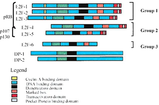
3.4 Classification ofthe E2F Family
42
1.GROUPI
42
2. GROUP II
42
3. GROUP III
43
3.5 Regulation ofE2F
43
3.5.1 The Pocket Protein Mediated Regulation ofE2F
44
3.5.2 Cyclin A-Mediated Regulation ofE2F
47
3.5.3 Acetylation-Mediated Regulation ofE2F
47
3.6 E2F and Apoptosis
47
3.7 E2f and DNA Damage
49
3.8 E2f and Tumorigenesis
50
3.9 E2F and Viruses
50
3.9.1 Human Papillomavirus (HPV)
51
3.9.2 Adenoviruses
51
3.9.4 Epstein Barr Virus (EBV)
52
3.9.5 Human Jmmunodeficiency Virus type 1 (HIV-1)
52
3.9.6 Herpes Simplex Virus 1 (HSV-1)
54
3.9.7 Hepatitis C Virus
54
3.9.8 Human Herpes Virus-6 (HHV-6)
54
CHAPTER II OBJECTIVES
56
CHAPTER III MATERIALS
AN])METHODS
58
3.1 CelIs
5$
3.2 Virus Preparation
58
3.3 CelI kifection
59
3.4 Nuclear and Cytoplasmic Protein Fractionation
59
3.5 Preparation ofE2F Specific Oligonucleotide Probes
60
3.6 E2f Gel Mobility Assays and Supershifl Assays
62
3.7 Western Blots
63
3.8 Antibodies and Reagents
64
CHAPTER W RESULTS
67
4.1 HHV-6 induced changes in the expression ofE2f
transcription factors
67
4.1.1
E2f-1
67
4.1.2
E2F-2
67
4.1.3
E2f-3
67
4.1.4
E2F-4
68
4.1.5
E2F-5
68
4.1.6
E2F-6
68
4.1.7
DP-1
69
4.1.8
DP-2
69
4.2 Alkaline Phosphatase treatrnent ofE2F
69
4.3 Effect ofvints infection on the DNA binding activities
ofE2F
7$
4.4 Identification ofthe E2F members bound to the cognate
DNA sequence
$4
CHAPTER V DISCUSSION AN]) CONCLUSIONS
94
CHAPTER VI BIBLIOGRAPHY
106
LIST 0F TABLES
Table 1:
Human Herpes viruses and the associated diseases
4
Table 2:
Classification of herpes viruses in different groups
5Table 3:
Important features ofHuman herpesvirus 6 (HHV-6)
11
LIST 0f FIGURES
Figure 1.
Schernatic representation of Human Herpes virus-6
6
Figure 2.Genornic organization of Herpes viruses
9
Figure 3. Replicationcycle ofHHV-6
21
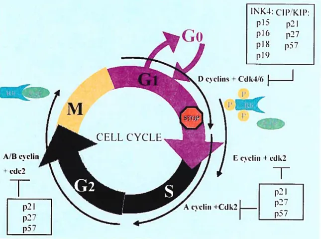
Figure 4.E2F and ceil cycle progression
31
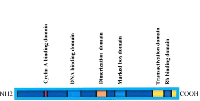
Figure
5
Structure of a prototypic E2F family member
33
Figure 6. Aschematic comparison ofthe structures ofdifferent E2f
35
famiÏy members and their interaction with different pocket
proteins
Figure
7. A schematic depiction ofthe E2F-7 structure
45
Figure 8.Activation ofE2F by different viral proteins
53
Figure 9.
The expression ofE2f-1 in the nuclear and cytoplasmic fractions of
70
HHV-6-infected and mock-infected celis
Figure 10.
E2F-2 immunoblot ofcytoplasmic and nuclear fractions of
71
mock and HHV-6 infected HSB-2 ceils
Figure il.
The expression of E2F-3 in the nuclear and cytoplasmic fractions of
72
HHV-6-infected and mock-infected ceils
Figure
12. E2F-4 immunoblot ofcytoplasrnic and nuclear fractions of
73
Figure 13.E2F-5 expression in cytoplasmic and nuclear fractions of
74
mock and HHV-6 infected HSB-2 celis
Figure
14. E2F-6 immunoblot ofcytoplasmic and nuclear fractions of
75
mock and HHV-6 infected HSB-2 ceils
figure
15. The expression ofDP-1 in the nuclear and cytoplasmic
76
fractions ofHHV-6-infected and mock-infected celis
Figure 16.
DP-2 immunoblot ofcytoplasmic and nuclear fractions of
77
mock and HHV-6 infected HSB-2 ceils
Figure
17. The effect of phosphatase treatment on the expression pattem of
79
E2F1 in the HHV-6-infected and mock-infected ceils
Figure 18.
The effect ofphosphatase treatment on the expression pattern of
80
E2F-2 in the HHV-6-infected and mock-infected ceils
Figure 19.
The effect ofphosphatase treatment on the expression pattern of
$1
E2F-4 in the HHV-6-infected and mock-infected celis
Figure
20. The effect ofphosphatase treatrnent on the expression pattem of
82
DP- 1 in the HHV-6-infected and mock-infected celis
Figure
21. Electrophoretic mobility shift assay for the nuclear extracts of
83
HHV-6- and rnock-infected HSB-2 ceils
Figure
22. E2F DNA Electrophoretic mobility super shift assay for the
$5
nuclear cxtract ofrnock infected and HHV-6 infected HSB-2
celis using antibodies against E2F- 1,-2,-3 and —4
Figure
23. E2F DNA Electrophoretic mobility super shift assay for the
$6
nuclear extract ofmock infected and HHV-6 infected HSB-2
celis using antibodies against E2F-5,-6,DP-1 and DP-2
Figure
24. E2F DNA Electrophoretic mobility super shift assay for the
87
nuclear extract ofrnock infected and HHV-6 infected HSB-2
ceils using antibody against pRB
Figure
25 E2F DNA Electrophoretic mobility super shift assay for the
88
nuclear extract ofmock infected and HHV-6 infected H$B-2
celis using antibody against p107
Figure
26 E2F DNA Electrophoretic mobility super shifi assay for the
89
nuclear extract ofmock infected and HHV-6 infected HSB-2
celis using antibody against p130
Figure
27 E2F DNA Electrophoretic mobility super shift assay for the
90
nuclear extract ofmock infected and HHV-6 infected H$B-2
celis using antibody against HDAC 1
Figure
2$ E2F DNA Electrophoretic mobility super shift assay for the
91
nuclear extract of mock infected and HHV-6 infected HSB-2
LIST 0F ABBREVIATIONS
AIDS
Acquired Immune Deficiency Syndrome
AP Buffer
Aikaline phosphatase Buffer
APAF1
Apoptotic protease activating factor-1
ATCC
American Type Culture Collection
ATM
Ataxia telangiectasia mutated
ATR
ATM and rad3 related
BCIP
5bromo-4 chloro-3 indolyl phosphate
bHLH
Basic helix-loop-helix
ClAP
Caif intestinal phosphatase
CMV
Cytomegalovirus
CNS
Central nervous system
C02
Carbon dioxide
CSF
Cerebrospinal fluid
DHfR
Dihydrofolate reductase
DR
Direct repeats
Ds DNA
Double stranded DNA
DTT
Dithiothreitol
EBV
Epstein-Barr virus
EDTA
Ethylenediarnine tetra acetic acid
EMSA
Electrophoretic mobility shift assay
fBS
Fetal bovine senim
HBLV
Human B-lymphotropic virus
HCMV
Human cytornegalovirus
HDAC
Histone de-acetylase
HHV-6
Human Herpes Virus-6
HW-1
Human immunodeficiency virus type 1
HPV
Human papillornavirus
HSB-2
Human T ceil leukemia celi line-2
HSPG
Heparin sulphate proteo glycan
HSV-l
Herpes simplex virus type 1
IFN
Interferons
IgG
Immunoglobulin G
IL
Interleukin
R
Inverted repeats
J JHAN
Human T ceil une
KC1
Potassium chloride
kDa
Kilo Dalton
KSHV
Kaposi’s sarcoma herpes virus
MAb
Monoclonal antibody
MDM2
Mouse double minute 2
Moi
Multiplicity of infection
MOLT-3
Human T cdl leukemia celi line
mRNA
Messenger ribonucleic acid
MS
Multiple scicrosis
NaF
Sodium fluoride
NET
Nitro blue tetrazolium
NF-KB
Nuclear factor KB
NK
Natural killer
OBP
Origin binding protein
ORF
Open reading frame
PBMC
Peripheral blood mononuclear celis
PBS
Phosphate buffer saline
PCR
Polymerase chain reaction
PCV
Packed ceil volumes
Pi
Post-infection
PVD
Poly vinyl dichloride
RANTES
Regulated upon activation, normal T cell expressed and secreted
SIDA
Syndrome immunodéficitaire acquis
SupT-1
Human lymphoma T ccli
TBE
Tris borate EDTA
TCID
Tissue culture infection dose
TfDP- 1
Transcription factor DP- 1
TGF-3
Transforming growth factor-3
TNF
Tumor necrosis factor
UL
Unique long region
US
Unique short region
UTR
Untranslated region
VIH-1
Virus de l’immunodéficience humaine
VZV
Varicella Zoster Virus
DEDICATED
My beloved father who is no more with me to sec this accomplisliment
ACKNOWLEDGEMENTS
I feel pleasure in expressing my heartiest gratitude to ail those who heiped and
contributed for the compietion of this project. I wish to express my heartfelt gratitude to
my mentor and research director Dr Ah Ahmad for accepting me in his laboratory as welÏ
as for his technical and financial support throughout this study.
I am highly thankful to ail my hab cohheagues speciahhy Dr Rasheed Ahmad, Dr Tanweer,
Paulo Cordeiro, Ohfa Debbeche, R’kia Dardari and Dr Jean Hughes for their academic
and social contributions during this study. Thanks are also extended to Elodie Martin,
Francoise Banga, Chenda Duong and Suzanne Samarani. I also thank Dr José Menezes
for lis moral support during my studies.
The personnel of the Research Center, Ste Justine Hospital and the Department of
Molecular Biology, University of Montreal are thanked for their constant
supportand
collaboration during my studies.
Special thanks
to
Madam
Vivianne Jodoin, secretary Department de Biologie
Moieculaire, Université de Montréal for her great support and help throughout my M Sc.
Her encouragement, invaluable guidance and gentleness can neyer be forgotten.
Lot of thanks to my wife and son Jabran for their patience and support through my
studies.
INTRODUCTION AND REVIEW
CHAPTER I
INTRODUCTION AND REVIEW 0F LITERATURE
The present study was conducted to investigate the effects of the Human Herpes Virus-6 (HHV-6) infection on the expression and functionai activity of E2F factors in human T celis. Therefore, the pertinent literature on the E2F factors and the virus is reviewed below:
1. THE HERPES VIRUSES
The name herpes is derived from the Greek word “herpin” meaning to crawl, to climb or to slip. The herpes viruses have been given this name because they crawl to latent and chronic infections. As of today, more that one hundred herpes viruses have been isolated. They are widely distributed in nature (Abiashi et ai, 1991). The history ofthe discovery of the herpes viruses goes back to the end of the Second World War when the first herpes virus, Varicella Zoster Virus (VZV), was described and found to be
the causative agent of chicken pox or shingles. At present it is also called HHV-3. The most recently discovered herpes virus is the Kaposi’s sarcoma herpes virus (KSHV) or HHV-8 discovered in 1994 (Chang et ai, 1994). On the basis of physiology and morphology, the herpes viruses have been grouped in a single family named Herpesviridae (reviewed by Roizmann et ai, 1992). They are ail doubie-stranded DNA vinises with relatively iarge and complex genomes. They replicate in the cell’s nucleus in a wide range of vertebrate hosts, including humans, horses, caille, mice, pigs, chickens, turties, iizards, fish, and even in some invertebrates, such as oystcrs. The viruses tend to have a restricted host range; oniy a fcw infect more than one species. At least one distinct herpes virus lias been isolated from most of the animal species. Some species may be infected with many herpes viruses. for example,
eight distinct herpes viruses have been isolated from humans (Table 1). The main characteristics of the viruses in this family are: the development of latency in the infected ceils, destruction of the infected ceils upon viral replication (lytic cycle), and
the replication of DNA and the assembly of the capsids in the nuclei of the infected ceils. In the latent state, the viral genome becomes a closed circular molecule and only a few viral genes are expressed. The herpes virus genomes encode a variety of
enzymes implicatcd in nucleic acid metabolisrn, DNA synthesis and protein translation. Based upon the arrangement of the terminal repeat sequences of >100 bp within their genomes, the herpes viruses have been classified into six (A-F) groups as shown in Table 2. Human herpes virus infections are endemic and sexual contact is a significant method of transmission for herpes simplex virus 1 and 2 (HSV-1, HSV-2), human cytomegalovirus (HHV-5) and likely for KSHV. HHV-6, however, is flot spread by sexual contact (see below).
1.1 WRION STRUCTURE
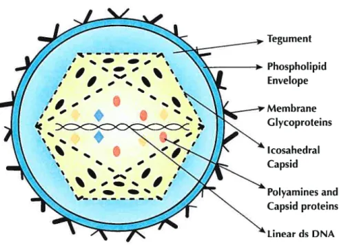
A typical herpes virus is composed of four structural elements as shown in Figure 1.
They include:
• Core. The core consists of a single linear molecule of double sfranded (ds) DNA in the form ofa toms.
• Capsid. Surrounding the core is an icosahedral capsid with a 100 nm diameter constmcted of 162 capsomeres.
• Tegument. Between the capsid and envelope, there is an amorphous, sometimes asymmetrical, feature namcd the tegument. It consists of viral enzymes, some of which are needed to modulate the cell’s biochemical
Table 1. Human Herpes viruses and the associated diseases
Common name Scïentific name ‘ Dïseases
Herpes simplex virus 1 Human herpes virus 1 Facial, labial and ocular
(HSV-1) (HHV-1) lesions or “cold sores”
Herpes simplex virus 2 Human herpes virus 2 Genital lesions
(HSV-2) (HHV-2)
Varicella-zoster virus Human herpes virus 3 Chickenpox andshingles
(VZV) (HHV-3)
Epstein-Ban virus Human herpes virus 4 Glandular fever or infectious
(EBV) (HHV-4) mononucleosis,
Hurnan cancers, e.g., Burkitt’s lymphoma, gastric cancer, undifferentiated NPC, Hodgkin’s disease.
Human cytomegalovirus Human herpes virus 5 Infectious mononucleosis
(HCMV) (HHV-5)
(no common names) Human herpesvirus6 Mild early childhood roseola (HHV-6) infantum, MS, CFS, several
lymplioproliferative disorders.
(
no common names) Human herpes virus 7 Rroseola infantum, pityriasis (HHV-7) rosea, “Socks and glovessyndrome”.
Kaposi’s sarcoma herpes virus Human herpes virus 8 Karposis sarcoma, (HHV-8) Castleman’s multicentric
disease.
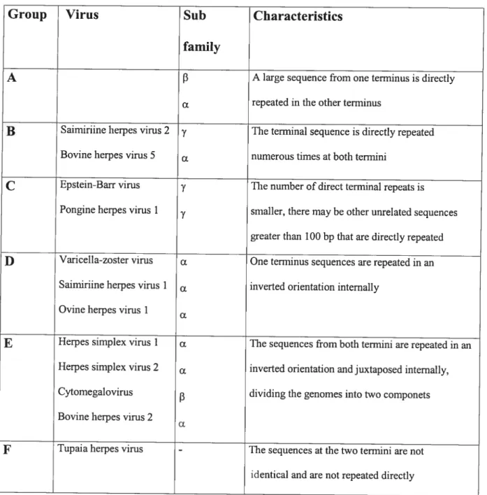
Table 2: Classification of Herpes vïruses in different groups
Group
Virus
Sub
Characteristics
family
A f3 A large sequence from one terminus is directly
(X repeated in the other terminus
B
Sainuriine herpes virus 2 y The terminal sequence is directly repeated Bovine herpes virus 5 numerous times at both terminiC
Epstein-Barr virus y Thenumber of direct terminal repeats is Pongine herpes virus 1y smaller, there may 5e other unrelated sequences
greaterthan 100 bp that are directly repeated
D
Varicella-zoster virus One terminus sequencesarerepeated in an Saimiriine herpes virus 1 inverted orientation intemallyOvine herpes virus I
E
Herpes simplex virus I o. Thesequences from both termini are repeated in anHerpes simplex virus 2 inverted orientationandjuxtaposed intemally,
Cytomegalovirus
f3 dividing the genomes intotwo componets Bovine herpes virus 2
F
Tupaia herpes virus - Thesequences at the two termini are flotidentical and are flot repeated directly
The classification is based upon the arrangement ofrepeat sequences within the genomes ofthe viruses.
Figure 1. Schematic representation of Human Herpes virus-6
I —— ‘ ,‘ s — I W I — %O
tc
ccc<
C ‘s % s ‘ s s S — s — h S — 4 ‘ _s F _s b• — _ I S Tegu ment Phosphol ipid Envelope Membrane Glycoproteins Icosahedral Capsid Polyamines and Capsid proteins Unear ds DNAThe spikes of membrane glycoproteins are projecting from the surface. Each viral
particle contains a copy of the viral genome in the form of a single double-stranded
DNA molecule. The viral components are flot drawn to the scale.
processes and viral repiication. Others are important to counter host celis immediate responses.
Envelope. The envelope is the outermost layer of the virion. It is derived from
the patches of the altered cellular membranes of the infccted celi into which
almost a dozen unique viral glycoproteins have been inserted. The viral glycoproteins appear as short spikes embedded in the lipid bilayer of the envelope in electron micrographs. There may be more than 1000 copies of each glycoprotein on a single virion.
1.2 GENOME CHARACTERISTICS:
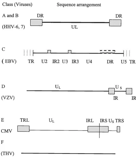
Herpes virus genomes range from 120 to 230 kbp in length with 31 to 75 % G+C content and contain 70 to 120 genes. Because replication takes place inside the nucleus, herpes viruses can use both the host’s transcription machinery and DNA repair enzymes to support a large genome with a compiex anay ofgenes. Herpes virus genes, like the genes oftheir eukaryotic hosts, arc flot ananged in operons and in most cases have individual promoters. However, unlike eukaryotic genes, only a few herpes virus genes are spliced. The genes are characterized as either essential or non-essential for growth in ccli culture. Essential genes regulate transcription and are needed for virus assembly. Non-essential or dispensable genes, for the most part, function to manipulate the cellular environment for virus production, to defend the virus from the host immune system and to promote celI-to-celI spread. The large number of dispensable genes is in reality required for a productive in vivo infection. Although the classification of Hcrpesviridae is based on the differences between the genomic sequences and viral proteins, they ail share the same genomic organization. A typical herpes viral genome consists of a unique long region (UL) and a unique short region (US) connected by inverted repeats (IR) as shown in Figure 2. However, HHV-6 and
HHV-7 have only a UL flanked on the right and on the left by direct repeats, which are calledDRRand DRL, respectively (Roizman and Pellete, 2001).
1.3
HERPES VIRIDAESURFAMILIES
As mentioned earlier, eight distinct herpes viruses have been isolated from humans. Based upon their tissue tropism and pathology, they are classified into following three subfamilies:
1.3.1 Mphaherpesvirinae. Members of this subfamily have a short reproductive cycle (—1 $ hr.) with efficient host ccli destruction and a variable
host range. They tend to become latent in sensory neurons. The subfarnily includes HSV-1, -2 and VZV.
1.3.2 Betaherpesvirinae. Members are lymphotropic. They have a long
reproductive cycle, and a restricted host range. The infected ceils become enlarged (cytomegalo). Hurnan Betaherpesvirinae include HCMV, HHV-6, and 7.
1.3.3 Gammaherpesvirinae. These herpes viruses are also lymphotropic;
however, they are specific for either T or B-lymphocytes. They rarely infect a species other than human beings. Members of the subfamily isolated from humans are EBV and KSHV.
2. HUMAN HERPES VIRUS-6
Human herpes virus 6 (HHV-6) was discovered for the first time in 1986 from six patients suffering from lymphoproliferative disorders, two of whom were HIV-seropositive. The virus was initially named as human B-lymphotropic virus (HBLV) as it was isolated from peripheral blood mononuclear celis of patients with B- cdl lymphoproliferative disorders (Salahuddin et aI, 1986). It
Figure 2. Genomic organization of Herpes viruses
Class (Vimses) Sequence arrangement
AandB DR DR (HHV-6, 7) UL I-1 (EBV) TR U2 1R2 U3 1R3 U4 DR U5 TR D UL DUS (VZV) IR IR
E TRI UL IRL IRS U TRS
CMVI
I
I
(THV)
DR: Direct repeat; IR: Inverted repeat; U[: Long unique region; Us: Short unique
region; TR: Terminal repeat; IRS: Short inverted repeat; IRL; Long inverted repeat;
TRL: Long terminal repeat.
was classifled as the sixth member of the herpes virus family and was placed in the gammaherpesvirinae subfamily in common with EBV. After it was realized that HHV-6 preferentially infected T lymphocytes rather than B-lymphocytes, it was reclassified in the betaherpesvirinae subfamily alongwith HCMV. The virus closely resembles HHV-7 and both cause roseola in chiidren, therefore, both were classified in a new genus called Roseola. Some important biological and pathological features of HHV-6 are sumrnarized in Table 3 and 4.
2.1 HHV-6 VARIANTS:
The first isolate of HHV-6 was terrned as GS or AJ. It was isolated from patients from Gambia (Lopez et al., 198$). Several other viral isolates have been obtained from patients from different geographical regions of the world. They include Ul 102 (from Uganda), Z29 (from Zambia), and HST (from Japan) (reviewed by Krueger and Ablashi, 2003). Based upon their reactivity with anti-HHV-6 monoclonal antibodies, RFLP, and in vitro tissue tropism, these isolates have been classified into two variant groups: A and B (Ablashi et al; 1993). The group A variants are represented by GS and U 1102, and B variants by Z29 and HST. The A variants replicate in HSB-2 and J JHAN celis and the B variants in MOLT-3 and MT-4. Both variants grow efficiently in vitro in IL-2 activated cord blood T ceils.
2.2 MOLECULAR BIOLOGY:
Complete genome sequences ofboth A (U1102) and B (Z29, HST) variants ofHHV-6 have been determined (Isegawa et al., 1999; Dominguez et al., 1999). The A and B
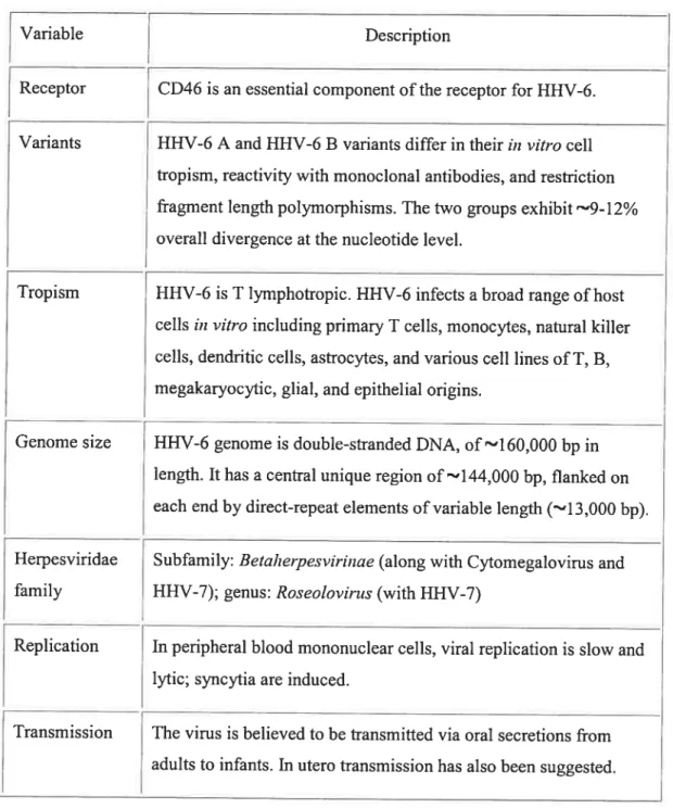
Table 3. Important features of Human herpes virus-6
Variable Description
t
Receptor CD46 is an essential component of the receptor forVariants HHV-6 A and HHV-6 B variants differ in their in vitro celi ftopism, reactivity with monoclonal antibodies, and restriction fragment length polymorphisms. The two groups exhibit r9l2% overal! divergence at the nucleotide level.
Tropism HHV-6 is T lympliotropic. 11HV-6 infects a broad range ofhost celis in vitro including primaty T celis, monocytes, natural killer ceils, dendritic ceils, astrocytes, and various ce!! !ines of T, B, megakaryocytic, g!ial, and epithelia! origins.
Genome size 1111V-6 genome is double-stranded DNA, of’l6O,OOO bp in length. It lias a centra! unique region of —‘144,000 bp, flanked on each end by direct-repeat elements of variable length (-‘13,000 bp). Herpesviridae Subfamily: Betaheipesvirinae (along with Cytomegalovirus and fami!y HHV-7); genus: RoseoÏovirus (with HHV-7)
[
Replication In peripheral blood mononuc!ear ceils, viral replication is slow andlytic; syncytia are induced.
Transmission The virus is believed to be transmitted via oral secretions from aduits to infants. In utero transmission has also been suggested.
Table 4: The pathogenesis and epidemiology of Human herpes virus-6
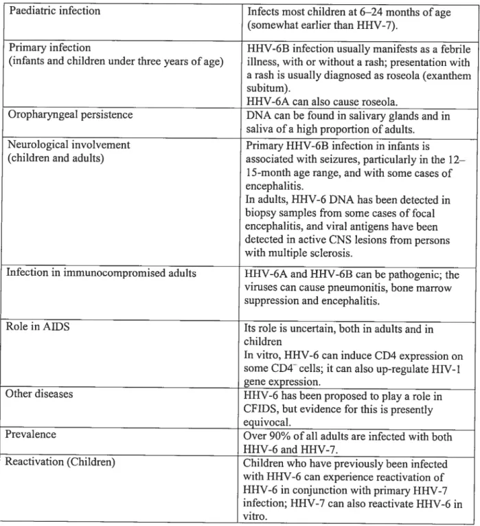
Paediatric infection Infects most chiidren at 6—24 months of age (somewhat earlier than HHV-7).
Primary infection HHV-6B infection usually manifests as a febrile (infants and chiidren under three years of age) illness, with or without a rash; presentation with a rash is usualiy diagnosed as roseola (exanthem subitum).
HHV-6A can also cause roseola.
Oropharyngeal persistence DNA can be found in salivary glands and in saliva of a high proportion of aduits.
Neurological involvement Primary HHV-6B infection in infants is
(chiidren and aduits) associated with seizures, particuiariy in the 12—
1 5-month age range, and with some cases of
encephalitis.
In adults, HHV-6 DNA has been detected in
biopsy samples from some cases of focal encephalitis, and viral antigens have been detected in active CNS lesions from persons with multiple scierosis.
Infection in immunocompromised aduits HHV-6A and HHV-6B can be pathogenic; the viruses can cause pneumonitis, bone marrow suppression and encephalitis.
Role in AIDS Its role is uncertain, both in aduits and in chiidren
In vitro, HHV-6 can induce CD4 expression on
some CD4 ceils; it can also up-reguiate HIV-l gene expression.
Other diseases HHV-6 has been proposed to play a role in CFIDS, but evidence for this is presently equivocal.
Prevalence Over 90% of ail aduits are infected with both HHV-6 and HHV-7.
Reactivation (Chiidren) Children who have previousiy been infected with HHV-6 can experience reactivation of HHV-6 in conjunction with primary HHV-7 infection; HHV-7 can also reactivate FLHV-6 in vitro.
AIDS Acquired immunodeficiency syndrome; CFIDS t Chronic fatigue
immunodeficiency syndrome; CNS t Centrai nervous system; HHV: Human herpes virus; HIV t Human Immunodeficiency virus.
variants are 88% identical at the nucleotide level. Their genomes are about 160-162 kb long. Each gcnome comprises a central unique (U) region of 143-144 kb length flanked by an 8-9 kb region of direct repeats (DR) on either end. The U region contains a hundred or more open reading frames (ORFs). The terminal and junctional regions of the DR contain human telomerase—like sequences of unknown function. The central region of the genome contains seven blocks of genes that are conserved in ail herpes viruses, and a group ofgenes (U2-U19) to the lefi ofthe seven-block region found only in the beta herpes viruses (Figure 2). The genome also contains genes that are only found in HHV-6 and 7. They are located to the left and right of the core genes. HHV-6A (Ui 102 strain) lias 110 ORFs whereas HHV-6B (Z29 and HST strains) contains 119 ORFs. Nine of the B variant (Z29) genes do flot have their counterparts in the A variant (Ui 102) genome and the vice versa is also true. The JE genes occur in two blocks, TE-A (U86-89) and lE-B (U16-19). Their spiicing pattem and temporal regulation may differ in two variant groups. The gene product of U$9, lEi, ofHHV-63 is phosphorylated on ser/thr residues, is surnoyiated and localizes to nucieus along with promyelocytic leukernia (PML) proteins (Gravel et al., 2002). The equivalent protein in HHV-6A is 62% identicai to it at amino acid level. Some U16/17.franscripts may appear late and act as late genes. U16 activates the LTR of HIV-i (Flebbe-Rehwaldt et al., 2000; Lusso et aI., 1989). The genes invoived in DNA replication (E genes) include U27, U41, U43/73/77 (reviewed by Clark, 2000). U94 is one of the only two HHV-6 genes that are not found in HHV-7. It is an lE gene, which encodes a homologue of the human AAV-2 rep gene. It plays a role in DNA replication and gene regulation.
It inhibits viral replication and is aiso expressed during latency (Thompson et ai, 1994). It bas been reported to inhibit HW-1 LTR and H-ras-mediated celi
transformation (Aroujo et al., 1997; Rotola et al., 1998).
0f the late genes, U39 and U48 encode surface glycoproteins gB and gH,
respectiveiy. The HHV-6 gB has 39% sequence homoiogy at amino acid level with HCMV gB, causing immunological cross reactivity between the two viruses. It plays
a roie in viral attachment and penetration and is conserved in ail herpes viruses. In
HHV-6A, gB is translated as a precursor molecule which is cleaved to give rise to functional sub units of 64 kd and 58 kD whereas HHV-6B gB forms about 102, 59 and 50 kD proteins (Takeda et ai, 1996).
The product of the gene U82, gL, complexes with gH, and plays a role in its transport and processing. The gH-gL compiex is invoived in the infection and fusion process (Mukai et ai, 1997). U72 encodes gM and U100 encodes gp82-100 complex due to differentiai spiicing. U100 of the two HHV-6 variants have oniy 72% sequence identity, suggesting variant specific roles in infection. gB, gH and gp$2-105 contain epitopes for virus neutralization. Ui 1 encodes a phosphoantigen p100, which is the
major structural antigen of HHV-6. There is only 80.1% amino acid sequence homology between p100 of A and B variants. The U53 encodes a viral protease, which is auto-cieaved at two sites and is necessary for virai assembiy and maturation.
A protein kinase encoded by U69 imparts sensitivity to gancyciovir (Ansari et al., 1999 ; reviewed by Clark, 2000.).
Like other herpes viruses, HHV-6 has usurped several host genes, e.g., U83 encodes a chemokine and two genes, U12 and U51, encode chemokine receptors. The DR7 gene encodes a protein, which binds and inactivates p53, transactivates HIV LTR and fransforms eukaryotic ceils (Kashanchi et ai, 1997; reviewed in Dockreli, 2003). It is
noteworthy that several of the HHV-6A ORFs arc translated into proteins, which are shorter than their HHV-63 counterparts.
The Roseolavirus-specific genes include U20-24, U24A, U26, and U$5 and U100. HHV-6A and HHV-6B variants have 94% amino acid identity in the seven-region conserved b!ock. They differ in the DR and a 24 kb segment to the right of U85 (except U94, which differs only by 2.4%). Because of the diffcrences in the genes between HHV-6A and HHV-6B, there are biological differences between the variants (Krueger and Ablashi, 2003).
2.3 1111V-6 BEHAVIOR AND MORPHOLOGY:
HHV-6 virions are 160—200 nm in diameter. Each consists of a central core containing a linear double-sfranded DNA, a capsid, and a tegument, which is surrounded by a membrane structure. The capsids are icosahedral and consist of approximately 162 capsomeres. Its tegument is amorphous and the core has smooth appearance. When HSB-2 (A human T ce!! leukemia ccli une; sec chaptcr III) ceils
are infected in vitro by HHV-6 (strain A), the virus binds to 50% of the ceils within
15 minutes and to 100% within 30 minutes. The virus binds to coated pits of the ccli membrane and is interna!ized by endocytosis within 6 hours (Tonisi et al., 1999; rcviewcd in Kruegcr and Ab!ashi, 2003). The viral DNA replication is initiated as early as 12 hours post infection. Newly formed uncoated nucleocapsids arc visible in
ce!! nuclei by day 3. The virus enve!ops in the nucicus, de-cnve!ops in thc cytosol and
re-envelops in the Golgi complex. By day 6 and later, enveloped viruses can be seen
in the endoplasmic reticu!um. It takes 6 to 10 days for the virus to appear in increasing
amounts in the culture medium. It is notcworthy that despite a high degree of infection, it is extremely difficuit to obtain cell-free high-titered viral stocks. This suggests that most of the newly formed virions may be non-infectious.
infected HSB-2 ceils undergo apoptosis, which progrcssively increases from 5% on day 1 to 30% on day 6 post-infection (reviewed in Krueger and Abiashi, 2003). The HHV-6A and HHV-63 variants infect and repiicate mainly in CD4+ human T ceils obtained from peripheral or cord blood, and in tissue culture-adapted unes, e. g., HSB-2 (Human T ccli leukemia ccli une), MOLT-3 (Human T ceil leukemia ccli une), SupT-1 (Human Iymphoma T celi) and J JHAN (Human T ccii une) (CiarkDa, 2000). HHV-6 infects and persists in human monocytes/macrophages in a latent state. The virus may also infect B ceils, neural celis, and human fibrobiasts; however the viral replication is very poor in these celis. Typicai cytopathic effects include the appearance of 2-5 times enlarged muitinucieated, giant celis, which are refractile and balioon-shaped (Taniguchi et al., 2000). The infected celis tend to aggregate in small to-medium clusters. The virus has also been reported to induce apoptosis in uninfected bystander T lymphocytes as weli as in naturai killer celis (Clark, 2000).
2.4 HHV-6 REPLICATION
2.4.1 Attacliment and Entry:
How HI-IV-6 attaches to host ceils and what is the mode of attachment are stiil not very clear. It is thought that, simiiar to HSV-1 gB, the HHV-6 gB binds to haparan suiphate proteogiycans (HSPG) on the ccii surface before attaching to the virai receptor, CD46. However, it is noteworthy that heparin, which inhibits interaction between HSPG and the HSV gB protein and consequently inhibits infection of the human ceiis by HSV-1, has no effect on HHV-6 infection (Pellet and Dominurez,
2001; Santoro et al., 1999). Aithough CD46 has been identified as a receptor for
HHV-6, its expression alone is flot sufficient for viral fusion and infection, suggesting that the virus requires other coreceptor (s) for infection. The chemokine receptors CXCR4 and CCR5 (which act as essential co-receptors for T-tropic and
monocytotropic HIV- 1 strains, respectively) were also studied and were found non essential for HI-W-6 infection (Yasukawa et al., 1999; reviewed in Dockrell, 2003). Furthermore, it was found that although HHV-6 preferentially infects CD4+T ceils, CD4 does flot serve as a viral receptor. The penetration of HHV-6 in the infected ceils
is rnediated by endocytosis and is pH sensitive. It can be inhibited by a monoclonal
antibody to the viral glycoprotein complex gplOO (Cirone et al., 1992; foa-Tomasi et
al., 1991).
2.4.2 Transcription:
HHV-6 genes belong to either latent or lytic category. The latent genes are expressed
in the latent phase of the infection and usually comprise a very restricted set of genes.
The lytic genes are expressed during a productive viral infection. Depending upon their temporal sequence of expression, they are classified into immediate early (lE), early (E) or late (L) genes (reviewed in Clark, 2000). The 1F genes are transcribed first to encode proteins needed for regulation of gene expression. lE genes are synthesized within minutes to hours post-infection and do flot require denovo protein
synthesis. Only virion-associated proteins may be sufficient for their expression. The
E genes encode proteins for DNA replication and the L genes encode structural
proteins necded for viral assembly. The transcription of E genes requires JE gene activity and transcription of L genes is dependent on viral DNA replication or the expression of E genes. U83 and U89/90 encode IF (A Iocus) genes in HHV-6 (Rapp
et al, 2000, French et aI, 1999). U16 through U19 have been designated as lE (B
locus) genes. U42 encodes a homolog of the HSV JE gene alpha27 and homolog of HCMV U69. However, it is not transcribed in the absence of de novo protein synthesis.
Some HHV-6 transcripts are spliced several times. Most important of them are IF locus and U100. The use of some spiice sites is kineticaily regulated whereas others may use non-canonical donor and acceptor sequences. In HHV-6 the DNA polymerase promoter has no TATA box and solely depends on the presence of a palindromic ATF/CREB transcription factor-binding site in the virus infected ceils (Agulnick et ai, 1994).
2.4.3 Genome Replication:
HHV-6 genes for lytic phase viral replication include DNA polymerase, DNA binding protein (DBP), the DNA polymerase processivity factor, a helicase/primase complex and origin binding protein (OBP) (Agulnick et al., 1993; reviewed in Pellet and Dominurez, 2001). HHV-6 lias onLyt region similar to other herpes viruses. The origin is located in the region between 5’end ofU4l and 3’end ofU42. U41 encodes
thc major DNA binding protein. There is a region in the centre of the oriLyt that
contains two sites, OBP-1 and OBP-2, separated by an AT rich region. The OBP binds to these sites. The protein is encoded by U73. It was shown in transient replication assays that both OBP- 1 and OBP-2 are required for efficient plasmid DNA replication (Dewhurst et ai, 1993; Dewhurst et ai, 1994; reviewed in Pellet and Dominurez, 2001).
2.4.4 Genome Packaging:
Circularized viral genomes, which are present in about 5% of the virai ncleocapsids, provide templates for roliing circle replication. The replication of virai DNA by rolling circie rnechanism resuits in the production of long concatemers of nascent DNA (Martin et al., 1991). The juxtaposition of DRR and DRL in a concatemer provides complete cleavage and packaging signal. This resuits in the packaging of a viral DNA unit into a single nucleocapsid.
2.4.5 Viral Assembly and Release:
In HHV-6, nucleocapsids are formed in the nucleus of the infected celi. Then, there is
a successive enveiopment, de-envelopment and re-envelopment when nascent virion moves from one celi compartment to other (Figure 3). Afier 3 days of infection we can see the nascent capsids containing DNA in celi nuclei (Black et ai, 1997). They
are enveioped as they pass through the inner nuclear membrane. The enveioped
capsids iose their envelopes in the cytopiasrn. At this stage abundant nonenveioped
and tegumented nucieocapsids can be seen in the cytoplasm (Torrisi et ai, 1999). In
some cases naked capsids may acquire tegument in the cytopiasrn. The virai giycoproteins gB, gH-gL and gp82-gp 105 are concenfrate in the annulate lamelia. The nucleocapsids acquire envelopes from the giycoprotein-studded aimuiate membranes. (Cardinaii et al., 1998; Torrisi et ai., 1999). The annulate lamellae are cytoplasmic structures re!ated to endopiasmic reticuium and are the sites for enveiopment and glycoprotein maturation. HHV-6—infected celis do flot express viral glycoproteins on their plasma (and nuclear) membranes. Therefore, virai capsids do flot envelope at the plasma membranes. The mature virions pass through the Golgi complex and are released by exocytosis or celi lysis, and flot by budding from the ce!! membrane.
2.5 Fate of the HHV-6-Infected Host Cells:
The infection causes several deieterious effects on the host celis. It shuts off DNA synthesis, marginates chromatin and stimuiates synthesis of macromo!ecuies in the infected ceils. The celis enlarge and can be recognized by the physica! characteristics like ballooning, refractility and the presence of muitinucleated and giant ce!!s. The vims-infected celis are anested in the G2/M phase of the ceil cycle (De Boue et al., 2004). The infected celis die by necrosis. The virus induces apoptosis in uninfected bystander ceils. However, in primary NK and T celis, the infection may cause
apoptosis. The infection inhibits proliferative responses of human peripheral blood mononuclear celis (PBMC) to antigens and mitogens. It inhibits IL-2 but enhances IFN-7 production from activated T ceils (f lamand et ai, 1995). The infection induces the expression of CD4 on CD4-negative celis, e.g., NK celis and CD8+ T ceils. This makes these celis susceptible to HW infection (Lusso et ai., 1991). The infection also down regulates expression ofCD3 and CXCR-4 molecules on T ceils (Secchiero et ai,
1997; Secchiero et ai, 1998; Yasukawa et ai, 1999) but enhances expression of many T celi adhesion molecules, e.g., HLA-DR, CD49d, CD44, CD11a and CD2. The
infection suppresses formation of erythroid and granulocyte-macrophage colonies by interacting with CD34 positive progenitor celis (Isomura et al., 2003; Carrigon and Knox, 1995).
2.6 BIOLOGICAL PROPERTIES 0F HHV-6:
2.6.1 CelI Tropism:
HHV-6 has the abiiity to infect a variety of ceils and celi unes but it is predominantly regarded as a T-celI tropic virus. The virus infects and replicates prefcrentiaily in activated CD4+ T ceils. However, it can aiso infect CD8 T lymphocytes, yE TCR+ T lymphocytes, dendritic celis, natural killer (NK) celis and monocytes ofthe peripherai biood. This virus can also infect established celi unes of the megakaryocytic, gliobiastomai, neurai, epithelial and fibrobiastic origin but replication is poor (Clark, 2000; Asada et al., 1999; Luppi et al., 1999). In vivo, the virus lias a wider tropism; HHV-6 genomes or antigens can 5e detected in lymph nodes, PBMC, tubular epithelial ceils, endothelial celis and histiocytes in kidney, salivary glands and CNS. Chimps and certain species of monkeys can be infected with HHV-6, which causes rashes in them (Dockrell, 2003).
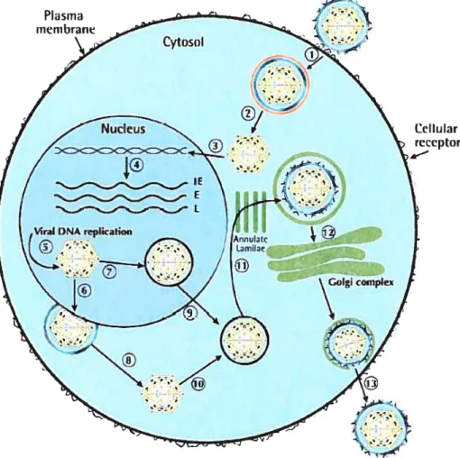
Figure 3. The reptication cycle of HHV-6
Cellular receptor
A singte viral particle is shown here initiating the infection process. However, it is more likely that severai viral particles are involved in this process. The process includes following steps: Attachrnent of the virus and its entry into the ccli by endocytosis (1); de-envelopment ofthe virus particle (2); transport ofthe capsid to the nucleus (3); transcription of immediate early (lE), early (E) and late (L) genes fotlowed by DNA repiication (4); packaging of viral DNA into icosahederai capsid (5); envelopment of capsid and egress to cytosol (6); packaging and tegusome formation (7); de-enveiopment ofthe virus to form capsid (8); transport ofthe virus particie into the cytoplasm (9); acquisition of tegusome (10); re-envelopment and acquisition of membrane glycoproteins while passing through ER (11); transport and glycosylation through Golgi compiex (12); egress oCthe virus from the infected ccli (13).The endosomal membrane is shown in red. ER, Endoplasmic reticulum.
2.6.2 Immune Response:
During a primary infection in chuidren, HHV-6-specific antibodies appear in serum in 3-7 days post-infection (pi). IgM tifre is high in the second week and can remain detectable for 2 months pi. IgG antibodies peak by two weeks. The infected persons remain seropositive for their entire lives (Dockrell et al., 1999). The infected host elicits both humoral and cellular immunity against the virus. Antiviral neutralizing antibodies recognize both linear and conformational epitopes in gB, gH and gp82-105. Ceil mediated immunity against HHV-6 is considered an important element in the
virus control by the host. Analysis of IFN-y production by T-lymphocyte clones in
response to f3-herpes viruses confirms that reacting clones respond to HHV-6 antigens. Individuals having defects in NK ceIl function are susceptible to repeated herpes virus infections. HHV-6 infection of human PBMC induces IL-15 production, which activates NK cclls and enhances NK activity of the PBMC (Flamand et ai.,
1996). Other virus-induced cytokines include IL-1$, IL-13, TNF- but flot IL-6.
HHV-6 suppresses mitogen-induced proliferation of human T celis and inhibits IL-2 secretion from them. It stimulates IFN-y production from CD4 T lymphocytes (Gosselin et ai, 1999).
HHV-6 infection causes CD4+ T ceii depletion. Both HHV-6 variants induce apoptosis in the CD4+ T-ceii unes in vitro. As mentioned earlier, apoptosis in these ceils is not because of viral replication as most of the apoptotic celis are uninfected
celis. However, the induction of apoptosis in CD4+ cord blood lymphocytes is caused
directly by the infected celis (Ichimi et ai., 1999). HHV-6A can also deplete CD4 T-lymphocyte by inducing CD46 mediated celi fusion (Mon et al., 2002). The viral receptor CD46 is down-regulated in HHV-6 infected celis. Since CD46 acts as a compiement regulatory protein, its down-regulation in the infected ceils resuits in
complement activation. Moreover HHV-6 decreases production of reactive oxygen in monocytes (Burd and Carrigan, 1993; reviewed in Dockrell, 2003).
2.6.3 Clinical Pathology:
Primary infections in chiidren usually cause roseola infantum, which is characterized by high fever and the deveiopment of a skin rash upon defervescence (Yamanishi et
ai., 1988; and reviewed by Abdel-Haq and Asmar, 2004). It is noteworthy that HHV-7 also causes roseola in chiidren. According to Hall et al. (1994), HHV-6 was the cause
of febrile illness in 21% of the 6-12 month old chiidren, who visited emergency units. The infection is usually seif-limiting. However, complications, e.g., febrile seizures, encephalitis, etc may also occur. The infection in aduits may cause undifferentiated febrile illness or infectious mononucleosis-like disease. The infection usually becomes latent and the infected persons usually become life-long virus carriers. Primary infections differ from reactivated ones in dlinical symptoms. HHV-6 B variants are ofien associated with roseola; however, A variants are a common cause of infections in chuidren in Zambia. Moreover the symptoms caused by type HHV-6A may vary than those ofHHV-6B. A variety ofclinical complications and diseases are associated with HHV-6 infection (sec Table 4).
2.7 HHV-6 IN THE IMMUNO COMPROMISED HOST:
2.7.1 111V-1 Infection:
HHV-6 is considered a co-factor in the development of AIDS in HIV-1 infected individuals. The HHV-6 U3 protein trans-activates the HIV-1 LTR in vitro (Mon et ai, 1998). It belongs to the HCMV US22 proteins and is expressed in the cytop]asm
and endoplasmic reticulum of HHV-6-infected ceils at one-day pi. HHV-6 infections
are frequently activated in. AIDS patients. Specific symptoms of the reactivation may
include pneumonitis and encephalitis. The infection contributes to the development of
immunosuppression by infecting and depleting CD4+ T celis. In this regard, HHV-6A but flot B variants induce fusion of infected CD4+ T celis with uninfected ceils via interaction between CD46 and gpB-H. As mentioned earlier, HHV-6 can also increase host ceils’ susceptibility to HIV-1 infection by inducing and up-regulating CD4 expression on them (Dockrcll et ai, 2003). The HHV-6 trans-activates HIV-1 LTR by
its several gene products, e.g., U16/17 and U3 (Fiebbe-Rehwaidt, 2000). It aiso
upregulates IL-1f3 and TNF-Πand other cytokines, that increase HIV-1 replication (Dockrel et al., 2003).
In the tissues coinfected with HHV-Ï and HHV-6, HIV-1 proviral DNA ievels are higher (Emery et al., 1999). In chiidren with vertically acquired HIV-1 infection, the primary HHV-6 infection has been associated with more rapid progression of the disease (Caserta et al., 2001). HIV-1 causes suppression of CCR5-tropic (M-tropic) virus by up-regulating RANTES, which is known to inhibit infection by CCR5-tropic viruses and enhance replication of CXCR4-fropic (T-tropic) virus (Griveri et al., 2001). Therefore, HHV-6 co-infection promotes evolution of HW-1 virus towards more pathogenic T-tropic syncytium-inducing viruses. HHV-6 reactivation lias also been implicated in the neuropathogenesis ofAIDS in chuidren.
It has been shown by using the immunomicroarray chip assay that HHV-6 modulates the inflammatory response to HIV infection. Both A and B variants of HHV-6 induce a type-1 response in the host T ceils (Mayne et ai., 2001). As a resuit ofthis infection,
IL-12, IL-15, IL-113, TNF-a and several members of the TNF-Πreceptor family are activated. The infection also up-regulates the transcription of the IL-8 gene and adhesion molecule in Jiver celi unes (reviewed in Ciark, 2000).
Chronic fatigue syndrome (CFS) is characterized by generalized fatigue accompanied by fevers, sore throat, myalgia, lymphadenopathy, sleeplessness, depression and neurocognitive difficulties. Patients suffer from a number of immunologie abnormalities including immunosuppression. It bas been observed that there is reactivation of HHV-6 viruses in the individuals having CfS. Thus, HHV-6 reactivation may be involved in the pathogenesis of this syndrome (reviewed in Abdel-Haq and Asmar, 2004).
2.7.3 Bone Marrow Transplantation:
The incidence of HHV-6 infection after bone marrow transplantationvaries from 28-75% depending upon the diagnostic method. The majority of the HHV-6 infections post-transplantation are due to reactivation of HHV-63 and the peak incidence of the infection is 2-4 weeks post transplantation (Dockrell and Paya, 2001). There is fever
and rash associated HHV-6 viremia after transplantation. Bone marrow suppression,
pneumonitis, encephalitis and grafi versus host disease have been seen in some individuals. The virus infects stem celis and exerts suppressive effects on engrafiment of these celis in transplant recipients. The reactivations may also increase pathogenicity of an existing viral infection or autoimmune condition without becoming a pathogen itself The use of OKT3 and anti-thymosine globulin in the transplantees to prevent grafi rejection is related to the reactivation (reviewed in Caserta et al., 2001). HHV-6 reactivation may also lead to HCMV and EBV reactivations.
2.7.4 Solld Organ Transplantation:
Up to 66% of renal transplant patients have reactivation of HHV-6 infection. As mentioned above in the case of bone manow transplantees, the reactivation occurs most commonly following treatment with OKT3 or antithymocyte globulin and may
be related to a significant degree of immunosuppression associated with these two products. HHV-6 reactivation lias also been described in liver transplant patients (Harma et al., 2003). Co-infections with HCV and HHV-6 may lead to enlianced fibrosis in liver.
2.8 HHV-6 AND 111E CNS
2.8.1 1111V-6 and Encephalitïs:
HHV-6 lias been implicated as a cause of enceplialitis in transplantation recipients (Singli et al., 2000). Its reactivation may cause meningitis and encephalitis in the immunocompetent individuais and the clinical outcomes in these HHV-6 associated encephalitis range from complete recovery to moderate impairment and death (Caserta et aI, 2001). Clinical features of encepliaiitis inciude headache, confusion, seizures, abnormal movernents and disturbance in higher mental function. Both HHV 6 variants can be detected in the specimens and HHV-6 DNA can be detected in the CSF. The presence of HHV-6 DNA in CSf conelates with the presence of central newous system symptoms.
2.8.2 Effect of HHV-6 Infection on CNS White Mafter:
HHV-6 lias a potential role in tlie demyelination of white matter of CNS. Multiple sclerosis (MS) is the most common demyeiinating disease of the human CNS. It lias been strongly associated witli HHV-6 infection. Tlie viral antigens can be detected in
the nuclci of oligodendrocytes from MS patients but flot from control subjects.
Increased levels of anti-HHV-6 IgM and soluble CD46 in MS patients have been detected. However, some workers were unable to support a role of this virus witli MS (Luppi et al., 1994; reviewed by Clark, 2000). The virus may also play a role in progressive multifocal leukoenceplialopathy, which is a demyelinating disease of the
2.9 FEBRILE SEIZURES:
HHV-6 infection causes febrile seizures in infants and young chiidren having more pronounced effect on chiidren of 12-15 months of age. Affected chiidren may have convulsions and other febrile diseases. Among the chiidren whose first febnle seizures were caused by HHV-6, the incidence of recunent febrile seizures was significant. Frequency of severe forms of convulsions and postictal paralysis is significantly higher among children with primary HHV-6 infection (Jee et al., 1998; reviewed by Abdel Haq and Asmar, 2004).
2.10 HHV-6 AND CARDIOVASCULAR SYSTEM:
HHV-6 infection has been detected in the endothelium of aorta, umbilical vein and capillaries ofthe bone manow (Takatsuka et ai., 2003). Thc infection is aiso related to thrombic microangiopathy (Toyabe et aI., 2002). Several reports have also associated primary HHV-6 infection with idiopathic thrombocytopenic purpura (Hashimoto et
al., 2002; reviewed in Koichi Yamanishi, 2001).
2.11.1111V-6 AND OTHER VIRUS INFECTION
HHV-6 can activate other viral infections, for instance those induced by EBV, HCV, measles, papillomavinis and parvovirus and may contribute to the pathologic effects of these viruses. Dual active infections frequently appear especially with other herpes viruses.
2.12. EPIDEMIOLOGY
HHV-6 is ubiquitous in human populations and up to 90% of the aduit population is seropositive ail over the world (reviewed by Krueger and Ablashi, 2003). It usually infects children in the later half of the flrst year of age, peaking at 3-9 months (Clark, 2000; Campadelii-Fiume et ai, 1999). Matemal antibodies decline by the age of 6 months and infants become increasingly susceptible to HHV-6 infection at this age. In
Europe and the USA, the HHV-6 seroprevalence is 72-95% both in aduits and children. In Africa, Asia and Latin America, the seroprevalence is 60-95%. Majority of the clinical infections in immunocompetent host are due to HHV-6B variants. The infections with A variants are frequently present in patients with immunosuppression and neurological manifestations (Hall et al., 199$). Infections usually occur via contaminated oral secretions (Krueger and Ablashi, 2003; Clark, 2000). In the salivary gland tissue, the more frequent strain is 6B. These variants are also more frequently found in the peripheral blood mononuclear celis. Co-infections with both A and B variants were detected in 22-34% oflung specimens (Clark, 2000; Cone et al., 1996).
2.13 ANTWIRAL TREATMENT
Many compounds effective against HHV-6 have been reported to date but no controlled clinical studies are available (reviewed by Clark, 2000). Gancyclovir and acyclovir have shown some inhibitory activity on HHV-6 viral infections. Gancyclovir blocks HHV-6 infection in bone marrow transplant patients with HHV-6 encephalitis. Valacyclovir is used for prophylaxis against HHV-6 reactivation. Foscamet and phosphonoacetic acid inhibit viral DNA polymerase. They have no effect on latent infections. Type 1 1EN has also been shown to reduce disease activity in patients with multiple sclerosis (Hong et aI, 2002).
3. E2F TRANSCRIPTION FACTORS
E2F factors comprise a family of related transcription factors that play a key role in the regulation of cdl cycle progression in many different species including mammals, flues, nematodes, amphibians and plants. The first E2F was originally identified as a factor with transcriptional activity that binds to the sequence
‘TTTCGCGC’
in theE2 promoter of the adenovirus (Kovesdi et ai, 1986). Hence the terrn E2F was coined; ‘E2’ cornes from the E2 promoter and ‘F’ stands for factor. Later on, simiiar
sequences, which are invoived in the reguiation of gene expression, were identified in
the promoters of several celluiar genes, e.g., dihydrofolate reductase (DHFR), c-myc and cyclin E, (Biake et ai, 1989). Most of these sequences are in the reguiatory
regions of the genes that moduiate growth, ccli cycle progression and DNA synthesis,
e.g., cyciin-dependent kinases (CDK), cyclins, proiiferating ccii nuclear antigen
(PCNA), DNA polyrnerase, etc. The activity of E2F transcription factors is under the control of pRB, and cyclins-CDKs as shown in Figure 4. The proiiferative E2Fs (E2F-1, E2F-2 and E2F-3) are aiso regulated by acetyiation.
E2Fs aiso regulate their own activity positively due to the presence of the E2F cognate sites in the E2F gene promoters. Recent studies have revealed that E2Fs target various gene functions not only during the GuS transition and DNA replication but also during mitosis and DNA damage and repair checkpoints (Ren et ai, 2002; reviewed in Cam and Dyniacht, 2003).
3.1 The E2F Family:
E2F famiiy is made up of DNA binding heterodimeric proteins containing one E2F subunit and one DP subunit. E2Fs bind DNA as dimers (Huber et ai, 1993). They may form homodimers among themseives or heterodimers with DP subunits. The homodimers of E2F eau bind to their cognate DNA sequence but this binding is very weak as compared with the E2F/DP heterodimers.
3.2 STRUCTURE 0F E2F AND 1W PROTEINS:
The structure of a typicai E2F family member is shown in Figure 5. It contains distinct domains for binding to DNA, cyclin A, and “pocket” proteins (sec