Study on the Toxic Mechanism of Prion
Protein Peptide 106–126 in Neuronal
and Non Neuronal Cells
Ingrid Dupiereux,1Willy Zorzi,1Walid Rachidi,2Danie`le Zorzi,1Olivier Pierard,1 Bernard Lhereux,3Ernst Heinen,1and Benaı¨ssa Elmoualij1*
1Department of Human Histology, CRPP, University of Lie`ge, Institute of Pharmacy-CHU, Sart Tilman, Lie`ge, Belgium
2Service de Ge´nomique Fonctionnelle, Baˆtiment Ge´nopole 2, CEA, Evry Cedex, France 3Laboratoire Roman Paı¨s, Nivelles, Belgium
A synthetic peptide corresponding to the 106–126 amy-loidogenic region of the cellular human prion protein (PrPc) is useful for in vitro study of prion-induced neuro-nal cell death. The aim of the present work was to examine the implication of the cellular prion protein in the toxicity mechanism induced by PrP 106–126. The effect of PrP 106–126 was investigated both on human neuroblastoma SH-SY5Y cells and on SH-SY5Y over-expressing murine cellular prions (wtPrP). We show by metabolic assay tests and ATP assays that PrPc ex-pression does not modulate the toxicity of the prion peptide. Moreover, we investigated the effect of this peptide on an established non neuronal model, rabbit kidney epithelial A74 cells that express a doxycycline-inducible murine PrPc gene. We show for the first time that the prion peptide 106–126 does not exert any toxic effect on this cell line in the presence or absence of doxycycline. Our results show that the PrP 106–126-induced cell alteration is independent of PrPc ex-pression. Rather, it seems to act via an interaction with lipidic components of the plasma membrane as strengthened by our results showing the differential susceptibility of neuronal and non neuronal cell lines that significantly differ by their membrane fatty acid composition. VVC 2006 Wiley-Liss, Inc.
Key words: cellular prion peptide; 106–126 prion pep-tide; lipid membrane; neurotoxicity
Prion diseases are fatal neurodegenerative disorders affecting the central nervous system (CNS) of humans and animals and characterized by a neuronal vacuolation, astrocytosis, and progressive neuronal degeneration (Gaj-dusek et al., 1966; Clinton et al., 1993; Fraser, 1993; Jeffrey et al., 2000). These diseases are caused by the in-tracerebral accumulation of an abnormal isoform of the cellular prion protein (PrPc), named PrPsc (PrP scrapie) (Caughey and Lansbury, 2003; Collins et al., 2004; Prusiner, 1998). Many lines of evidence suggest that PrPc acts as a template that promotes the conversion of PrPcto PrPsc(Caughey and Raymond, 1991). The plasma
membrane seems to play a key role in the molecular mechanism implicated in this conversion. Supporting this hypothesis is the subcellular site for the formation of PrPsc; indeed, the conversion occurs after PrPcreaches the plasma membrane (Taraboulos et al., 1995). Moreover, Baron et al. (2002) have shown, using purified raft mem-branes, that the conversion of raft-associated GPI-anch-ored PrPc to PrPsc requires the insertion of PrPsc into the lipid membrane. However, the mode of interaction of prion proteins with the membranes has not as yet been elucidated: does the interaction occur via a direct insertion of the prion protein into the lipid bilayer or via a putative membrane receptor?
We have shown previously that PrP (106–126), a peptide largely used as a model to study the PrPsc in-duced neurotoxicity, destabilizes lipid vesicles mimicking the composition of neuronal membranes (liposomes) and induces liposome fusion (Dupiereux et al., 2005a). This destabilization mechanism occurs at low concentrations of the peptide (from 2–10 lM) and via a membrane in-teraction, as supported by the integrity membrane assay measuring the release of the intracellular lactate dehydro-genase (LDH assay). These results are in agreement with other previous reports. Indeed, several studies have shown that PrP 106–126 forms ion channels in planar lipid bilayers (Arispe et al., 1996; Kawahara et al., 2000) and that it increases the membrane microviscosity of
Contract grant sponsor: Re´gion Wallonne; Contract grant number: BA4 114915, EPH331030000092-430001; Contract grant sponsor: Fonds Social Europe´en; Contract grant number: W2002134; Contract grant sponsor: Commisariat a` l’Energie Atomique (CEA); Contract grant spon-sor: LaboratoireRoman Paı¨s. AQ1 *Correspondence to: Benaı¨ssa Elmoualij, Department of Human Histol-ogy, CRPP, University of Lie`ge, Institute of Pharmacy-CHU, 1, avenue de l’Hoˆpital, Sart Tilman, 4000 Lie`ge Belgium.
E-mail: b.elmoualij@ulg.ac.be
Received 20 December 2005; Revised 21 February 2006, 22 March 2006; Accepted 26 April 2006
Published online 00 Month 2006 in Wiley InterScience (www. interscience.wiley.com). DOI: 10.1002/jnr.20965
neurons and astrocytes (Diomede et al., 1996; Salmona et al., 1997). Nevertheless, the role of the cellular prion protein in the membrane destabilization mechanism in-duced by this peptide remains as yet non-elucidated. Several works, using knock-out cell lines, show that PrPc expression is necessary for the cellular toxicity in-duced by this peptide (Brown et al., 1994; Chabry et al., 2003a; Fioriti et al., 2005b). These results indicate that the membrane destabilization induced by PrP 106–126 could arise from a dual mechanism: a direct peptide in-sertion in the lipid bilayer or an interaction with PrPc.
To investigate the role of PrPc in the neurotoxicity induced by the 106–126 prion peptide we have examined whether an overexpression of murine PrPc(wtPrP) (Walms-ley et al., 2001a) changes the susceptibility of neurons to the peptide. Our results indicate that the toxicity induced by the 106–126 peptide is independent of PrPc expression level. Moreover, the addition of an anti-prion antibody (SAF 34), able to decrease the basal level of PrPc, induces, in our ex-perimental conditions, non-significant changes in toxicity, showing that the PrPc expression is not necessary for the PrP 106–126 toxicity. Additionally, to study the potential relationship between PrP 106–126 and lipidic components of the plasma membrane, we have investigated the effect of the peptide on non-neuronal cells, rabbit kidney epithelial (A74) cells, which express a doxycycline-inducible murine PrPc gene. We have shown that this cell line is resistant to the PrP 106–126 toxicity confirming that the toxicity of this peptide is independent of PrPc expression levels. We there-fore suggest that this toxicity might be in relation with the lipidic composition of the cell membranes as strengthened by our results after the comparison of the fatty acid compo-sition between neuronal and non-neuronal cells by HPLC fatty acid analysis.
MATERIALS AND METHODS AQ2
Chemicals
PrP106–126, derived from amino residues 106–126 of the human prion protein sequence (sequence: Lys-Thr-Asn- Met-Lys-His-Met-Ala-Gly-Ala-Ala-Ala-Ala-Gly-Ala-Val-Val-Gly-Gly-Leu-Gly) was purchased from Eurogentec SA
AQ3 and
scrambled peptide containing the same amino acids in a ran-dom order (sequence: Asn-Gly-Ala-Lys-Ala-Leu-Met-Gly-Gly-His-Gly-Ala-Thr-Lys-Val-Met-Val-Gly-Ala-Ala-Ala) was purchased from Bachem
AQ3 . Palmitic acid was purchased from
Sigma Aldrich
AQ3 . The secondary antibody rabbit antimouse IgG, A, M/FITC was purchased from Serotec
AQ3 and Streptavidine/
FITC from Pharmingen
AQ3 . Rabbit polyclonal antibody P45-66
was supplied by Dr. D. Harris (Washington University, St. Louis, MO) and the secondary antibody HRP-coupled rabbit IgG was from Sigma (St. Louis, MO). The mouse anti-PrPC SAF34 was kindly provided by Dr. J. Grassi from CEA of Paris. All cell culture supplies were purchased from Life Tech-nologies Inc.
AQ3 and fatty acid supplies were from VWR Interna-tional
AQ3 and Sigma-Aldrich.
Cell Culture
The human neuroblastoma cell line SH-SY5Y, kindly provided by Professor N.M. Hooper, and the same line stably transfected with the wild-type murine PrP (wtPrP) (Walmsley et al., 2001b) were cultured in Dulbecco’s modified Eagle’s medium (DMEM) (Life Technologies Inc.) supplemented AQ3 with 10% fetal bovine serum (FBS) (Life Technologies Inc.), 1% penicillin/streptomycin (Life Technologies Inc.). Cells were maintained at 378C in a humidified incubator with 95% air and 5% CO2. For experiments, cells were maintained in FBS-free DMEM medium containing the neuroblastoma growth supplement N2 (Life Technologies Inc.) and 1% penicillin/strep-tomycin.
The A74 cell line, initially generated in the laboratory of Drs. Vilette and Laude (INRA, Jouy-en-Josas, France), was established by transfecting rabbit kidney epithelial cells with murine PrPc. The expression of the murine PrP is doxycy-cline-inducible via a tetracydoxycy-cline-inducible (tet-on) system (Vilette et al., 2001d). The stable transfectants were selected in the presence of puromycin (10 lg/ml). A74 were cultured in modified Eagle’s medium (MEM) supplemented with 10% heat-inactivated FBS and 1% penicillin/streptomycin. Cells were grown at 378C in a humidified incubator with 95% air and 5% CO2. For experiments, cells were maintained in MEM medium containing 2% FBS and 1% penicillin/strep-tomycin.
Cell Metabolism Assays
SH-SY5Y cells were seeded into 96-well culture plates. Sixteen hours after seeding, the medium was replaced with se-rum-free DMEM containing the neuroblastoma growth sup-plement N2 and the cells were treated for 24 hr with different concentrations of PrP 106–126 (10–200 lM). The treatment with the anti-prion antibody SAF 34 was carried out by incu-bating the cells with an optimized concentration of antibody SAF 34 (1 lg/ml) and after 3 hr, the medium was replaced with fresh medium and cotreated for an additional 24 hr with PrP 106–126 (200 lM) and SAF 34 (1 lg/ml).
A74 cell line in 96-well culture plates were grown over-night in MEM supplemented with 2% FBS. The next day the medium was changed and the cells were treated with doxycy-cline at 500 ng/ml to induce PrPcexpression. After 24 hr the cells were incubated with PrP 106–126 (50–100 lM) for an additional 24 hr.
To study the effect of lipidic factors, such as palmitic acid, on the toxicity effect of the 106–126 peptide, both the neuronal (SH-SY5Y) and non neuronal cells (A74) were incu-bated with palmitic acid at a final concentration of 25 lM for 16 hr. Cells were then treated with the palmitic acid (25 lM) and PrP 106–126 (200 lM) for additional 24 hr.
The cell proliferation was measured using the CellTiter 96 AQueousNon-Radioactive Cell Proliferation Assay (Prom-ega) according to the manufacturer’s instruction. The cell tox- AQ3 icity was assessed quantitatively by MTS assay in the presence of phenazine methosulfate (PMS). After addition of 20 ll of the combined MTS/PMS solution in each well, the plates were incubated at 378C in a humidified atmosphere
contain-ing 5% CO2 for 2 hr. The absorbance was measured at 490 nm (EL 312e microplate Bio-Tek Instruments
AQ3 ).
All MTS assays were carried out in triplicate. MTS assay is a sensitive indicator of mitochondrial activity.
Cell Toxicity Test
Measurement of ATP levels allows to estimate cellular suffering. Human SH-SY5Y cells were seeded into 96-well culture plates. Sixteen hours after seeding, the medium was replaced with serum-free medium containing the neuroblastoma growth supplement N2 and the cells were treated for 24 hr with different concentrations of PrP 106–126 (10–200 lM).
ATP was quantified using the CellTiter-Glo Lumines-cent Cell Viability Assay (Promega) according to the manufac-turer’s instruction. The reagent is a buffered solution contain-ing detergents to break the cell membrane, releascontain-ing ATP im-mediately on addition to wells and ATPase inhibitors to stabilize ATP once it is released from cells. After 106–126 peptide treatment, assay plates were removed from the incuba-tor and 100 ll of CellTiter-Glo Assay reagent, equilibrated to room temperature, was added to each well. Plates were shaken for 2 min to mix the contents of the wells. After a 10–15 min incubation at room temperature, luminescence was deter-mined using a Microplate Luminometer MPL2 (Berthold). PrPc Labeling for Confocal Microscopy
A total of 20,000 SH-SY5Y or wtPrP cells were seeded and left to adhere for 5 hr in 50 ll DMEM 10% FBS and 1% penicillin/streptomycin on uncoated 12-mm circular glass coverslips placed in 24-well culture plates (Greiner). Fresh medium (450 ll) was added. Cells were cultured for 24 hr then fixed during 20 min in 1:1 acetone/methanol at 208C. The acetone/methanol was discarded and the coverslips were kept at 208C until use. The cells were then dehydrated at room temperature and rehydrated in phosphate buffered saline (PBS) before labeling. PrPCwas revealed
AQ6 at room temperature
as follows: cells were incubated 1 hr with SAF34 10 lg/ml in PBS, washed 3 times with PBS, incubated 30 min with the rabbit anti-mouse IgG, A, M/FITC 1/1,000 in PBS, followed by washing 3 times in PBS. The coverslips were mounted on microscopy glass slides using DAKO fluorescence mounting medium before observation with a Leica TCS SL SP2 confo-cal microscope (Leica, Belgium) equipped with Argon, Ne/ Kr, and He/Ne lasers. One-step scans were carried out. A se-ries of 20 images were acquired and stacked using the Maxi-mum Projection.
Western Blotting
A74 cells (seeding density 5 3 106) were grown in T75 cm2 flasks. The next day the cells were treated for an additional 24 hr with different concentrations of doxycycline (10–500 ng/ml). After 24 hr, the cells were washed twice with cold PBS, calcium- and magnesium-free, and lysed for 30 min at 48C in Triton-deoxycholate lysis buffer (13 buffer is 150 mM NaCl, 0.5% Triton X-100, 0.5% sodium deoxy-cholate, and 50 mM Tris-HCl, pH 7.5) containing protease inhibitors. After 1 min of centrifugation at 10,000g, the super-natant was collected, and its protein concentration was
meas-ured by the BCA assay (Pierce). The equivalent of 20 lg of total protein in SDS loading buffer was subjected to 12% SDS-PAGE electrophoresis followed by electroblotting on polyvinylidene difluoride in Tris-glycine buffer containing 20% methanol. The membrane was blocked with 5% non-fat dry milk in TBST (0.1% Tween 20, 100 mM NaCl, 10 mM Tris-HCL, pH 7.8) for 1 hr at room temperature, and PrP was detected by immunoblotting with P45-66 antibody. After adding the second antibody (horseradish peroxidase-coupled rabbit IgG), immunoreactive proteins were detected with the ECL Western blot system.
Flow Cytometry
Human SH-SY5Y cells seeded in 6-well plates (1.6 3 106 cells/well) were incubated for 24 hr in the presence or absence of SAF34 at 1 lg/ml. The cells were then rinsed in PBS and collected after incubation at 378C for 10 min with 500 ll of cell dissociation buffer (enzyme free; Invitrogen). Samples of 100 ll containing 1 3 106 SH-SY5Y cells were incubated with the biotinylated antibody SAF34 (10 lg/ml) for 30 min at 48C in PBS, washed and then incubated for an additional 30 min with a fluorescein isothiocyanate-conjugated streptavidin (diluted 1/1,000). The control cells were not in-cubated with the antibody (biotinylated SAF34). After rinsing, the resuspended cells were immediately analyzed in a FACS-can (Becton-Dickinson, Sunnyvale, CA).
Fatty Acid Analysis
Phospholipid extraction was carried out on SH-SY5Y and A74 cells. The cells were placed in 0.1 ml of a mixture of butylated hydroxy toluene (BHT)/CH2Cl2 (0.0204 mg of BHT in 1 ml of CH2Cl2) and 4 g of Na2SO4 and crushed with a cooled mortar. After 1 min, Celite 545 (3 g) was added and the cells were further crushed for 1 min. The mixture was transferred in a chromatography column filed with glass wool and 2 g of a mixture of CaHPO4.2H20 and Celite 545 AQ4 in a proportion of 1:9. The mortar was washed with 15 ml of the mixture CH2Cl2/CH3OH (90:10) and transferred onto the column. About 50 ml of the mixture CH2Cl2/CH3OH was added and the eluent was collected and diluted in 1 ml of ethyl acetate and the phospholipid fatty acids were separated with a SPE method (solid phase extraction). The trans-esterifi-cation procedure of the carboxylic group with methanol in hot acid medium produced the fatty acid methyl esters (FAME). The FAME were separated by gas chromatography (GC), Agi-lent 6890, on a capillary column with helium as the carrier gas. The analysis was realized with a flame ionization detector. The chem station software, on the GC Agilent 6890, measured the height of the different peaks and calculated the concentrations of the different FAME.
RESULTS
Toxicity of PrP 106–126 Is Independent of the Level of Cellular PrP Expression
To study the relationship between the toxicity of PrP 106–126 and the expression levels of the cellular prion protein, we compared the effect of the peptide on SH-SY5Y neuroblastoma cells, expressing a basal level of
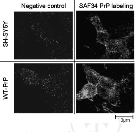
human PrP, with the same SH-SY5Y cells overexpress-ing a murine prion protein (wtPrP). The level of PrP expression was analyzed by confocal microscopy using the SAF 34 antibody. As shown in Figure
F1 1, a more
intense immunostaining was observed on wtPrP cells, whereas a weak staining was detected on SH-SY5Y cells. These results confirm that the wtPrP cells overexpress PrP in comparison with the SH-SY5Y cells that express a basal level of PrP.
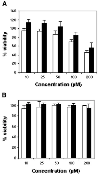
Neuronal injuries induced by the 106–126 peptide were monitored by measuring the reduction of the mi-tochondrial activity using the MTS assay and the ATP assay monitoring cell alteration. MTS is converted to a formazan product by dehydrogenase enzymes, which become inactive as the cell suffers. Measurement of this formazan product is an indicator of cell metabolism and viability. As shown in Figure
F2 2A, PrP 106–126
treat-ment (10–200 lM) induces a concentration-dependent toxicity for both SH-SY5Y and wtPrP cells. No signifi-cant difference in the toxicity induced by the peptide was observed between the two cell lines; indeed, PrP 106–126 caused a 50% decrease at 200 lM in SH-SY5Y and wtPrP (45.9 6 3.6% and 55.8 6 11.2%, respec-tively). In contrast, 200 lM of scrambled peptide had no effect (99.5 6 0.96%) (Fig. 2B). These data indicate that the toxicity induced by the peptide 106–126 is inde-pendent of the level of PrP expression.
By using the ATP assay, the effect of PrP 106–126 on the intracellular content of ATP was measured in relation with the level of PrP expression (Fig. 3). A F3 24 hr treatment with different concentrations of the pep-tide (10–200 lM) induced a dose-dependent decrease in the ATP level without any difference between the two tested cell lines (SH-SY5Y and WtPrP). These data are in agreement with those obtained with the MTS assay confirming that the toxicity induced by PrP 106–126 is independent of the level of PrPc expression.
To elucidate whether the neurotoxic effect of PrP 106–126 observed on the neuroblastoma cell lines is mediated by the PrP basal level expression, we blocked PrPc surface expression with an anti-PrP antibody. We used SAF34, known to interact with PrPc and prevent the interaction between PrPc and PrPsc (Perrier et al., 2004; Feraudet et al., 2005a).
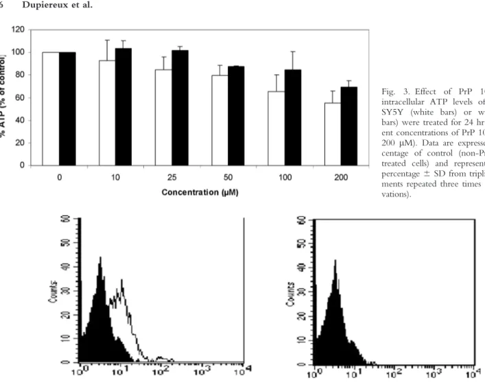
To investigate the effect of the SAF34 antibody on PrPc expression, we analyzed by flow cytometry the level of PrPc on SH-SY5Y neuroblastoma cells incubated for 24 hr in the presence or absence of the SAF34 antibody. As shown in Figure 4a, in the absence of SAF34 treat- F4 ment, the neuroblastoma cells SH-SY5Y present a posi-tive immunostaining indicating that they express a basal level of PrPc as shown by confocal microscopy (Fig. 1). Conversely, when the cells were pre-incubated with the SAF34 antibody for 24 hr (Fig. 4b) no staining was
Fig. 1. Immunofluorescence detection of the prion protein on SH-SY5Y and wtPrP cells. PrPc was detected using SAF 34 antibody (10 lg/ml) followed by a FITC-conjugated secondary anti-body. Cells were viewed by a Leica laser scanning confocal microscope. A more pronounced staining was ob-served on wtPrP cells indicating a high expression of PrP. Figure can be viewed in color online via www.interscience. wiley.com.
observed, suggesting that PrPc was either cleared or masked by the interaction with SAF34.
To study the effect of the antibody treatment on the toxicity induced by the prion peptide 106–126, we have carried out toxicity assays on cells treated for 24 hr with the antibody. The MTS assay (Fig.
F5 5) showed that
the measured values remained equivalent in the presence of the SAF antibody suggesting that the toxicity of the peptide is independent of the cellular prion protein.
We have shown previously that the PrP 106–126 induced a destabilization of lipidic liposomal vesicles indicating that the toxicity effect of this peptide could occur via a membrane interaction (Dupiereux et al., 2005b); this in turn suggests that lipidic components
could play a key role in the toxicity of PrP 106–126 peptide.
Rabbit Kidney Epithelial Cells Are Resistant to Prion Peptide 106–126 Toxicity
To further investigate the relationship between the basal expression of PrPc and the neurotoxicity induced by the peptide 106–126 we used a cell model (rabbit kidney epithelial cells) that expresses low levels of PrPc but in which murine PrPc can be selectively induced by doxycycline (Vilette et al., 2001c).
Treatment with doxycycline (10–1,000 ng/ml) in-duced the expression of PrPc, as shown by Western blot-ting (Fig. 6). Conversely, in the absence of doxycycline F6 no endogenous PrPc was detected in these cells (Vilette et al., 2001b).
In these cells, we have shown that the absence or the presence of doxycycline (absence or presence of PrPC expression), does not change the PrP 106–126 tox-icity (Fig. 7). The incompatibility between the human F7 sequence of PrP 106–126 and the murine PrP can be discarded; indeed, we have shown by MTS assay, that the same peptide is neurotoxic for murine neuroblastoma cells (N2a) expressing a murine PrP in the same concen-tration range of PrP 106–126 inducing 50% cell death at 200 lM (data not shown). These data indicate that the PrP expression is not required for PrP106–126 toxicity.
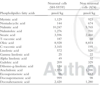
The difference in the susceptibility of these two tested cell lines (neuronal and non neuronal), toward the PrP 106–126 peptide, might be related to the composi-tion of the lipidic cell membrane. To study the putative implication of the lipids, the fatty acid composition of the cellular membrane of SH-SY5Y and A74 was com-pared (Table I). Significant differences were observed T1 between these two cell lines and in particular in the composition of their unsaturated fatty acids. Indeed, the content in unsaturated fatty acids in the neuroblastoma cells was significantly higher than in the rabbit kidney epithelial cells. The most unsaturated fatty acids in the neuronal cells were palmitoleic acid (16:1), vaccenic acid (18:1), arachidonic acid (20:4), docosapentaenoic acid (22:5), and docosahexaenoic acid (22:6). These data sug-gest that the susceptibility to the prion peptide 106–126 could be related to the fatty acid composition of the cel-lular membranes. Our observation reinforces the hypoth-esis of a direct interaction of the prion 106–126 peptide with the lipidic membrane.
Enhanced Toxicity of PrP 106–126 on the Neuroblastoma Cell Line in the
Presence of Palmitic Acid
Based on fatty acid analysis we observed different saturated and non saturated fatty acid composition between the neuronal and non neuronal cell lines used in this study. Indeed, amongst others, palmitic acid, vac-cenic acid and docosahexaenoic acid were present in low amounts in the PrP 106–126 resistant non neuronal
Fig. 2. PrP 106–126 neurotoxicity in SH-SY5Y and wtPrP cells. PrP 106–126 (A) or scrambled (control) PrP 106–126 (B) was added at concentrations of 10–200 lM. Cell metabolism (viability) was measured 24 hr later using the MTS assay. PrP 106–126, but not scrambled peptide, altered the metabolic activity of SH-SY5Y (white bars) and wtPrP (black bers) cells in a dose-dependent manner. No significant differences were observed in relation with the level of PrPc expression. Each value represents the mean percentage 6 SD from triplicate experiments repeated three times (nine observations).
cell line. In the present work we were mainly interested on the potential implication of palmitic acid, a represen-tative cell membrane fatty acid, in this resistance.
To evaluate whether palmitic acid triggered PrP 106–126 cytotoxicity, we determined the synergistic toxic effect of palmitic acid and prion peptide on the neuronal (SH-SY5Y) and non neuronal (A74) cell lines. Cell viability as measured by MTS assay is shown in Fig-ure
F8 8. We compared the viability of the palmitic acid pre-treated cells after a 24 hr exposure to 200 lM PrP 106–126. As expected, PrP 106–126 led to a 50% sur-vival for the neuronal cell line (SH-SY5Y) whereas no toxicity was observed for the rabbit kidney epithelial cell line (A74). In contrast, SH-SY5Y pre-treated with pal-mitic acid showed significant susceptibility to the toxic effect of prion peptide with a cell death increase of 10%, whereas palmitic acid alone did not affect cell viability. Surprisingly, the non neuronal A74 cell line pre-treated with palmitic acid remained resistant to the PrP 106– 126 toxicity.
DISCUSSION
PrP 106–126 peptide is a useful model for the in vitro study of prion-induced cell suffering. Indeed, this peptide exhibits some of the pathogenic and physico-chemical properties of PrPsc. It is able to form protease-resistant fibrils, induce neuronal toxicity, and has a rela-tively high b-sheet content (Tagliavini et al., 2001). However the mechanism by which this peptide induces alterations in neuronal cells is not well understood. Does this occur by direct membrane interactions or via mem-brane receptors?
We have studied the relationship between the expression of PrPc, the lipidic membrane composition, and the toxicity induced by the prion peptide 106–126.
It is widely accepted that PrPc is necessary for the toxicity of PrPsc (Bueler et al., 1993; Brandner et al., 1996). This dependence on PrPc and toxicity of PrP 106–126 has also been indicated (Brown et al., 1994; Chabry et al., 2003b; Fioriti et al., 2005d). Nevertheless,
Fig. 3. Effect of PrP 106–126 on intracellular ATP levels of cells. SH-SY5Y (white bars) or wtPrP (black bars) were treated for 24 hr with differ-ent concdiffer-entrations of PrP 106–126 (10– 200 lM). Data are expressed as a per-centage of control (non-PrP 106–126 treated cells) and represent the mean percentage 6 SD from triplicate experi-ments repeated three times (nine obser-vations).
Fig. 4. Flow cytometry detection of PrPc expression in the absence (A) or in the presence (B) of SAF 34. SH-SY5Y cells were incubated for 24 hr with the anti-prion antibody SAF 34 and PrPc was detected with a biotinylated antibody SAF34 (10 lg/ml) for 30 min at 48C (white histogram). The control cells were not incubated with the antibody biotinylated SAF34 (black histogram).
Fig. 6. Doxycycline-dependent induction of PrPc expression in A74 cells. Dox was added at different concentrations (0, 10, 25, 50, 100, 500, and 1,000 ng/ml) to medium for 24 hr, and PrPc expression was determined in A74 cells by Western blot. The equivalent of 20 lg of protein were subjected to SDS-PAGE and PrPc was
detected with antibody P45-66 raised against the N terminus of the protein. PrPc expression reaches a maximum at 500 ng/ml of dox; after this concentration we reach a plateau. Molecular mass markers are indicated on the left in kDa.
Fig. 7. Effect of PrP 106–126 on a rab-bit kidney epithelial cells (A74) in rela-tion with the expression level of PrPc. A74 cells were seeded at 5,000 cells/ well and stimulated with doxycycline (50 and 500 ng/ml) 24 hr before PrP 106–126 exposure at different concen-trations: 50 lM (white bars); 100 lM (black bars). Viability was measured by MTS assay. Each value represents the mean percentage 6 SD from triplicate experiments repeated three times (nine observations).
Fig. 5. Effect of SAF 34 on the toxicity induced by PrP106–126 in SH-SY5Y and wtPrP cells. The viability was mea-sured in untransfected SH-SY5Y cells (white bars) or wtPrP-expressing cells (black bars) both exposed to PrP 106– 126 at 200 lM for 24 hr after a pre-incubation of 3 hr in the presence or absence of SAF 34. Each value repre-sents the mean percentage 6 SD from triplicate experiments repeated three times (nine observations).
the mechanism of toxicity remains unclear and contra-dictory data about the relation between PrPc expression and the toxicity induced by PrPsc or PrP 106–126 have been reported. Several works have shown that the prion peptide 106–126 is toxic to cultured neurons and that neurons derived from Prnp / mice are resistant to this peptide (Brown et al., 1994; Chabry et al., 2003c). Fior-iti et al. (2005a) using cerebellar granule neurons derived from wild-type mice and Tg mice overexpressing mouse PrP, did not detect any difference in the toxicity of PrP 106–126 on both cell lines. Conversely, they did not observe any toxicity of the peptide on the knock-out cell line. These data suggest that the expression of PrPc is necessary for the toxicity induced by the peptide but that the level of expression does not modulate this neu-rotoxicity. The authors propose to explain the toxicity of PrP 106–126 by the physiologic loss of PrPc.
Further-more, Gu et al. (2001) have shown that in a human neuronal cell line resistant to the PrP 106–126 toxicity, the prion peptide binds at the cell surface and that some of the resistant neuronal cells internalize the peptide that accumulate in intracellular compartments. They con-cluded that the resistance to the toxicity induced by PrP 106–126 seemed to be related to an aberrant binding of the peptide with the membranes. Currently, it is known that such an aberrant binding with the cellular mem-brane can be induced by an alteration in the memmem-brane lipidic composition.
In the present study, we have analyzed the toxicity of the peptide on a neuroblastoma cell line expressing different levels of PrPc. Our results show that the toxic-ity of PrP 106–126 is independent of the expression level of the cellular prion protein, as shown by Fioriti and collaborators (Fioriti et al., 2005c). To study the implication of the endogenous levels of cellular prion protein in the toxicity induced by the peptide, we have used an original strategy based on the ability of an anti-prion antibody (SAF34) to inhibit PrPsc replication and to decrease the levels of total PrPc and PrPsc in an infected cell model (Feraudet et al., 2005b). We have shown by MTS assay that an incubation of SH-SY5Y with SAF34 did not reduce the toxic effect of the pep-tide 106–126 indicating that endogenous levels of the cellular prion protein is not directly implicated in the toxicity of the peptide. Because we cannot exclude the possibility that residual PrPc is left in our cells after the antibody treatment, we propose that a sub-physiological level or an appropriated lipid composition could be required for peptide toxicity.
Our finding is consistent with other reports indi-cating that the toxicity of PrPsc and PrP 106–126 is in-dependent of the expression of PrPc. Indeed, it has been shown that PrPsc and PrP 106–126 may cause an upre-gulation of the MAP kinases signalling pathway inducing neurotoxicity in Prnp / cells (Gavin et al., 2005; Mar-ella et al., 2005). Furthermore, McHattie et al. (1999) have studied the cellular trafficking of the prion peptide 106–126 and have observed that cultured cells are capa-TABLE I. Comparative Study of Fatty Acid Composition of the
Cellular Membrane of SH-SY5Y and A74 Cells
Phospholipidics fatty acids
Neuronal cells (SH-SY5Y) Non neuronal cells (A74) lmol/kg lmol/kg Myristic acid 1,129 923 Nentadecyclic acid 144 674 Nalmitic acid 10,247 5,765 Nalmitoleic acid 1,276 701 Stearic acid 3,596 2,460 T-vaccenic acid 187 68 Oleic acid 8,928 8,814 C-vaccenic acid 3,165 195 Linolenic acid 572 520 Gamma linolenic acid 25 22 Alpha linolenic acid 49 32
Gadoleic acid 209 329 Dihomo-g-linolenic acid 124 177 Arachidonic acid 1,974 1,390 Eicosapentaenoic acid 96 612 Docosapentaenoic acid 999 98 Docosahexaenoic acid 2,420 1,280
Fig. 8. Effect of palmitic acid on the toxicity induced by PrP 106–126 in SH-SY5Y and A74 cells. The viability was measured in SH-SY5Y cells (white bars) and A74 cells (black bars) both exposed to PrP 106–126 at 200 lM for 24 hr after a pre-incubation of 16 hr in the presence or absence of palmitic acid (25 lM). Each value represents the mean percentage 6 SD from triplicate experiments repeated three times (nine observations).
ble of sequestering the peptide independent of PrPc expression.
These results suggest that the toxic mechanism of this peptide is independent of a direct interaction with PrPc and indicate that a direct insertion of PrP 106–126 into the cell membrane, particularly in cholesterol-rich lipid domains, named rafts, could induce disturbances. We have shown recently that the 106–126 peptide de-stabilizes lipid vesicles mimicking the composition of neuronal membranes and that it induces liposome fusion. This destabilization occurred via a membrane interaction as was shown by the integrity membrane assay (Dupier-eux et al., 2005c).
Using a non-neuronal cell line in which the expression of PrPc could be selectively induced by doxy-cycline (Vilette et al., 2001a), no toxicity was observed in the presence and in the absence of PrPc. These data are in agreement with the results obtained on the neuro-blastoma cells and suggest that the toxicity induced by PrP 106–126 could be dependent on the composition of the cellular membrane as was shown by the fatty acids analysis. We have observed that the effect of toxicity induced by the PrP 106–126 prion peptide was more pronounced in the presence of palmitic acid whereas, no effect was observed for the non-neuronal cell line A74. The mechanism as to how palmitic acid could increase the prion peptide toxicity was not investigated in the present study and needs to be further explored; these results must be thoroughly studied by testing different other lipid factors but at the current state of the work, we can suggest that the palmitic acid enhances binding or internalization of the peptide into the lipid mem-brane. It has been reported that fatty acids with longer acyl chains (C12–18) cause a reduction in the fluidity of the cellular membranes affecting cell membrane func-tion (Johnson et al., 2003) and inducing apoptosis. Our results seem to indicate that the cellular membrane fatty acid composition could be an important modulator factor in the neurodegeneration observed in prion pa-thology. This is probably by altering the physicochemi-cal properties of cell membranes favoring the membrane interaction of pathologic prion protein with the cell membranes
AQ5 .
In conclusion, we reinforce the hypothesis that the toxicity induced by the prion peptide 106–126 may not be due only to direct interaction with PrPc and that the key event in this toxicity could be a direct interaction with the lipidic components of cell membranes. A desta-bilization of membrane lipids could interfere with PrPc function and may facilitate access of the peptide to intra-cellular targets.
Our current results reinforce previous observations (Chen et al., 1995; Jeffrey et al., 2004b) proposing that, during prion diseases, the novo-transconformed PrPsc could be cleaved in toxic prion protein fragments re-sponsible of neuronal dysfunction and death. Even more, these peptides may diffuse and intoxicate adjacent PrP-negative cells; this hypothesis might explain neuronal death in transgenic mice knock-out for the Prnp gene
except in astrocytes after PrPsc infection (Jeffrey et al., 2004a).
ACKNOWLEDGMENTS
We thank Professor G. Deby and Dr. A. Mouithys-Mickalad for fruitful discussions, Professor D. Harris for providing the rabbit polyclonal antibody P45-66, and Professor N.M. Hooper for providing the neuroblastoma cell line.
REFERENCES
Arispe N, Pollard HB, Rojas E. 1996. Zn2+ interaction with Alzheimer amyloid beta protein calcium channels. Proc Natl Acad Sci USA 93: 1710–1715.
Baron GS, Wehrly K, Dorward DW, Chesebro B, Caughey B. 2002. Conversion of raft associated prion protein to the protease-resistant state requires insertion of PrP-res (PrP(Sc)) into contiguous membranes. EMBO J 21:1031–1040.
Brandner S, Isenmann S, Raeber A, Fischer M, Sailer A, Kobayashi Y, Marino S, Weissmann C, Aguzzi A. 1996. Normal host prion protein necessary for scrapie-induced neurotoxicity. Nature 379:339–343. Brown DR, Herms J, Kretzschmar HA. 1994. Mouse cortical cells
lack-ing cellular PrP survive in culture with a neurotoxic PrP fragment. Neuroreport 5:2057–2060.
Bueler H, Aguzzi A, Sailer A, Greiner RA, Autenried P, Aguet M, Weissmann C. 1993. Mice devoid of PrP are resistant to scrapie. Cell 73: 1339–1347.
Caughey B, Lansbury PT. 2003. Protofibrils, pores, fibrils, and neurode-generation: separating the responsible protein aggregates from the inno-cent bystanders. Annu Rev Neurosci 26:267–298.
Caughey B, Raymond GJ. 1991. The scrapie-associated form of PrP is made from a cell surface precursor that is both protease- and phospholi-pase-sensitive. J Biol Chem 266:18217–18223.
Chabry J, Ratsimanohatra C, Sponne I, Elena PP, Vincent JP, Pillot T. 2003. In vivo and in vitro neurotoxicity of the human prion protein (PrP) fragment P118–135 independently of PrP expression. J Neurosci 23:462–469.
Chen SG, Teplow DB, Parchi P, Teller JK, Gambetti P, Autilio-Gam-betti L. 1995. Truncated forms of the human prion protein in normal brain and in prion diseases. J Biol Chem 270:19173–19180.
Clinton J, Forsyth C, Royston MC, Roberts GW. 1993. Synaptic degen-eration is the primary neuropathological feature in prion disease: a pre-liminary study. Neuroreport 4:65–68.
Collins SJ, Lawson VA, Masters CL. 2004. Transmissible spongiform encephalopathies. Lancet 363:51–61.
Diomede L, Sozzani S, Luini W, Algeri M, De GL, Chiesa R, Lievens PM, Bugiani O, Forloni G, Tagliavini F, Salmona M. 1996. Activation effects of a prion protein fragment [PrP-(106–126)] on human leuco-cytes. Biochem J 320:563–570.
Dupiereux I, Zorzi W, Lins L, Brasseur R, Colson P, Heinen E, Elmoualij B. 2005. Interaction of the 106–126 prion peptide with lipid membranes and potential implication for neurotoxicity. Biochem Bio-phys Res Commun 331:894–901.
Feraudet C, Morel N, Simon S, Volland H, Frobert Y, Creminon C, Vilette D, Lehmann S, Grassi J. 2005. Screening of 145 anti-PrP monoclonal antibodies for their capacity to inhibit PrPSc replication in infected cells. J Biol Chem 280:11247–11258.
Fioriti L, Quaglio E, Massignan T, Colombo L, Stewart RS, Salmona M, Harris DA, Forloni G, Chiesa R. 2005. The neurotoxicity of prion protein (PrP) peptide 106–126 is independent of the expression level of PrP and is not mediated by abnormal PrP species. Mol Cell Neurosci 28:165–176. Fraser H. 1993. Diversity in the neuropathology of scrapie-like diseases
Gajdusek DC, Gibbs CJ, Alpers M. 1966. Experimental transmission of a Kuru-like syndrome to chimpanzees. Nature 209:794–796.
Gavin R, Braun N, Nicolas O, Parra B, Urena JM, Mingorance A, Soriano E, Torres JM, Aguzzi A, del Rio JA. 2005. PrP(106–126) acti-vates neuronal intracellular kinases and Egr1 synthesis through activation of NADPH-oxidase independently of PrPc. FEBS Lett 579:4099–4106. Gu Y, Jing Y, Kumar A, Sharma Y, Fujioka H, Singh N. 2001. Isolation
of human neuronal cells resistant to toxicity by the prion protein pep-tide 106–126. J Alzheimers Dis 3:169–180.
Jeffrey M, Goodsir CM, Race RE, Chesebro B. 2004. Scrapie-specific neuronal lesions are independent of neuronal PrP expression. Ann Neu-rol 55:781–792.
Jeffrey M, Halliday WG, Bell J, Johnston AR, MacLeod NK, Ingham C, Sayers AR, Brown DA, Fraser JR. 2000. Synapse loss associated with abnormal PrP precedes neuronal degeneration in the scrapie-infected murine hippocampus. Neuropathol Appl Neurobiol 26:41–54. Johnson RA, Hamilton JA, Worgall TS, Deckelbaum RJ. 2003. Free
fatty acids modulate intermembrane trafficking of cholesterol by in-creasing lipid mobilities: novel 13C NMR analyses of free cholesterol partitioning. Biochemistry 42:1637–1645.
Kawahara M, Kuroda Y, Arispe N, Rojas E. 2000. Alzheimer’s beta-amyloid, human islet amylin, and prion protein fragment evoke intra-cellular free calcium elevations by a common mechanism in a hypo-thalamic GnRH neuronal cell line. J Biol Chem 275:14077–14083. Marella M, Gaggioli C, Batoz M, Deckert M, Tartare-Deckert S, Chabry
J. 2005. Pathological prion protein exposure switches on neuronal
mitogen-activated protein kinase pathway resulting in microglia recruit-ment. J Biol Chem 280:1529–1534.
McHattie SJ, Brown DR, Bird MM. 1999. Cellular uptake of the prion protein fragment PrP106–126 in vitro. J Neurocytol 28:149–159. Perrier V, Solassol J, Crozet C, Frobert Y, Mourton-Gilles C, Grassi J,
Lehmann S. 2004. Anti-PrP antibodies block PrPSc replication in prion-infected cell cultures by accelerating PrPC degradation. J Neuro-chem 89:454–463.
Prusiner SB. 1998. Prions. Proc Natl Acad Sci USA 95:13363–13383. Salmona M, Forloni G, Diomede L, Algeri M, De GL, Angeretti N,
Giaccone G, Tagliavini F, Bugiani O. 1997. A neurotoxic and glio-tropic fragment of the prion protein increases plasma membrane micro-viscosity. Neurobiol Dis 4:47–57.
Tagliavini F, Forloni G, D’Ursi P, Bugiani O, Salmona M. 2001. Studies on peptide fragments of prion proteins. Adv Protein Chem 57:171–201. Taraboulos A, Scott M, Semenov A, Avrahami D, Laszlo L, Prusiner SB.
1995. Cholesterol depletion and modification of COOH-terminal tar-geting sequence of the prion protein inhibit formation of the scrapie isoform. J Cell Biol 129:121–132.
Vilette D, Andreoletti O, Archer F, Madelaine MF, Vilotte JL, Lehmann S, Laude H. 2001. Ex vivo propagation of infectious sheep scrapie agent in heterologous epithelial cells expressing ovine prion protein. Proc Natl Acad Sci USA 98:4055–4059.
Walmsley AR, Zeng F, Hooper NM. 2001. Membrane topology influ-ences N-glycosylation of the prion protein. EMBO J 20:703–712.

![[DOC] Télécharger tutoriel complet d’ADA en doc | Cours informatique](data:image/gif;base64,R0lGODlhAQABAIAAAP///wAAACH5BAEAAAAALAAAAAABAAEAAAICRAEAOw==)